báo cáo hóa học: "Lower trunk motion and speed-dependence during walking" pptx
Bạn đang xem bản rút gọn của tài liệu. Xem và tải ngay bản đầy đủ của tài liệu tại đây (370.26 KB, 10 trang )
BioMed Central
Page 1 of 10
(page number not for citation purposes)
Journal of NeuroEngineering and
Rehabilitation
Open Access
Research
Lower trunk motion and speed-dependence during walking
Justin J Kavanagh
Address: School of Physiotherapy and Exercise Science, Griffith Health, Griffith University, Gold Coast, Queensland, Australia
Email: Justin J Kavanagh -
Abstract
Background: There is a limited understanding about how gait speed influences the control of
upper body motion during walking. Therefore, the primary purpose of this study was to examine
how gait speed influences healthy individual's lower trunk motion during overground walking. The
secondary purpose was to assess if Principal Component Analysis (PCA) can be used to gain further
insight into postural responses that occur at different walking speeds.
Methods: Thirteen healthy subjects (23 ± 3 years) performed 5 straight-line walking trials at self
selected slow, preferred, and fast walking speeds. Accelerations of the lower trunk were measured
in the anterior-posterior (AP), vertical (VT), and mediolateral (ML) directions using a triaxial
accelerometer. Stride-to-stride acceleration amplitude, regularity and repeatability were examined
with RMS acceleration, Approximate Entropy and Coefficient of Multiple determination
respectively. Coupling between acceleration directions were calculated using Cross Approximate
Entropy. PCA was used to reveal the dimensionality of trunk accelerations during walking at slow
and preferred speeds, and preferred and fast speeds.
Results: RMS acceleration amplitude increased with gait speed in all directions. ML and VT trunk
accelerations had less signal regularity and repeatability during the slow compared to preferred
speed. However, stride-to-stride acceleration regularity and repeatability did not differ between
the preferred and fast walking speed conditions, partly due to an increase in coupling between
frontal plane accelerations. The percentage of variance accounted for by each trunk acceleration
Principal Component (PC) did not differ between grouped slow and preferred, and preferred and
fast walking speed acceleration data.
Conclusion: The main finding of this study was that walking at speeds slower than preferred
primarily alters lower trunk accelerations in the frontal plane. Despite greater amplitudes of trunk
acceleration at fast speeds, the lack of regularity and repeatability differences between preferred
and fast speeds suggest that features of trunk motion are preserved between the same conditions.
While PCA indicated that features of trunk motion are preserved between slow and preferred, and
preferred and fast speeds, the discriminatory ability of PCA to detect speed-dependent differences
in walking patterns is limited compared to measures of signal regularity, repeatability, and coupling.
Background
As the upper body accounts for a large proportion of total
body mass and the bipedal base of support is continually
changing, the central nervous system (CNS) is challenged
to adjust motor output to remain upright and stable dur-
ing walking. Perhaps the greatest challenge to regulating
Published: 9 April 2009
Journal of NeuroEngineering and Rehabilitation 2009, 6:9 doi:10.1186/1743-0003-6-9
Received: 4 March 2008
Accepted: 9 April 2009
This article is available from: />© 2009 Kavanagh; licensee BioMed Central Ltd.
This is an Open Access article distributed under the terms of the Creative Commons Attribution License ( />),
which permits unrestricted use, distribution, and reproduction in any medium, provided the original work is properly cited.
Journal of NeuroEngineering and Rehabilitation 2009, 6:9 />Page 2 of 10
(page number not for citation purposes)
upper body motion is the potentially perturbing events
that surround the foot contacting the ground. In particu-
lar, following foot contact the upper body tends to rotate
forward and over the stance leg causing rapid stride-to-
stride horizontal accelerations of the trunk [1]. In the
plane of progression, erector spinae activity [2,3] and
almost equal and opposite hip extensor moments [4] are
generated to prevent unbalancing of the trunk early in the
stance phase. In contrast, the primary source of lateral bal-
ance is proposed to be foot placement and medial-lateral
moments generated about the ankle, with secondary con-
tributions from the hip abductors to ensure trunk orienta-
tion [5]. These balance mechanisms combined with
coordinated motion of torso and pelvis rotation, and arm
and leg swing, cause the upper body to oscillate in a rhyth-
mical and semi-predictable manner during unperturbed
walking [6,7]. The general ability to regulate balance dur-
ing walking is reflected in the acceleration profile of upper
body motion [1]. In particular, acceleration patterns of a
segment in close proximity to the body's centre of mass
(i.e. the lower trunk) have provided valuable insight
about how balance is maintained in health and disease
[8].
It is generally recognised that altering the speed that an
individual walks is reflected by systematic changes in tem-
poral, kinematic, and kinetic parameters measured for the
lower limb. However, considerably less is known about
how gait speed influences upper body motion during
walking. Of the limited data available, two contrasting
observations concerning the speed-dependent responses
of trunk accelerations have been identified. Firstly,
increasing gait speed corresponds to increases in move-
ment amplitude across the gait cycle. For instance, at a
range of self-imposed slow to fast speeds, walking speed
corresponds to an almost linear increase in the RMS
amplitude of lower trunk accelerations [9,10]. Alterna-
tively a U-shaped response in movement amplitude may
be observed, where suboptimal responses are evident at
an individual's non-preferred walking speeds. Using the
ratio of even to odd signal harmonics, the rhythmicity of
3D lower trunk accelerations for healthy individuals have
been reported to be greatest at the self-selected preferred
walking speed and step frequency, with declines in rhyth-
micity occurring at non-preferred speeds [11]. The above-
mentioned discrepancy in observations using RMS
acceleration and harmonic ratio highlights the need to
employ a battery of tests to reveal speed-dependent pos-
tural processes.
A dichotomy many investigators face is determining
whether inter- and intra-individual differences in motor
output are an inherent property of the neuromuscular sys-
tem, or simply due to variations in gait speed. As such,
insights into postural responses are being gained with
analyses that have less emphasis on the amplitude of seg-
mental and joint motion, and more emphasis on the spa-
tial and temporal variability of movement in relation to
walking speed [12,13]. In an effort to understand how the
CNS regulates motor output for a given task, importance
should not only be placed on the degree of variability, but
also the structure of movement variability [14]. An
increasing number of motor control studies are exploring
the structure of movement variability, discovering that
hidden features in motor output previously believed to be
noise are actually meaningful data that relates to function-
ality of the system. One such analysis that reveals under-
lying structure within a data set is Principal Component
Analysis (PCA). PCA has typically been used as a dimen-
sionality reduction tool, with a view of decreasing redun-
dant information in multidimensional data sets by
representing the original data as a few orthogonal Princi-
pal Components (PC's) [15,16]. However if PCA is
applied to univariate data sets such as trunk accelerations,
the extracted PC's will reveal patterns that are embedded
in the waveforms of the original data set. If a relatively
small number of PC's explain the majority of variance in
a data set grouped across different walking speeds, then
common components of walking variability exist across
walking speeds.
The primary purpose of this study was to examine how
gait speed influences healthy individual's trunk motion
during overground walking. Lower trunk accelerations in
the anterior-posterior (AP), mediolateral (ML) and verti-
cal (VT) direction were examined using analyses that
address the amplitude of motion as well as the stride-to-
stride structure of trunk accelerations. It was anticipated
that the amplitude of accelerations would increase sys-
tematically with walking speed. However, the structure of
acceleration profiles in terms of rhythmicity, repeatability
and regularity would be greatest at an individual's self-
selected preferred walking speed. The secondary purpose
was to perform a preliminary study to assess if PCA can be
used to gain further insight into postural responses that
occur at different speeds during overground walking. It
was expected that a relatively small number of PC's would
explain the majority of variance in lower trunk accelera-
tion across different walking speeds.
Methods
Subjects
Thirteen healthy subjects (7 male, 6 female, age: 23 ± 3
years, height: 1.71 ± 0.11 m, mass: 71 ± 11 kg) with no
history of musculoskeletal pathology or injury were
recruited from the university community. Written
informed consent was obtained from each subject prior to
testing. All experimental procedures complied with the
guidelines of the Griffith University Ethics Committee for
Human Research.
Journal of NeuroEngineering and Rehabilitation 2009, 6:9 />Page 3 of 10
(page number not for citation purposes)
Experimental protocol
Subjects were required to perform 5 straight-line walking
trials along a 30 m level walkway at self-selected slow, pre-
ferred, and fast walking speeds. The sequence in which
walking speeds were performed was counter-balanced by
randomly allocating each subject with a walking
sequence; e.g. fast followed by slow followed by preferred
speed walking trials. For the preferred speed, subjects were
instructed in colloquial language to 'walk at a normal
comfortable speed that you would use in everyday life'.
For the slow and fast speeds, subjects were instructed to
'walk at a pace much slower/faster than you normally
walk at, but not so slow/fast that you feel unsteady and
may lose balance'.
Although head orientation and gaze fixation were not the
focus of this study, subjects were encouraged to avoid
looking around the laboratory, as excessive head motion
will affect natural thoracic and pelvic movement patterns.
Furthermore, in an effort to represent the natural oscilla-
tory properties of the trunk, subjects performed the walk-
ing task unshod to avoid artificially damping oscillations
that arise from foot contact events. Gait velocity was mon-
itored using 3 pairs of Omron (E3JK-R4M2) photoelectric
light gates spaced at 5 m intervals along the middle of the
walkway. Walking trials were accepted for inclusion if gait
velocity was with ± 5% for each 5 m interval recorded
using the light gate system. Trials not meeting this crite-
rion were re-performed.
Instrumentation
Three triaxial accelerometers (Crossbow CXL02LF3, range
± 2 g) were used to measure AP, VT, and ML accelerations
of the lower trunk and shanks during walking. A single
accelerometer was fixed with rigid sports tape over the L3
spinous process, a region suggested to have low transverse
plane rotation relative to axial rotation of the pelvis and
thorax [17]. The other two accelerometers were attached
to the left and right legs with sports tape, 3 cm proximal
to the lateral malleolus. Before testing, subjects were
encouraged to walk around the laboratory at a non-spe-
cific speed and duration until they confirmed that they felt
comfortable wearing the apparatus. Prior to all testing ses-
sions, each accelerometer axis was statically calibrated
using a horizontal reference surface to ensure vertical axis
output was -1 g and horizontal axes outputs were 0 g.
Accelerometer data were sampled at 512 Hz using a port-
able data logger (Valitec AD2012 Ready DAQ) which was
attached to a waist belt worn by the subject.
Data analysis
Analog data were downloaded from the data logger onto
a PC using Valitec configuration and analysis software
(Version 2.5) and analysed using custom Matlab software
(MathWorks, Version 6.0). Data were low-pass filtered
using dual-pass zero-lag Butterworth filter with a cut-off
frequency set at 20 Hz.
Following data collection, a tilt correction was applied to
all acceleration data to account for any deviation in accel-
erometers axes from global vertical and horizontal planes
whilst attached to the subject's body. The degree of axes
misalignment was determined from acceleration data col-
lected during quiet stance prior to each walking trial as per
Kavanagh et al. [6]. Under static conditions, the output of
each accelerometer reflects the degree of tilt in the device,
which can be determined, and corrected for, using basic
trigonometry. After tilt correction, axes from all acceler-
ometers corresponded to the global AP, VT and ML axes
with the subject standing in anatomical position.
Foot contact detection
Accelerometers were fixed to the lower shank for the pur-
pose of identifying foot contact. The site of attachment
has minimal underlying subcutaneous tissue, and is close
to the point of impact. Reducing the distance between the
point of impact and the attachment site on the body will
enhance accuracy of foot contact detection, as there is
minimal opportunity for the musculoskeletal system to
attenuate the acceleration signal.
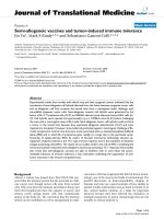
Foot contact was calculated from the acceleration zero
crossing following peak negative accelerations for the
shank in the AP direction (Figure 1). Raw AP accelerations
of the shank were differentiated to produce jerk, from
which an algorithm was applied to identify peaks in the
signal (Figure 1). A similar foot contact peak was observed
for the jerk profile for the ML acceleration of the shank,
however it was lower in amplitude and more difficult to
extract compared to the AP acceleration. It should be
noted that peaks in the AP jerk profile correspond to peak
trunk accelerations in the AP direction, which have previ-
ously been used to determine foot contact events during
walking [18,19]. Data were divided into step cycles,
defined as the period between left and right foot contact
events (and vice-versa for the subsequent step). Two con-
secutive steps constituted a stride. The middle 20 step
cycles were the basis of data analysis in the current study.
Root mean square acceleration
The amplitude of trunk accelerations during each trial
were assessed using RMS amplitude computed over indi-
vidual strides.
Signal repeatability
Acceleration repeatability was assessed in the present
study by calculating the adjusted Coefficient of Multiple
Determination (CMD), which indirectly quantifies the
percentage variance accounted for within the data [20,21].
While the CMD has previously been used to report relia-
Journal of NeuroEngineering and Rehabilitation 2009, 6:9 />Page 4 of 10
(page number not for citation purposes)
bility of gait data collected from different testing sessions,
the present study calculated CMD between strides for
individual trials as a measure of stride-to-stride repeatabil-
ity [22]. Waveforms that are similar return CMD values
that approach 1, whereas dissimilar waveforms result in
the CMD values approaching zero [21]. As the CMD is
influenced by the magnitude of the signals under exami-
nation, and acceleration amplitude is correlated to walk-
ing speed, raw trunk acceleration data were normalised to
each subjects preferred speed RMS acceleration.
Signal regularity
The degree of acceleration signal regularity within each
stride was determined using Approximate Entropy
(ApEn). ApEn is a probability statistic based on the loga-
rithmic likelihood that a sample of data will remain
within a tolerance window defined as 20% of the standard
deviation (r = 0.2) in subsequent data increments of one
data point (m = 1) within a serial signal [23,24]. ApEn
analysis returns a scalar value which approaches zero with
increased signal regularity, and approaches two with
increased signal irregularity [23,25]. Increased regularity
in a signal, or a signal containing a large degree of repeat-
able pattern features such as a pure low frequency sine
wave, will return a low ApEn value. In contrast, an irregu-
lar signal where time series events are unrelated to previ-
ous event (such as white noise) ApEn will return a high
value. Similar to the CMD, as ApEn is influenced by the
magnitude of the accelerations, the amplitude of raw
trunk acceleration data were normalised to each subjects's
preferred speed RMS acceleration.
Directional coupling
The degree of coupling between movement directions
within each stride was calculated by applying Cross ApEn
to trunk acceleration signals in each direction (ie. VT-AP,
VT-ML, and AP-ML coupling) [26,27]. Cross ApEn quanti-
fies the degree of signal regularity between standardised
serial signals. While Cross ApEn has similarities with corre-
lation analysis, there is evidence to suggest that the former
is more sensitive to identifying and grading subtle serial sig-
nal evolutions [24,28]. A higher Cross ApEn value is indic-
ative of weaker coupling between paired acceleration
signals, whilst a value that approaches zero indicates a
stronger degree of signal congruence and coupling [24,28].
Principal component analysis
The most common applications of PCA have been with
large multivariate data sets, where the number of variables
that are required to explain a biological process are
reduced whilst retaining much of the variation present in
the data set [16]. The reduced set of variables (PC's) are
orthogonal and uncorrelated, indicating that each PC rep-
resents a different dimension present in the data. In the
present study, PCA was used with a view of identifying the
main sources of step-to-step variance for gait-related trunk
accelerations. The procedures used to perform PCA on
continuous serial data collected during human walking
have been outlined in detail [15,29-31], and will only be
briefly described here.
Raw acceleration data were divided by steps and normal-
ised to 151 points. Two 150 × 151 column vector matrices
were created for each subject and acceleration direction
that represented trunk accelerations grouped for the slow
and preferred walking speeds, and the preferred and fast
walking speeds. In this study, 20 steps from each of the
subjects 5 trials were used as the basis of the PCA calcula-
tions. Trunk accelerations in the AP and VT directions are
biphasic during the stride cycle and similar in profile
when raw data is divided into steps (for example of AP
direction refer to Figure 1). In contrast, accelerations in
the ML direction are monophasic during the stride cycle
[6,7], and dividing accelerations into steps results in half
of the raw data being positive (eg left step) and the
remaining data being negative (eg right step). Therefore,
accelerations that were negative during the step cycle in
the ML direction were inverted so that all ML accelerations
in the PCA were consistent in profile.
Following the construction of the acceleration data
matrix, a mean-adjusted covariance matrix was calculated,
which was the basis of the PCA. In the present study, the
Representative data illustrating the temporal relationship between raw trunk and shank acceleration, and shank jerk in the anterior-posterior (AP) directionFigure 1
Representative data illustrating the temporal rela-
tionship between raw trunk and shank acceleration,
and shank jerk in the anterior-posterior (AP) direc-
tion. Dashed vertical lines indicate foot contact calculated
from peaks in shank jerk profile.
Journal of NeuroEngineering and Rehabilitation 2009, 6:9 />Page 5 of 10
(page number not for citation purposes)
covariance matrix was favoured to the correlation matrix;
the latter of which is more appropriate when the variables
under consideration have different scaling or units
[32,33]. To extract PC's, eigenvector decomposition was
performed on the covariance matrix. PC's were ordered so
that the variance (eigenvalues) exhibited in PC
1
> variance
PC
2
> variance PC
3
> variance PCn, where n is equal to
the number of variables in the original association matrix.
Often, extracted PC's contain multiple peaks and variance
components which make interpretation of results diffi-
cult, or even impossible [16]. Therefore, an orthogonal
Varimax rotation procedure was applied to the original
PCA solution. Varimax rotation has the effect of amplify-
ing higher loadings and suppressing lower loadings so
that PC's have a simpler structure [16], often resulting in
a reduced number of peaks in the extracted eigenvectors.
Although a large number of PC's were extracted, only a
few were retained for subsequent analysis. In the present
study, a PC was only retained if its eigenvalue accounted
for greater than 1% of variance in the acceleration data set.
Statistical analysis
All statistical analyses were performed in SAS for Win-
dows (Release 9.1). Using the Mixed procedure and Con-
trast statement, two-way Analysis of Variance (ANOVA)
was used to determine whether the dependent variables of
RMS acceleration, CMD, ApEn, and Cross ApEn were dif-
ferent according the speed of walking (slow, preferred,
fast). Speed by direction (VT, AP, ML) interactions were
selectively examined according to direction using planned
contrasts applied to incremented walking speed condi-
tions. For example, contrasts were used to identify if accel-
erations in the VT direction were different between slow
and preferred speeds, and preferred and fast speeds for
each dependent variable. Main effects were not reported
in the present study, as averaging dependent variables into
a walking speed condition, or alternatively the accelera-
tion direction, reduces the capacity to interpret how the
dependent variables were influenced by different walking
speeds. Using the GLM procedure, MANOVA were used to
determine if PC's differed between grouped walking speed
conditions (slow-preferred, preferred-fast). As individual
PC's are uncorrelated, each PC was entered into the
MANOVA as a separate dependent variable. In the event
of a significant main effect of walking speed condition or
acceleration direction, post hoc analyses were performed.
The level of significance for all statistical analysis in the
present study was 0.05.
Results
Basic gait parameters
Data for gait velocity, stride duration, cadence and step
length for each walking speed condition are presented in
table 1. A significant main effect for speed was identified
for all basic gait parameters. The average gait velocity,
cadence, and step length increased from the slow to pre-
ferred to fast walking speed conditions, whereas stride
duration decreased across the same conditions.
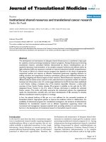
Root mean square acceleration
A significant interaction effect of walking speed and accel-
eration direction was identified for RMS acceleration (F(8,
96) = 189.92, p < 0.01). Contrasts revealed that RMS accel-
eration was greater for the preferred speed compared to
the slow speed for the VT (F(1, 96) = 87.09, p < 0.01), AP
(F(1, 96) = 30.47, p < 0.01), and ML directions (F(1, 96)
= 19.56, p < 0.01), and greater for the fast speed compared
to the preferred speed for the VT (F(1, 96) = 319.59, p <
0.01), AP (F(1, 96) = 108.20, p < 0.01), and ML direction
(F(1, 96) = 110.54, p < 0.01, Figure 2a).
Signal repeatability
A significant interaction effect of walking speed and accel-
eration direction was identified for CMD (F(8, 96) =
189.92, p < 0.01). Contrasts revealed that CMD was
greater for the preferred speed compared to the slow speed
for the VT (F(1, 96) = 6.00, p = 0.01) and ML directions
(F(1, 96) = 7.99, p < 0.01, Figure 2b).
Signal regularity
A significant interaction effect of walking speed and accel-
eration direction was identified for ApEn (F(8, 96) =
39.03, p < 0.01). Contrasts revealed that ApEn was lower
for the preferred speed compared to the slow speed for the
VT (F(1, 96) = 3.99, p = 0.04), AP (F(1, 96) = 5.95, p =
0.02) and ML directions (F(1, 96) = 21.24, p < 0.01, Figure
2c)
Directional coupling
A significant interaction effect of walking speed and accel-
eration direction was identified for Cross ApEn (F(8, 96)
= 3.21, p < 0.01). Contrasts revealed that Cross ApEn was
lower for the fast walking speed compared to the preferred
walking speed for VT-ML coupling (F(1, 96) = 4.82, p =
0.04, Figure 2d).
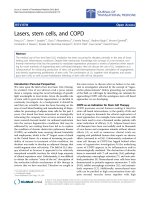
Principal component analysis
The number of PC's retained for analysis were 10, 10, and
11 for both the slow-preferred speed data and the pre-
ferred-fast speed data in the VT, AP and ML directions
respectively. For the slow-preferred speeds the retained
PC's accounted for 95.2 ± 1.1%, 94.8 ± 1.1% and 95.6 ±
1.0%, and for the preferred-fast speed the retained PC's
accounted for 96.3 ± 1.0%, 95.2 ± 1.2% and 97.2 ± 1.0%
in the VT, AP and ML directions respectively. No signifi-
cant differences were identified for PC data between
grouped walking speed conditions. Normalised eigenval-
ues calculated for each PC for the VT, AP and ML direc-
tions are presented in Figure 3.
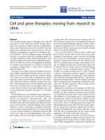
Representative eigenvectors for a single subject are pre-
sented in Figure 4, illustrating the profiles of PC1 to PC4
Journal of NeuroEngineering and Rehabilitation 2009, 6:9 />Page 6 of 10
(page number not for citation purposes)
for the slow-preferred walking speed data and the pre-
ferred-fast walking speed data. In general, peak PC's pre-
dominantly occurred between 0–20% and 80–100% of
the step cycle for each direction. Peaks emerged between
20–80% of the step cycle only at higher PC's (e.g. > 6)
where the variance accounted for in the acceleration data
was relatively low compared to the first few PC's.
Discussion
The primary purpose of this study was to examine how
gait speed influences healthy individual's trunk motion
during overground walking. Lower trunk accelerations
were examined using analyses that address the amplitude
of motion as well as the stride-to-stride structure of trunk
accelerations. This investigation was also a preliminary
study to assess if PCA can be used to gain further insight
into postural responses that occur at different speeds dur-
ing overground walking.
The basic gait parameters of gait velocity, cadence, and step
length systematically increased from the slow to preferred
to fast walking speed conditions, and as could be expected
stride duration decreased during the same conditions. The
different walking speeds were also found to alter the abso-
lute amplitude of trunk, as RMS acceleration increased with
gait velocity in the AP, VT, and ML directions. RMS ampli-
tude of trunk accelerations may be a reasonable indicator of
the inertial properties of the upper body that must be over-
come during walking, however it provides limited insight
to how motor output is regulated [11].
A noteworthy finding in the present study was that trunk
accelerations in the ML and VT directions had less signal
regularity and repeatability during the slow walking speed
compared to preferred walking speed. However, stride-to-
stride acceleration signal regularity and repeatability did
not statistically differ between the preferred and fast walk-
ing speed conditions. These findings are in contrast to the
U-shaped response in movement amplitude that was
expected with changes in walking speed. As the funda-
mental requirements of human walking include progres-
sion, support and balance, marked reductions in the
speed of progression may have produced added challenge
to the individuals support and balance mechanisms [34].
Under normal walking conditions, mechanical models
and biological data indicate that lateral balance is an
active rather than passive control process, which results in
greater motor output variability than the AP direction [35-
37]. Therefore, the less repeatable and regular accelera-
tions at slow speeds may be a factor of altered motor activ-
ity and variability in sensorimotor mechanisms that
facilitate frontal plane balance.
In light of the greater amplitude of trunk acceleration for
the fast speed compared to the preferred speed, the lack of
regularity and repeatability differences between the same
conditions suggest that features of trunk motion are pre-
served at speeds that are faster than preferred. However
this finding should be taken with caution, as the subjects
in the present study were walking at self-selected fast
speeds. Increasing speed to beyond what is perceived as a
comfortable level by the subjects would most likely result
in added perturbation to the upper body. The results of
the Cross ApEn analysis between acceleration directions
revealed a change in coordination dynamics that assisted
in controlling trunk motion at fast speeds. At the fast
walking speed the coupling between accelerations in the
ML and VT direction increased, thus placing a greater
importance on regulating the global motion of the trunk
in the frontal plane rather than independently regulating
accelerations according to direction. Similar coupling
dynamics have been reported for gait-related head acceler-
ations in healthy older individuals, presumably to assist
in maintaining head stability in the presence of reduced
postural control [26].
Acceleration RMS amplitude, regularity, repeatability, and
coupling were used collectively to characterise the speed-
dependent trunk motion across the gait cycle. However,
additional information about how motor output is regulated
can be gained by examining the complexity of motor output
[38]. In regards to gait-related trunk motion, there is evi-
dence to suggest that the variability and complexity of trunk
acceleration signals corresponds to an individual's health
status. Post-stroke hemiplegic patients [39], Parkinsonian
patients [40], and healthy older individuals [41] have greater
complexity in gait-related trunk motion than younger indi-
viduals, which suggests that increased dimensionality may
be a characteristic of reduced postural stability.
In the current study, a criterion was set where PC's were
only considered for analysis when their variance was
Table 1: Basic gait parameters for self-selected slow, preferred and fast walking speeds (mean ± SD).
Variable Walking condition Effect of speed
Slow Preferred Fast
Gait velocity (m.s
-1
) 0.93 ± 0.11 1.32 ± 0.18 1.78 ± 0.29 F(2, 24) = 443.32, p < 0.01)
Stride duration (s) 0.63 ± 0.05 0.53 ± 0.03 0.47 ± 0.03 F(2, 24) = 444.62, p < 0.01)
Cadence (steps.min
-1
) 95.72 ± 8.59 111.85 ± 6.69 126.49 ± 7.53 F(2, 24) = 519.11, p < 0.01)
Step length (m) 0.59 ± 0.06 0.71 ± 0.09 0.84 ± 0.11 F(2, 24) = 352.35, p < 0.01)
Journal of NeuroEngineering and Rehabilitation 2009, 6:9 />Page 7 of 10
(page number not for citation purposes)
greater than 1% of the total variance in the lower trunk
accelerations. Consequently, 10 PC's for the AP, 10 PC's for
the VT, and 11 PC's for the ML direction were retained in
the analysis. The number of PC's required to account for
greater than 95% of variance was relatively low compared
to the 300 dimensional set of absolute possibilities. The
interpretation of dimensionality within a univariate data
set is one of conjecture, partly because measurements
involving a small number of biomechanical degrees of free-
dom (such as a single accelerometer) are reflective of
dimensional organisation at other levels of the control sys-
tem [42]. Sanger [43] examined the trajectory of hand
motion when performing a practiced tracking task in a Car-
tesian coordinate system, and found that the smooth pla-
nar movement associated with copying a target trajectory
was low-dimensional. It was suggested that the low-dimen-
sional output may be a strategy to simplify the interaction
between CNS control and musculoskeletal mechanics.
However, the author stressed that such conclusions were
speculative as applying PCA to the output of a motor task
RMS acceleration (a), Coefficient of Multiple Determination (b), and Approximate Entropy (c), derived from lower trunk accel-erations in the vertical (VT), anterior-posterior (AP), and mediolateral (ML) directionFigure 2
RMS acceleration (a), Coefficient of Multiple Determination (b), and Approximate Entropy (c), derived from
lower trunk accelerations in the vertical (VT), anterior-posterior (AP), and mediolateral (ML) direction. Cross
Approximate Entropy (d) representing strength of coupling between accelerations in the vertical and anterior-posterior (VT-
AP), anterior-posterior and mediolateral (AP-ML), and vertical and mediolateral (VT-ML) directions. All data is presented for
slow, preferred, and fast walking speeds. Error bars represent one standard error of the mean.
Journal of NeuroEngineering and Rehabilitation 2009, 6:9 />Page 8 of 10
(page number not for citation purposes)
Percentage of variance accounted for by the first ten Principal Components (PC) of lower trunk accelerations measured for the vertical, anterior-posterior, and mediolateral directionsFigure 3
Percentage of variance accounted for by the first ten Principal Components (PC) of lower trunk accelerations
measured for the vertical, anterior-posterior, and mediolateral directions. Error bars represent one standard error
of the mean.
Journal of NeuroEngineering and Rehabilitation 2009, 6:9 />Page 9 of 10
(page number not for citation purposes)
only provides a description of movement and not causality
of movement. The absence of significant differences
between the slow-preferred and preferred-fast PC data in
the present study suggests that variance within the step
cycle was similar at different walking speeds, and features of
trunk motion are retained despite changes in walking
speed. However, the discriminatory ability of PCA to detect
speed-dependent differences in walking patterns appears to
be limited compared to measures of signal regularity,
repeatability, and coupling. Interestingly, PC peaks gener-
ally occurred following foot contact, suggesting that vari-
ance is greatest during the weight acceptance phase of
walking. To date, upper body motion is yet to be examined
in detail regarding how acceleration of the trunk is influ-
enced by foot contact events or the swing and stance phase.
Conclusion
The main finding of this study was that walking at speeds
slower than preferred primarily alters lower trunk acceler-
ations in the frontal plane. Despite greater amplitudes of
trunk acceleration at fast speeds, the lack of regularity and
repeatability differences between preferred and fast speeds
suggest that features of trunk motion are preserved
between the same conditions. This was partly due to an
Representative Principal Components (PC) for a single subject plotted across the step cycleFigure 4
Representative Principal Components (PC) for a single subject plotted across the step cycle. Data represents
PC's extracted from lower trunk accelerations measured for the vertical, anterior-posterior, and mediolateral directions. Solid
lines are PC's extracted from grouped data for slow and preferred walking speeds, and dashed lines are PC's from grouped
preferred and fast walking speeds.
Journal of NeuroEngineering and Rehabilitation 2009, 6:9 />Page 10 of 10
(page number not for citation purposes)
increase in coupling between accelerations in the ML and
VT direction, which places a greater importance on regu-
lating the global motion of the trunk in the frontal plane
instead of independently regulating accelerations accord-
ing to direction. PCA provided useful insight to the
dimensionality of lower trunk motion, identifying that a
relatively low number of PC's explained the majority of
variance in acceleration data across different walking
speeds. Further research needs to be undertaken to deter-
mine if a higher number of PC's correlates to conditions
of postural instability. If this is the case, PCA may be a tool
that can be employed in a rehabilitation setting to moni-
tor improvement or declines in postural control.
Competing interests
The author declares that he has no competing interests.
References
1. Winter DA, Patla AE, Frank JS: Assessment of balance control in
humans. Medical Progress Through Technology 1990, 16(1–2):31-51.
2. Anders C, Wagner H, Puta C, Grassme R, Petrovitch A, Scholle HC:
Trunk muscle activation patterns during walking at different
speeds. Journal of Electromyography and Kinesiology 2007, 17:245-252.
3. Thorstensson A, Carlson H, Zomlefer MR, Nilsson J: Lumber back
muscle activity in relation to trunk movements during loco-
motion in man. Acta Physiologica Scandinavica 1982, 116:13-20.
4. Winter DA, MacKinnon CD, Ruder GK, Wieman C: An integrated
EMG/biomechanical model of upper body balance and pos-
ture during human gait. Progress in Brain Research 1993,
97:359-367.
5. MacKinnon CD, Winter DA: Control of whole body balance in
the frontal plane during human walking. Journal of Biomechanics
1993, 26(6):633-644.
6. Kavanagh JJ, Barrett RS, Morrison S: Upper body accelerations
during walking in healthy young and elderly men. Gait Posture
2004, 20(3):291-298.
7. Menz HB, Lord SR, Fitzpatrick RC: Acceleration patterns of the
head and pelvis when walking on level and irregular surfaces.
Gait Posture 2003, 18(1):35-46.
8. Kavanagh JJ, Menz HB: Accelerometry: A technique for quanti-
fying movement patterns during walking. Gait Posture 2008,
28:1-15.
9. Menz HB, Lord SR, Fitzpatrick RC: Acceleration patterns of the
head and pelvis when walking on level and irregular surfaces.
Gait Posture 2003, 18:35-46.
10. Moe-Nilssen R: A new method for evaluating motor control in
gait under real-life environmental conditions. Part 2: Gait
analysis. Clinical Biomechanics 1998, 13(4–5):328-335.
11. Latt MD, Menz HB, Fung VS, Lord SR: Walking speed, cadence
and step length are selected to optimize the stability of head
and pelvis accelerations. Experimental Brain Research 2008,
184(2):201-209.
12. Kang HG, Dingwell JB: Separating the effects of age and walking
speed on gait variability. Gait Posture 2008, 27(4):572-577.
13. England SA, Granata KP: The influence of gait speed on local
dynamic stability of walking. Gait Posture 2007, 25(2):172-178.
14. Newell KM, Slifkin AB: The nature of movement variability. In
Motor behavior and human skill Edited by: Piek JJ. Human Kinetics:
Champaign, IL; 1998.
15. Shemmell J, Johansson J, Portra V, Gottlieb GL, Thomas JS, Corcos
DM: Control of interjoint coordination during the swing
phase of normal gait at different speeds. Journal of NeuroEngi-
neering and Rehabilitation 2007, 4:10.
16. Chau T: A review of analytical techniques for gait data. Part
1: fuzzy, statistical and fractal methods. Gait Posture 2001,
13:49-66.
17. Moe-Nilssen R: A new method for evaluating motor control in
gait under real-life environmental conditions. Part 1: The
instrument. Clinical Biomechanics 1998, 13(4–5):320-327.
18. Zijlstra W, Hof AL: Assessment of spatio-temporal gait param-
eters from trunk accelerations during human walking. Gait
Posture 2003, 18(2):1-10.
19. Zijlstra W: Assessment of spatio-temporal parameters during
unconstrained walking. European Journal of Applied Physiology 2004,
92(1–2):39-44.
20. Besier TF, Sturnieks DL, Alderson JA, Lloyd DG: Repeatability of
gait data using a functional hip joint centre and a mean heli-
cal knee axis. Journal of Biomechanics 2003, 36(8):1159-1168.
21. Kadaba MP, Ramakrishnan HK, Wootten ME, Gainey J, Gorton G,
Cochran GV: Repeatability of kinematic, kinetic, and electro-
myographic data in normal adult gait. Journal of Orthopaedic
Research 1989, 7:849-860.
22. Kavanagh JJ, Morrison S, James DA, Barrett R: Reliability of seg-
mental accelerations measured using a new wireless gait
analysis system. Journal of Biomechanics 2006, 39:2863-2872.
23. Pincus SM:
Approximate entropy as a measure of system
complexity. Proceedings of the National Academy of Sciences of the
United States of America 1991, 88:2297-2301.
24. Pincus SM, Singer BH: Randomness and degrees of irregularity.
Proceedings of the National Academy of Sciences of the United States of
America 1996, 93:2083-2088.
25. Pincus SM: Approximate entropy (ApEn) as a complexity
measure. Chaos 1995, 5(1):110-117.
26. Kavanagh JJ, Barrett RS, Morrison S: Age-related differences in
head and trunk coordination during walking. Human Movement
Science 2005, 24(4):574-587.
27. Kavanagh JJ, Morrison S, Barrett RS: Coordination of head and
trunk accelerations during walking. European Journal of Applied
Physiology 2005, 94(4):468-475.
28. Pincus SM: Irregularity and asynchrony in biologic network
signals. Methods in Enzymology 2000, 321:149-182.
29. Ivanenko YP, Grasso R, Zago M, Molinari M, Scivoletto G, Castellano
V, Macellari V, Lacquaniti F: Temporal components of the motor
patterns expressed by the human spinal cord reflect foot kin-
ematics. Journal of Neurophysiology 2003, 90(5):3555-3565.
30. Deluzio KJ, Astephen JL: Biomechanical features of gait wave-
form data associated with knee osteoarthritis: An applica-
tion of Principal Component Analysis. Gait Posture 2007,
25:86-93.
31. Cappellini G, Ivanenko YP, Poppele RE, Lacquaniti F: Motor pat-
terns in human walking and running. Journal of Neurophysiology
2006, 95:3426-3437.
32. Jackson JE: A User's Guide to Principal Components. 1st edi-
tion. Wiley-Interscience; 1991.
33. Kayser J, Tenke CE: Optimizing PCA methodology for ERP
component identification and measurement: theoretical
rationale and empirical evaluation. Clinical Neurophysiology 2003,
114:2307-2325.
34. den Otter AR, Geurts AC, Mulder T, Duysens J: Speed related
changes in muscle activity from normal to very slow walking
speeds. Gait Posture 2004, 19:270-278.
35. Bauby CE, Kuo AD: Active control of lateral balance in human
walking. Journal of Biomechanics 2000, 33:1433-1440.
36. Kuo AD: Stabilization of lateral motion in passive dynamic
walking. International Journal of Robotics Research 1999, 18:917-930.
37. Dean JC, Alexander NB, Kuo AD: The effect of lateral stabiliza-
tion on walking in young and old adults. Transactions on Biological
Engineering 2007, 45(11):1919-1926.
38. Hausdorff JM: Gait variability: methods, modeling and mean-
ing. Journal of Neuroengineering and Rehabilitation 2005, 2:19.
39. Akay M, Sekine M, Tamura T, Higashi Y, Fujimoto T: Fractal dynam-
ics of body motion in post-stroke hemiplegic patients during
walking. Journal of Neural Engineering 2004, 1(2):111-116.
40. Sekine M, Akay M, Tamura T, Higashi Y, Fujimoto T: Fractal dynam-
ics of body motion in patients with Parkinson's disease. Jour-
nal of Neural Engineering 2004, 1:8-15.
41. Sekine M, Tamura T, Akay M, Fujimoto T, Togawa T, Fukui Y: Dis-
crimination of walking patterns using wavelet-based fractal
analysis. IEEE Transactions on Neural Systems and Rehabilitation Engi-
neering 2002, 10(3):188-196.
42. Newell KM, Vaillancourt DE: Dimensional change in motor
learning. Human Movement Science 2001, 20(4–5):695-715.
43. Sanger TD: Human arm movements described by a low-
dimensional superposition of Principal Components. Journal
of Neuroscience 2000, 20(3):1066-1072.