báo cáo hóa học: "Infrared thermography as an access pathway for individuals with severe motor impairments" docx
Bạn đang xem bản rút gọn của tài liệu. Xem và tải ngay bản đầy đủ của tài liệu tại đây (786.58 KB, 8 trang )
BioMed Central
Page 1 of 8
(page number not for citation purposes)
Journal of NeuroEngineering and
Rehabilitation
Open Access
Research
Infrared thermography as an access pathway for individuals with
severe motor impairments
Negar Memarian
1,2
, Anastasios N Venetsanopoulos
3,4
and Tom Chau*
1,2
Address:
1
Institute of Biomaterials and Biomedical Engineering, University of Toronto, Toronto, Canada,
2
Bloorview Research Institute, Bloorview
Kids Rehab, Toronto, Canada,
3
Department of Electrical and Computer Engineering, University of Toronto, Toronto, Canada and
4
Department of
Electrical and Computer Engineering, Ryerson University, Toronto, Canada
Email: Negar Memarian - ; Anastasios N Venetsanopoulos - ;
Tom Chau* -
* Corresponding author
Abstract
Background: People with severe motor impairments often require an alternative access pathway,
such as a binary switch, to communicate and to interact with their environment. A wide range of
access pathways have been developed from simple mechanical switches to sophisticated
physiological ones. In this manuscript we report the inaugural investigation of infrared
thermography as a non-invasive and non-contact access pathway by which individuals with
disabilities can interact and perhaps eventually communicate.
Methods: Our method exploits the local temperature changes associated with mouth opening/
closing to enable a highly sensitive and specific binary switch. Ten participants (two with severe
disabilities) provided examples of mouth opening and closing. Thermographic videos of each
participant were recorded with an infrared thermal camera and processed using a computerized
algorithm. The algorithm detected a mouth open-close pattern using a combination of adaptive
thermal intensity filtering, motion tracking and morphological analysis.
Results: High detection sensitivity and low error rate were achieved for the majority of the
participants (mean sensitivity of all participants: 88.5% ± 11.3; mean specificity of all participants:
99.4% ± 0.7). The algorithm performance was robust against participant motion and changes in the
background scene.
Conclusion: Our findings suggest that further research on the infrared thermographic access
pathway is warranted. Flexible camera location, convenience of use and robustness to ambient
lighting levels, changes in background scene and extraneous body movements make this a potential
new access modality that can be used night or day in unconstrained environments.
Background
Alternative access pathways
Individuals with severe physical impairments who are
unable to communicate through speech or gestures
require an alternative means to convey their intentions. In
the rehabilitation engineering context, these alternative
channels are called access pathways and they constitute
the critical front end of an access solution [1]. Some recent
efforts have set out to non-invasively translate physiolog-
ical signals such as the electrical [2,3] and hemodynamic
Published: 16 April 2009
Journal of NeuroEngineering and Rehabilitation 2009, 6:11 doi:10.1186/1743-0003-6-11
Received: 15 September 2008
Accepted: 16 April 2009
This article is available from: />© 2009 Memarian et al; licensee BioMed Central Ltd.
This is an Open Access article distributed under the terms of the Creative Commons Attribution License ( />),
which permits unrestricted use, distribution, and reproduction in any medium, provided the original work is properly cited.
Journal of NeuroEngineering and Rehabilitation 2009, 6:11 />Page 2 of 8
(page number not for citation purposes)
activity [4-6] of the brain or the electrodermal response of
the skin [7,8] into functional communication. A compre-
hensive review of emerging access technologies can be
found in [1].
Biomedical applications of thermal imaging
Infrared thermography refers to the measurement of the
radiation emitted by the surface of an object in the infra-
red range of the electromagnetic spectrum, i.e., between
wavelengths of 0.8 μm and 1.0 mm [9]. Infrared cameras
use specialized lenses manufactured from materials such
as germanium to focus thermal radiation onto a focal
plane array of infrared detectors [10]. Thermal cameras
yield an image that is a spatial, two-dimensional (2-D)
map of the 3-D temperature distribution of the object
[11].
Infrared thermography has been widely applied in health
research, including, for example, breast cancer detection
[12,13], brain surgery [14,15], heart surgery [16], diagno-
sis of vascular disorders [17], arthritis [18], pain assess-
ment [19] and post-surgical follow-up in ophthalmology
[20].
Recently, Murthy and Pavlidis non-invasively measured
human breathing using infrared imaging and a statistical
methodology based on multinormal distributions, the
method of moments, and Jeffreys divergence measure
[21]. Their study was based on the fact that exhaled gases
have a higher temperature than the typical background of
indoor environments. They achieved high detection accu-
racy on a small set of subjects and suggested potential
applications in polygraphy, sleep studies, sport training,
and patient monitoring [21].
Thermal imaging as an access pathway
The goal of this paper is to investigate the potential of
thermal imaging as an access pathway. In particular, we
introduce a thermographic binary switch activated by vol-
untary mouth opening. Expired air and the oral cavity are
generally warmer than the surrounding tissue and envi-
ronment while cyclic jaw movements do not cause signif-
icant increases in facial temperatures over time [22].
Therefore localized temperature changes due to mouth
opening and closing may be detectable using video and
image processing of thermographic data. Examples of
patient groups that may benefit from this access pathway
are people with high level spinal cord injuries resulting in
quadriplegia and individuals with spastic quadriplegic
cerebral palsy or general hypotonia.
Like computer vision-based access pathways [23], thermal
imaging is non-invasive and does not require any sensor
attachment to the user. However, thermography over-
comes some of the major limitations of conventional
computer vision-based access pathways. Firstly, thermog-
raphy is skin colour invariant since there is no difference
in emissivity between black, white and burnt skin, in vivo
or in vitro [24]. Human skin has an emissivity of about
0.98. Thermal radiation from the skin originates in the
epidermis and is independent of race; it depends therefore
only on the surface temperature [9,11]. Secondly, thermal
image quality is independent of ambient lighting condi-
tions and can thus be effective both night and day. Con-
ceivably, this non-contact, non-invasive access pathway
could be tailored to the user's unique motor capacity,
whether that be mouth opening, eye blinking or simply
deep breathing. These are all motor activities that may
generate measurable, local temperature changes. Further-
more, given that the key information is thermal variation,
a frontal view of the user may not be necessary, facilitating
more flexible and unobtrusive placement of the camera.
Methods
Participants
Eight able-bodied participants and two individuals with
quadriplegia (one with a C1-C2 incomplete spinal cord
injury and the other with severe spastic quadriplegic cere-
bral palsy) participated in this study. All participants pro-
vided written consent. The experimental protocol was
approved by the research ethics board of the university
and affiliated hospital.
Instrumentation and setup
A THERMAL-EYE 2000B thermal video camera by L-3
Communications with thermal sensitivity ≤100 mK [25]
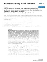
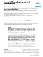
Components of the proposed mouth opening detection algorithmFigure 1
Components of the proposed mouth opening detection algorithm.
Journal of NeuroEngineering and Rehabilitation 2009, 6:11 />Page 3 of 8
(page number not for citation purposes)
was connected via an NTSC to USB TV convertor (Dazzle
Multimedia). Videos were recorded as 240 × 320 AVI files
(30 fps) and processed offline in MATLAB & Simulink
(version R2007b).
Participants were comfortably seated within a laboratory
environment. Those with disability remained in their
wheelchairs. The thermal camera was positioned anterior
and lateral to the participant at a 45° angle. This camera
location was chosen over the often-used frontal view,
keeping in mind the eventual application as an access
switch where the user's field of view ought to be unob-
structed. In the 45° angle condition, infrared thermo-
grams only exhibit a small error in recorded temperatures
[9]. Each participant was cued to open his or her mouth
and to hold it ajar for one second before closing the
mouth. Participants were given an auditory prompt upon
every open and close action. The end of each mouth clos-
ing was followed by a 3 second rest before the onset of the
next mouth opening. The participants were instructed to
maintain a constant head position, so that their mouth
movement stayed within the camera's field of view.
The thermal sensitivity of the infrared camera we used was
well beyond what was needed to detect the temperature
change due to mouth opening. We are looking at temper-
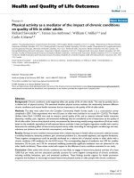
The action of the different modules of the mouth opening detection algorithmFigure 2
The action of the different modules of the mouth opening detection algorithm. (a) Input thermal video frame, (b)
Segmented face region, (c) Warm facial zones, (d) Moving facial zones, (e) Intersection of warm and moving objects within the
face region, (f) After morphological, size variation, and anthropometric filtering, (g) Final output; detected mouth open is high-
lighted on the original video with a hollow box.
Journal of NeuroEngineering and Rehabilitation 2009, 6:11 />Page 4 of 8
(page number not for citation purposes)
ature difference of about 1.5 to 3°C between when mouth
is closed and when it is open, while the thermal sensitivity
of our infrared camera was ≤100 mK.
Thermal video processing
Figure 1 shows a schematic of our algorithm for detecting
mouth openings from the thermal video data. The system
consisted of three main components, namely face seg-
mentation, thermal intensity-motion filtering and false
positive removal. Each component will be discussed
below. To begin, the boundary pixels of each video frame
(the first and last pixels of every column and every row)
were set to zero to detach objects that may be connected
to the borders.
Face segmentation
In addition to the participant's head and facial region,
other body parts such as the participant's neck, thorax and
upper limbs also appeared in the videos. For the partici-
pants with disability, parts of their wheelchairs were also
captured on thermal video. Objects in the background,
and in a couple of instances people moving around the
participant were also recorded. It was thus essential to seg-
ment the participant's face region from all other non-tar-
get body parts and objects. Each frame of the video was
binarized. Given that facial temperature distributions vary
within and among individuals [26], we adopted Otsu's
method to determine an adaptive rather than fixed inten-
sity threshold which minimized, on a frame by frame
basis, the intra-class variance of the grayscale values of the
pixels to be binarized [27].
The binarized frames were then morphologically opened
with a disk structuring element of radius 5 pixels to
remove small objects, break thin connections, remove
thin protrusions, and smooth object contours [28]. In the
resulting image, the object with maximum area (presum-
ably the face region) was retained and the object's interior
holes were filled by morphological closing with a disk
structuring element of radius 20 pixels. The camera-user
distance and the user's head size affect the dimension of
the above mentioned structuring elements. In a real life
application, the camera will be mounted on the user's
wheelchair at a fixed distance from the user's face. Hence,
once the appropriate parameters are selected in the initial
calibration, they do not need to be changed for subse-
quent use. An example of a segmented face region is
depicted in Figure 2(b).
Thermal intensity-motion filtering
All subsequent processing was applied to the intensity
image and confined to the identified face region. The
region of interest (ROI) was the participant's mouth and
the task of interest was mouth opening. A combination of
temperature thresholding and motion tracking was used
to perceive mouth opening. Warm zones inside the facial
region were extracted by thresholding the segmented face
with a scaled version of Otsu's threshold [27] to favour
higher intensity (i.e., warmer) pixels. The scale factor was
empirically derived as
and typically ranged from 2.5 to 3. This segmentation
yielded a warm zone mask which served to detect
instances of mouth opening. However, there were occa-
sions where nearby facial regions had similar tempera-
tures as those of the oral cavity. A corroborating cue was
therefore required to accurately pinpoint a mouth open-
ing event.
Since mouth opening involves motion, optical flow was
utilized to estimate the direction and speed of motion
from one video frame to the next using the Horn-Schunck
method [29]. Motion vectors in each frame of the video
sequence were computed by solving the optical flow con-
straint equation
where I
x
, I
y
and I
t
are the spatiotemporal image brightness
derivatives, u is the horizontal optical flow and v is the
Scale factor mean intensity in face region=− −3 150 50()/
(1)
Iu Iv I
xyt
++=0
(2)
Table 1: Performance of the proposed mouth opening detection algorithm
Participant Video length (sec) Total Video frames Actual # of mouth openings Sensitivity Specificity
1 256 7662 50 88% 100%
2 252 7546 50 96% 100%
3 254 7621 50 96% 100%
4 252 7481 50 98% 100%
5 244 7424 50 88% 99%
6 243 7594 50 92% 98%
7 245 7664 50 94% 99%
8 243 7613 50 80% 100%
9* 153 4592 30 93% 99%
10* 272 8160 15 60% 99%
*Participant with severe disability.
Journal of NeuroEngineering and Rehabilitation 2009, 6:11 />Page 5 of 8
(page number not for citation purposes)
vertical optical flow. By assuming that the optical flow is
smooth over the entire image, the Horn-Schunck method
computes an estimate of the velocity field, [u v ]
T
, that
minimizes this equation:
In this equation and are the spatial deriva-
tives of the optical velocity component u, and
α
scales the
global smoothness term [29]. Motion vectors with veloc-
ity magnitude exceeding the mean velocity (i.e., the aver-
age of velocity magnitudes across the most recent five
frames) per frame across time were retained, yielding a
motion mask. The intersection of this motion mask and
the warm zone mask, introduced above, yielded all the
regions of the face that were both warm and moving.
False positive removal
Despite the combination of motion and thermal cues, the
processed frames occasionally contained non-mouth
objects (false positives) such as parts of the chin, forehead
and the periorbital regions. These non-mouth objects
E I u I v I dxdy
u
x
u
y
v
x
xyt
=∫∫ + +
()
+∫∫
∂
∂
⎛
⎝
⎜
⎞
⎠
⎟
+
∂
∂
⎛
⎝
⎜
⎞
⎠
⎟
+
∂
∂
⎛
⎝
⎜
⎞
⎠
⎟
+
2
a
∂∂
∂
⎛
⎝
⎜
⎞
⎠
⎟
⎧
⎨
⎪
⎩
⎪
⎫
⎬
⎪
⎭
⎪
v
y
dxdy
(3)
∂
∂
()
u
x
∂
∂
()
u
y
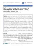
Robustness of the proposed algorithm to motion artefacts and changes in the backgroundFigure 3
Robustness of the proposed algorithm to motion artefacts and changes in the background. (a) Robustness to
motion artefacts. Top row from left to right shows input thermal video of an able-bodied participant moving his arm to his
head (frames 63, 66, 70, and 74). Bottom row depicts face segmentation in the corresponding frames. (b) Robustness to
changes in the background. Top row from left to right is an input thermal video of a participant with disability while a passerby
traverses the scene in the background (frames 1759, 1765, 1779, 1790). The corresponding face segmentation results are pre-
sented in the bottom row.
Journal of NeuroEngineering and Rehabilitation 2009, 6:11 />Page 6 of 8
(page number not for citation purposes)
were also warm and moving and were therefore retained
subsequent to the thermal intensity and motion filters. An
example is the forehead, which according to the literature,
is the warmest part of the human body with a temperature
(34.5°C) close to that inside the mouth [30]. Therefore
motion of the forehead may result in a false positive.
To deal with these false positives, we deployed a series of
additional filters based on morphology, size variation
between frames, and facial anthropometry. Objects that
did not meet the following morphological conditions
were deemed as false positives and removed.
1. 30 pixels < Area < 150 pixels
2. Eccentricity ≤ 0.9.
3.
The first condition rejects objects which are either too
small or too large to be candidate mouth openings. Like-
wise, the second condition removes regions that are too
elongated to qualify as mouth regions while the third con-
dition eliminates hollow regions as the mouth is expected
to be solid. The constants in these morphological filters
were selected to resemble the shape of the open mouth
and were empirically defined. In addition, objects whose
size varied less than 25% between the current frame and
the frame occurring ten frames earlier were considered
static warm facial regions (e.g., forehead, chin, around the
eyes, neck) and were also discarded. This constitutes the
size variation filter in Figure 1.
Finally we exploited the fact that facial anatomy is static
(i.e., unlikely to change over time). Based on human face
anthropometry, the mouth is located in the lower half of
the menton-sellion length [31,32]. When we partitioned
the facial ROI along its major axis into four strips, we
noticed that indeed the mouth was usually located in the
second strip from the bottom. With this anthropometric
filter, we dismissed candidate ROIs outside of the second
facial quarter. Figures 2(c)–(g) demonstrate the action of
the different processing modules.
Algorithm evaluation
To facilitate algorithm evaluation, a truth set was prepared
manually for each recorded thermal video. The truth set
contained the frame numbers corresponding to the begin-
ning and ending of each mouth opening, the end points
of the line maximally spanning the width of the mouth at
the onset of opening and the end points of the line maxi-
mally spanning the height of the mouth when fully ajar.
This truth set served as the gold standard for automatic
algorithm evaluation. A true positive was defined as the
detection of a ROI temporally within the range of frames
corresponding to a gold standard mouth opening, and
spatially situated within the bounding box defined by the
endpoints extracted above. All other detected objects were
considered false positives. A mouth opening that was
missed by the algorithm was counted as a false negative. A
true negative occurred when there was no mouth opening
and the algorithm concluded the same. Sensitivity and
specificity values were estimated.
Results and discussion
The performance of the proposed algorithm on the ther-
mal video of ten participants is summarized in Table 1.
Detection of mouth opening is generally achieved with
very high sensitivity and specificity. The exception is the
poorer result for participant 10, which is mainly due to
participant's posture, frequent involuntary head rotation
away from the camera, and suboptimal camera place-
ment. This participant had an awkward position in his
wheelchair (See Figure 3(b)) which forced us to position
the thermal camera at an angle and distance from the par-
ticipant that was not consistent with the other partici-
pants. Several improvements can be made to enhance the
results in situations like this: (1) The algorithm can be
updated to track and focus on the region of interest (par-
ticipant's face) more accurately; (2) Multiple cameras can
be used to capture participant's facial region from differ-
ent angles, so that the problem of participant mouth leav-
ing the camera's field of view will be mitigated; and (3)
The user can be trained. Figures reported in the present
paper are the result of just one test session. Training is
expected to have a positive effect on user performance.
Specificity is generally higher than sensitivity as the algo-
rithm was tuned to minimize false positives, again keep-
ing in mind the alternative access application where
inadvertent switch activations are arguably more costly
than missed activations. Most of the false positives were
repeated detections of the same non-mouth object in mul-
tiple frames. The chin was the source of the majority of the
false positives, which tended to occur during actual
mouth openings. This is perhaps not surprising given that
the chin is proximal to the mouth and moves as the jaw
descends to open the mouth. Further, the chin is report-
edly the warmest facial area after the forehead [33] when
measured by thermography.
The proposed algorithm is robust against participant
motion and changes to the background scene. Figure 3(a)
demonstrates an example of one of the participants mov-
ing his arm towards his face. Although the arm is both
warm and moving, and even touches the participant's face
in some frames, it was correctly disregarded by the algo-
rithm. Figure 3(b) depicts an example of a person entering
and leaving the background scene. The algorithm success-
Area of object
Area of bounding box
> 05.
Journal of NeuroEngineering and Rehabilitation 2009, 6:11 />Page 7 of 8
(page number not for citation purposes)
fully rejected the background activity and did not generate
any false positives.
The proposed combination of filters is location and posi-
tion invariant; regardless of where in the frame the user
moves his or her head within the camera's field of view
and independent of the user's position (sitting or semi-
supine), mouth opening could generally be located rela-
tive to the segmented face region.
If one can voluntary control mouth open and close action,
sip and puff technology, EMG based switches, and com-
puter vision based switches can also be used. The advan-
tage of the proposed thermography based access pathway
over sip and puff and EMG based switches is that it is non-
invasive and non-contact, i.e., does not require attach-
ment of any sensor or external object to the user. Hence it
is more hygienic and safe, as the risk of choking is also
eliminated. Its advantage over visible light computer
vision based access pathways is that it is independent of
lighting/color and can thus be used both night and day,
indoor and outdoor.
Despite these encouraging findings, thermal imaging does
have its limitations. Infrared thermal cameras are more
expensive than conventional (visible light) cameras.
However, recent innovations in affordable, pocket sized,
portable thermal cameras [34] may eventually eliminate
the cost issue. Thermal image quality is susceptible to fluc-
tuations in ambient temperature, humidity and regional
air circulation [9]. A robust thermographic access pathway
may need to dynamically compensate for changes in these
contextual factors. A final limitation of thermal imaging is
the relatively low resolution of infrared cameras and the
inherent difficulty in discriminating between fine facial
features. These issues may be mitigated by fusing thermal
videos with simultaneously recorded visible spectrum
imagery [35].
Conclusion
We have demonstrated that infrared thermography can be
used as a non-contact and non-invasive access pathway
for individuals who retain voluntary mouth opening and
closing. Our analyses suggest that the thermographic
access pathway may be robust to various lighting levels,
different body postures, extraneous user movements, and
background variations.
Competing interests
The authors declare that they have no competing interests.
Authors' contributions
NM designed and implemented the video processing
algorithm, performed the thermographic data analysis,
and drafted the manuscript. ANV read the manuscript and
commented on the methods. TC conceived the study and
edited the manuscript. All authors read and approved the
final manuscript.
Acknowledgements
The authors would like to acknowledge the Natural Sciences and Engineer-
ing Research Council of Canada, Ministry of Health and Long Term Care,
and Whipper Watson Scholarship from Bloorview Kids Rehab. The authors
would also like to thank Mr. Russel Rasquinha and Ms. Denise Dar-
mawikarta for their assistance in thermal video recording and preparation
of the truth sets, respectively. Written consent for publication was
obtained from the patient or their relative.
References
1. Tai K, Blain S, Chau T: A review of emerging access technolo-
gies for individuals with severe motor impairments. Assistive
Technology 2008, 20:204-219.
2. Sellers EW, Kubler A, Donchin E: Brain-computer interface
research at the university of South Florida cognitive psycho-
physiology laboratory: the P300 speller. IEEE Transactions on
Neurological Systems and Rehabilitation Engineering (Special Issue on
Brain-Computer Inerfaces) 2006, 14(2):221-224.
3. Piccione F, Giorgi F, Tonin P, Priftis K, Giove S, Silvoni S, Palmas G,
Beverina F: P300-based brain computer interface: Reliability
and performance in healthy and paralyzed participants. Clin-
ical Neurophysiology 2006, 117(3):531-537.
4. Coyle SM, Ward TE, Markham CM: Brain-computer interface
using a simplified functional near-infrared spectroscopy sys-
tem. J Neural Eng 2007, 4:219-226.
5. Sitaram R, Zhang H, Guan C, Thulasidas M, Hoshi Y, Ishikawa A,
Shimizu K, Birbaumer N: Temporal classification of multichan-
nel near-infrared spectroscopy signals of motor imagery for
developing a brain-computer interface. NeuroImage 2007,
34:1416-1427.
6. Naito M, Michioka Y, Ozawa K, Ito Y, Kiguchi M, Kanazawa T: A
communication means for totally locked-in ALS patients
based on changes in cerebral blood volume measured with
near-infrared light. IEICE Transactions on Information and Systems,
E90D(7) 2007:1028-1037.
7. Blain S, Mihailidis A, Chau T: Assessing the potential of electro-
dermal activity as an alternative access pathway. Medical engi-
neering & physics 2008, 30(4):498-505.
8. Tsukahara R, Aoki H: Skin potential response in letter recogni-
tion task as an alternative communication channel for indi-
viduals with severe motor disability. Clinical Neurophysiology
2002, 113:1723-1733.
9. Jones BF: A reappraisal of the use of infrared thermal image
analysis in medicine. IEEE Transactions on Medical Imaging 1998,
17(6):1019-1027.
10. Lupo J, Balcerak R: The physical basis of thermal imaging. Proc.
22nd Annual Conf. IEEE Engineering in Medicine and Biology Society. Chi-
cago 2000.
11. Jones BF, Plassmann P: Digital infrared thermal imaging of
human skin. IEEE Eng Med Biol Mag 2002, 21:41-48.
12. Qi H, Diakides NA: Thermal Infrared Imaging in Early Breast
Cancer Detection – A Survey of Recent Research. Proceedings
of the 25' Annual International Conference of the IEEE EMBS. Cancun,
Mexico 2003:17-21.
13. Gautherie M: Atlas of breast themogmphy with specific guidelines for
eramination and interpretation Milan, Italy, PAPUSA; 1989.
14. Okada Y, Kawamata T, Kawashima A, Hori T: Intraoperative appli-
cation of thermography in extracranial-intracranial bypass
surgery. Neurosurgery 2007, 60(4 Suppl 2):362-365.
15. Watson JC, Gorbach AM, Pluta RM, Rak R, Heiss JD, Oldfield EH:
Real-time detection of vascular occlusion and reperfusion of
the brain during surgery by using infrared imaging. J Neuro-
surg 2002, 96:918-923.
16. Madjid M, Willerson JT, Casscells SW: Intracoronary thermogra-
phy for detection of high-risk vulnerable plaques. J Am Coll Car-
diol 2006, 47(8 Suppl):C80-C85.
17. Ring F, Harding R: Infrared thermal imaging in peripheral vas-
cular diseases. World Congress on Medical Physics and Biomedical Engi-
neering. Chicago 2000.
Publish with BioMed Central and every
scientist can read your work free of charge
"BioMed Central will be the most significant development for
disseminating the results of biomedical research in our lifetime."
Sir Paul Nurse, Cancer Research UK
Your research papers will be:
available free of charge to the entire biomedical community
peer reviewed and published immediately upon acceptance
cited in PubMed and archived on PubMed Central
yours — you keep the copyright
Submit your manuscript here:
/>BioMedcentral
Journal of NeuroEngineering and Rehabilitation 2009, 6:11 />Page 8 of 8
(page number not for citation purposes)
18. Spalding SJ, Kwoh CK, Boudreau R, Enama J, Lunich J, Huber D, Denes
L, Hirsch R: Three-dimensional and thermal surface imaging
produces reliable measures of joint shape and temperature:
a potential tool for quantifying arthritis. Arthritis Research &
Therapy 2008, 10:R10.
19. Herry CL, Frize M: Quantitative assessment of pain-related
thermal dysfunction through clinical digital infrared thermal
imaging. BioMedical Engineering OnLine 2004, 3:19.
20. Rose AD, Kanade V: Thermal imaging study comparing
phacoemulsification with the sovereign with WhiteStar Sys-
tem to the legacy with AdvanTec and NeoSoniX System.
American Journal of Ophthalmology 2006, 141(2):322-326.
21. Murthy R, Pavlidis I: Non-contact measurement of breathing
function. IEEE Engineering in Medicine and Biology Magazine 2006,
25(3):57-67.
22. Morimoto T, Takada K, Huiya H, Yasuda Y, Sakuda M: Changes in
facial skin temperature associated with chewing efforts in
man: a thermographic evaluation. Archs oral Biol 1991,
36(9):665-760.
23. Tu J, Tao H, Huang T: Face as mouse through visual face track-
ing. Computer Vision and Image Understanding 2007, 108:35-40.
24. Steketee J: Spectral emissivity of the skin and pericardium.
Phys Med Biol 1973, 18(5):686-694.
25. L-3 Communications Infrared Products, Thermal-Eye
2000B/300D [ />]
26. Zaproudina N, Varmavuo V, Airaksinen O, Narhi M: Reproducibil-
ity of infrared thermography measurements in healthy indi-
viduals. Physiological Measurement 2008, 29(4):515-524.
27. Otsu N: A Threshold Selection Method from Gray-Level His-
tograms. IEEE Transactions on Systems, Man, and Cybernetics 1979,
9:62-66.
28. Gonzalez RC, Woods RE, Eddins SL: Digital Image Processing Using
MATLAB New Jersey: Pearson Prentice Hall; 2004.
29. Barron JL, Fleet DJ, Beauchemin SS, Burkitt TA: Performance of
optical flow techniques. Proceedings of IEEE Computer Society Con-
ference on Computer Vision and Patter Recognition (CVPR): 1992; Los
Alamitos, CA 1996:236-242.
30. Uematsu S: Symmetry of skin temperature comparing one
side of the body to the other. Thermology 1986, 1:4-7.
31. Bailar JC, Meyer EA, Pool R, Editors: Assessment of the NIOSH head-
and-face anthropometric survey of U.S. respirator users The National Aca-
demic Press; 2007.
32. DeCarlo D, Metaxas D, Stone M: An anthropometric face model
using variational techniques. Proceedings SIGGRAPH; 1998;
Orlando, FL 1998:67-74.
33. Moriyamashi T, Tagucihi H, Mishima Y: Relation between the
brain waves, face temperature and blood pressure using
nonintrusive blood pressure monitor and the environments.
Proceedings of the 35th SICE Annual Conference. International Session
Papers: July 24–26 1996; Tottori, Japan 1996:1205-1208.
34. MobIR® M4 Thermal Camera User Manual China: Wuhan Guide Infra-
red Technology Co., Ltd. Wuhan; 2005.
35. Wang J, Sung E: Facial feature extraction in an infrared image
by proxy with a visible face image. IEEE Transactions on Instru-
mentation and Measurement 2007, 56(4):2057-2066.