báo cáo hóa học: "Effect of auditory feedback differs according to side of hemiparesis: a comparative pilot study" pdf
Bạn đang xem bản rút gọn của tài liệu. Xem và tải ngay bản đầy đủ của tài liệu tại đây (845.3 KB, 11 trang )
BioMed Central
Page 1 of 11
(page number not for citation purposes)
Journal of NeuroEngineering and
Rehabilitation
Open Access
Research
Effect of auditory feedback differs according to side of hemiparesis:
a comparative pilot study
Johanna VG Robertson*
1,2
, Thomas Hoellinger
1
, Påvel Lindberg
3
,
Djamel Bensmail
1,2
, Sylvain Hanneton
1
and Agnès Roby-Brami
1,2
Address:
1
Laboratoire de Neurophysique et Physiologie, Université Paris Descartes, CNRS UMR 8119, 45 rue des St Pères, 75006 Paris, France,
2
Department of Physical Medicine and Rehabilitation, University of Versailles Saint-Quentin R. Poincaré Hospital, AP-HP, 104 Bd R. Poincaré,
92380 Garches, France and
3
Laboratoire de Neurobiologie des Réseaux Sensorimoteurs, Université Paris Descartes, CNRS UMR 7060, 45 rue des
St Pères, 75006 Paris, France
Email: Johanna VG Robertson* - ; Thomas Hoellinger - ;
Påvel Lindberg - ; Djamel Bensmail - ;
Sylvain Hanneton - ; Agnès Roby-Brami -
* Corresponding author
Abstract
Background: Following stroke, patients frequently demonstrate loss of motor control and
function and altered kinematic parameters of reaching movements. Feedback is an essential
component of rehabilitation and auditory feedback of kinematic parameters may be a useful tool
for rehabilitation of reaching movements at the impairment level. The aim of this study was to
investigate the effect of 2 types of auditory feedback on the kinematics of reaching movements in
hemiparetic stroke patients and to compare differences between patients with right (RHD) and left
hemisphere damage (LHD).
Methods: 10 healthy controls, 8 stroke patients with LHD and 8 with RHD were included. Patient
groups had similar levels of upper limb function. Two types of auditory feedback (spatial and simple)
were developed and provided online during reaching movements to 9 targets in the workspace.
Kinematics of the upper limb were recorded with an electromagnetic system. Kinematics were
compared between groups (Mann Whitney test) and the effect of auditory feedback on kinematics
was tested within each patient group (Friedman test).
Results: In the patient groups, peak hand velocity was lower, the number of velocity peaks was
higher and movements were more curved than in the healthy group. Despite having a similar clinical
level, kinematics differed between LHD and RHD groups. Peak velocity was similar but LHD
patients had fewer velocity peaks and less curved movements than RHD patients. The addition of
auditory feedback improved the curvature index in patients with RHD and deteriorated peak
velocity, the number of velocity peaks and curvature index in LHD patients. No difference between
types of feedback was found in either patient group.
Conclusion: In stroke patients, side of lesion should be considered when examining arm reaching
kinematics. Further studies are necessary to evaluate differences in responses to auditory feedback
between patients with lesions in opposite cerebral hemispheres.
Published: 17 December 2009
Journal of NeuroEngineering and Rehabilitation 2009, 6:45 doi:10.1186/1743-0003-6-45
Received: 24 February 2009
Accepted: 17 December 2009
This article is available from: />© 2009 Robertson et al; licensee BioMed Central Ltd.
This is an Open Access article distributed under the terms of the Creative Commons Attribution License ( />),
which permits unrestricted use, distribution, and reproduction in any medium, provided the original work is properly cited.
Journal of NeuroEngineering and Rehabilitation 2009, 6:45 />Page 2 of 11
(page number not for citation purposes)
Background
Less than half of stroke patients regain functional use of
their arm [1] making recovery of upper limb function a
major aim of stroke rehabilitation. Studies using move-
ment analysis techniques have shown alterations in
movement patterns following stroke, including: decreased
velocity, alterations in the shape of the velocity curve, loss
of smoothness and loss of inter-joint coordination [2,3].
These impairments may result as a direct consequence of
the lesion however secondary compensatory strategies are
also observed [2].
Rehabilitation aims to improve function but training at
the impairment level may be necessary so that patients
reach their full potential [4]. Analysing movement kine-
matics may allow identification of important movement
parameters for training. The addition of augmented feed-
back may help to improve movement performance and
thus complement conventional therapy.
Augmented feedback is the addition of a feedback not
normally present in the environment, as distinct from
intrinsic feedback which refers to a person's own sensory-
perceptual information that is available as a result of the
movement being performed. Feedback may be given to
enhance knowledge of how the task is performed (knowl-
edge of performance, KP) or regarding goal achievement
(knowledge of results, KR) [5]. Since following a stroke,
intrinsic feedback mechanisms are frequently compro-
mised, the provision of extrinsic feedback may be benefi-
cial [6] and different types of feedback (KP or KR) may be
used depending on the aims of rehabilitation. In a review,
Van Vliet and Wulf [6] concluded that although evidence
for the use of augmented feedback in stroke rehabilitation
is lacking, auditory and visual feedback appear to be ben-
eficial. Certain criteria appear to affect the effectiveness of
the feedback such as: (i) the number of trials with feed-
back (less than 100% of trials is more effective) and (ii) if
the feedback induces an external focus (i.e., that the
patient increases attention to target position etc) [6]. It has
been suggested that the provision of specific impairment-
related feedback may be able to elicit motor learning and
to affect motor recovery even in chronic stroke [4]. KP has
been shown to be more effective than KR for generalisa-
tion of learning to different tasks in chronic stroke
patients [7,8].
Few studies have evaluated the use of auditory feedback to
guide upper limb movements in stroke patients. Maulucci
et al. [9] used auditory feedback which informed subjects
of the deviation of their hand from the ideal path by use
of a tone which was emitted if the hand strayed out-with
the 'normal reach zone'. The frequency of the tone
increased with distance from the normal zone. After 6
weeks of training, hand path was significantly closer to the
normal path and changes in movement direction were sig-
nificantly decreased in the experimental group. The con-
trol group, who practiced the same movements with no
feedback, showed fewer improvements.
Huang et al. [10] evaluated a novel musical feedback relat-
ing to movement smoothness in two stroke patients. The
feedback consisted of a musical phrase (piano) which was
only recognisable if hand motion was smooth. Compen-
sation by use of trunk movements was discouraged by
interference of other instruments (violins) which occurred
if the trunk was flexed beyond a predetermined distance.
In this small pilot study, they found that, when the musi-
cal feedback was added to the visual feedback provided by
means of virtual reality, hand trajectories became
smoother.
In order to further study the potential of auditory feed-
back during upper limb movements after stroke we devel-
oped a method that delivered auditory feedback during
reaching movements. In this pilot study we wanted to
investigate whether the auditory feedback could modify
the quality of the hand trajectory during a reaching move-
ment in stroke patients. We chose to provide the auditory
feedback during the movement for several reasons: (i) it
can be delivered easily online; (ii) it can induce an exter-
nal focus to movement, (iii) since adding auditory feed-
back might be complementary without interfering with
normal visual or proprioceptive feedback processes. Two
types of auditory feedback during arm reaching were
developed: (i) simple feedback, which gives information
regarding the distance (by increasing or decreasing vol-
ume); and (ii) spatial feedback, which gives information
regarding the direction of the target (by spatial distribu-
tion of sound in either ear). The feedback was provided on
all trials since the aim of this study was not to evaluate
learning but to test the immediate effect of the feedback.
Work within our team on sensory substitution (visual to
auditory) demonstrated that healthy subjects could use an
auditory feedback to explore their environment with no
prior knowledge of the characteristics of the feedback
[11]. This suggests that the human brain is able to directly
extract spatial information from natural sound sources.
We therefore hypothesized that this kind of feedback
could be used directly by patients to obtain information
about the hand trajectory and that such feedback may
improve the trajectory in stroke patients.
In this pilot study we were also interested in investigating
whether the effects of auditory feedback differed depend-
ing on side of stroke lesion. In stroke patients, reaching for
targets of different distances with the ipsilesional arm
results in different modulations of acceleration amplitude
and duration, according to side of brain lesion [12]. This
suggests that kinematics during reaching with the paretic
Journal of NeuroEngineering and Rehabilitation 2009, 6:45 />Page 3 of 11
(page number not for citation purposes)
arm (contralesional) may also differ depending on side of
the stroke. However, to our knowledge, this has not been
tested directly. We also considered it likely that side of the
lesion would influence response to auditory feedback
since auditory processing may be lateralised in the brain
[13]. Therefore we also postulated that performance dur-
ing reaching for a target and effect of auditory feedback
would differ depending on the side of hemisphere dam-
age.
Method
Subjects
Ethical approval for the study was obtained and patients
(or their family in one case) gave informed consent.
Patients were included if they were over 18 years and had
hemiparesis of vascular origin with sufficient recovery to
complete the task. They were excluded if they had multi-
ple cerebral lesions, acute algoneurodystrophy, cerebellar
involvement, comprehension deficits preventing partici-
pation in the experiment or hearing deficits. Hearing def-
icits were assessed with a home-made hearing test
(validated informally in 10 healthy subjects). This
involved listening to 12 tones ranging from 125 Hz to
15000 Hz played in each ear via headphones. The volume
was set to a comfortable level for each subject. Subjects
were asked in which ear they heard the tone. Subjects were
excluded if they had less than 10/12 correct responses in
each ear. A total of 16 hemiparetic patients were included
in the study, eight with left hemisphere damage (LHD)
and eight with right hemisphere damage (RHD) following
a first ischemic or hemorrhagic stroke with cortical and/or
subcortical lesions (Table 1). In the LHD group, three sub-
jects were female and the average age was 57 years (range
46-79). In the RHD group, two were female and average
age was 48 years (range 28-78). There was no statistically
significant difference between the ages of the two groups.
Subjects used their hemiparetic arms for the experiment.
All the LHD patients were right handed and so used their
dominant hands for the experiment. 6 out of 8 RHD
patients were right handed and therefore used their non-
dominant hands for the experiment while the 2 left-
handed RHD patients used their dominant hands.
10 healthy subjects also performed the task in order to
have reference values of hand kinematics with which to
compare the patients. No auditory feedback was provided
to the healthy subjects as it was assumed that it would
have no effect on 'normal' movements. All healthy sub-
jects were right handed and had no neurological or ortho-
paedic pathology of the upper limb. Mean age was 41
years (range 25-69). Healthy subjects performed reaching
movements with their right hands.
Clinical evaluation
Scores of routine clinical tests were used to compare the
level of impairment and functional ability across patient
groups: Action Research Arm Test (ARAT) [14], Box and
Block test [15] and Barthel Index [16] (Table 2). These
Table 1: Subject details
Subject Age Sex Time since onset
(mths)
Lesion site Type of stroke Dominant hand Neglect/Aphasia/
Apraxia/
1 28 M 11 R deep MCA Isch Left nil
2 78 M 1 R paraventricular Isch Right nil
3 32 M 27 R sup + deep MCA Isch Right nil
4 33 F 4 R capsulo thalamic Haem Right nil
5 55 F 3 R Pontine Isch Left nil
6 53 M 1 R sup MCA Isch Right nil
7 66 M 6 R parieto-occipital Haem Right Mild neglect
8 35 M 3 R fronto-parietal focal Haem Right Mild neglect
9 69 M 5 L MCA Isch Right Apraxia (++)
Mild neglect
Aphasia (1)
10 52 M 1.5 L sup + deep MCA Isch Right Apraxia(++)
Aphasia (2)
11 58 M 6 L anterior cereb + MCA Isch Right Apraxia(++)
Mild neglect
Aphasia (1)
12 79 F 3 L capsulo-thalamic Haem Right nil
13 53 F 5 L MCA Haem Right Apraxia(++)
Aphasia (3)
14 50 F 9 L choroidial artery Isch Right nil
15 46 M 2 L sup+ deep MCA Isch Right Aphasia (3)
16 52 M 1.5 L post lenticulaire Isch Right Aphasia (5)
Abreviations: M = male, F = female, R = right, L = left, Mca = middle cerebral artery, sup = superficial, Isch = ischemic, Haem = haemorrhagic,
Apraxia(+) = mild, Apraxia(++) = interferes with ADL, Aphasia score according to the Boston Diagnostic Aphasia Examination.
Journal of NeuroEngineering and Rehabilitation 2009, 6:45 />Page 4 of 11
(page number not for citation purposes)
tests were all carried out by the patient's individual thera-
pist, independently of the study. The ARAT measures arm
and hand function, the Box and Block tests dexterity and
gross motor coordination and the Barthel Index is meas-
ure of functioning in basic activities of daily living. In
Tables 1 and 2, patients are ranked according to level of
impairment as measured by the ARAT. There were no sig-
nificant differences in clinical scores between LHD and
RHD patient groups for ARAT (p = 0.83), Box and Block
(p = 0.25) or Barthel Index (p > 0.99).
Symptoms such as aphasia, apraxia and neglect were also
noted (Table 1). Aphasia was scored according to the Bos-
ton Diagnostic Aphasia Examination [17]. This is a scale
from 0 to 5 on which a score of 0 indicates no intelligible
expression or oral comprehension and 5 indicates a
hardly noticeable disability which cannot be objectively
measured. Apraxia was scored as mild or severe according
to if it interfered or not with activities of daily living
(ADL). Neglect was scored using the Bell Test [18]. None
of the patients found to have neglect actually neglected
more than 10 bells; this was considered as mild neglect.
Protocol
Patients were asked to carry out reciprocal pointing move-
ments with their hemiparetic arm in three conditions: no
feedback, simple auditory feedback and spatial auditory
feedback. The two types of feedback were tested on differ-
ent days in order to 'washout' any effect one might have
on the other. On each day, a no feedback condition was
also carried out as a control. The order of the sessions was
randomized and the randomization procedure was strati-
fied according to left or right hemiparesis. Patients could
be randomized into one of four 'session orders' (Table 3).
In each 'session order', there were two LHD and two RHD
patients.
Experimental set up and task
The task consisted of making three reciprocal movements
to each of nine targets (a total of 27 movements per con-
dition). The starting position was with the hand on the
abdomen, the elbow flexed at approximately 90° and the
shoulder in approximately 0° flexion and slight abduc-
tion. The instruction given was to place the palm of the
hand on the target and return the hand to the abdomen
three times consecutively at a comfortable speed. So as not
to interfere with natural movements, the starting position
of the arm was not checked during the three consecutive
movements. Patients were, however, instructed before
beginning to place their hand on the same part of the
abdomen after each movement. Targets were presented in
a standardized order.
The nine targets were positioned on a table in front of the
subject. Target distance was adjusted for each patient,
depending on arm length, measured from the acromion
to the centre of the palm (since the palm was the 'working
point'). This measurement was used to position the tar-
gets for each individual. Six targets were positioned on the
surface of the table: 3 close (60% arm length), and 3 far
(90% of arm length), and three were high (17 cm above
the corresponding far target, on a removable support).
Targets were arranged on three lines, one in the saggital
plane, in line with the subject's shoulder, and the other
Table 2: Clinical scores
RHD ARAT Box and Block Barthel LHD ARAT Box and Block Barthel
1 931009 15090
2 27 18 65 10 18995
3 2888511 33 17 25
4 38 16 95 12 36 74 85
5 43 33 70 13 49 30 90
6 51 28 95 14 52 41 90
7 52 29 90 15 56 53 100
8 57 34 100 16 57 51 100
MEAN (SD) 38.1 (16.1) 21.1 (11.7) 87.5 (13.4) MEAN (SD) 39.5 (16.7) 34.4 (25.0) 84.4 (24.6)
Table 3: Four possible session orders
Day 1 Day 2
Session1 Session2 Session3 Session4
No feedback Simple feedback No feedback Spatial feedback
No feedback Spatial feedback No feedback Simple feedback
Simple feedback No feedback Spatial feedback No feedback
Spatial feedback No feedback Simple feedback No feedback
Journal of NeuroEngineering and Rehabilitation 2009, 6:45 />Page 5 of 11
(page number not for citation purposes)
two on lines angled 30° to the left or right of the saggital
line (Figure 1). The movement distance to reach the inter-
nal targets was shorter than for external targets because of
the starting position of the hand (as indicated by the thick
arrows in Figure 1). Target positions for left-sided hemi-
paretic patients were the mirror image of those of the
right-sided patients.
Patients were comfortably seated on a chair adjusted so
that the table was approximately level with the navel. Tar-
get position and chair height and position were marked so
as to ensure that the same positions were used during the
two visits. Patients wore headphones for the feedback
delivery.
Data collection
Recordings were made using an electromagnetic Pol-
hemus system (acquisition frequency = 30 Hz) with 4 sen-
sors and an emitting source fixed underneath the table.
The sensors were placed on the sternum (just below the
manubrium), acromion, upper arm (at the level of the
deltoid insertion) and on the dorsum of the hand, on the
middle of the third metacarpal bone. The Polhemus sys-
tem gives displacement data and Euler angles (azimut,
elevation, roll) for each sensor. Only data from the hand
sensor will be presented here. A small splint was used to
prevent wrist motion.
Auditory feedback
An OpenAl software library was used to create an audio-
motor coupling. The sound was a complex "buzzing"
sound similar to a fly whose envelop varied between 100-
3000 Hz). The data of the Polhemus sensor fixed to the
hand were processed on line to modulate the sound heard
in the headphones. Two types of feedback were tested;
simple and spatial. In the case of the simple feedback, the
volume increased as the hand approached the target. For
the spatial feedback, as well as increasing volume with
proximity to the target, the sound perceived depended on
3D orientation of the hand relative to the target. The spa-
tial auditory model simulates binaural spatial cues like
interaural level differences and interaural time differences
[19]. In the horizontal plane, the sound was equally bal-
anced if the hand pointed directly towards the target. If
the hand was not orientated directly towards the target,
the sound was 'muffled' in one ear in the same manner as
it would be in the right ear when listing to a radio on the
left side of the body (Figure 1).
Patients were not informed of the particularities of the
feedback. They were simply told that they would hear a
sound. Before each feedback session, they were given a
chance to explore the workspace with the feedback
switched on for as long as they desired (usually less than
one minute).
Data analysis
Velocity curves of the hand sensor were calculated by der-
ivation of the displacement data. A Labview routine was
used to automatically detect the beginning and end of
movements (with a threshold of 0.05 m/s), these were
then visually checked. Only movements towards the tar-
gets were analysed and not the return movements.
Three kinematic variables were analyzed, all relating to
hand trajectory: peak movement velocity, movement
smoothness (number of velocity peaks) and global trajec-
tory curvature (curve index, calculated as the ratio
between the actual hand path length and the direct length
from the starting to finishing points [20]).
Statistics
Because the data were not normally distributed, non par-
ametric tests were used. For multiple, paired data, a Fried-
man test was used. If the result was significant, the
Wilcoxon sign test was used to determine which pairs dif-
fered. For comparison of unpaired data, the Mann-Whit-
ney test was used. p < 0.05 was taken as significant in each
case.
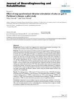
Position of subject and targets and pictorial indication of spa-tial sound for one targetFigure 1
Position of subject and targets and pictorial indica-
tion of spatial sound for one target. The position of the
targets relative to a subject with right hemiparesis is shown.
Six targets were positioned on the surface of the table and
three were on a removable support. Targets were arranged
on three lines (saggital and 30° to the left and to the right).
Thick arrows indicate movement distance. The intensity and
frequency of the sound in each ear depended on the direc-
tion and position of the hand relative to the target.
Journal of NeuroEngineering and Rehabilitation 2009, 6:45 />Page 6 of 11
(page number not for citation purposes)
Results
Movement kinematics
Graphs for the 3 kinematic variables analysed are pro-
vided in Figure 2a, b, c. Data for healthy subjects and the
two patient groups (RHD and LHD) are superimposed.
All data displayed in these figures was collected in the no-
feedback condition. Comparison of healthy subjects and
patients showed that peak hand velocity was much greater
in healthy subjects (p < 0.0001) and the number of veloc-
ity peaks (p < 0.0001) and curve index (p < 0.0001) were
much lower. Healthy subjects also displayed much less
variability. Peak hand velocity was scaled according to tar-
get distance in healthy subjects as well as in patients with
RHD and LHD, although to a lesser extent in the patient
groups.
Targets were grouped into 'near', 'far' and 'high' (distance
condition) and 'internal', 'middle' and 'external' (direc-
tion condition). Peak velocity increased significantly
between near and far (p < 0.0001) and near and high tar-
gets in healthy subjects and both patient groups (p <
0.0001) and also between high and far targets in the LHD
group (p = 0.005). Peak velocity was significantly higher
for external targets compared with internal (p < 0.0001),
and middle (p < 0.0001) in healthy subjects and both
patient groups (Figure 2a). There were no significant dif-
ferences between the target conditions for the number of
velocity peaks in healthy subjects or either of the patient
groups (Figure 2b). Trajectories were significantly less
curved for high compared with near targets (p = 0.003)
and high compared with far targets (p = 0.02) in healthy
subjects and for far compared with near targets for both
RHD (p = 0.006) and LHD groups (p = 0.0003) (Figure
2c). In the LHD group, trajectories to far targets were also
significantly less curved than to high targets (p = 0.002).
Trajectories were significantly more curved for external
compared to middle targets in healthy subjects (p = 0.02)
but were less curved in both patient groups for external
compared to internal targets (RHD p < 0.0001, LHD p <
0.0001)
LHD and RHD groups were compared by grouping all tar-
gets together. Peak velocity did not differ between groups
(p = 0.85). The RHD group had significantly more velocity
peaks than the LHD group (p = 0.004) and significantly
greater trajectory curvature (p < 0.0001).
Effect of feedback
In order to compare the effect of the simple and spatial
feedbacks on movement parameters, the percentage dif-
ference between the auditory feedback sessions and the
control (no feedback) sessions carried out on the same
day was calculated for each variable. The percentage dif-
ference relating to the simple feedback was then com-
pared with that relating to the spatial feedback. No
significant differences were found for any of the parame-
ters analysed. Mean and SD data for each kinematic varia-
ble in the different feedback conditions are presented in
table 4. Each subject was asked to describe the nature of
the sound after the sessions. Only one subject was aware
of the spatial nature of the feedback, he was a musician
(Subject 3).
Because there was no difference between the effects of the
different types of feedback, the data were pooled into a
'feedback' condition and a 'no-feedback' condition for
further analysis (Figure 3a, b, c). The addition of auditory
feedback had different effects on LHD and RHD groups.
Although peak velocity did not change significantly, a
generally beneficial effect was noted in the RHD group
Comparison of kinematic variables between subject groupsFigure 2
Comparison of kinematic variables between subject groups. Mean values and standard errors are presented (healthy
group = black triangles, RHD = red squares, LHD = blue open circles). All data are from the 'no-feedback' condition. a) peak
velocity b) number of velocity peaks c) curve index. Int = internal; mid = middle; ext = external. Kinematic performance of
healthy subjects was significantly better than patients and performance of LHD patients was significantly better than RHD
patients.
Journal of NeuroEngineering and Rehabilitation 2009, 6:45 />Page 7 of 11
(page number not for citation purposes)
with a significant decrease in the number of velocity peaks
(p = 0.0003) (Figure 3b) and a significant decrease in tra-
jectory curve index (p = 0.005) (Figure 4c). The opposite
effect was noted for the LHD group: significant decrease in
peak velocity (p < 0.0001) (Figure 3a), significant increase
in the number of peaks in the velocity curve (p < 0.0001)
(Figure 3b) and significant increase in curve index (p =
0.02) (Figure 3c).
We also examined individual responses to feedback to
check if patients in each group followed the same tenden-
cies (Figure 4). It appeared that the kinematic perform-
ance of the majority of LHD patients did worsen in the
presence of feedback while in the RHD group, patients
changed less except for one subject who was a musician
(subject 3 denoted by green dotted lines in Figure 4) who
had a much greater response to the feedback than the oth-
ers. When statistical analysis was repeated without this
patient, the effect of feedback on peak velocity remained
non-significant (p = 0.13), the decrease in the number of
velocity peaks was no longer significant but tended
towards significance (p = 0.067) and the decrease in curve
index remained significant (p = 0.005) (results in figures
and tables include all patients).
Dominant versus non-dominant hand
Since two of the patients in the RHD group were left-
handed, we examined individual data in order to deter-
mine if the differences in response to feedback between
the two groups were the result of individual differences
related to the use of dominant versus non dominant hand
or left versus right hemiparesis. The two left handed
patients in the RHD group (marked with arrows in Figure
4) used their dominant hands and had movement param-
eters at the lower level of the group, but the effect of feed-
back appeared similar to that observed in the other RHD
patients (no or little change) while movement parameters
of most LHD patients worsened (Figure 4).
Discussion
The main findings of this pilot study were that, despite
having similar functional capacity and similar movement
velocity, the RHD and LHD patients exhibited differences
in movement smoothness and curvature and showed
opposite responses to the feedback. Patients in the RHD
group showed a consistent improvement in curvature
with the addition of auditory feedback whereas patients in
the LHD group showed a consistent deterioration of all
movement parameters.
Table 4: Mean (SD) values of parameters evaluated in each condition. NF = no feedback
Same day Same day
NF Simple NF Spatial
LHD Peak velocity 0.73 ± 0.28 0.68 ± 0.25 0.70 ± 0.23 0.64 ± 0.23
N° vel. peaks 2.87 ± 1.42 3.54 ± 2.10 3.11 ± 1.59 3.53 ± 2.04
Curve index 1.10 ± 0.08 1.12 ± 0.09 1.10 ± 0.08 1.11 ± 0.10
RHD Peak velocity 0.70 ± 0.27 0.69 ± 0.26 0.67 ± 0.24 0.72 ± 0.27
N° vel. peaks 4.36 ± 2.66 3.92 ± 2.23 3.85 ± 2.23 3.31 ± 1.73
Curve index 1.17 ± 0.13 1.15 ± 0.13 1.16 ± 0.13 1.15 ± 0.12
Comparison of kinematic variables with and without auditory feedbackFigure 3
Comparison of kinematic variables with and without auditory feedback. Mean values and standard errors are pre-
sented (RHD = red squares, LHD = blue open circles). a) peak velocity b) number of velocity peaks c) curve index. * indicates
a significant difference between conditions for LHD group, # indicates a significant difference between conditions for RHD
group. The presence of feedback improved performance in the RHD group and degraded the LHD group.
Journal of NeuroEngineering and Rehabilitation 2009, 6:45 />Page 8 of 11
(page number not for citation purposes)
Kinematic characteristics
We observed low peak velocities, lack of smoothness and
increased curvature of the hand trajectories of the stroke
patients compared with the healthy subjects. This is in
agreement with previous studies [2,3]. Peak hand velocity
was scaled with target distance in both healthy subjects
and patients consistent with previous reports [21]. Peak
velocity was significantly higher for external compared
with internal targets in healthy subjects and patients. This
is likely to be related to the fact that the movement dis-
tance was greater to the external targets but it has also
been shown in healthy subjects that hands move faster in
their own hemispace [22,23]. In the patient groups, the
curve index was significantly lower for external than inter-
nal targets while the opposite was true for the healthy sub-
jects. Desmurget et al. [24] reported similar results in
healthy subjects, the curve index increased as targets
became more external. Perhaps this indicates a particular
control problem for hemiparetic patients in the internal
part of the workspace. Indeed, Levin [3] found greater dis-
ruption in shoulder-elbow coordination in hemiparetic
patients for internal targets compared with external tar-
gets.
To our knowledge, this is the first study to compare kine-
matics of the contralesional hand between left and right
hemispheric lesions. We found that smoothness and cur-
vature of the hand trajectory were different between the
LHD group and the RHD group. The LHD group had gen-
erally less curved and smoother movements than the RHD
group, denoting better kinematic movement quality. This
difference cannot be explained by different levels of
impairment between the groups since clinical scores and
peak hand velocity were not significantly different
between the groups. It is possible that the kinematic dif-
ferences between our groups reflect some specialization of
the lesioned cerebral hemisphere. We speculate that
movement smoothness and curvature may be predomi-
nantly controlled in the right hemisphere. This is indi-
rectly supported from findings in studies comparing hand
performance in healthy subjects [22,25] and ipsilesional
hand performance between left and right hemispheric
lesions [12,26,27]. The left hemisphere has been linked
with an open-loop form of control [26], specialized in the
control of limb dynamics [28,29] and temporal process-
ing [30]. The right hemisphere is believed to function in a
closed loop, specialised in the control of on-line visual
processing [26] and final limb posture. Right hemisphere
damage has been found to reduce final position accuracy
of the right hand while it does not reduce accuracy of the
left hand [12]. It seems likely that the kinematic differ-
ences we found between patients with LHD and RHD
reflect differences in hemispheric specialization. Further
study is warranted to confirm this.
The presence of apraxic patients within the LHD group
may be a confounding factor in this study. However, kin-
ematic errors tend to increase in apraxic patients with task
difficulty (decreased visual feedback and target size) [31]
while our task was simple requiring little precision.
Hermsdörfer et al. [32] also showed that errors linked to
apraxia were not correlated with kinematic errors. Also,
our LHD patients, including apraxic patients, demon-
strated better kinematics than the RHD group. Therefore
we consider it unlikely that presence of apraxia could
completely explain the kinematic differences found
between patient groups in this study.
Effect of feedback
The addition of auditory feedback had the opposite effect
in each group. The group mean for each variable analyzed
Individual kinematic data in RHD and LHD patient groups with and without feedbackFigure 4
Individual kinematic data in RHD and LHD patient groups with and without feedback. Comparison of condition
without (NF = no feedback) and with feedback (FB = feedback). Each shape represents an RHD and an LHD subject (see ID on
Figure legend). Arrows indicate the two left handed patients (subjects 1 and 5) and the dashed green line indicates one patient
who responded differently from his group (subject 3).
Journal of NeuroEngineering and Rehabilitation 2009, 6:45 />Page 9 of 11
(page number not for citation purposes)
improved in the RHD group while it deteriorated in the
LHD group when the feedback was added.
Visual analysis of individual responses to the feedback
(Figure 4) showed that one subject in the RHD group had
a much greater response than other patients in his group.
This patient was a musician and therefore had highly
developed auditory function. However, even when he was
excluded from the analysis, the results remained essen-
tially the same (although the decrease in number of veloc-
ity peaks then only tended to significance). Although
these improvements were not consistent across all param-
eters, they suggest that auditory feedback could be a useful
tool to improve movement kinematics in RHD patients.
However, this remains to be tested with different types of
feedback.
It is known that patients with aphasia can also have defi-
cits in non-verbal sound processing [33] and five of our
eight LHD patients had receptive aphasia which may
explain why the auditory feedback was disruptive in this
group. The particular characteristic of our feedback was
the sensation of motion. Several studies have demon-
strated specific deficits in motion detection or auditory
spatial localisation following right hemisphere lesions
which are linked with neglect [34]. However, on compar-
ison of deficits arising from lesions in opposite hemi-
spheres Adriani et al. [35] found no difference in sound
localisation or motion perception deficits between
patients with LHD or RHD. It is not possible to ascertain
if the detrimental effect of the feedback was linked to the
degree of aphasia because of we did not quantify the
degree of aphasia and too few LHD patients were included
for further subgroup analysis. Degree of aphasia may thus
be a confounding factor in this study and further investi-
gation into the relation between degree of aphasia and
reaching kinematics is indicated.
Degree of spatial attention deficits may be another possi-
ble explanation for different effects of feedback depending
on side of lesion. Our auditory feedback task could be
considered similar to a dual task since patients were
required to perform reaching movements while listening
to feedback. This may have had greater attentional
requirements than carrying out reaching movements
alone. Hyndman et al. [36], however found that patients
with RHD tended to have worse divided attention than
LHD so the dual task hypothesis does not seem to explain
the difference in our groups. However in the same study,
the LHD exhibited slightly worse auditory selective atten-
tion (patients were asked to count low tones while ignor-
ing high tones) at discharge than RHD (the differences
were trends). Perhaps the difference between our groups
is therefore related to the processing of the feedback itself.
Some evidence suggests that the left-hemisphere may be
superior with regard to on-line feedback processing dur-
ing goal-directed movements although this evidence
tends to come from studies using visual feedback [37].
Lesions of the left hemisphere may therefore disrupt feed-
back processing capacity.
In short, our results demonstrated a difference in the effect
of the auditory feedback depending on side of lesion. It is
not possible, however, to determine if this is related to a
difference in processing of auditory information or the
fact that each hemisphere has a different role in move-
ment control or an interaction of the two factors.
Lack of effect of spatial feedback
Our group previously showed that healthy subjects are
able to use spatial feedback to locate unseen static virtual
objects using spatial feedback [38]. We therefore hoped
that this unusual feedback could be integrated online in a
similar manner to visual feedback [39] and guide hand
orientation in patients. We suggest three explanations for
the lack of effect of the spatial nature of the feedback.
1) It is possible that the directional component of our spa-
tial feedback was either too complex or too subtle to be
integrated implicitly in patients with cerebral lesions.
However, a similar type of spatial feedback was success-
fully used for sensory substitution in patients with vestib-
ular disorders [40] but in this study the patients were
aware of the nature of the feedback and they did not have
cerebral lesions.
2) Perhaps auditory feedback is poorly adapted for spatial
guidance of the hand. It has previously been shown that
subjects rely more on visual than proprioceptive feedback
and adjust movement trajectories so as to ensure visually
constant movements [41]. We allowed use of vision since
we wanted to assess an augmented feedback, not a substi-
tution. However, the auditory feedback may have been
superfluous if patients gained sufficient intrinsic feedback
(visual and proprioceptive). Auditory feedback may be
better adapted for temporal guidance, such as in that
developed by Huang et al. [10] (described in the introduc-
tion), since temporal parameters are well coded in the
auditory cortex while the visual cortex codes predomi-
nantly spatial information.
3) Although moving sounds can be detected by a single
hemisphere, for accurate discrimination of sound motion,
interaction between both hemispheres may be necessary
for the interpretation of interaural differences [34]. In
patients with cerebral lesions of one hemisphere, capacity
to process moving sounds might be reduced. Indeed, only
one of the sixteen patients actually became aware of the
spatial nature of the sound, he was a musician.
Journal of NeuroEngineering and Rehabilitation 2009, 6:45 />Page 10 of 11
(page number not for citation purposes)
Conclusion, limitations and perspectives
Studies of stroke patients usually restrict subject inclusion
to right handed patients with left hemisphere damage or
they do not make comparisons between patient groups.
Until now, no study has compared the effect of feedback
in patients with left versus right hemisphere damage. We
found that patients with left hemisphere damage made
smoother, less curved movements than patients with right
hemisphere damage despite having a similar level of
impairment and peak hand velocity. The kinematic per-
formance of the LHD group was degraded by the presence
of auditory feedback while that of the RHD group was not.
These results demonstrate a need for thorough investiga-
tion of differences in motor abilities in a variety of envi-
ronments and conditions between patients with left and
right hemisphere lesions before developing complex reha-
bilitation methods such as virtual reality.
It is important to note, however, that the small sample
size and heterogenous population, including patients
with neuropscychological deficits mean that our results
must be interpreted with caution. Equally, the presence of
2 left-handed patients within the RHD group may have
confounded the results although the role of each hemi-
sphere may be independent of hand preference [22,25].
In stroke patients, auditory feedback may not be suitable
for the provision of knowledge of performance when dis-
crimination of features of the sound is necessary. The
manner in which different aspects of sound are processed
is not yet fully understood and the presence of cerebral
lesions may render perception of changes in sound diffi-
cult for patients. Perhaps visual feedback is a more appro-
priate mode of provision of knowledge of performance of
spatial aspects during hand movement while auditory
feedback may be better adapted for the provision of tem-
poral information or knowledge of results or to warn of
errors. Further study is indicated in the use of auditory
feedback in stroke patients.
Competing interests
The authors declare that they have no competing interests.
Authors' contributions
JVGR and ARB participated in the conception and design
of the protocol, analysis and interpretation of data and
drafting the article, TH and SH participated in the concep-
tion and design of the protocol and created the feedback,
PL and DB were involved in data interpretation and
helped to draft the article. All authors gave final approval
of the version submitted.
Acknowledgements
Agnès Roby-Brami is supported by INSERM.
This project was supported by national clinical research project funding
(PHRC): 'Comprendre et reduire le handicap moteur' (Understanding and
reducing motor handicap).
We wish to thank all the subjects who kindly participated in the study.
References
1. Jorgensen HS, Nakayama H, Raaschou HO, Vive-Larsen J, Stoier M,
Olsen TS: Outcome and time course of recovery in stroke.
Part I: Outcome. The Copenhagen Stroke Study. Arch Phys
Med Rehabil 1995, 76:399-405.
2. Roby-Brami A, Feydy A, Combeaud M, Biryukova EV, Bussel B, Levin
MF: Motor compensation and recovery for reaching in stroke
patients. Acta Neurol Scand 2003, 107:369-381.
3. Levin MF: Interjoint coordination during pointing movements
is disrupted in spastic hemiparesis. Brain 1996, 119:281-293.
4. Michaelsen SM, Dannenbaum R, Levin MF: Task-specific training
with trunk restraint on arm recovery in stroke: randomized
control trial. Stroke 2006, 37:186-192.
5. Schmidt R, Wrisberg C: Motor learning and performance Leeds, Eng-
land: Human Kinetics; 2004.
6. van Vliet PM, Wulf G: Extrinsic feedback for motor learning
after stroke: what is the evidence? Disabil Rehabil 2006,
28:831-840.
7. Cirstea CM, Ptito A, Levin MF: Feedback and cognition in arm
motor skill reacquisition after stroke. Stroke 2006,
37:1237-1242.
8. Cirstea MC, Levin MF: Improvement of Arm Movement Pat-
terns and Endpoint Control Depends on Type of Feedback
During Practice in Stroke Survivors. Neurorehabil Neural Repair
2007, 21:398-411.
9. Maulucci RA, Eckhouse RH: Retraining reaching in chronic
stroke with real-time auditory feedback. NeuroRehabilitation
2001, 16:171-182.
10. Huang H: Interactive multimodal biofeedback for task-orien-
tated neural rehabilitation. 27th Annual International Conference of
the IEEE Engineering in Medicine and Biology Society Shanghai
2005:2547-2550.
11. Auvray M, Hanneton S, O'Regan JK: Learning to perceive with a
visuo-auditory substitution system: localisation and object
recognition with 'the vOICe'. Perception 2007, 36:416-430.
12. Schaefer SY, Haaland KY, Sainburg RL: Ipsilesional motor deficits
following stroke reflect hemispheric specializations for
movement control.
Brain 2007, 130:2146-2158.
13. Alain C, He Y, Grady C: The contribution of the inferior pari-
etal lobe to auditory spatial working memory. J Cogn Neurosci
2008, 20:285-295.
14. Lee JH Van der, De Groot V, Beckerman H, Wagenaar RC, Lankhorst
GJ, Bouter LM: The intra- and interrater reliability of the
action research arm test: a practical test of upper extremity
function in patients with stroke. Arch Phys Med Rehabil 2001,
82:14-19.
15. Platz T, Pinkowski C, van Wijck F, Kim IH, di Bella P, Johnson G: Reli-
ability and validity of arm function assessment with stand-
ardized guidelines for the Fugl-Meyer Test, Action Research
Arm Test and Box and Block Test: a multicentre study. Clin
Rehabil 2005, 19:404-411.
16. Wade DT, Collin C: The Barthel ADL Index: a standard meas-
ure of physical disability? Int Disabil Stud 1988, 10:64-67.
17. Goodglass H, Kaplan E: Boston diagnostic aphasia examination Philidel-
phia: Williams & Wilkins; 1983.
18. Gauthier L, Dehaut F, Joanett J: The Bell Test: A quantitative and
qualitative test for visual neglect. International Journal of Clinical
Neuropsychology 1989, 11:49-54.
19. Group I A S I: 3D audio rendering and evaluation guidelines Los Angeles
CA: MIDI Manufacturers Association Incorporated; 1998.
20. Cirstea MC, Levin MF: Compensatory strategies for reaching in
stroke. Brain 2000, 123(Pt 5):940-953.
21. Roby-Brami A, Fuchs S, Mokhtari M, Bussel B: Reaching and grasp-
ing strategies in hemiparetic patients. Motor Control 1997,
1:72-91.
22. Boulinguez P, Nougier V, Velay JL: Manual asymmetries in reach-
ing movement control. I: Study of right-handers. Cortex 2001,
37:101-122.
Publish with BioMed Central and every
scientist can read your work free of charge
"BioMed Central will be the most significant development for
disseminating the results of biomedical research in our lifetime."
Sir Paul Nurse, Cancer Research UK
Your research papers will be:
available free of charge to the entire biomedical community
peer reviewed and published immediately upon acceptance
cited in PubMed and archived on PubMed Central
yours — you keep the copyright
Submit your manuscript here:
/>BioMedcentral
Journal of NeuroEngineering and Rehabilitation 2009, 6:45 />Page 11 of 11
(page number not for citation purposes)
23. Hodges NJ, Lyons J, Cockell D, Reed A, Elliott D: Hand, space and
attentional asymmetries in goal-directed manual aiming.
Cortex 1997, 33:251-269.
24. Desmurget M, Jordan M, Prablanc C, Jeannerod M: Constrained
and unconstrained movements involve different control
strategies. J Neurophysiol 1997, 77:1644-1650.
25. Boulinguez P, Velay JL, Nougier V: Manual asymmetries in reach-
ing movement control. II: Study of left-handers. Cortex 2001,
37:123-138.
26. Winstein CJ, Pohl PS: Effects of unilateral brain damage on the
control of goal-directed hand movements. Exp Brain Res 1995,
105:163-174.
27. Bagesteiro LB, Sainburg RL: Nondominant arm advantages in
load compensation during rapid elbow joint movements. J
Neurophysiol 2003, 90:1503-1513.
28. Haaland KY, Schaefer SY, Knight RT, Adair J, Magalhaes A, Sadek J,
Sainburg RL: Ipsilesional trajectory control is related to cont-
ralesional arm paralysis after left hemisphere damage. Exp
Brain Res 2009, 196(2):195-204.
29. Bagesteiro LB, Sainburg RL: Handedness: dominant arm advan-
tages in control of limb dynamics. J Neurophysiol 2002,
88:2408-2421.
30. Haaland KY, Prestopnik JL, Knight RT, Lee RR: Hemispheric asym-
metries for kinematic and positional aspects of reaching.
Brain 2004, 127:1145-1158.
31. Haaland KY, Harrington DL, Knight RT: Spatial deficits in ideomo-
tor limb apraxia. A kinematic analysis of aiming movements.
Brain 1999, 122(Pt 6):1169-1182.
32. Hermsdorfer J, Blankenfeld H, Goldenberg G: The dependence of
ipsilesional aiming deficits on task demands, lesioned hemi-
sphere, and apraxia. Neuropsychologia 2003, 41:1628-1643.
33. Saygin AP, Dick F, Wilson SM, Dronkers NF, Bates E: Neural
resources for processing language and environmental
sounds: evidence from aphasia. Brain 2003, 126:928-945.
34. Ducommun CY, Murray MM, Thut G, Bellmann A, Viaud-Delmon I,
Clarke S, Michel CM: Segregated processing of auditory motion
and auditory location: an ERP mapping study. Neuroimage
2002, 16:76-88.
35. Adriani M, Maeder P, Meuli R, Thiran AB, Frischknecht R, Villemure
JG, Mayer J, Annoni JM, Bogousslavsky J, Fornari E, Thiran JP, Clarke
S: Sound recognition and localization in man: specialized cor-
tical networks and effects of acute circumscribed lesions. Exp
Brain Res 2003, 153:591-604.
36. Hyndman D, Pickering RM, Ashburn A: The influence of attention
deficits on functional recovery post stroke during the first 12
months after discharge from hospital. J Neurol Neurosurg Psychi-
atry 2008, 79:656-663.
37. Keulen RF, Adam JJ, Fischer MH, Kuipers H, Jolles J: Distractor
interference in selective reaching: effects of hemispace,
movement direction, and type of movement. Cortex 2007,
43:531-541.
38. Hoellinger T, Auvray M, Roby-Brami A, Hanneton S: Localisation
tasks with a three-dimensional audio-motor coupling based
on an electromagnetic motion capture device. International
Multisensory Research Forum: July 16-19. Hamburg, Germany 2008.
39. Pisella L, Grea H, Tilikete C, Vighetto A, Desmurget M, Rode G, Bois-
son D, Rossetti Y: An 'automatic pilot' for the hand in human
posterior parietal cortex: toward reinterpreting optic
ataxia. Nat Neurosci 2000, 3:729-736.
40. Dozza M, Horak FB, Chiari L: Auditory biofeedback substitutes
for loss of sensory information in maintaining stance. Exp
Brain Res 2007, 178:37-48.
41. Wolpert DM, Ghahramani Z, Jordan MI: Are arm trajectories
planned in kinematic or dynamic coordinates? An adaptation
study. Exp Brain Res 1995, 103:460-470.