báo cáo hóa học: "Kinematic aspects of trunk motion and gender effect in normal adults" pdf
Bạn đang xem bản rút gọn của tài liệu. Xem và tải ngay bản đầy đủ của tài liệu tại đây (315.75 KB, 7 trang )
RESEARC H Open Access
Kinematic aspects of trunk motion and gender
effect in normal adults
Chin Youb Chung
1
, Moon Seok Park
1
, Sang Hyeong Lee
1
, Se Jin Kong
2
, Kyoung Min Lee
1*
Abstract
Background: The purpose of this study was to analyze kinematic trunk motion data in normal adults and to
investigate gender effect.
Methods: Kinematic trunk motion data were obtained for 20 healthy subjects (11 men and 9 women; age from 21
to 40 years) during walking a 9 m long lane at a self selected speed, namely, motions in the sagittal (tilt), coronal
(obliquity), and transverse (rotation) planes, which were all expressed as motions in global (relative to the groun d)
and those in pelvic reference frame (relative to pelvis), i.e., tilt (G), obliquity (G), rotation (G), tilt (P), obliquity (P),
rotation (P).
Results: Range of tilt (G), obliquity (G) and rotation (G) showed smaller motion than that of tilt (P), obliquity (P)
and rotation (P), respectively. When genders were compared, female trunks showed a 5 degree more extended
posture during gait than male trunks (p = 0.002), which appeared to be caused by different lumbar lordosis.
Ranges of coronal and transverse plane motion appeared to be correlated. In gait cycle, the trunk motion
appeared to counterbalance the lower extremity during swing phase in sagittal plane, and to reduce the angular
velocity toward the contralateral side immediate before the contralateral heel strike in the coronal plane.
Conclusions: Men and women showed different lumbar lordosis during normal gait, which might be partly
responsible for the different prevalence of lumbar diseases between genders. However, this needs further
investigation.
Background
Trunk motion has not attracted much attention from
those interested in three dimensional gait analysis,
because this motion is relatively small and is gen erally
thought to be passive and to depend on lower extremity
motion. However, some recent studies have shown that
trunk posture and motion can influence gait patterns of
the lowe r extremity [1] and alter energy expenditure in
the pathologic gait compared to a normal gait [2].
Moreover, the role of tr unk motion in b alanc e and pro-
prioceptive function in gait [3,4] is being investigated by
studying pathologic gait in patients with neurological,
vestibular, or musculoskeletal diseases [5-7]. However,
three dimensional gait analysis studies that have f ocus-
ing on normal trunk m otion have been somewhat
limited, and as far as we know, no study has examined
gender associated differences in trunk motion. We
undertook this study to identify the kinematic aspects of
normal trunk motion using three dimensional gait ana-
lysis and to determine whether the trunk motions of
men and women are different, w hich may provide us
with a possible explanatory clue for the different preva-
lences of spinal diseases between genders [8,9].
Methods
Inclusion criteria and data acquisition from three
dimensional gait analysis
This study was approved by the institutional review
board at our institute. Healthy adult volunteers, without
musculoskeletal, neurological or cardiopulmonary disor-
ders that could potentially have affected normal gait,
were included in this study. Anthropometric parameters,
such as, height, weight and BMI were recorded. Those
volunteers who deviated from the population norms
(<3% or >97%, SD 1.88) for height and weight were
excluded, as were those with a BMI >27 kg/m
2
or <18
kg/m
2
. Pelvic markers and trunk markers were attached
* Correspondence:
1
Department of Orthopedic Surgery, Seoul National University Bundang
Hospital, 300 Gumi-Dong, Bundang-Gu, Sungnam, Kyungki 463-707, Korea
Chung et al. Journal of NeuroEngineering and Rehabilitation 2010, 7:9
/>JNER
JOURNAL OF NEUROENGINEERING
AND REHABILITATION
© 2010 Chung et al; licensee BioMed Central Ltd. This is an Open Access article distributed under the terms of the Creative Commons
Attribution License (http://creativecom mons.org/licenses/by/2.0), which permits unrestricted use, distribution, and reproduction in
any medium, provided the original work is properly cited.
to volunteers, as follows. Three pelvic markers were
placed on the right ASIS (anterior superior iliac spine),
left ASIS, and sacrum in the middle of left and right
PSIS (posterior superior iliac spine), respectively, and
four trunk markers were located on the spinous process
of the 7
th
cervical vertebra, the spinous process of the
10
th
thoracic vertebra, the jugular notch where clavicles
meet the sternum, and at the xiphoid process of the
sternum, respectively. Additional foot and ankle markers
were placed to acquire data on gait cycles a nd walking
speeds. Marker placement was performed as described
for the Plug In Gait model (Vicon Motion Systems)
[10], and was done by a single experience d operator. All
subjects walked with bare feet along a 9 mete r long
straight lane at a self-selected speed with markers
attached. Seven VICON CCD cameras (Oxford Metrics,
Oxford, England) captured marker movements at a sam-
pling rate of 60 Hz, and three trials were averaged to a
single data set. For each trial (9-meter walk) one gait
cycle, which was not in the initial step or in last step,
was selected by one author. Three g ait cycles selected
from three trials were averaged to a gait cycle for one
person, and the kinematic gait data was retrieved from
the averaged gait cycle. The gait information obtained
was processed using VICON Workstation (Version 3.1,
Oxford Metrics, Oxford, England) in which Euler angle
[11] was employed for the kinematic data. To display
gait data, one gait cycle was represented using a 100%
scale and the angular values of motions were collected
at 2% intervals. The gait cycle was defined as an interval
from one heel contact to the next contact made by the
same heel; heel strike and toe off information was also
recorded. Kinematic trunk motion data were presented
for the sagittal, coronal and transverse planes, which
were defined as tilt, obliquity, and rotation, respectively.
For all subjects, both trunk motion in the global refer-
ence frame (motion (G), i.e., to the ground) and trunk
motion in the pelvic reference frame (motion (P), i.e.,
relative to the plane defined by the three pelvic markers)
were obtained [7,12]. We referred to motions in the
three planes in those two reference frames as tilt (G),
obliquity (G), rotation (G), tilt (P), obliquity (P) and
rotation (P). Positive angular values were defined for
forward bending in tilt, bending to the ipsilateral side in
obliquity, external rotation in rotation; negative values
represent the opposite movements, where the angular
definition of movement in the global reference frame
was converted to the opposite direction of the Euler
angle [11] for a better understanding. The kinematic
and basic gait data such as walking speed, cadence, and
stride length were obtained separately for the right and
left sides, and overall 40 sets of data were included for
statistical analysis. Basic gait data were normalized by
ad hoc normalization [13], where the data were divided
by leg length or square root of leg length. Variables,
such as, mean and range of trunk motion were recorded
in all planes. To describe relative phase movements, we
determined points of percentage in the gait cycle [14]
when movement angular values were at a maximum or
minimum.
Sample size estimation and Statistical analysis
Prior sample size estimation was performed. When we
assumed 5 degrees of difference between genders was
significant and set standard deviations to be 2.5 degrees,
sample size was ca lculated to be 8 subjects in each gen-
der group (a-error 0.05, b-error 0.8).
Descriptive analysis was performed separately for all
sets of data in all motion planes. Kinematic trunk
motion data in global and pelvic reference frames were
compared using the paired t-test or Wilcoxon’ ssigned
rank test depending on data set normality which was
determined using Kolmogorov-Smirnov test. Analysis of
covariance (ANCOVA) was performed to compare the
kinematic variables between genders. Correlations
between the trunk motion variables were evaluated
using Pearson’s or Spearman’s correlation tests. Statisti-
cal significance wa s accepted for P values of < 0.05
except for the correlation test which was adjusted for
family wise error. All statistical analyses were c arried
out using SPSS 11.0 (SPSS, Chicago, Illinois, USA).
Results
Twenty volunteers were recruited for this study. Of the
volunteers, 11 were male and nine were female. BMIs
ranged from 18.4 kg/m
2
to 26.5 kg/m
2
. The heights,
weights and BMIs of all subjects were between the 3
and 97 percentiles. Heights, weights, and BMIs were
different between genders although ages were not signif-
icantly different. Walking speeds were not significantly
different between genders, while normalized walking
speeds showed s ignificant difference b etween genders
(p = 0.025) (Table 1).
Table 1 Anthropometric Data and Walking Speeds
Male
(N = 11)
Female
(N = 9)
Difference P value
Age (years) 31.9 (6.4) 28.6 (5.5) 3.3 0.230
Height (cm) 169.5 (3.9) 160.8 (4.4) 8.7 <0.001
Weight (kg) 68.9 (5.7) 54.4 (6.1) 14.5 <0.001
BMI (kg/m
2
) 24.0 (1.4) 21.1 (2.8) 2.9 <0.001
Walking speed (m/sec) 1.18 (0.06) 1.21 (0.09) 0.03 0.240
Walking speed/√L
0
1.27 (0.07) 1.34 (0.12) 0.07 0.025
Cadence (No./min) 107.6 (4.3) 114.5 (7.6) 6.9 0.002
Cadence × √L
0
100.2 (3.0) 103.8 (6.5) 3.6 0.039
Stride length (m) 1.31 (0.06) 1.26 (0.06) 0.05 0.018
Stride length/L
0
1.51 (0.08) 1.54 (0.10) 0.03 0.369
L
0
: Leg length
Chung et al. Journal of NeuroEngineering and Rehabilitation 2010, 7:9
/>Page 2 of 7
Normal values of trunk motion, comparison between
trunk motion in pelvic reference frame versus global
reference frame
Mean tilt (P) was about 1 0 degrees l ess than mea n tilt
(G), suggesting that the pelvis was anteriorly tilted at 10
degr ees in the sagittal plane during gait. Mean obliquity
and rotation were near 0 degrees accor ding to both pel-
vic and global reference frames whilst walking, as was
expected. Ranges of motions in global reference frame
were smaller than those in pelvic reference frame. Range
of rotation (P) was greatest and range of tilt (P) was
smallest for motions in the pelvic reference frame. In
terms of motions in the global reference frame, range of
rotation (G) was the largest and range of obliquity (G)
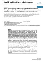
was the smallest (Table 2). In terms of relative phasic
motion i n gait cycle curves, tilt (P) and tilt (G) showed
near reciprocal movement, obliquity (P) and obliquity
(G) were synchronous, and rotation (P) followed rota-
tion (G), which was delayed by 15% of the gait cycle
(Figure 1).
Comparisons between men and women
The most prominent result was observed in the sagittal
plane. Both mean tilt (P) and mean tilt (G) of women
were about 5 degrees less than those of men, meaning a
more extended trunk posture in women (P = 0.002).
Ranges of tilt (P) and tilt (G) were not significantly dif-
ferent between gender. Range of obliquity (P) in women
was larger than in men (P = 0.026), but no significant
difference in obliquity (G) was observed between men
and women, which concurred with the result of a pre-
vious study which suggested larger coronal mot ion of
the female pelvis than male [15]. These results are
detailedinTable3.Nodifferenceinrelativephase
motion was observed between men and women. For the
significantly different variables between genders
(Table 3), an ANCOVA test was performed to exclude
the confounding effect of the different BMI and normal-
ized walking speed between genders (Table 1). The fixed
factor was gender, and the covar iates were the BMI and
normalized speed. The dependent variables were tilt (P),
tilt (G), and the range of obliquity (P). The kinematic
data was found to have an equality of error variances on
the Levene’s test. Tilt (G) was significantly different
between genders (p < 0.001) after excluding the effects
of the normalized walking speed (p = 0.132) and BMI
(p = 0.147) on the ANCOVA test. Tilt (P) was similar in
both genders (p = 0.415), while the normalized walking
speed (p = 0.004) and BMI (p = 0.040) had a significant
effect on tilt (P). The range of obliquity (P) was found
to be affected significant ly by the normalized walking
speed (p = 0.004), gender (p = 0.026), and BMI
(p = 0.039).
Correlation between motion planes in trunk motion
Trunk motion (G) tended to be correlated w ith its
counterpart trunk motion (P). Range of rotation (P) and
range of obliquity (P) were found to be correlated
(r = 0.617; P < 0.001), as were range of rotation (P) and
range of obliquity (G) (r = 0.610; P <0.001)(Table4).
Therefore range of trunk motion in c oronal plane was
correlated with that in transverse plane. In the correla-
tion test, the numbe r of pa irs by w hich the alpha-error
was devided was 15. Therefore, the statistical signifi-
cance was set to P < 0.003, which was adjusted for
family wise error.
Discussion
Trunk motions to the ground showed narrow ranges in
all three planes, whereas trunk motions relative to the
pelvis tended to be larger than those to the ground,
which concurs with the results of previous studies
[14,16]. Women’ strunksshowed5degreesmore
extended posture during gait than men’s trunks. Range
of trunk motion in coronal plane appeared to be corre-
lated with range of trunk motion in transverse plane.
This study has some limitations that require consid-
eration. First t he number of cases was quite small and
the generalization of our results requires c onfirmation
by further study although prior sample size was calcu-
lated in this study. Second, our trunk model did not
take the intra-truncal movement into account, and con-
sidered the trunk to be a rigid segment. The lumbar
lordosis was not actually measured but calculated. The
motion or posture that we considered lumbar lordosis
might have originate d in part from the intratruncal
movement. Third, there were significant variations in
the subjects’ height and weight, which could affect the
basic gait data. Fourth, the normalized walking speed
was different between genders, which could be a con-
founding factor when comparing the gender differences
even though we performed a ANCOVA test to excluded
the different effects of BMI and normalized walking
speed between gen ders. Fifth, the small differences
between groups were statistically significant. However,
Table 2 Comparison of Trunk motion (P) vs Trunk motion
(G) in degrees
Motion Value Trunk motion
(P)
Trunk motion
(G)
Difference P
value
Trunk Mean -10.2 (5.5) -0.2 (3.6) 10.0 <0.001
Tilt Range 4.7 (2.2) 4.0 (1.8) 0.7 0.004*
Trunk Mean -0.0 (2.3) -0.0 (1.3) 0.0 0.985
Obliquity Range 13.0 (4.5) 3.3 (1.4) 9.7 <0.001
Trunk Mean 0.1 (2.3) -0.1 (2.1) 0.2 0.738
Rotation Range 13.7 (4.9) 6.9 (2.9) 6.8 <0.001
*, nonparametric method (Wilcoxon signed rank test)
Chung et al. Journal of NeuroEngineering and Rehabilitation 2010, 7:9
/>Page 3 of 7
these results might have been caused by variabilities o f
marker placement at l east in part, an d care should be
taken when interpreting the clinical implications.
Posterior tilting of the trunk (T ilt (G) graph in Figure
1) begins with the initiation of the single limb support
phase (gait cyle 10%, 60%), which is approxima tely the
opposite movement of lower extremity during swing
phase. It appears that sagittal trunk motion counterba-
lances the lower extremity during the single limb sup-
port phase. On the other hand, the trunk started to
bend anteriorly from just before heel strike through the
double limb support phase, which appears to enhance
forward progression when the body is st abilized by dou-
ble support. Trunk motion in the sagitt al plane is two
repetitive movement and each shape of the two motions
in one gait cycle seems quite similar (Tilt (P) and Tilt
(G) graph in Figure 1), which was also shown in other
Figure 1 Trunk motions in three planes. In each graph, the transverse axis represents the phase of the gait cycle as percentages of gait cycle
and the vertical axis represents angular values. The graphs depict trunk motion in each plane using global and pelvic reference frames. Relative
phase of motions between two reference frames were almost reciprocal in the sagittal plane, synchronous in the coronal plane, and 15%
different phase in the transverse plane. Note two repetitive motions in tilt (G) and tilt (P), and the slight differences between maxima (asterisks)
and minima (arrow heads) during first and second motions, which are believed to be influenced by motions of other planes. The bars on the
transverse axis represent double limb support phases.
Table 3 Comparison between Male (N = 11, 22 sides) and
Female (N = 9, 18 sides) trunk motions (in degrees)
Motion Value Male Female Difference P value
Trunk Mean -7.8 (5.0) -13.0 (4.9) 5.2 0.002
Tilt (P) Range 4.4 (2.4) 5.0 (1.7) 0.6 0.373
Trunk Mean -0.1 (2.6) -0.0 (2.1) 0.0 0.954
Obliquity (P) Range 11.6 (4.0) 14.8 (4.6) 3.1 0.026
Trunk Mean 0.2 (2.2) 0.0 (2.4) 0.2 0.821
Rotation (P) Range 13.9 (5.2) 14.1 (4.7) 0.2 0.598
Trunk Mean 2.3 (2.4) -3.1 (2.2) 5.4 <0.001
Tilt (G) Range 4.0 (2.4) 3.9 (0.7) 0.1 0.922*
Trunk Mean -0.0 (1.2) 0.0 (1.5) 0.0 0.909
Obliquity (G) Range 3.5 (1.4) 3.1 (1.3) 0.3 0.431
Trunk Mean -0.1 (2.1) -0.0 (2.2) 0.1 0.914
Rotation (G) Range 6.3 (1.7) 7.6 (3.8) 1.3 0.202
*, nonparametric method (Mann-Whitney test)
Chung et al. Journal of NeuroEngineering and Rehabilitation 2010, 7:9
/>Page 4 of 7
studies [4,16]. However, despite the similar shapes of the
two repetitive motions, their angular values are slightly
different (asterisks and arrow heads in Tilt (P) and Tilt
(G) of Figure 1), because different rotation or obliquity
positions caused different positions in sagittal plane.
Indeed, the same degree of sagittal tilt would appear
smaller than the real value in some degrees of axial
rotation, and appear larger in some degrees of coronal
obliquity if the rotation and obliquity were between 0
and 90 degrees. This has some implications when kine-
matic trunk motion data is measured or analyzed,
because if the phases of gait cycles or motions in other
planes are not considered at the same time, kinematic
data could be distorted.
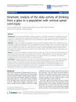
At the curve of obliquity (G) (Figure 2), the trunk
starts to bend contralaterally right after the single limb
support phase commenced (gait cycle 13%). During this
coronal motion bending to the contralateral side, t here
is slightly lowered angular velocity portion (Figure 2)
just before heel strike of the opposite foot (gait cycle
50%), which appears to be an effort to reduce the
impact from the heel strike.
Rotational motion in the transverse plane showed the
largest motion range in both the pelvic and global refer-
ence frames. A relative phase difference of 15% was
observed between rotation (P) and rotation (G), which
might be a means of conserving angular momentum, as
was described in a previous study [17]. According to
other studies [18,19], t he rotational motion of trunk
played an important role in adapting to the cha nges in
walking speed. However, in this study, there was no ten-
dency or changes in rotational motion according to the
walking speed, which might be due to the relatively nar-
rower range of walking speed than those of other studies.
Table 4 Correlation coefficients between Ranges of Trunk Motions
Tilt (P) Obliquity (P) Rotation (P) Tilt (G) Obliquity (G) Rotation (G)
Tilt (P)
Obliquity (P) 0.054
Rotation (P) 0.065 0.617*
Tilt (G) 0.748* -0.322 -0.226
Obliquity (G) 0.412 0.495* 0.610* 0.131
Rotation (G) 0.066 0.008 0.346 0.105 -0.033
*, P < 0.003 (P-value adjusted for famil y wise error)
Figure 2 Trunk motion in the coronal plane. After beginning the single limb support phase (a), the trunk moves to the contralateral side (f
and s). This motion decelerates slightly (s) while approaching the heel strike of the opposite foot (c, gait cycle 50%), which appears to be an
effort to reduce the impact of the heel strike. The difference between the slopes of f and s represents the difference in angular velocity.
Chung et al. Journal of NeuroEngineering and Rehabilitation 2010, 7:9
/>Page 5 of 7
On comparing the genders, no significant difference in
self-selected walking speed was observed. However, after
normalization, each gender show ed a significantly differ-
ent walking speed and cadence (Table 1). Therefore,
ANCOVA test was performed to exclude the confound-
ing effect of the different normalized walking speed and
BMI between genders. The mean tilt (G) appeared to be
influenced significantly by gender after eliminating the
confounding effect of the normalized walking speed and
BMI, while mean til t (P) was significantly affected by
normalized walking speed and BMI. The range of obli-
quity (P) appeared to be influenced significantly by gen-
der, normalized walking speed and BMI. Therefore, after
excluding the confounding effect of the normalized
walking speed and body size, the most prominent gen-
der difference in the kinematic data of trunk motion is
believed to be the more extended trunk posture in
women, which is r epresented by the mean tilt (G). Dur-
ing normal gait, women’s trunks were approximately 5
degrees more extended posture than men’s. A previous
study [15] suggested that the female pelvis is more ante-
riorly tilted throughout the gait cycle, but our data
showed no significant difference in mean pelvic tilt
between men (mean 10.10°, SD 3.47°) and women
(mean 9.89°, SD 3.82°). Therefore we believe that the 5
degrees of differ ence in trunk tilt betw een men and
women c ame from the different lumbar lordosis , which
means that women have 5 degrees more lumbar lordosis
than men. This might explain the different prevalence of
lumbar diseases [8,9] between genders in part through
further investigation, but this topic is beyond the scope
of this study.
The range of rotation (P) showed some relationship
with the obliquity (P) and obliquity (G) (correlation
coefficient, 0.617 and 0.610, respectively) (Table 4). We
consider that sagittal trunk motion was more indepen-
dent than the other tw o plane motions, and coronal
motion and transverse p lane motion are possibly inter-
connected in three dimensional space. This concurs
with the findings of a previous study, in which coupling
between lateral bending and axial rotation of the lumbar
spi ne wa s suggested [20]. The vector of the spinal mus-
cles or axis of lumbar spinal joint might explain the cor-
relation between the coronal trunk motion a nd
transverse trunk motion, but more study will be needed
to better understand this result.
In the present study, we mainly focused on kinematic
trunk motion data. More comprehensive studies, which
include o ther body parts, kinetic data, EMG, and varia-
tions in walking speed, are recommended before we are
able to understand trunk motion better. Additionally, it
should be noted that some of the results of the present
study differ from those of previous studies because of
different numbers of cases, walking conditions (treadmill
vs. ground walking) [21], trunk marker protocols, equip-
ment, definitions of positive angular values of motion.
Conclusions
Women showed 5 degrees more extended trunk posture
during gait than men, which appeared to be caused by
diff erent lumb er lordosis. This different lumbar lordosis
could possibly explain the different prevalences of lum-
bar diseases between gender, which needs further inves-
tigation. Coronal trunk motion and transverse trunk
motion were correlated. Kinematic trunk motion sug-
gested its role to counterbalance the lower extremity
during swing phase in sagittal plane and to reduce the
angula r velocity toward the contralateral side immediat e
before the contralateral heel strike in the coronal plane.
Acknowledgements
The authors thank Mi Seon Ryu for data collection.
This study was conducted at Seoul National University Bundang Hosptial.
Author details
1
Department of Orthopedic Surgery, Seoul National University Bundang
Hospital, 300 Gumi-Dong, Bundang-Gu, Sungnam, Kyungki 463-707, Korea.
2
DooRee Motion Research Center, 223-17 Jamsilbon-Dong, Songpa-Gu,
Seoul, 138-863, Korea.
Authors’ contributions
All authors were fully involved in the study and preparation of the
manuscript. Each of the authors has read and concurs with the content in
the final manuscript. Nobody who qualifies for authorship has been omitted
from the list.
Competing interests
No benefits in any form have been received or will be received from a
commercial party related directly or indirectly to the subject of this article.
Received: 17 May 2009 Accepted: 15 February 2010
Published: 15 February 2010
References
1. Saha D, Gard S, Fatone S: The effect of trunk flexion on able-bodied gait.
Gait & Posture 2008, 27:650-660.
2. Nankaku M, Tsuboyama T, Kakinoki R, Kwanabe K, Kanzaki H, Mito Y,
Nakaura T: Gait analysis of patients in early stages after total hip
arthroplasty: effect of lateral trunk displacement on walking efficiency.
Journal of Orthopaedic Science 2007, 12:550-554.
3. Allum JH, Bloem BR, Carpenter MG, Hulliger M, Hadders-Algra M:
Proprioceptive control of posture: a review of new concepts. Gait &
Posture 1998, 8:214-242.
4. Thorstensson ALF, Nilsson J, Carlson H, Zomlefer MR: Trunk movements in
human locomotion. Acta Physiologica Scandinavica 1984, 121:9-22.
5. Adkin AL, Bloem BR, Allum JH: Trunk sway measurements during stance
and gait tasks in Parkinson’s disease. Gait & Posture 2005, 22:240-249.
6. Goldberg A, Russel JW, Alexander NB: Standing balance and trunk
position sense in impaired glucose tolerance (IGT)-related peripheral
neuropathy. Journal of the Neurolgical Sciences 2008, 270:165-171.
7. Romkes J, Peeters W, Oosterom AM, Molenaar S, Bakels I, Brunner R:
Evaluating upper body movements during gait in healthy children and
children with diplegic cerebral palsy. Journal of Pediatric Orthopaedics B
2007, 16:175-180.
Chung et al. Journal of NeuroEngineering and Rehabilitation 2010, 7:9
/>Page 6 of 7
8. Muraki S, Oka H, Akune T, Mabuchi A, En-Yo Y, Yoshida M, Saika A, Suzuki T,
Yoshida H, Ishibashi H, Yamamoto S, Nakamura K, Kawaguchi H,
Yoshimura N: Prevalence of radiographic lumbar spondylosis and its
association with low back pain in the elderly of population-based
cohorts: the ROAD study. Annals of the Rheumatic Diseases 2009,
68:1401-6.
9. Kalichman L, Kim DH, Li L, Guermazi A, Berkin V, Hunter DJ: Spondylolysis
and spondylolisthesis: prevalence and association with low back pain in
the adult community-based population. Spine 2009, 34:199-205.
10. Gutierrez EM, Bartonek A, Haglund-Akerlind Y, Saraste H: Centre of mass
motion during gait in persons with myelomeningocele. Gait & Posture
2003, 18:37-46.
11. Davis RB, Ounpuu S, Tyburski D, Gage JR: A gait analysis data collection
and reduction technique. Human Movement Science 1991, 10:575-587.
12. Taylor NF, Goldie PA, Evans OM: Angular movements of the pelvis and
lumbar spine during self-selected and slow walking speeds. Gait &
Posture 1999, 9:88-94.
13. Pierrynowsky MR, Galea V: Enhancing the ability of gait analyses to
differentiate between groups: scaling gait data to body size. Gait &
Posture 2001, 13:193-201.
14. Goujon-Phillet H, Sapin E, Fode P, Lavaste F: Three-dimensional motions of
trunk and pelvis during transfemoral amputee gait. Archives of Physical
Medicine and Rehabilitation 2008, 89:87-94.
15. Cho SH, Park JM, Kwon OY: Gender differences in three dimensional gait
analysis data from 98 healthy Korean adults. Clinical Biomechanics 2004,
19:145-152.
16. Sartor C, Alderink G, Greenwald H, Elders L: Critical kinematic events
occurring in the trunk during walking. Human Movement Science 1999,
18:669-679.
17. Crosbie J, Vachalathiti R, Smith R: Patterns of spinal motion during
walking. Gait & Posture 1997, 5:6-12.
18. Bruijn SM, Meijer OJ, Van Dieen JH, Kingma I, Lamoth CJ: Coordination of
leg swing, thorax rotations, and pelvis rotations during gait: the
organisation of total body angular momentum. Gait & Posture 2008,
27:455-462.
19. Van Emmerik RE, McDermott WJ, Haddad JM, Van Wegen EE: Age-related
changes in upper body adaptation to walking speed in human
locomotion. Gait & Posture 2005, 22:233-239.
20. Whittle MW, Levine D: Three-dimensional relationships between the
movements of the pelvis and lumbar spine during normal gait. Human
Movement Science 1999, 18:681-692.
21. Vogt L, Pfeifer K, Banzer W: Comparison of angular lumbar spine and
pelvis kinematics during treadmill and overground locomotion. Clinical
Biomechanics 2002, 17 :162-165.
doi:10.1186/1743-0003-7-9
Cite this article as: Chung et al.: Kinematic aspects of trunk motion and
gender effect in normal adults. Journal of NeuroEngineering and
Rehabilitation 2010 7:9.
Submit your next manuscript to BioMed Central
and take full advantage of:
• Convenient online submission
• Thorough peer review
• No space constraints or color figure charges
• Immediate publication on acceptance
• Inclusion in PubMed, CAS, Scopus and Google Scholar
• Research which is freely available for redistribution
Submit your manuscript at
www.biomedcentral.com/submit
Chung et al. Journal of NeuroEngineering and Rehabilitation 2010, 7:9
/>Page 7 of 7