Environmental Monitoring Part 11 pptx
Bạn đang xem bản rút gọn của tài liệu. Xem và tải ngay bản đầy đủ của tài liệu tại đây (2.75 MB, 35 trang )
20
Environmental Monitoring of
Opportunistic Protozoa in Rivers and Lakes:
Relevance to Public Health in the Neotropics
Sônia de Fátima Oliveira Santos
1,2
, Hugo Delleon da Silva
1,2
,
Carlos Eduardo Anunciação
2
and Marco Tulio Antonio García-Zapata
1
1
Instituto de Patologia Tropical e Saúde Pública (IPTSP), Núcleo de Pesquisas em Agentes
Emergentes e Re-emergentes, Universidade Federal de Goiás
2
Laboratório de Diagnóstico Genético e Molecular, Instituto de Ciências Biológicas II,
Universidade Federal de Goiás
Brazil
1. Introduction
Water is a natural resource of vital importance to living beings, but due to anthropic action
several microorganisms are disseminated into aquatic environments. In developing
countries, over one billion people do not have access to clean, properly treated water and
approximately three billion people do not have access to adequate sanitary facilities
(Kraszewski et al., 2001) This scenery is probably a consequence of the increased
environmental degradation, depletion of water resources, and constant contamination of
bodies of water with wastewater and industrial effluents (Pedro & Germano, 2001), causing
microorganisms from soil, faeces, decomposing organic matter, and other pollutant sources
to spread into water.
Goiania, the capital of the state of Goiás, located in the Midwestern Region of Brazil,
has ca. 1.221.654 inhabitants and is considered a regional metropolis, among the major
Brazilian cities that receive a large number of migrants (Alves & Chaveiro 2007). As a result,
the city faces problems of disorderly and unsustainable urban growth with a consequent
increase in superficial waste, which is a continuous source of contamination of water
courses.
The current sources of public water supply for the city of Goiania, the Meia Ponte river
basin and its tributary river João Leite, are constantly submitted to degradation processes
due to anthropic action, such as agriculture, intensive livestock production, and
urbanization. And although all the water supplies of Goiânia come from this basin (52%
from the Joa˜o Leite River and 48% from the Meia Ponte River), this municipality is its
largest polluter (Silva et al., 2010).
Among the microorganisms that contaminate the aquatic environment, special attention
should be given to opportunistic protozoa, such as Coccidea (Cryptosporidium parvum,
Isospora belli, Sarcocystis sp., and Cyclospora sp.) and Microsporidia that infect the
Environmental Monitoring
342
gastrointestinal tract, are considered emergents (Gomes et al., 2002), and also Giardia sp.,
which causes diarrhea episodes (States et al., 1997), can be spread through water.
The magnitude of enteric protozoan to public health should be emphasized because of their
high prevalence, cosmopolitan distribution, and deleterious effects on the individuals’
nutritional status and immune system. Although children are the most susceptible
individuals to these pathogens, they also affect people from other age groups (Geldreich,
1996), mainly in subtropical and tropical areas.
According to Fayer et al. (2000) the Cryptosporidium is a protozoan parasite of vertebrates
that causes diarrhea in humans in Different Geographical Regions of the world. Through
molecular techniques, it is accepted that the C. parvum comprises at least two genotypes: 1
or H - only infectious for humans (anthroponotic), 2 or C - infecting cattle, men and
various animals, confirming the zoonotic potential initially attributed to protozoa (Kosek
et al. 2001).
Among the various water-borne pathogens (viruses, bacteria, fungi and parasites) are noted
protozoa Giardia duodenalis (synonym Giardia lamblia and Giardia intestinalis) Thompson
(2000) and Cryptosporidium sp., which cause gastroenteritis in humans and animals. These
infectious agents are derived mainly from infected people and other warm-blooded animals,
which undoubtedly pollute water (Gomes et al., 2002), highlighting some that are
considered emerging, such as coccidia, Cryptosporidium parvum, Isospora belli, Sarcocystis sp.,
Cyclospora sp. and Microsporidia sp. (Garcia-Zapata et al., 2003).
For many years, C. parvum was considered the only emerging agent of opportunistic
human infection. Recently, using molecular techniques was possible to prove that other
animals and other genotypes also affect humans, such as C. felis (Caccio et al., 2002), C.
Muris (Katsumata et al., 2001) or C. meleagridis (Pedraza-dias et al., 2000), thus showing
that other species may also have an impact on public health, especially for people with
immune system changes, such as patients infected with the AIDS (Acquired
Immunodeficiency Syndrome), transplant recipients or patients undergoing
chemotherapy, diabetics, elderly and very young children (Fayer et al., 2000). In
developing countries, over one billion people do not have access to clean, properly treated
water and approximately three billion people do not have access to adequate sanitary
facilities (Kraszewski, 2001). This scenery is probably a consequence of the increased
environmental degradation, depletion of water resources, and constant contamination of
bodies of water with wastewater and industrial effluents (Pedro & Germano, 2001),
causing microorganisms from soil, faeces, decomposing organic matter, and other
pollutant sources to spread into water.
The magnitude of enteric protozoan to public health should be emphasized because of their
high prevalence, cosmopolitan distribution, and deleterious effects on the individuals’
nutritional status and immune system. Although children are the most susceptible
individuals to these pathogens, they also affect people from other age groups (Geldreich,
1996), mainly in subtropical and tropical areas.
Criptosporidiosis is an important parasitic disease that can become a public health problem
(Cimerman et al., 2000). The main modes of Cryptosporidium sp. transmission are frequently
associated to contaminated water, which could be either treated or non-treated superficial
water, treated water contaminated along the distribution systems, or inappropriate treated
water, usually using only a simple chlorination method (Solo-gabriele & Neumeister, 1996).
Environmental Monitoring of
Opportunistic Protozoa in Rivers and Lakes: Relevance to Public Health in the Neotropics
343
Human health is likely to be affected either directly by drinking water contaminated with
biological agents such as bacteria, viruses, and parasites, indirectly by consuming food or
drinks prepared with contaminated water, or accidentally during recreational or
professional activities.
A massive waterborne outbreak of cryptosporidiosis occurred in 1993, in Milwaukee,
Wisconsin, in the United States. Approximately 403.000 people experienced illness, 4.400 of
them were hospitalized, and 100 deaths were registered (Corso et al., 2003). In 1996, the
United States American Environmental Protection Agency (U.S. EPA) started a program to
identify, standardize, and validate new methods for the detection of Giardia sp. cysts and
Cryptosporidium sp. oocysts in water environments.
From 1984 to 2000, 76 outbreaks of waterborne Cryptosporidium sp. have been associated
with in countries like USA, England, Northern Ireland, Canada, Japan, Italy, New Zealand
and Australia, affecting about 481.026 people, of these 59.2% were related to drinking
water and 40.7% to the recreational use of water (Fayer et al., 2000; Fricker et al. 1998;
Glaberman et al., 20; Howe et al., 2002). The most frequent causes of contamination
are due to operational failures of treatment systems and water contact with sewage or
faecal accident in the case of recreational waters In the U.S., factors such as deterioration
in raw water quality and decrease the effectiveness of the process of coagulation
and filtration of one of the local water supply companies showed an increase in
turbidity of treated water and inadequate removal of Cryptosporidium sp. (Kramer et al.,
1996).
Programs to monitor these pathogens in water have been spontaneously carried out in some
countries such as the United States and the United Kingdom (Clancy et al., 1999). Since this,
methods 1622 and 1623 (USEPA, 1999) have been used as reference procedures in the United
States (Clancy et al., 2003; Franco, 2004).
In Brazil, the concern about water quality prompted the Health Ministry to issue one
Decree - Ordinance 518 (Brasil, 2004) - establishing procedures and responsibilities
regarding the control and surveillance of water quality for human consumption and
pattern of potability, and other measures. Nowadays, in Brazil, routine monitoring of
protozoa is not performed in bodies of water used for the abstraction of water intended
for human consumption. Nonetheless, the Brazilian Health Ministry recommends the
inclusion of methods for the detection of Giardia sp. cysts and Cryptosporidium sp. oocysts
aiming to reach a standard in which the water supplied to the population must be free of
these pathogens.
It should be emphasized that the detection of cysts and oocysts in superficial water is a
crucial component to control these pathogens. However, the current methods present
high variability of recovery efficiency of Cryptosporidium sp. oocysts and Giardia sp. cysts
(Hsu et al., 2001), leading to the need of aggregating other types of methodology to
guarantee that water potability achieves a higher degree of reliability. Due to lack of
specific techniques for detection of Microsporidia and Coccidea in water and food, the
analysis has been carried out by adaptations of methods used for clinical testing
(Thurston-enriquez et al., 2002).
The goal of this study was to optimize and use parasitological and molecular techniques in
the analysis and seasonal monitoring of opportunistic protozoa in water from fluvial
systems for human usage in the municipality of Goiânia, the capital of the state of Goiás, in
Environmental Monitoring
344
the Midwestern Region of Brazil, focusing on Cryptosporidium sp., Cyclospora cayetanensis,
Isopora belli and Microsporidia.
2. Materials and methods
This is a descriptive observational study approved by the Human and Animal Research
Ethics Committee at Hospital das Clínicas of Universidade Federal de Goiás.
2.1 Spatial and temporal sample delimitation
A total of 72 samples were collected on a monthly basis for one year (February 2006
to January 2007), from one point in the center of each of the following bodies of water:
Meia Ponte river, João Leite river, Vaca Brava Park lake, Bosque dos Buritis
lake.
Meia Ponte river
In this river two sites were selected for sampling: the first, 1 km after the emission of
wastewater treated by the municipal wastewater treatment plant of Goiânia, located at
16°37'40.94"S latitude and 49°16'13.41"W longitude (MP1), and the second, located at
16°38'22.39"S latitude and 49°15'50.68"W longitude (MP2) (Figure 1).
Fig. 1. Photograph of Meia Ponte river at the time of sampling during the rainy season,
showing the high volume of water and its coloring (Santos et al., 2008).
Environmental Monitoring of
Opportunistic Protozoa in Rivers and Lakes: Relevance to Public Health in the Neotropics
345
João Leite river
In this river two sites were selected for sampling: one located at 16°37'40.18"S latitude and
49°14'26.08"W longitude (JL1) (Figure 2), when this body of water reaches Goiânia, and the
other located at 16°19'37.52"S latitude and 49°13'24.53"W longitude (JL2), before Goiânia.
Figure 3 shows hydrographic map with the four sampling points in the rivers under study:
João Leite (JL1 and JL2) and Meia Ponte (MP1 and MP2).
Fig. 2. João Leite river upstream of Goiania, after interbreeding Jurubatuba stream with the
Posse stream, municipality of Goianapolis (Santos et al., 2008).
Vaca Brava Park lake
This park encompasses an area of approximately 72.7 thousand m
2
, distributed among green
areas, walking and jogging tracks, sports courts, playground, and exercise facilities. The site
selected for sampling is located at 16°42'31.18"S latitude and 49°16'15.67"W longitude (VB)
(Figure 4).
Bosque dos Buritis lake
Bosque dos Buritis is an urban park encompassing an area of approximately 125 m
2
with
three artificial lakes supplied by Buriti stream. The site selected for sampling is located at
16°40'58.51"S latitude and 49°15'38.35"W longitude (BB) (Figure 5)
Environmental Monitoring
346
Fig. 3. Hydrographic map showing the four sampling points in the rivers under study: João
Leite (JL1 and JL2) and Meia Ponte (MP1 and MP2).
Environmental Monitoring of
Opportunistic Protozoa in Rivers and Lakes: Relevance to Public Health in the Neotropics
347
Fig. 4. Photography of Vaca Brava lake, demonstrating the puopulsion system of water
(Santos et al., 2008).
Fig. 5. Bosque dos Buritis lake, where we observe the dark Green water (an indicator of
eutrophication) (Santos et al., 2008).
Environmental Monitoring
348
2.2 Sample concentration
Each sample was taken in a clean 10-L polyethylene container from one point in the center
of the bodies of water approximately 20 cm under the surface and sent within 2 h to the
Laboratório de Genética Molecular e Citogenética (Genetics and Molecular Diagnostic
Laboratory) of the Universidade Federal de Goiás, and concentrated according to Silva et al.
(2010).
Briefly, water samples were pre-filtered in a vacuum filter with qualitative paper filter, a
process also called clarification, aiming to remove excessive amounts of organic matter, such
as algae, plants, and other organisms, and immediately submitted to microfiltration using a
positively nylon membrane with 0.45µm porosity with 47 mm of diameter (Hybond TM-N+,
Amersham Pharmacia). The material adsorbed to the membrane was eluted by 5 ml of TE
buffer (10 mM Tris-HCl, pH 8.0; 1mM EDTA) and 0.02% Tween-20, aliquoted and stored at -
20°C.
2.3 Parasitological analysis
Aliquots of 10 µL of concentrated material were employed to prepare smears in two series of
two slides each using the modified Ziehl-Neelsen-stain technique and the Kinyoun hot
staining method, fixed in alcohol 70%, and processed for specific detection of Coccidea
(Cryptosporidium sp., Isospora belli, and Cyclospora caytanensis).In order to detect enteral
Microsporidia, the modified hot-chromotrope technique was used (Kokoskin et al., 1994).
All the slides were analyzed in duplicate using a common optical microscope with a 100x oil
immersion objective.
2.4 DNA extraction and amplification
The modified method of Boom et al., (1990) was used to extract the genetic material, based
on cationic exchange resin processes, simultaneously with the phenol/chloroform method
of Sambrook & Russel (2001).
The detection of DNA was performed using Nested-PCR, a variation of the polymerase
chain reaction (PCR). The literature was searched to find primers flanking site-specific
regions of these opportunistic protozoan genomes (Table 1). The Nested-PCR method was
applied only to the positive and/or doubtful samples detected by parasitological
methods.
Three primer pairs were used: XIAF/XIAR (Cryptosporidium sp. and C. parvum), flanking a
region of approximately 1325 bp; AWA995f/AWA1206R (Cryptosporidium sp.), amplifying a
region of approximately 211 bp; LAX469F/LAX869R (C. parvum), amplifying a
chromosomal region of approximately 451 pb.
A conventional PCR was carried out using primers XIAF/XIAR and two aliquots were
taken from the resulting product, one for detection of protozoan genera via Nested-PCR,
using primers AWA995f/AWA1206R, (Awad-el-Kariem, 1994) and the other for the
detection of C. parvum/C. hominis using primers LAX469F/LAX869R.
PCR using primers XIAF/XIAR and 28 μL extracted DNA was performed in a final volume
of 50 μL with the following reagents: 5.0 μL buffer 10X, 2.0 mM Mg, 200 μM dNTP (dATP,
dCTP, dTTP, and dGTP), 0.5 μM of each primer, and 1.25 U Taq DNA polymerase. The
reaction conditions were an initial denaturation step for 4 min followed by another
denaturation step of 35 cycles of 94°C for 1 min, annealing at 55°C for 45 s, extension at 72°C
for 1 min, and final extension at 72°C for 7 min (Xiao, et al., 1999).
Environmental Monitoring of
Opportunistic Protozoa in Rivers and Lakes: Relevance to Public Health in the Neotropics
349
Microorganism Primer Sequence
Cryptosporidium sp. and
C. parvum
XIAF
XIAR
5’-TTCTAGAGCTAATACATCCG-3’
5’-CCCATTTCCTTGAA ACAGGA-3’
Cryptosporidium sp.
AWA995F
AWA1206R
5’-TAGAGATTGGAGGTTGTTCCT-3’
5’-CTCCACCACTA AGAACGGCC-3’
C. parvum
C. hominis
LAX469F
LAX869R
5’-CCGAGTTTGATCCAAAAAGTTACGA-3’
5’-TAGCTCCTCATATGCCTTATTGAGTA-3’
Table 1. Primers selected to be used in confirmation/specification of protozoa detected by
parasitological methods
PCR using primers AWA995f/AWA1206R and 14 μL DNA amplified by primers
XIAF/XIAR was performed in a final volume of 25 μL with the following reagents: 2.5 μL
buffer 10X, 1.5 mM Mg, 200 μM dNTP (dATP, dCTP, dTTP, and dGTP), 0.5 μM of each
primer, and 1.25 U Taq DNA polymerase. The reaction conditions were an initial
denaturation step for 7 min followed by another denaturation step of 40 cycles of 94°C for 1
min, annealing at 54°C for 1 min, extension at 72°C for 3 min, and final extension at 72°C for
7 min.
PCR using primers LAX469F/LAX869R Laxer, (1991) and 14 μL DNA amplified by primers
XIAF/XIAR was performed in a final volume of 25 μL with the following reagents: 2.5 μL
buffer 10X, 2.0 mM Mg, 200 μM dNTP (dATP, dCTP, dTTP, and dGTP), 0.5 μM of each
primer, and 1.25 U Taq DNA polymerase. The reaction conditions were an initial
denaturation step for 7 min followed by another denaturation step of 40 cycles of 94°C for 1
min, annealing at 52°C for 1 min, extension at 72°C for 1 min, and final extension at 72°C for
7 min.
The PCR products were separated by electrophoresis on 8% acrylamide gels stained with
silver nitrate and on 1.5% agarose gels stained with ethidium bromide. Samples presenting
211-bp and 451-bp bands were considered positive.
2.5 Direct immunofluorescence assay kit
One aliquot of each sample concentrate was tested employing the MERIFLUOR® direct
immunofluorescence assay kit using homologous monoclonal antibodies for the detection of
Cryptosporidium sp. and Giardia sp. Each sample was analyzed in duplicate; however, due to
a shortage of reagents, this technique was applied to 50% (36/72) of the samples taken at
random and the positive samples detected by parasitological methods.
2.6 Statistical analyses
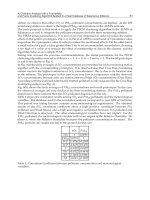
The results obtained in this study were digitalized in spreadsheets using the software
Microsoft Office Excel 2007. Statistical analyses were performed using the chi-squared test
and the logistic regression analysis. Statistical significance level was set at p < 0.05 using the
Statistical Package for the Social Sciences (SPSS) version 10.0.
3. Results
Among the 72 samples processed, 8.33% (6/72) were positive for the protozoa researched.
Using the MERIFLUOR® direct immunofluorescence assay kit, we found six positive
Environmental Monitoring
350
samples: two at JL2 in September and November, one at JL1 in August, two at MP1 in July,
and one at VB in September.
Using the modified Ziehl-Neelsen-stain technique, 2.7% (2/72) samples were positive for
Coccidea, and the presence of Cryptosporidium sp. was detected in two samples and
confirmed by the MERIFLUOR® direct immunofluorescence assay kit Figure 6 shows a
Cryptosporidium sp. oocyst and Figure 7 displays a Cryptosporidium parvum oocyst, which is
approximately 5 µm in diameter, whereas Cryptosporidium hominis oocyst is approximately 4
µm in diameter.
Fig. 6. Cryptosporidium sp. oocyst stained by the modified Ziehl-Neelsen (magnitude
100x)technique and confirmed by the MERIFLUOR® direct immunofluorescence assay kit
and PCR (Santos et al., 2010).
Using primers AWA995f/AWA1206R we demonstrated that the samples belonged to the
genus Cryptosporidium sp., and using primers LAX469F/LAX869R, we showed that just the
sample collected in July was identified as Cryptosporidium parvum. As we detected only two
positive samples for Cryptosporidium sp., the molecular detection was processed exclusively
for them.
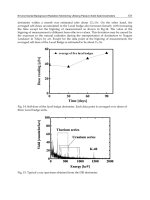
Using the Kinyoun hot staining method and the hot-chromotrope method for the detection
of protozoa, no samples were found to be positive. Table 2 shows the results of each test
carried out for the six sampling sites. Table 3 presents the frequency of protozoa detected in
each sampling site.
Environmental Monitoring of
Opportunistic Protozoa in Rivers and Lakes: Relevance to Public Health in the Neotropics
351
Fig. 7. Cryptosporidium parvum oocyst stained by the modified Ziehl-Neelsen technique
(magnitude 100x) and confirmed by the MERIFLUOR® direct immunofluorescence assay kit
and PCR (Santos et al., 2010).
Sampling
site
Method
Ziehl-Neelsen Kinyoun
Hot-
chromotrope
MERIFLUOR®
MP1 C. parvum* Negative Negative Giardia sp.
MP2 Negative Negative Negative Negative
JL1 Negative Negative Negative Giardia sp.
JL2 Negative Negative Negative Giardia sp.**
VB Cryptosporidium sp.* Negative Negative Negative
BB Negative Negative Negative Negative
MP1: Meia Ponte river, at 16°37'40.94"S latitude and 49°16'13.41"W longitude; MP2: Meia Ponte river at
16°38'22.39"S latitude and 49°15'50.68"W longitude; JL1: João Leite river, at 16°37'40.18"S latitude and
49°14'26.08"W longitude; JL2: João Leite river, at 16°19'37.52"S latitude and 49°13'24.53"W longitude;
VB: Vaca Brava Park lake, at 16°42'31.18"S latitude and 49°16'15.67"W longitude; BB: Bosque dos Buritis
lake, at 16°40'58.51"S latitude and 49°15'38.35"W longitude. *Confirmation by PCR; ** Two positive
samples.
Table 2. Results according to the six sampling sites and the methods used to analyze the 12
samples in each site monitored, in a total of 72 samples (2006/2007)
Environmental Monitoring
352
Protozoa
Sampling site
MP1 MP2 JL1 JL2 VB BB
n % n % n % n % n % n %
Negative 12 100.0 10 83.4 11 91,7 10 83,3 11 91.7 12 100.0
Cryptosporidium
sp.
0 0.0 0 83.4 0 0,0 0 0,0 1 8.3 0 0.0
C. parvum
0 0.0 1 8.3 0 0,0 0 0,0 0 0.0 0 0.0
Giardia lamblia
0 0.0 1 8.3 1 8,3 2 16,7 0 0.0 0 0.0
Total 12 100.0 12 100.0 12 100,0 12 100,0 12 100.0 12 100.0
MP1: Meia Ponte river, at 16°37'40.94"S latitude and 49°16'13.41"W longitude; MP2: Meia Ponte river at
16°38'22.39"S latitude and 49°15'50.68"W longitude; JL1: João Leite river, at 16°37'40.18"S latitude and
49°14'26.08"W longitude; JL2: João Leite river, at 16°19'37.52"S latitude and 49°13'24.53"W longitude;
VB: Vaca Brava Park lake, at 16°42'31.18"S latitude and 49°16'15.67"W longitude; BB: Bosque dos Buritis
lake, at 16°40'58.51"S latitude and 49°15'38.35"W longitude.
Table 3. General distribution of samples in the six sites according to the presence of
protozoa, from February 2006 to January 2007
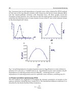
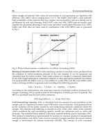
Average temperature in the period of protozoa occurrence was 26.8ºC, while in the period
showing no register of this pathogen, it was 25.6ºC. The logistic regression analysis for
temperature revealed p = 0.262 and OR = 1.227 (Table 4).
Average relative humidity in the period of protozoa occurrence was 42.3%, whereas in the
period showing no register of this pathogen, it was 56.3%, a not significant value since the
logistic regression analysis for relative humidity revealed p = 0.060 and OR = 0.944 (Table 4).
Protozoa n Mean Standard deviation p OR
Temperature
Negative 66 25.6 2.5
Positive 6 26.8 1.5 0.262 1.227
Relative humidity
Negative 66 56.3 16.0
Positive 6 42.3 14.6 0.060 0.944
Table 4. Mean and standard deviation of temperature and relative humidity according to the
presence of protozoa in the bodies of water sampled in Goiania during February 2006 to
January 2007
(logistic regression analysis)
4. Discussion
This study revealed that the water in all sampling sites monitored during the research is not
suitable for human consumption. Despite this evidence, we could observe the presence of
people collecting water for human consumption, bathing, washing clothes, and even fishing.
This fact is highly worrying because various waterborne diseases, not only related to
opportunistic protozoa, but also to several other biological agents, can be transmitted
through these contaminated bodies of water. Some sources of pollution observed in the
Environmental Monitoring of
Opportunistic Protozoa in Rivers and Lakes: Relevance to Public Health in the Neotropics
353
sampling sites were: clandestine sewage discharges, livestock and poultry farms,
slaughterhouses, meat processing plants, landfills, among others.
Nonetheless, we detected low recovery efficiency of opportunistic protozoa cysts and/or
oocysts, which might be related to environmental influence and physical-chemical factors,
such as water pH and turbidity, among others, since the influence of physical-chemical
factors on sampling was reported by other researchers (Fricker & Crabb, 1998, McCuin &
Clancy, 2003). The influence of physical-chemical factors on sampling was reported by other
researchers (Fricker et al., 1998; Clency et al., 2003). Adverse environmental factors have
been proven to alter the morphology of cysts and oocysts (Orgerth & Stibbs, 1987) , ; thus
justifying the low positivity found in the present study using parasitological methods. Other
factors might have had influence as well, such as the concentration of Cryptosporidium sp.
oocysts, based almost exclusively on particle size (Fricker, 1998). The parasitological
techniques employed in our study are not specific and, consequently, concentrate a large
amount of several materials that may be present in the water, such as organic and inorganic
particles, bacteria, yeast, and algae, which interfere in the detection of the parasites.
However, the methods used in the present study are in accordance with those
recommended for concentration and detection of microorganisms by the Standard Methods
for the Examination of Water and Wastewater (Clesceri et al., 1998). They are easily applied,
do not pose a great risk to the technician, and are low cost techniques, which can be
employed by technicians trained to monitor water for human consumption.
Hall and Croll (1997) evaluated the performance of some rapid gravity filters in England
using turbidity measurement and particle counts in filtered water as parameters for
monitoring and controlling Cryptosporidium sp. oocysts as an indicator microorganism, a
method similar to the one used in this study.
Some studies have demonstrated that Cryptosporidium sp. prevalence is approximately 6% in
developed countries (6), around 2-6% in immunodepressed adults (Goldman & Ausiello 2004),
and shows a great variation in underdeveloped countries (Casemore, 1990). In industrialized
countries, the seroprevalence of oocyst antigens is between 17% and 32% (Goldman & Ausiello
2004). In Canada, a study showed that 21% of the water samples collected were contaminated
with Giardia sp. cysts and 4.5% with Cryptosporidium sp. oocysts (Wallis, 1996). However, in
the United States, the contamination of 65% to 97% of superficial water with Cryptosporidium
sp. oocysts and Giardia sp. cysts was reported (Kirkpatrick & Green, 1985), and it was also
estimated that 80% of superficial water and 26% of treated water contains oocysts, although
their infectivity has not been investigated (Goldman & Ausiello 2004). Nevertheless, we found
contamination of 8.33% (6/72) of the samples in the present study, much inferior to the
American data, which might be explained by the method applied. Therefore, new
methodologies should be tested in order to compare the results in terms of specificity and
efficiency to be employed in environmental monitoring of protozoa of public health interest.
Since our sampling points are located before the municipal wastewater treatment plant of
Goiânia, the results of this study were considered within the tolerable levels, due to the low
protozoan positivity according to the method used, in spite of the clandestine sewage
discharges. It is worth mentioning that the water from all sources analyzed in this research
is improper for usage in natura, because it meets neither the Brazilian standard (Brasil, 2004),
which establishes that water for human consumption ought to be free from Giardia sp. and
Cryptospridium sp., nor the American one (McCuin & Clancy, 2003).
The parasitological techniques employed in our study are not specific and, consequently,
concentrate a large amount of several materials that may be present in the water, such as
organic and inorganic particles, bacteria, yeast, and algae, which interfere in the detection of
Environmental Monitoring
354
the parasites. However, the methods used in this study are in accordance with those
recommended for concentration and detection of microorganisms by the Standard Methods
for the Examination of Water and Wastewater (Clesceri, 1998). They are easily applied, do not
pose a great risk to the technician, and are low cost techniques, which can be employed by
technicians trained to monitor water for human consumption.
The performance of some rapid gravity filters was evaluated in England, using turbidity
measurement and particle counts in filtered water as parameters for monitoring and
controlling Cryptosporidium sp. oocysts as an indicator microorganism (Geldreich, 1996), a
method similar to the one used in our study.
The in vitro amplification of DNA fragments of Cryptosporidium sp. obtained sensibility and
specificity. Nevertheless, the amplification was only possible using Nested-PCR primers
(AWA995f/AWA1206R and LAX469F/LAX869R). The primer LAX469F/LAX869R
amplifies the regions of C. parvum/C. hominis, but C. parvum diagnosis was confirmed by the
difference in diameter, since its oocyst is approximately 5 µm in diameter, while C. hominis
oocyst is approximately 4 µm in diameter.
Nested-PCR presents the advantage of concentrating a smaller quantity of PCR inhibitors
(Kirkpatrick & Green, 1985). In environmental samples, there are several Taq DNA
polymerase inhibitors, such as fecal hemoglobin and phenolic compounds, and it might
have been the case of the samples processed in the present research.
It was possible to obtain satisfactory amplification with the two methods of DNA extraction
applied. Furthermore, they are quick and low-cost, although close attention should be paid
to the phenol/chloroform method since it is toxic and corrosive.
As adverse environmental factors have been proven to alter the morphology of cysts and
oocysts (Hsu BM, 2001), making their detection more difficult, this may justify the low
positivity found in the present study using parasitological methods. Other factors might
have had influence as well, such as the concentration of Cryptosporidium sp. oocysts, based
almost exclusively on particle size (Hsu, 2001). Also, the level of protozoa may vary
according to the season, and an increase in their resistant forms in rainy periods, winter and
beginning of spring has already been reported (Atherholt 1998, Ong et al. 2002)
Temperature has also been considered a factor that influences protozoa and autochthonous
microorganism survival in rivers (Howe, 2002). In this study, we observed just small
variations of water temperature in the rivers and lakes sampled during the period of study,
although within the limits that allow the survival and viability of protozoa. Using univariate
logistic regression (p = 0.066), we demonstrated that temperature was not a statistically
significant variable, whereas humidity (p = 0.958) was. In the region of sample collection
there are two well-defined seasons, the dry (from April to September) and the rainy (from
October to March) seasons, the latter characterized by torrential rain and runoff, which
certainly makes the detection of parasites more difficult.
Due to the low number of protozoa found in this work, i.e. two Cryptosporidium sp. and four
Giardia sp., we could not infer if the protozoan levels vary by season, but only observe the
qualitative inference of their presence in the bodies of water monitored.
5. Conclusion
The rivers and lakes of Goiânia are contaminated with opportunistic protozoa;
Standardization and application of parasitological and molecular techniques in the
analysis and seasonal monitoring of opportunistic protozoa were successfully carried
out for environmental samples;
Environmental Monitoring of
Opportunistic Protozoa in Rivers and Lakes: Relevance to Public Health in the Neotropics
355
During seasonal monitoring of opportunistic protozoa, with emphasis on Coccidia
Cryptosporidium sp., Cyclospora cayetanensis, Isospora belli and Microsporidia, it was
possible to detectic Cryptosporidium parvum and Cryptosporidium sp. using PCR and
Nested-PCR, respectively
The parasitological and molecular techniques applied are quick, low-cost, and can be
employed in laboratories that monitor the microbiological quality of water for human
consumption. Considering that the microorganisms studied herein are opportunistic, their
persistent contact with humans may generate new parasites able to breach the immune
barrier of normal individuals and to produce more aggressive cycles. Our results point to
the need for efficient programs to prevent, treat, and monitor the presence of these
parasites in rivers and lakes used for abstraction of water intended for human
consumption and/or for recreational purposes all over the world. Furthermore, more
efficient parasitological techniques, such as PCR, should be adopted in routine analyses in
the laboratories of environmental monitoring, water for human consumption should be
purified with UV radiation, and the activated sludge generated by wastewater treatment
plants and intended for use in agriculture should be monitored.
6. Concluding remarks
Cryptosporidium is considered a coccidia resistant (Carey et al. 2004), because oocysts have
characteristics that favor its rapid spread in the environment, such as the ability to
withstand the action of commonly used disinfectants (formaldehyde, phenol, ethanol, lysol),
able to cross some water filtration systems due to its small size, the ability to float, remain in
the environment by a few weeks or months and tolerance in certain temperatures and
salinity (Fayer et al. 2004). Given the scope of the aquatic environment coupled with the
wide distribution of different species in Brazilian waters, make the control measures of
Cryptosporidium limited.
Therefore, to minimize the risks inherent in the spread of cryptosporidiosis in the
populations of free-living mammals, it is of fundamental importance to environmental
control, through the adoption of agricultural practices to prevent pollution of rivers by the
faeces of animals (Graczyk et al. 2000), as well as encouraging the adequacy of sanitation
facilities, protection of water sources, education and guidance on waste discharges from
vessels during nautical activities. Regarding the control measures of captive aquatic
mammals, so as to minimize or eliminate the risks inherent in the spread of coccidian,
several studies should be adopted.
Finally, it must be remembered that currently monitoring systems treated water are based
on the frequency of fecal coliforms and Escherichia coli as indicators of pollution, and that
this methodology is insufficient to predict the presence of other pathogens such as parasites.
Thus, it is imperative the use of alternative methods for the diagnosis, investigation and
monitoring of large amounts of water of these pathogens. For in this way can be proposed
reorganization measures that contribute to reducing the incidence of opportunistic diseases
emerging in water of human use, especially for children, elderly, immunocompromised and
immunosuppressed patients.
7. References
Alves, T. M., & Chaveiro, E.F. Metamorfose urbana: a conurbação Goiânia-Goianira e suas
implicacões sócio-espaciais [Urban metamorphosis: the conurbation of Goiânia and
Environmental Monitoring
356
Goianira cities and its socio-spatial implications]. Revista Geográfica Acadêmica,
1 :95–107, 2007.
Atherholt, T.B.; LeChevallier M.W.; Norton W.D.; & Rosen J.S. Effect of rainfall on giardia
and crypto. J Am Water Works Assoc, 90: 66–80, 1998.
Awad-el-Kariem, F.M.; Warhurst D.C.; & McDonald V. Detection and species identification
of Cryptosporidium oocysts using a system based on PCR and endonuclease
restriction. Parasitology, 109: 19–22, 1994.
Boom, R.; Sol C.J.A.; Salimans M.M.M.; Jansen C.L.; Wertheim-van-Dillen P.M.E.; & van der
Noordaa J. Rapid and simple method for purification of nucleic acids. J Clin
Microbiol, 28: 495–503, 1990.
Brasil. Ministério de Saúde. Portaria 518 de 25 de Março de 2004. Estabelece os
procedimentos e responsabilidades relativos ao controle e vigilância da qualidade
da água para consumo humano e seu padrão de potabilidade, e dá outras
providências. Diário Oficial da União, Brasília-DF, 2004.
Carey, C. M.; Lee, H.; & Trevors, J.T. Biology, persistence and detection of Cryptosporidium
parvum and Cryptosporidium hominis oocyst. Water research, 38 : 818-862, 2004.
Caccio, S.; Pinter, E.; Fantini, R.; Mezzarona, I.; & Pozio, E. Human infection with
Cryptosporidium felis: case report and literature review. Emerging Infectious
Diseases, 8: 263-268, 2002
Casemore, D.P. Epidemiological aspects of human cryptosporidiosis. Epidemiol Infect, 104: 1–
28, 1990.
Cimerman, S.; Castañeda C.G.; Iuliano W.A; & Palacios R. Perfil das enteroparasitoses
diagnosticadas em pacientes com infecção pelo vírus HIV na era da terapia
antiretroviral potente em um centro de referência em São Paulo, Brasil. Parasitol
Latinoam, 57: 111–118, 2002.
Clancy, J.L.; Bukhari, Z.; McCuin, R.M.; Matheson, Z.; & Fricker, C.R. USEPA Method
1622. J Am Water Works Assoc, 91: 60–68, 1999.
Clancy, J.L.; Connel, K.; McCuin, R.M. Implementing PBMS improvements to USEPA`S
Cryptosporidium and Giardia methods. J Am Water Works Assoc, 95: 80–93, 2003.
Clesceri, L.S.; Greenberg, A.E.; & Eaton, A.D. Standard methods for the examination of
water and wastewater. 20th ed. Washington, D.C., American Public Health
Association, 1998.
Corso, P.S.; Kramer, M.H.; & Blair, K.A. Addiss DG, Davis JP, Haddix AC. Cost of illness in
the 1993 waterborne Cryptosporidium outbreak, Milwaukee, Wisconsin. Emerg
Infect Dis, 9: 426–431, 2003.
Fayer, R.; Morgan, U.; & Upton, S.J. Epidemiology of Cryptosporidium: transmission,
detection, and identification. International Journal for Parasitology, 30:1305-1322, 2000.
Franco, R. M.B. (Docente ): Método 1623: Evolução e Análise Crítica; 2004; Palestra; ;
Unicamp/Comitê de Bacias Hidrográficas e Sociedade Paulista de Parasitologia;
Hotel Premium Norte; Campinas; BR; Meio digital;
www.ib.unicamp.br/parasito/seminario2004.
Fricker, C.R.; & Crabb, J.H. Water-borne cryptosporidiosis: detection methods and treatment
options. Adv. Parasitol, 40: 242-278, 1998.
Garcia-Zapata, M.; T.A.; Passo, A.; Ruano, A.L.; Souza júnior, E.S.; Cechetto, F. H.; &
Manzi, R.S. Ciclosporíase intestinal: relato dos primeiros casos humanos no estado
de Goiás, Goiânia, Brasil. Revista de Patologia Tropical, 32 : 121-130, 2003.
Environmental Monitoring of
Opportunistic Protozoa in Rivers and Lakes: Relevance to Public Health in the Neotropics
357
Geldreich, E.E. Amenaza mundial de los agentes patógenos transmitidos por el agua. In:
Craun GF, Castro R. (Ed.) Calidad del agua potable en América Latina:
ponderación de los riesgos microbiológicos contra los riesgos de los subproductos
de la desinfección química. Washington, DC, ILSI Press, p. 21–49, 1996.
Glaberman, S.; Moore, J.E.; Lowery, C.J.; Chalmers, R.M.; Sulaiman, I.; Eiwin, K.; Rooney,
P.J.; Millar, BC.; Dooley, J.S.G., Lal, A.A. & Xiao, L. Three drinking water-associated
cryptosporidiosis outbreaks, Northern Ireland. Emerg. Inf. Dis, 8 (6): 631-633, 2002.
Goldman L, Ausiello D. Cryptosporidiosis. In: Goldman L, Bennett JC (Ed.) Cecil textbook of
medicine. 22nd ed. Philadelphia, WB Saunders, p. 2092-2095, 2004.
Gomes, A.H.S.; Pacheco, M.A.S.R.; Fonseca, Y.S.K.; Cesar, N.P.A.; Dias, H.G.G.; Silva, R.P.
Pesquisa de Cryptosporidium sp em águas de fontes naturais e comparação com
análises bacteriológicas. Rev Inst Adolfo Lutz, 61: 59–63, 2002.
Graczyk, T.K., Evans, B.M., Zif, C.J., Karreman, H.J. & Patz, J.A. Environmental and
geographical factores contributing to watershed contamination with
Cryptosporidium parvum oocysts. Environ. Res, 82(3):263-271, 2000.
Hall, T. ;Croll, B.Particle counters as tools for managing Cryptosporidium risk in water
treatment. Water Scien Technol, 36, 143–149, 1997.
Howe, A.D.; Forster, S.; Morton, S.; Marshall, R.; Osborn, K. S.; Wright, P & Hunter, P. R.
Cryptosporidium oocysts in a water supply associated with a cryptosporidiosis
outbreak. Emerg. Inf. Dis, 8 (6): 619-624, 2002.
Hsu, B.M.; Huang, C.; Lai, Y.C.; Tai, H.S.; & Chung, Y.C. Evaluation of immunomagnetic
separation method for detection of Giardia for different reaction times and reaction
volumes. Parasitol Res, 87: 472–474, 2001.
Katsumata, T.; Hosea, D.; Ranuh, I.G.; Uga, S.; Yanagi, & T.; Khono, S. Short report: possible
Cryptosporidium muris infection in humans. American Journal Tropical Medicine
Hygiene, 62: 70-72, 2001.
Kosek, M.; Alcantara, C.; Lima, A.A.M.; & Guerrant, R.L. Cryptosporidiosis: na update. The
Lancet Infectous Diseases, 1:262-269, 2001.
Kirkpatrick, C.E. ; & Green, G.A. IV. Susceptibility of domestic cats to infections with
Giardia lamblia cysts and trophozoites from human sources. J Clin Microbiol, 21:
678–680, 1985.
Kramer, ; M.H.; Herwaldt,; B.L.; Craun, G.F.; Calderon, R.L.; & Juranek, D.D. Surveillance
for waterborne-disease outbreaks, United States, 1993-1994. MMWR 45(SS-1): 1-33,
1996.
Kokoskin, E.; Gyorkos, T.W.; Camus, A.; Cedilotte, L.; Purtill, T.; Ward, B. Modified
technique for efficient detection of microsporidia. J Clin Microbiol, 32: 1074–1075,
1994.
Kraszewski J. Water for people supports small systems for impoverished people worldwide.
J Am Water Works Assoc, 93: 36–37, 2001.
Laxer, M.A.; Timblin, B.K.; & Patel, R.J. DNA sequences for the specific detection of
Cryptosporidium parvum by the Polymerase Chain Reaction. Am J Trop Med Hyg,
45: 688–694, 1991.
McCuin, R.M.; Clancy, J.L. Modifications to United States Environmental Protection Agency
Methods 1622 and 1623 for detection of Cryptosporidium oocysts and Giardia cysts
in water. Appl Environ Microbiol, 69: 267–274, 2003.
Environmental Monitoring
358
Ong, C.S.L.; Eisler, D.L.; Alikhani, A.; Fung, V.W.K.; Tomblin, J.; BowniE, W.R.; & Issac-
Renton, J.L. Novel Cryptosporidium genotypes in sporadic cryptosporidioisis
cases: first report of human infection with a corvine genotype. Emerging Infectious
Diseases, 8:263-268, 2002.
Ongerth, J. E. ; Stibbs, H. H. Identification of Cryptosporidium oocysts in river water. Appl
Environ Microbiology, 53 : 672–676, 1987.
Pedraza-Dias, S.; AmaR, C.; & Mclauchlin, J. The identification and characterization of an
unusual genotype of Cryptosporidium from human faeces as Cryptosporidium
meleagridis. FEMS Microbiology Letters, 189:189-194, 2000.
Pedro, M.L.G.; & Germano, M.I.S. A água: um problema de segurança nacional. Rev Hig
Alim, 15: 15–18, 2001.
Sambrook, J.; & Russel, D. Molecular cloning: a laboratory manual. 3rd ed. v. 1, v. 2, v. 3.
New York, Cold Spring Harbor Laboratory Press Section, 2001.
Santos, Sônia de Fátima Oliveira. Estudo dos parasitos oportunistas em águas fluviais de
uso humano no município de Goiânia-Goiás, Brasil, 2006/2007. Dissertation
(Mestrado em Ciências da Saúde - Master in Health Sciences) – Pós-graduação em
Ciências da Saúde, Universidade Federal de Goiás, Goiânia, 2008.
Santos, S.F.O., Silva, H.D., Souza-Junior, E.S., Anunciação, C. E., Silveira-Lacerda, E. P.,
Vilanova-Costa, C.A.S.T., Garcia-Zapata, M.T.A. Environmental Monitoring of
Opportunistic Protozoa in Rivers and Lakes in the Neotropics Based on Yearly
Monitoring. Water Quality, Exposure and Health, v.2, p.1 - 8, 2010.
Silva, H.D., Wosnjuk, L.A.C., Santos, S.F.O., Vilanova-Costa, C.A.S.T., Pereira, F.C., Silveira-
Lacerda, E.P., Garcia-Zapata, M.T.A., Anunciação, C.E. Molecular Detection of
Adenoviruses in Lakes and Rivers of Goiânia, Goiás, Brazil. Food and Environmental
Virology, v.2, p.35 - 40, 2010.
Solo-Gabriele, H.; & Neumeister, S. US outbreaks of cryptosporidiosis. J Am Water Works
Assoc, 88: 76–86, 1996.
States, S.; Stadterman, K.; Ammon, L.; Vogel, P.; Baldizar, J.; Wright, D.; Conley, L.; &
Sykora, J. Protozoa in river water: sources, occurrence, and treatment. J Am Water
Works Assoc, 89: 74–83, 1997.
Thompson, R.C.A. Giardiasis as a re-emerging infectious disease and its zoonotic potential.
International Journal for Parasitology, 30:1259-1267, 2000.
Thurston-enriquez, J.A.; Watt, P.; Dowd, S.E.; Enriquez, R.; Pepper, I. L.; Gerba, C.P.
Detection of protozoan parasites and microsporidia in irrigation waters used for
crop prodution. J Food Prot. US Department of Agriculture, Agricultural Research
Service, University of Nebraska. United States, Feb. 2002.
Wallis, P.M.; Erlandsen, S.L.; Isaac-Renton, J.L.; Olson, M.E.; Robertson, W.J.; Van Keulen,
H. Prevalence of Giardia cysts and Cryptosporidium oocysts and characterization
of Giardia spp. isolated from drinking water in Canada. Appl Environ Microbiol, 62:
2789–2797, 1996.
Xiao, L.; Escalante, L.; Yang, C.; Sulaiman, I.; Escalante, A.A.; Montali, R.J.; Fayer, R.; & Lal,
A.A. Phylogenetic analysis of Cryptosporidium parasites based on the small-
subnunit rRNA gene locus. Appl Environ Microbiol, 65: 1578–1583, 1999.
Part 3
Environmental Monitoring with
Wireless Sensor Network Technology
21
Biosensor Arrays for Environmental Monitoring
Wei Song
1
, Si Wei
2
, Hong-Xia Yu
2
, Maika Vuki
3
and Danke Xu
1
1
State Key Laboratory of Analytical Chemistry for Life Science,
School of Chemistry and Chemical Engineering, Nanjing University,
2
State Key Laboratory of Pollution Control and Resource Reuse,
School of the Environment, Nanjing University,
3
College of Natural and Applied Sciences, University of Guam, Mangilao, Guam,
1,2
China
3
USA
1. Introduction
Environmental monitoring involves several steps such as sampling, sample handling and
sample transportation to specialized laboratories, sample preparation and analysis.
Traditional environmental monitoring approaches are based on discrete sampling methods
followed by laboratory analysis. These approaches do not improve our understanding of the
natural processes governing chemical species behavior, their transport and bioavailability,
or the relationship between anthropogenic releases and their long-term impact on aquatic
systems
[1]
. The challenge of environmental monitoring in situ requires new and improved
analytical devices featuring precision, sensitivity, specificity, rapidity, and ease of operation
to detect decreasing concentrations of an ever growing array of pollutants. Such devices
must be comparable to or better than traditional analytical systems, and must be simple to
handle, small, cheap, able to provide reliable information in real-time, and must be sensitive
and selective for the analyte of interest, and suitable for in situ monitoring
[2]
. Biosensors not
only fulfill all these requirements but also have applications in many areas such as clinical
diagnostics, forensic chemistry, pharmaceutical studies, food quality control and
environmental monitoring.
A biosensor is an analytical device for the detection of an analyte that combines a biological
component with a physicochemical detector component. It consists of 3 parts: (1) the
sensitive biological element (biological material such as tissue, microorganisms, organelles,
cell receptors, enzymes, antibodies, nucleic acids, etc.), a biologically derived material or
biomimic; (2) the transducer or the detector element (works in a physicochemical way;
optical, piezoelectric, electrochemical, etc.) that transforms the signal resulting from the
interaction of the analyte with the biological element into another signal that can be more
easily measured and quantified; (3) associated electronics or signal processors that are
primarily responsible for the display of the results in a user-friendly way
[3]
. Depending on
the type of transduction mechanism applied and the bio-recognition element employed, the
potential for these devices for detection can be enormous. The technological development
and the success in single analyte detection propelled advances in the miniaturization of
sensors along with multi-analyte detection with sensitivities ranging in the nano-mole to
Environmental Monitoring
362
atto-mole range. With advances in techniques for biosensor construction, it has been
possible to miniaturize the whole biosensor system on a chip to fabricate biosensor arrays.
The Biosensor arrays developed at the Naval Research Laboratory (NRL) has successfully
been used in the detection of a variety of protein toxins, organic molecules, physiological
health markers, a virus and a number of bacteria, initially in buffer but increasingly in food,
biological and environmental matrices
[4]
. These developed biosensors are rapid, simple to
perform and require little-to-no sample pretreatment prior to analysis, even for more
complex sample matrices. In addition, the two-dimensional nature of the slide sensing
surface facilitates simultaneous analysis of multiple samples for multiple analytes. Research
on biosensor arrays as multi-analyte bio-systems has generated increased interest in the last
decade. The main feature of the micro-array technology is the ability to simultaneously
detect multiple analytes in one sample by an affinity-binding event at a surface interface.
Fifteen years ago, the gene expression analysis of cDNA on micro-arrays was one of the first
applications that successfully detected thousands of labeled target DNA molecules in
parallel. Also the first immuno-analytical biosensor array was described at the same time. In
the meantime, a great variety of target analytes capable of interacting selectively with a bio-
molecular receptor has been adapted to arrays
[5]
. The biosensor arrays have been envisioned
as a tool for rapid, on-site screening of pollutants in whatever location they might be found.
The goals of automation, weight reduction, minimal size, ease of use, and reliability have
remained paramount as the system has been developed
[6]
.
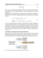
The challenge of continuous in situ monitoring of environmental pollution requires
instruments that are robust and with sufficient sensitivity and long lifetime. Commonly used
conventional methods are time-consuming, expensive, require skilled operators, and lack the
required selectivity. Biosensor arrays have the advantage of being simple, uniform whole
structures featuring direct transduction, high bio-selectivity, high sensitivity, miniaturization,
electrical/optoelectronic readout, continuous monitoring, ease of use, and cost effectiveness.
User advantages include low price, reliability, no sample preparation, disposability, and clean
technology. Hence, biosensor arrays show the potential to complement both laboratory-based
and field analytical methods for environmental monitoring. Biosensor arrays are based on one
general principle—certain bio-molecular recognition elements are defined on a heterogeneous
matrix. Each element is dedicated to an analyte and contains quantitative information. The
matrix is a patterned surface where the recognition molecules are immobilized by micro-
printing such as screen printed technique, micro fluidic or other micro-structuring processes
[5]
.
The type of biosensor arrays involves DNA-based biosensor array, antibody-based biosensor
array, aptamer-based biosensor array, enzyme-based biosensor array, and microorganism-
based biosensor. Recent progress in the development of analytical detection methods for
antibody arrays, enzyme arrays and aptamer arrays as well as microbial arrays are
summarized in this review, and their applications in the environment monitoring are also
discussed. Detection approach is focused on electrochemical and optical measurements
including various electrochemical or florescent probes as well as label-free approach. The
numerous fabrication methods of DNA capture probes, antibodies and aptamer for
multiplexed biological targets are also discussed.
2. DNA-based biosensor arrays
Deoxyribonucleic acids (DNA) are arguably the most important of all bio-molecules. The
unique complementary structure of DNA between the base pairs adenine/thymine and
Biosensor Arrays for Environmental Monitoring
363
cytosine/guanine has been the basis for genetic analysis over the last few decades. The
ability of a single stranded DNA (ssDNA) molecule to ‘seek out’, or hybridize to, its
complementary strand in a sample is the foundation of DNA-based detection systems. There
is a great potential market for simple, cheap, rapid, and quantitative detection of specific
genes. Areas of application include clinical, veterinary, medico-legal, environmental, and
the food industry
[7]
. Development of DNA biosensors and DNA biosensor arrays has
increased tremendously over the past few years as demonstrated by the large number of
scientific publications. Numerous DNA detection systems based on the hybridization
between a DNA target and its complementary probe, which is present either in solution or
on a solid support, have been described
[8]
. Homogeneous assays allowing the determination
of DNA sequences have been developed. These systems can be based on optical
[9]
or
electrochemical
[10]
detection. However, they do not allow easy continuous monitoring and
miniaturization. Heterogeneous DNA biosensors and DNA biosensor arrays offer promising
alternatives to these methods. They allow continuous, fast, sensitive, and selective detection
of DNA hybridization, and they also can be reused. DNA biosensors arrays (commonly
called gene chips, DNA chips, or biochips) exploit the preferential binding of
complementary single-stranded nucleic acid sequences. This system usually relies on the
immobilization of a single-stranded DNA (ssDNA) probe onto a surface to recognize its
complementary DNA target sequence by hybridization. Transduction of hybridization of
DNA can be measured optically, electrochemically, or using other devices. The detection
process is schematized in Figure 1
[8]
.
Fig. 1. Steps involved in the detection of a DNA sequence. Reprinted from ref. 8 with
permission by the American Chemical Society.
In the case of DNA biosensors arrays, the immobilization of a DNA probe is achieved
directly onto a transducer surface. DNA biosensor arrays are made from glass, plastic, or
silicon supports and are constituted of tens to thousands of 10–100 μm reaction zones onto
which individual oligonucleotide sequences have been immobilized. The exact number of
DNA probes varies in accordance with the application. DNA biosensor arrays allow
multiple parallel detection and analysis of the patterns of expression of thousands of genes
in a single experiment.
Environmental Monitoring
364
We have presented an ultra-sensitive and direct electrochemical DNA biosensor array based
on Ag aggregate tag and differential pulse voltammtery
[11]
. The scheme of detection is shown
in Figure 2. The silver tags consist of Conjugate 1 (functionalized with capture probes and
oligo A and Conjugate 2 (modified with oligo T). Hybridization between complementary oligo
(d) A and oligo (d) T anchored on the silver nanoparticles produced aggregate tags. The
hybridization-induced tags are successfully applied to bind with the DNA target via sandwich
hybridization format and offer direct and amplified readout by differential pulse voltammetric
method. We have found that the detection sensitivity by use of the aggregate tags can be
improved by 3 orders of magnitude as compared to the single silver nanoparticle labels and a
detection limit of 5 amol/L could be obtained.
Fig. 2. Schematic illustration of the electrochemical Assay (a) and Multiplexed Assay (b)
with silver nanoparticle Conjugates; Preparation of the aggregates is shown as well.
Reprinted from ref. 11 with permission by the American Chemical Society.
Biosensor Arrays for Environmental Monitoring
365
Environmental applications of DNA biosensor arrays are in the field of species
identification. For instance, DNA biosensor arrays are extensively exploited in the detection
of pathogenic microorganisms relevant to food, bio-defense and environmental
contamination applications. Mainly, DNA biosensor arrays have been coupled to PCR, as a
specific detection method of the amplified base sequence. Zhang et al.
[12]
have developed a
label-free electrochemical DNA biosensor array as a model system for simultaneous
detection of multiplexed DNAs using micro-liters of sample. A novel multi-electrode array
was comprised of six gold working electrodes and a gold auxiliary electrode, which were
fabricated by gold sputtering technology, and a printed Ag/AgCl reference electrode was
fabricated by screen-printing technology. The DNA biosensor array for simultaneous
detection of the human immunodeficiency virus (HIV) oligonucleotide sequences, HIV-1
and HIV-2, was fabricated in sequence by self-assembling each of two kinds of thiolated
hairpin-DNA probes onto the surfaces of the corresponding three working electrodes,
respectively. The hybridization events were monitored by square wave voltammetry using
methylene blue (MB) as a hybridization redox indicator. The oxidation currents of MB
accumulated on the array decreased with increasing the concentration of HIVs due to higher
affinity of MB for single strand rather than double strands of DNA. Under the optimized
conditions, the peak currents were linear over ranges from 20 to 100 nmol/L for HIV-1 and
HIV-2, with the same detection limits of 0.1 nmol/L (S/N= 3), respectively. The detection
process is illustrated in Figure 3. The biosensor array showed a good specificity without the
obvious cross-interference. Furthermore, single-base mutation oligonucleotides and random
oligonucleotides can be easily discriminated from complementary target DNAs. Their work
demonstrates that different hairpin-DNA probes can be used to design the label-free
electrochemical biosensor array for simultaneous detection of multiplexed DNA sequences
for various applications.
Fig. 3. Schematic diagrams of multi-electrode array and representation of biosensor array
with fabrication steps and performance. Reprinted from ref. 12 with permission by the
Elsevier.