Human Musculoskeletal Biomechanics Part 3 potx
Bạn đang xem bản rút gọn của tài liệu. Xem và tải ngay bản đầy đủ của tài liệu tại đây (2.25 MB, 20 trang )
European Braces for Conservative Scoliosis Treatment
31
Key elements of the recommendations are efficacy and compliance. The latter stem from the
planned treatment, as well as from the responses of the patient and family. It is also highly
related to behaviour of the treating team.
The recommendations concerning the standards of management of idiopathic scoliosis with
bracing, with the aim to increase efficacy and compliance to treatment are extensively
described in a recent SOSORT Consensus paper. It is recommended to professionals
engaged in patient care to follow the guidelines of this Consensus in their clinical practice.
The SOSORT criteria should also be used along with the published criteria for bracing
proposed by SRS, [Negrini et al 2009b,e, Thompson and Richards 2008, Richards et al. 2005].
Several other major issues in brace management apart compliance are currently also in
discussion, namely, the pressure being applied, the treatment time and the bending
radiographs. These topics are also discussed in the recently published editorial on
“Scoliosis” Journal Brace Technology Thematic Series by Negrini & Grivas 2010.
4. European braces for conservative scoliosis treatment
4.1 Cheneau brace
Dr Jacques Chêneau built the brace during the 60’s. In 1972 the first patient’s results were
obtained and officially presented in 1979 at Bratislava. Initially the brace was named
Cheneau-Toulouse-Munster Brace as well. Now it is accepted and used worldwide. Useful
information on the brace and its philosophy can be found in o. It is a
rigid brace providing three-dimensional correction, Figure 1. The mechanisms of Chêneau
Brace correction are a) passive mechanisms, namely 1) convex to concave tissue transfer,
achieved by multiple three-point system acting in 3D, with the aim of curve
hypercorrection, 2) elongation and unloading by the “cherry stone” effect, 3) Derotation of
the thorax, 4) bending and b) active mechanisms, namely 1) vertebral growth acting as a
corrective factor, 2) asymmetrically guided respiratory movements of the rib-cage, 3)
repositioning of the spatial arrangement of the trunk muscles to provide their physiological
action and 4) anti-gravitational effect, [Kotwicki & Cheneau 2008 a,b]. This brace opens
anteriorly. After some modifications made by Dr Jacques Chêneau, since 1996 the brace is
divided in 54 zones and provides large free spaces opposite to pressure sites. The hump
should be pressed on 1/3 of the surface of apex. The corresponding dodging site involves
4/5 of the surface of the concave side of curve. Each of the remaining two pressure parts of
the three-point system presses on 1/5 of the surface of the concave side. They are the apexes
of the neighboring curves. Dodging opposite the latter sites allows movements and
straightening of the curve in an active way. It is not permitted to hinder any of the three
dodging areas, that is, the middle 4/5 of concave side and the 1/3 over and under the apex.
Regarding the outcomes of brace application, the Cheneau-Toulouse-Munster brace has
been found to decrease the coronal shift forward, the coronal tilt, the axial rotation, and to
increase the sagittal shift forward and the sagittal vertebral tilt (3-D correction), [Périé et al.
2001], obtaining an average primary correction 41% (thoracic, lumbar, double) (n = 52
patients) and a long term correction 14.2% thoracic, 9.2% lumbar double curves: 5.5% in
thoracic & 5.6% in lumbar, [Hopf
& Heine 1985]. In a recent report, at the end of treatment
there was an improvement of Cobb angle correction of about 23% and after 5 years there
was a stabilization of about 15 % (p value < 0.05). Therefore, based on this study, it could be
stated that conservative treatment with Cheneau brace not only stops progression, but it
also reverses the scoliotic curve [Cinnella et al. 2009]. The effectiveness of Cheneau brace in
Human Musculoskeletal Biomechanics
32
the management of IS was also recently analysed in a prospective observational study,
[Zaborowska-Sapeta et al 2011]. It reports the results of treatment according to SOSORT and
SRS recommendations on 79 patients (58 girls and 21 boys) with progressive IS, treated with
Cheneau brace and physiotherapy, with initial Cobb angle between 20 and 45 degrees, no
previous brace treatment, Risser 4 or more at the final evaluation and minimum one year
follow-up after weaning the brace. Achieving 50 degrees of Cobb angle was considered
surgical recommendation. At follow-up 20 patients (25.3%) improved, 18 patients (22.8%) were
stable, 31 patients (39.2%) progressed below 50 degrees and 10 patients (12.7%) progressed
beyond 50 degrees (2 of these 10 patients progressed beyond 60 degrees). Progression
concerned the younger and less skeletally mature patients, [Zaborowska-Sapeta et al 2011].
Fig. 1. The Cheneau brace
4.2 Cheneau brace derivatives ( Rigo system Cheneau brace, ScoliOlogiC® “Chêneau
light” and the Gensingen brace™)
4.2.1 Rigo system cheneau brace
This brace was developed by Dr Manuel Rigo during the early 90s in Instituto Èlena Salvá in
Barcelona, Spain. The German - Spanish collaboration for brace production and information
on manufacturing can be obtained at: The
RSCB, Figure 2, is based on the Chêneau Brace, and it is able to produce the required
combined forces to correct scoliosis in 3D. The blueprint of the brace is based on the
idiopathic scoliosis curve classification correlated with brace treatment introduced by Dr
Rigo [Rigo et al 2010a]. The classification includes radiological as well as clinical criteria. The
radiological criteria are utilized to differentiate five basic types of curves including: (I)
imbalanced thoracic (or three curves pattern), (II) true double (or four curve pattern), (III)
balanced thoracic and false double (non 3 non 4), (IV) single lumbar and (V) single
thoracolumbar. In addition to the radiological criteria, the Rigo Classification incorporates
the curve pattern according to SRS terminology, the balance/imbalance at the transitional
point, and L4-5 counter-tilting. The principles of correction of the five basic types of curves
European Braces for Conservative Scoliosis Treatment
33
are also described by Dr. Rigo, [Rigo et al 2010a]. Biomechanically the RSC brace offers
regional derotation. The rib cage and spine are de-rotated. The brace derotates the thoracic
section against the lumbar section, with a counter-rotation pad at the upper thoracic region
[Rigo and Weiss 2008]. The brace also produces physiological sagittal profile. Initial reports
on outcomes using this brace indicated a 31.1% primary Cobb angle correction and 22.2%
primary torsion angle correction. At a follow up of 16.8 months 54% of curves were stable,
27% improved and 19% progressed, [Rigo et al 2002]. In patients with long thoracic curves
treated with a recently described RSC brace design (three-curve-scoliosis brace with pelvis
open) there was 76.7 % in-brace Cobb angle correction and 55.9% in-brace axial rotation
correction [Rigo & Gallo 2009]. The latter pattern is easy to correct according to the
principles and it can not be compared to “Chêneau light” cohort, which in addition contains
double curve patterns which correct least [Weiss et al 2007].
Fig. 2. Rigo System Cheneau Brace
4.2.2 ScoliOlogiC® “Chêneau light”
The brace, Figure 3, was invented by Dr. Hans-Rudolf Weiss. The application for the patent
was presented in April 2005 and the first braces were built in May 2005. Useful information
on the brace can be obtained in and [Weiss
et al 2007 and Weiss & Werkmann 2010].
The ScoliOlogiC® off the shelf bracing system enables the CPO to construct a light brace for
scoliosis correction from a variety of pattern specific shells to be connected to an anterior
and a posterior upright. This brace, when finally adjusted is called Chêneau light™ brace.
The advantage of this new bracing system is that the brace is available immediately, is easily
adjustable and that it can also be easily modified. This avoids construction periods of
sometimes more than 6 weeks, where the curve may drastically increase during periods of
fast growth. The disadvantage of this bracing system is that there is a wide variability of
possibilities to arrange the different shells during adjustment, [Weiss & Werkmann 2010].
Weiss et al, 2007, reported 51% correction of Cobb angle (Cobb angle in the whole group of
patients was reduced by an average of 16,4 degrees), 62 % correction for lumbar &
thoracolumbar curve pattern, 36 % correction for thoracic scoliosis and 50 % correction for
Human Musculoskeletal Biomechanics
34
double major curve pattern. The correction effect correlated negatively with age (r = -0,24; p
= 0,014), negatively with the Risser stage (-0,29; p = 0,0096) and negatively with Cobb angle
before treatment (r = -0,43; p < 0,0001) [Weiss et al 2007].
The reduction of material in the Chêneau light® brace seems to increase patient's comfort
and reduces the stress patients may suffer from whilst in the brace. 80% of the adolescent
population of scoliosis patients can be braced with the Chêneau light™ brace. In certain
patterns of curvature and in the younger population with an age of less than 11 years, other
approaches have to be used, such as plaster based bracing or the application of CAD/CAM
based orthoses [ Weiss & Werkmann 2010].
There are blueprints to build a RSC® or a Chêneau light® brace according to the conservative
treatment of AIS classification by Dr M. Rigo and Dr. HR Weiss [Rigo & Weiss 2008].
Fig. 3. ScoliOlogiC® “Chêneau light”
4.2.3 The Gensingen brace™
From the experience obtained through the Chêneau light® brace a new CAD/CAM brace
has been designed and applied by Dr. Weiss since 2009 which is called the Gensingen
brace®. It is extensively described, [Weiss 2010a,b]
This is a new asymmetric Chêneau style CAD/CAM derivate. It has been designed to
overcome some problems the designer experienced with other Chêneau CAD/CAM
systems over his medical praxis during the recent years, Figure 4.
Fig. 4. Different Gensingen braces™ on the patient’s body, from Weiss 2010b].
European Braces for Conservative Scoliosis Treatment
35
The Gensingen brace™ is adjusted according to the same principles of correction as the
Chêneau light™ brace, therefore similar results in both brace types are expected, [Weiss
2010b].
As it is reported, the majority of patients treated by Dr Weiss choose the Chêneau light™
brace. However the Gensingen brace™ is used in curvature patterns a Chêneau light™
brace is not available for, or for curvatures exceeding 50°. Therefore, a direct comparison of
the results achieved with both brace types will not be possible in the near future. The so far
documented results are based on case reports showing sufficient in-brace corrections in
certain curve patterns and in bigger curves as well, [Weiss 2010b].
According to the patients’ reports the Gensingen brace™ is comfortable to wear, when
adjusted properly, [Weiss 2010b].
4.3 Lyonnaise brace
It is an adjustable rigid brace, without any collar, Figure 5. The Lyon Brace was created by
Pierre Stagnara in 1947. Allègre and Lecante modified it to its present form using aluminium
bars and plexidur (a high rigidity material) in 1958.
The brace features several characteristics in order to allow for the child’s growth of up to
seven centimeters and increase in weight of seven kilograms. It is active because of the
rigidity of the PMM (polymetacrylate of methyl) structure. The child’s body shape is
stimulated and the active axial auto correction decreases the pressures of the valve on the
trunk. It is decompressive due to the effect of extension between the two pelvic and scapular
girdles which decreases the pressure on the intervertebral disc and allows a better
effectiveness of the pushes in the other planes. It is symmetrical and additionally to the
aesthetic aspect, the brace is easier to build. It is stable and its stability of both the shoulder
and pelvic girdles facilitates the intermediate 3D corrections. It is transparent and usually, it
is not necessary to use “pads”; so the pressure of the shells on the skin can be directly
controlled, [de Mauroy et al. 2011].
Fig. 5. The Lyonnaise Brace
The bars of the brace are made of radio see-through duralumin, the faceplate and joint of
high steel and the thermo malleable plastic is made of polymetacrylate of methyl. The
treatment using Lyonnaise Brace is based on two main principles of treatment. An initial
plaster cast to stretch the deep ligaments before the application of Lyon brace and the
subsequent application of the adjustable brace. The blueprint is designed according to
Human Musculoskeletal Biomechanics
36
Lenke’s idiopathic scoliosis classification and there are 14 design types, figure 6. The
indications for this brace are scoliotics 11-15 years old. It is not applied
earlier to prevent tubular deformation of the thorax. The reported results detail an
effectivity index (results of 1338 scoliosis treated in France and in Italy based on SRS -
SOSORT treatment criteria 2 years after the weaning of the brace) 0,97 for lumbar curve, 0,88
for thoraco-lumbar curve and 0,80 for thoracic curve. The Cobb angle correction is reported
for thoracic (n=285 cases) correction 12%, double major (n=351 cases) 10% and 25%
respectively, thoraco-lumbar (n=279 cases) 24%, lumbar (n= 450 cases) 36%. Results are also
obtained on cosmesis (hump in mm). The rib hump is better corrected than the Cobb angle,
which is reduced by 1/3 at the thoracic level and by more than 50% at the lumbar level. The
esthetical aspect is always better than the radiographs. In 1338 treated scoliotics, 67.19 %
improved, 27.80 % were stable and 5.00 % deteriorated, [de Mauroy et al 2008a,b].
Fig. 6. Blueprints corresponding to 14 types of the Lenke’s classification for a right thoracic
and left lumbar scoliosis
4.4 Dynamic Derotating Brace (DDB)
The dynamic derotation brace (DDB) was designed in Greece in 1982, as a modification of
the Boston brace. It is a custom-made, underarm spinal orthosis featuring aluminium blades
set to produce derotating and anti-rotating effects on the thorax and trunk of patients with
scoliosis. It is indicated for the non-operative correction of most curves, barring the very
high thoracic ones, (when the apex vertebra is T5 or above) [Grivas et al. 2010].
This brace was developed by the late Dr D. Antoniou and Dr J Valavanis at Athens. The first
official announcement of Dynamic Derotating Brace (DDB) took place at the 21st common
meeting of SRS and BSS, 1986. It is made of polypropylene with a soft foam polyethylene
lining, Figure 7a,b. This brace opens posteriorly, [Antoniou et al 1986].
European Braces for Conservative Scoliosis Treatment
37
The key feature of the DDB is the addition of the aluminium-made derotating blades
posteriorly. These function as a force couple, which is added to the side forces exerted by the
brace itself. Corrective forces are also directed through pads. One or more of previously
proposed pathomechanical models of scoliosis may underline the corrective function of the
DDB: it may act directly on the apical intervertebral disc, effecting correction through the
Heuter-Volkman principle; the blades may produce an anti-rotatory element against the
deforming “spiral composite muscle trunk rotator"; or it may alter the neuro-motor response
by constantly providing new somatosensory input to the patient.
Fig. 7. a,b. The dynamic derotation brace (DDB). (7a: anterior and posterior view), The DDB
extends from underneath the axillae to the pelvis, 7b. The dynamic derotation brace (DDB),
lateral view of a DDB.
More specifically, in this TLSO type brace, the anti-rotatory blades act as springs - anti-
rotatory devices, maintaining constant correcting forces at the pressure areas of the brace
Human Musculoskeletal Biomechanics
38
and, at the same time, produce movements in opposite directions of the two side-halves of
the brace. The de-rotating metal blades are attached to the rear side of the brace
corresponding to the most protruding part of the thorax (hump) or the trunk of the patient.
They become active when their free ends are located underneath the opposite side of the
brace and the brace is tightened by its straps, [Valavanis et al 1995]. The forces applied by
the de-rotating blades are added to the side forces exerted by the brace, and changing of the
backward angle of the blades can modify them.
There are three main types of DDB designs. The thoracic/thoracolumbar module, whose main
indications are thoracic or thoracolumbar curves. It encompasses one or two de-rotatory
blades, attached opposite to the thoracic or thoracolumbar hump, figure 8 The lumbar module,
used in lumbar curves, is constructed with one de-rotatory blade, located opposite to the
lumbar loin hump, figure 9 [Grivas et al. 2010] The double curve module, figure 10, used in
patients with double major curves, is supplied with two de-rotatory blades, placed over the
thoracic hump and lumbar loin hump each. Each blade acts on the contralateral posterior half
of the brace. A major difference of the lumbar curve pattern module with the
thoracic/thoracolumbar curve pattern and double major curve pattern modules is the longer
trochanteric and the reduced thoracic extension of the former, as seen in figure 6, compared
with the later two modules (see figures 8,9 and 10). The positioning of the derotation blade
also differs according to the curve pattern module as described. As noted previously, the pads
are always placed against the apex of the hump. [Grivas et al. 2010].
Fig. 8. The dynamic derotation brace (DDB), the thoracic/thoracolumbar module.
Fig. 9. The dynamic derotation brace (DDB), the Lumbar module.
European Braces for Conservative Scoliosis Treatment
39
Fig. 10. Dynamic derotation brace (DDB), the double curve module
The conservative treatment of IS using the DDB has shown favorable results. The published
outcomes reports detail an overall initial Cobb angle correction of 49.54% and at 2 years
follow up a correction of 44.10%, [Andoniou et al. 1992 , Valavanis et al 1995]. It was also
reported that the overall 35.70% of curves improved, 46,42% were stable and 7.83%
worsened – increased, [Grivas et al 2003]. Thoracic curves appear more resistant to both
angular and rotatory correction. As far as the cosmesis is concerned (Angle Trunk
Inclination – ATI – hump), DDB improves the cosmetic appearance of the back of IS children
with all but right thoracic curves, [Grivas et al 2008b]. Study on quality of life after
conservative treatment of AIS using DDB with the Brace Questionnaire (BrQ), which is
specific for conservative treatment, revealed an influence on school activity and social
functioning, but not on general health perception, physical functioning, emotional
functioning, vitality, bodily pain, self-esteem or aesthetics, [Vasiliadis & Grivas 2008,
Vasiliadis et al 2006a, Vasiliadis et al 2006b].
The published outcome data on the DDB support the authors’ belief that the incorporation of
aluminium blades to other orthoses would likely improve their efficacy, [Grivas et al. 2010].
4.5 TriaC brace
This brace was developed by Dr Albert Gerrit Veldhuizen in Nederland. The name TriaC
derives from the three C’s of Comfort, Control, and Cosmesis. The TriaC orthosis has a
flexible coupling module connecting a thoracic and a lumbar part, Figure 11. The TriaC
brace exerts a transverse force system, consisting of an anterior progression force
counteracted by a posterior force and torque, acts on the vertebrae of a scoliotic spine. In the
frontal plane the force system in the TriaC brace is in accordance with the force system of
the conventional braces. However, in the sagittal plane the force system only acts in the
thoracic region. As a result, there is no pelvic tilt, and it provides flexibility without affecting
the correction forces during body motion, [Veldhuizen 1985, Veldhuizen et al 2002]. The
introducers suggest that the inclusion criteria are: IS with a Cobb-angle between 20 and 40
degrees, in skeletally immature scoliotics, with Risser 0–1 status, pre-menarche, post-
menarche\1 year, in primary thoracic apex between the 7th and 11th thoracic vertebra and
primary lumbar apex between the 2nd and 5th lumbar vertebra, in flexible spinal column as
evidenced by at least 40% correction on bending films [Bulthuis et al 2008].
Some other studies suggest that the TriaC™-Brace represents an alternative exclusively for
the correction of lumbar curves [Zeh et al 2008]. An initial 22% correction is reported for the
Human Musculoskeletal Biomechanics
40
primary curves within the brace and 35% for the secondary curves. The improvement
remained after bracing and in a mean follow up of 1.6 years, as long as it was above a
threshold of 20%. In 76% of the patients there was control or net correction of IS curves
[Bulthuis et al 2008]. It is stated that the TriaC brace significantly alters the predicted natural
history of AIS, [Bulthuis et al 2008].
Fig. 11. The TriaC brace
4.6 Sforzesco brace
The Sforzesco brace was developed by Stefano Negrini together with the CPO Gianfranco
Marchini in 2004, in Milan, Italy, based on the SPoRT concept (Symmetric, Patient- Oriented,
Rigid, Three-Dimensional, Active). The Sforzesco brace combines characteristics of the
Risser cast and the Lyon, Chêneau-Sibilla and Milwaukee braces, Figure 12.
Fig. 12. The Sforzesco brace
European Braces for Conservative Scoliosis Treatment
41
Its main action is to push scoliosis from the pelvis up, so to deflex, derotate and restore the
sagittal plane (three-dimensional action). For more information please visit
Results have been published superior to the Lyon
brace [Negrini & Marchini 2007] and similar to the Risser cast with less side-effects [Negrini
et al 2008a], making of the Sforzesco brace, according to authors, an instrument for worst
cases [Negrini et al 2008a, Negrini et al 2009d]. It is based on the efficacy and acceptability
correction principles. 1. Efficacy: a) the active brace: the patient is allowed (encouraged) to
move freely, b) mechanical efficacy, achieved through pushes, escapes, stops and drivers
(the last being a newly developed concept with this brace) c) versatility and adaptability; d)
teamwork: MDs, CPOs, PTs patient & family, e) compliance. 2. Acceptability: a) body design
and minimal visibility, b) maximal freedom in the Activities of Daily Life, c) assumption of
responsibility and d) a cognitive-behavioural approach. The authors reported results on
various outcomes (Cobb degrees and aesthetics) [Negrini et al 2009d, Negrini et al 2009b,
Negrini et al 2008b, Negrini et al 2009e, Zaina et al 2009].
4.7 Progressive Action Short Brace (PASB)
The Progressive Action Short Brace (PASB) is used since 1976, for the treatment of thoraco-
lumbar and lumbar idiopathic curves. It is a custom-made thoraco-lumbar-sacral orthosis
(TLSO) brace of original design, devised by Dr. Lorenzo Aulisa, in Italy, Figure 13. The
PASB is only indicated for the treatment of thoraco-lumbar and lumbar curves. The brace is
informed by the principle that a constrained spine dynamics can achieve correction of a
Fig. 13. The Progressive Action Short Brace (PASB)
curve, by inverting the abnormal load distribution during growth. The practical application
of the biomechanical principles of the PASB is achieved through two operative phases. A
plaster cast phase precedes the brace application. At this stage, external forces are exerted to
correct the deformity that is elongation, lateral deflection and derotation. This procedure
allows obtaining transversal sections represented by asymmetric ellipsis. The finishing
touch of the cast establishes the real geometry of the plastic brace. One or sometimes two
casts, in relation to the curve rigidity, are manufactured before switching to the custom-
Human Musculoskeletal Biomechanics
42
made polypropylene orthosis of the second phase of treatment, [Di Benedetto et al 1981,
Aulisa et al 2009]. Aulisa et al, 2009 reported Cobb angle and Perdriole torsion angle
readings of the treated thoraco-lumbar and lumbar curves. The pre treatment Cobb mean
value was 29,30 degrees ± 5,16 SD and the initial apical rotation 12.70 degrees ± 6,14 SD. The
immediate Cobb correction was 14,67 ± 7,65 SD and the apical rotation correction at follow
up 8,95 degrees ± 5,82. Overall curve correction was noted at 94% of patients, curve
stabilization in 6% of patients, [Aulisa et al 2009].
5. Conclusions
The treatment of adolescent idiopathic scoliosis (AIS) aims to stop the progression of the
deformity and to improve the aesthetic appearance, trunk balance and quality of life
[Negrini et al 2006]. Several centers in Europe offer full treatment, ranging from prevention
(School screening), bracing with or without the use of exercises and surgery. The study and
improvement of braces will ultimately improve the outcomes using the specific braces. As
far as the conservative treatment with braces is concerned, there is a variety of outcomes
reported in literature, [Rigo et al 2003, Maruyama et al 2003, Negrini et al 2008b, Weiss &
Goodall 2008, Dolan & Weinstein 2007]. Poor results can be due to poor bracing and this
could be verified through in-brace radiographs to assess the obtained correction. Poor
results can also be due to improper management of the patient, a factor that can ultimately
influence compliance. The latter has not been yet sufficiently stressed in literature despite its
critical role in the efficacy of any treatment [Landauer et al. 2003, Negrini et al. 2009a].
Finally the documentation of all the critical aspects (history, design rationale, indications,
biomechanics, outcomes and comparison between braces) of the European braces widely
used will enable to draw attention to their pros and cons with the final aim not only to
improve the braces, but also to offer a better conservative treatment for scoliosis.
6. Acknowledgements
We thank: Dr Jacques Chêneau and Dr Jean Claude de Mauroy for their consensus on the
text, Dr Hans-Rudolf Weiss, Dr Stefano Nergini, and Prof. Lorenzo Aulisa and Dr Angelo
Aulisa who read the text and made useful suggestions. We also express our appreciation for
allowing the use of figures depicting their braces.
7. References
Andoniou D, Valavanis J et al: The effectiveness of our bracing system in the conservative
treatment of idiopathic scoliosis. J Bone Joint Surg, 74-B. Suppl, I, (1992), 86.
Antoniou D, Valavanis J, Zachariou C, Smyrnis P. 1986 “Dynamic Derotation Brace (DDB).
A new aspect for the conservative treatment of Idiopathic Scoliosis” (1986),
Presentation in 21st common meeting of SRS and BSS.
Aulisa AG, Guzzanti V, Galli M, Perisano C, Falciglia F and Aulisa L. Treatment of thoraco-
lumbar curves in adolescent females affected by idiopathic scoliosis with a
progressive action short brace (PASB): assessment of results according to the SRS
European Braces for Conservative Scoliosis Treatment
43
committee on bracing and nonoperative management standardization criteria
Scoliosis, (2009), 4:21 doi: 10.1186/1748-7161-4-21.
Bagnall KM, Grivas TB, Alos N, Asher M, Aubin CE, Burwell RG, Dangerfield PH, Edouard
T, Hill D, Lou E, et al: The International Research Society of Spinal Deformities
(IRSSD) and its contribution to science. Scoliosis 2009, 4(1):28.
Bulthuis GJ , Veldhuizen AG, Ζ Nijenbanning G. Clinical effect of continuous corrective
force delivery in the non-operative treatment of idiopathic scoliosis: a prospective
cohort study of the triac-brace Eur Spine J 17 (2008), 231–239.
Castro FP Jr: Adolescent idiopathic scoliosis, bracing, and the Hueter-Volkmann principle.
Spine J 2003, 3(3):180-185.
Cinnella P. Muratore M. Testa E. Bondente P.G. The Treatment of adolescent idiopathic
scoliosis with Cheneau brace: long term outcome. Oral Presentation at Lyon 2009
SOSORT Meeting.
Coillard C, Leroux MA, Badeaux J, Rivard CH: SPINECOR: a new therapeutic approach for
idiopathic scoliosis. Stud Health Technol Inform 2002, 88:215-217.
Coillard C, Leroux MA, Zabjek KF, Rivard CH: SpineCor–a non-rigid brace for the
treatment of idiopathic scoliosis: post-treatment results. Eur Spine J 2003, 12(2):141-
148.
Danielsson AJ, Romberg K, Nachemson AL. Spinal range of motion, muscle endurance, and
back pain and function at least 20 years after fusion or brace treatment for
adolescent idiopathic scoliosis: a case-control study. Spine, 31 (2006), 275–283.
Danielsson, A.J.; Hasserius, R.; Ohlin, A. & Nachemson A.L. (2007). A prospective study of
brace treatment versus observation alone in adolescent idiopathic scoliosis: a
follow-up mean of 16 years after maturity. Spine, (2007 Sep 15), Vol.32, No.20, pp.
2198-2207, ISSN 0362-2436
de Mauroy JC, Fender P, Tato B, Lusenti P, Ferracane G. Lyon brace. Stud Health Technol
Inform. 135 (2008), 327-340.
de Mauroy JC, Lecante C, Barral F, Daureu D, Gualerzi S, Gagliano R. The Lyon brace.
Disabil Rehabil Assist Technol. 3(3) (2008), 139-145.
de Mauroy JC, Lecante C, Barral F (2011) "Brace Technology" Thematic Series –The Lyon
approach to the conservative treatment of scoliosis. Scoliosis 2011,
Di Benedetto A, Vinciguerra A, Pennestri' E, Aulisa L: Biomechanics of Scoliosis Using a
New Type of Brace. Proceedings of the 8th Canadian Congress of Applied Mechanics,
Moncton, N B., Canada, 7-12 June, (1981), 785-786.
Dolan LA, Weinstein SL: Surgical rates after observation and bracing for adolescent
idiopathic scoliosis: an evidence based review. Spine, 32(19 Suppl) (2007), S91-S100.
Grivas TB, Vasiliadis E, Chatziargiropoulos T, Polyzois VD, Gatos K: The effect of a
modified Boston brace with anti-rotatory blades on the progression of curves in
idiopathic scoliosis: aetiologic implications. Pediatr Rehabil 2003, 6(3-4):237-242.
Grivas TB, Vasiliadis E, Malakasis M, Mouzakis V, Segos D: Intervertebral disc
biomechanics in the pathogenesis of idiopathic scoliosis. Stud Health Technol
Inform 2006, 123:80-83.
Human Musculoskeletal Biomechanics
44
Grivas TB, Vasiliadis ES, Rodopoulos G, Bardakos N: The role of the intervertebral disc in
correction of scoliotic curves. A theoretical model of idiopathic scoliosis
pathogenesis. Stud Health Technol Inform 2008a, 140:33-36.
Grivas TB and Vasiliadis ES. Cosmetic outcome after conservative treatment of idiopathic
scoliosis with a dynamic derotation brace. Stud Health Technol Inform. 135, 2008b,
387-392.
Grivas T, Vasiliadis ES, Triantafyllopoulos G, Kaspiris A: A comprehensive model of
idiopathic scoliosis (IS) progression, based on the pathobiomechanics of the
deforming “three joint complex”. Scoliosis 2009, 4(Suppl 2):O10.
Grivas TB, Bountis A, Vrasami A, Bardakos NV (2010) Brace technology thematic series: the
dynamic derotation brace. Scoliosis 2010, 5:20
Hopf C, Heine J. Long-term results of the conservative treatment of scoliosis using the
Chêneau brace. Z Orthop Ihre Grenzgeb. 123(3) (1985), 312-322.
Kotwicki T, Cheneau J. Biomechanical action of a corrective brace on thoracic idiopathic
scoliosis: Cheneau 2000 orthosis. Disabil Rehabil Assist Technol. 3(3), (2008), 146-53.
Kotwicki T, Cheneau J. Passive and active mechanisms of correction of thoracic idiopathic
scoliosis with a rigid brace. Stud Health Technol Inform. 135 (2008), 320-326.
Landauer F, Wimmer C, Behensky H: Estimating the final outcome of brace treatment for
idiopathic thoracic scoliosis at 6-month follow-up. Pediatr Rehabil 6(3–4) (2003), 201-
207.
Lupparelli S, Pola E, Pitta L, Mazza O, De Santis V, Aulisa L: Biomechanical factors affecting
progression of structural scoliotic curves of the spine. Stud Health Technol Inform
2002, 91:81-85.
Maruyama T, Kitagawa T, Takeshita K, Mochizuki K, Nakamura K: Conservative treatment
for adolescent idiopathic scoliosis: can it reduce the incidence of surgical treatment?
Pediatr Rehabil 6(3–4), (2003), 215-219.
Negrini S, Grivas TB, Kotwicki T, Maruyama T, Rigo M, Weiss HR, the members of the
Scientific society On Scoliosis Orthopaedic and Rehabilitation Treatment
(SOSORT): Why do we treat adolescent idiopathic scoliosis? What we want to
obtain and to avoid for our patients. SOSORT 2005 Consensus paper. Scoliosis,
(2006a), 1:4.
Negrini S, Marchini G, Tomaello L: The Sforzesco brace and SPoRT concept (Symmetric,
Patient-oriented, Rigid, Three-dimensional) versus the Lyon brace and 3-point
systems for bracing idiopathic scoliosis. Stud Health Technol Inform 2006b,
123:245-249.
Negrini S, Marchini G. Efficacy of the symmetric, patient-oriented, rigid, three-dimensional,
active (SPoRT) concept of bracing for scoliosis: a prospective study of the Sforzesco
versus Lyon brace. Eura Medicophys. 43(2) (2007), 171- 181.
Negrini S, Atanasio S, Negrini F, Zaina F, Marchini G. The Sforzesco brace can replace cast
in the correction of adolescent idiopathic scoliosis: A controlled prospective cohort
study. Scoliosis. (2008a) Oct 31;3:15.
Negrini S, Atanasio S, Zaina F, Romano M, Parzini S, Negrini A. End-growth results of
bracing and exercises for adolescent idiopathic scoliosis. Prospective worst-case
analysis. Stud Health Technol Inform. 135 (2008b), 395-408.
European Braces for Conservative Scoliosis Treatment
45
Negrini S, Grivas TB, Kotwicki T, Rigo M, Zaina F and the international Society on Scoliosis
Orthopaedic and Rehabilitation Treatment (SOSORT). Guidelines on "Standards of
management of idiopathic scoliosis with corrective braces in everyday clinics and
in clinical research": SOSORT Consensus 2008. Scoliosis 2009a, 4:2
doi:10.1186/1748-7161-4-2.
Negrini S, Atanasio S, Fusco C and Zaina F. Efficacy of conservative treatment of adolescent
idiopathic scoliosis: end-growth results respecting SRS and SOSORT criteria.
Scoliosis, 2009b, 4(Suppl 2):O48.
Negrini S, Atanasio S, Fusco C and Zaina F. Efficacy of bracing immediately after the end of
growth: final results of a retrospective case series. Scoliosis 2009c, 4(Suppl 2):O49.
Negrini S, Atanasio S, Fusco C and Zaina F. Efficacy of bracing in worst cases (over 45°) end-
growth results of a retrospective case-series. Scoliosis 2009d, 4(Suppl 2):O50
Negrini S, Atanasio S, Fusco C, Zaina F. Effectiveness of complete conservative treatment for
adolescent idiopathic scoliosis (bracing and exercises) based on SOSORT
management criteria: results according to the SRS criteria for bracing studies -
SOSORT Award 2009 Winner. Scoliosis. 2009e Sep 4;4:19.
Negrini S, Minozzi S, Bettany-Saltikov J, Zaina F, Chockalingam N, Grivas TB, Kotwicki T,
Maruyama T, Romano M, Vasiliadis ES. Braces for idiopathic scoliosis in
adolescents. Cochrane Database of Systematic Reviews 2009f, Issue 1.
Negrini S, Minozzi S, Bettany-Saltikov J, Zaina F, Chockalingam N, Grivas TB, Kotwicki T,
Maruyama T, Romano M, Vasiliadis ES: Braces for idiopathic scoliosis in
adolescents. Cochrane Database Syst Rev 2010a, 1: CD006850.
Negrini S, Minozzi S, Bettany-Saltikov J, Zaina F, Chockalingam N, Grivas TB, Kotwicki T,
Maruyama T, Romano M, Vasiliadis ES (2010b): Braces for Idiopathic Scoliosis in
Adolescents. Spine (Phila Pa 1976). 2010b Jun 1;35(13):1285-93. Review.
Negrini S and Grivas TB (2010) Introduction to the “Scoliosis” Journal Brace Technology
Thematic Series: increasing existing knowledge and promoting future
developments Scoliosis 2010, 5:2
Odermatt D, Mathieu PA, Beausejour M, Labelle H, Aubin CE: Electromyography of
scoliotic patients treated with a brace. J Orthop Res 2003, 21(5):931-936.
Périé D, Sales De Gauzy J, Sévely A, Hobatho MC. In vivo geometrical evaluation of
Cheneau-Toulouse-Munster brace effect on scoliotic spine using MRI method. Clin
Biomech (Bristol, Avon). 16(2) (2001), 129-137.
Richards BS, Bernstein RM, D’Amato CR, Thompson GH: Standardization of criteria for
adolescent idiopathic scoliosis brace studies: SRS Committee on Bracing and
Nonoperative Management. Spine 2005, 30(18):2068-2075, discussion 2076-2067.
Rigo M, Quera-Salvá G, Puigdevall N, Martínez M. et al 2002 (Retrospective results in
immature idiopathic scoliotic patients treated with a Chêneau brace. Stud Health
Technol Inform. 88 (2002), 241-245.
Rigo M, Reiter C, Weiss HR: Effect of conservative management on the prevalence of
surgery in patients with adolescent idiopathic scoliosis. Pediatr Rehabil (2003), 6(3–
4):209-214.
Human Musculoskeletal Biomechanics
46
Rigo M, Negrini S, Weiss H, Grivas T, Maruyama T, Kotwicki T: SOSORT consensus paper
on brace action: TLSO biomechanics of correction (investigating the rationale for
force vector selection). Scoliosis 2006, 1:11.
Rigo MD, Weiss HR 2008 The Chêneau concept of bracing-biomechanical aspects. Stud
Health Technol Inform. 135 (2008), 303-319
Rigo M and Gallo D. 2009 A new RSC brace design to treat single long thoracic scoliosis.
Comparison of the in-brace correction in two groups treated with the new and the
classical models. Oral Presentation at Lyon 2009 SOSORT Meeting.
Rigo M, Villagrasa M and Gallo D (2010a) A specific scoliosis classification correlating with
brace treatment: description and reliability. Scoliosis 2010a, 5:1
Rigo M and Grivas TB (2010b) 'Rehabilitation schools for scoliosis' thematic series:
describing the methods and results. Scoliosis 2010b, 5:27 doi:10.1186/1748-7161-5-
27
Rowe DE. The Scoliosis Research Society Brace Manual. Milwaukee, WI: Scoliosis Research
Society; (1998), 1–9.
Sankar WN, Albrektson J, Lerman L, Tolo VT, Skaggs DL: Scoliosis in-brace curve correction
and patient preference of CAD/CAM versus plaster molded TLSOs. J Child Orthop
2007, 1(6):345-349.
Schiller JR, Thakur NA, Eberson CP Brace Management in Adolescent Idiopathic Scoliosis.
Clin Orthop Relat Res. 2010 Mar; 468(3):670-8. Epub 2009 May 30PMID: 19484317 .
Smania N, Picelli A, Romano M, Negrini S: Neurophysiological basis of rehabilitation of
adolescent idiopathic scoliosis. Disabil Rehabil 2008, 30(10):763-771.
Stokes IA, Burwell RG, Dangerfield PH: Biomechanical spinal growth modulation and
progressive adolescent scoliosis - a test of the ‘vicious cycle’ pathogenetic
hypothesis: Summary of an electronic focus group debate of the IBSE. Scoliosis
2006, 1:16.
Stokes IA: Mechanical modulation of spinal growth and progression of adolescent scoliosis.
Stud Health Technol Inform 2008, 135:75-83.
The Scoliosis Research Society Brace Manual. Introduction. http://www.
srs.org/professionals/bracing_manuals/section1.pdf.
Thompson GH, Richards BS III: Inclusion and assessment criteria for conservative scoliosis
treatment. Stud Health Technol Inform 2008, 135:157-163.
Valavanis J, Bountis A, Zachariou C, Kokkonis D, Anagnostou D, Giahos D, Daskalakis E.
Three-Dimensional Brace Treatment for Idiopathic Scoliosis. In: Three Dimensional
Analysis of Spinal Deformities M D'Amico et al (Eds.) IOS Press, (1995), 337-341.
Vasiliadis E and Grivas TB. Quality of life after conservative treatment of adolescent
idiopathic scoliosis. Stud Health Technol Inform. 135 (2008), 409-413.
Vasiliadis E, Grivas TB, Gkoltsiou K. Development and preliminary validation of Brace
Questionnaire (BrQ): a new instrument for measuring quality of life of brace treated
scoliotics. Scoliosis. (2006) May 20;1:7.
Vasiliadis E, Grivas TB, Savvidou O, Triantafyllopoulos G. The influence of brace on quality
of life of adolescents with idiopathic scoliosis. Stud Health Technol Inform. 123 (2006),
352-356.
European Braces for Conservative Scoliosis Treatment
47
Veldhuizen AG. Idiopathic Scoliosis. A Biomechanical and Functional Anatomical Study.
Thesis, University of Groningen. Groningen, the Netherlands: (1985).
Veldhuizen AG, Cheung J, Bulthuis GJ, Nijenbanning G. A new orthotic device in the non-
operative treatment of idiopathic scoliosis. Medical Engineering & Physics, 24 (2002),
209–218.
Weinstein SL, Dolan LA, Spratt KF, Peterson KK, Spoonamore MJ, Ponseti IV. Health and
function of patients with untreated idiopathic scoliosis: a 50-year natural history
study. JAMA, 289 (2003), 559–567.
Weinstein SL, Ponseti IV. Curve progression in idiopathic scoliosis. J Bone Joint Surg Am. 65
(1983), 447–455.
Weinstein SL, Zavala DC, Ponseti IV. Idiopathic scoliosis: long-term follow-up and
prognosis in untreated patients. J Bone Joint Surg Am. 63 (1981), 702–712.
Weiss HR, Hawes MC (2004): Adolescent idiopathic scoliosis, bracing and the Hueter-
Volkmann principle. Spine J 2004, 4(4):484-485.
Weiss HR, Werkmann M, Stephan C. Correction effects of the ScoliOlogiC® "Chêneau light"
brace in patients with scoliosis. Scoliosis. 2007 Jan 26;2:2.
Weiss HR, Goodall D (2008): The treatment of adolescent idiopathic scoliosis (AIS)
according to present evidence. A systematic review. Eur J Phys Rehabil Med 44(2)
(2008), 177-193.
Weiss HR, Rigo M (2008). The Chêneau concept of bracing actual standards. Stud Health
Technol Inform. 135 (2008), 291-302.
Weiss HR: Best Practice in conservative scoliosis care. 3
rd
. edition, Pflaum, Munich, 2010.
Weiss HR and Werkmann M (2010). "Brace Technology" Thematic Series - The ScoliOlogiC
®
Chêneau light™ brace in the treatment of scoliosis. Scoliosis 2010,
5:19doi:10.1186/1748-7161-5-19.
Weiss RH 2010: “Brace technology” thematic series – the Gensingen brace™ in the treatment
of scoliosis. Scoliosis 2010, 5:22
Wong MS, Cheng JC, Lam TP, Ng BK, Sin SW, Lee-Shum SL, Chow DH, Tam SY: The effect
of rigid versus flexible spinal orthosis on the clinical efficacy and acceptance of the
patients with adolescent idiopathic scoliosis. Spine 2008, 33(12):1360-1365.
Wong MS, Cheng JC, Wong MW, So SF: A work study of the CAD/CAM method and
conventional manual method in the fabrication of spinal orthoses for patients with
adolescent idiopathic scoliosis. Prosthet Orthot Int 2005a, 29(1):93-104.
Wong MS, Cheng JC, Lo KH: A comparison of treatment effectiveness between the
CAD/CAM method and the manual method for managing adolescent idiopathic
scoliosis. Prosthet Orthot Int 2005b, 29(1):105-111.
Zaborowska-Sapeta K, Kowalski IM, Kotwicki T, Protasiewicz-Faldowska H, Kiebzak
W.(2011) Effectiveness of Cheneau brace treatment for idiopathic scoliosis:
prospective study in 79 patients followed to skeletal maturity. Scoliosis. 2011 Jan
25;6(1):2.
Zaina F, Negrini S, Fusco C, Atanasio S. How to improve aesthetics in patients with
Adolescent Idiopathic Scoliosis (AIS): a SPoRT brace treatment according to
SOSORT management criteria. Scoliosis. (2009) Sep 1;4:18.
Human Musculoskeletal Biomechanics
48
Zeh A, Planert M, Klimas S, Hein W, Wohlrab D The flexible Triac™-Brace for conservative
treatment of idiopathic scoliosis. An alternative treatment option ? Acta Orthop.
Belg, 74 (2008), 512-521.
3
Motion Preservation and Shock Absorbing in
Cervical and Lumbar Spine: A New Device for
Anterior Cervical Arthroplasty, for Anterior or
Posterior Lumbar Arthroplasty
Giuseppe Maida
Division of Neurosurgery,Departement of Neurosciences and Reabilithation
S.Anna Hospital
School of Medicine, Ferrara University
Italy
1. Introduction
In spine surgery, developing an anatomical, artificial disc prosthesis is one of the most
difficult technological goals.
After surgical intervertebral disc removal, many pathologies are possible: to perform a
vertebral fusion or a vertebral non fusion, depending on pathology that was treated, and in
general, spinal condition in which these pathology were collocated.
While non fusion is the surgical option preferred, disc prosthesis is the preferred device.
Motion preservation, shock absorbing, biocompatibility, minimally invasive surgery for
placement, and magnetic resonance imaging (MRI) compatibility, are essential technological
aspects to satisfy.
Until now it was very difficult, if not impossible, to guarantee shock absorbing.
Shock absorbing, for these kinds of devices, is probably the most important biomechanical
aspect.
A new device was recently ideated to resolve these problems.
Titanium and peek, materials of the devices, guarantee biocompatibility and MRI
compatibility, much more resistance.
The particular shape of the device, reproducing a “molla a tazza” (“spring-cup”), the
collocation and the alternation of the different materials in the device’s construction,
guarantee the motion preservation and, most importantly, shock absorption characteristics.
All these elements reduce device dimensions.
Disc prosthesis, in fact, until now was implanted by an anterior surgical approach.
For lumbar surgery, much more than cervical surgery, the anterior approach is potentially a
very invasive surgical way.
The possibility to perform a surgical lumbar disc prosthesis placement by a posterior
approach is, potentially, a “revolution” in spine surgery, for the minimally invasive spine
surgery goal.
After about 40 years of research and development, artificial disc technology is considered a
real option in spine surgery.
Human Musculoskeletal Biomechanics
50
Motion preservation, load sharing and cushioning are three of the most important aspects of
the technology. They make the artificial device similar to the natural intervertebral disc
becoming, with spinal kinematics and histologic osseointegration at the prosthetic-bone
interface, a very hard research challenge.
Two vertebral bodies and the corresponding intervertebral disc are called Spinal Functional
Unit (SFU): is a three-articular-complex, in which translation and rotation are allowed by X-
Z-Y axis.
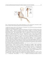
In flexion, extension and lateral bending, there is a variation of the SFU’s instantaneous
centre of rotation that, by an elliptical instantaneous axis, falls in the posterior half and
inferior discal margin (picture 1).
Picture 1. SFU’s instantaneous centre of rotation in X-Y-Z axis motion.
The artificial intervertebral disc, in a stand alone way, under ordinary and extraordinary
load, has to perform and guarantee all these.
The Kinematical classification of the intervertebral disc prosthesis, based on the own
different free motion degree, organizes the devices in 3 categories.
unconstrained
constrained, and,
semiconstrained
Devices in each of the above 3 groups have advantages and disadvantages.
The first have a variable centre of rotation.
They permit anterior/posterior/lateral translation, rotation, guarantee a more physiological
centre of rotation, but give more stress to the articular joints.
Prefer the preservation of the posterior longitudinal ligament (PLL) during their collocation
,
“forgiven” minimally positional imperfections.
The second have a fix centre of rotation.
They permit only rotation, have a less physiological centre of rotation, and give less stress to
the articular joints.
Another example of classification is reported in the picture 2.