Modern Telemetry Part 3 doc
Bạn đang xem bản rút gọn của tài liệu. Xem và tải ngay bản đầy đủ của tài liệu tại đây (1.47 MB, 30 trang )
Communication Strategies for Various Types of Swallowable Telemetry Capsules
51
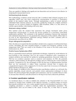
The MPE is the highest power or energy density of an RF source that is considered safe, i.e.
that has a negligible probability for creating damage. Since the MPE is regulated from the
outside of the body, it could be used as a guideline for the amount of RF radiation inside the
body.
Fig. 17. The maximum permissible exposure regulation.
In order to determine the RF band, the body attenuation, MPE, and data rate have to be
considered. Since the antenna efficiency is extremely low at low frequencies (<100 MHz), the
length of the antenna has to be longer than the size of the small pill. However, the low
frequency modulation requires less power consumption and radiation power because the
human body does not attenuate the low frequency. Therefore, early capsule type telemetry
systems were designed to use the FM method and used a long and flexible antenna. Since
early telemetry systems did not require a high data rate, this was sufficient except for the
repulsion of its shape.
With the advent of capsule endoscopy, the data rate has to be increased so as to be sufficient
enough for transmission of gastrointestinal images. Fig. 18 shows an example of capsule
endoscopes. The analog type can transmit the National Television Standards Committee
(NTSC) format, which is widely used for analog TV transmission, and the physician can
monitor the inside of the gastrointestinal tract as if watching an analog television. Since the
NTSC uses the analog transmission technology, it could provide a high fame rate (30
frame/s) but it is weak to channel noise; further, restoration of the data is impossible. Fig 18
(b) shows digital type capsule endoscope that could transmit 640×480×8 resolution images
by using a digital transmitter. Since a digital receiver can restore the data from
environmental noises, the frame rate of the capsule is reduced to 1 frame/s. Fig. 18 (c) shows
images taken from the ileum and esophagus by using a digital type transmitter capsule.
In order to transmit at a high data rate, the RF frequency has to be increased so as the make
the antenna effective. For capsule endoscopy, the 430 and 1200 MHz the industrial, scientific
and medical (ISM) bands are widely used to transmit the signal. These bands can transmit
higher data rate than the FM band and the human body attenuation is moderate enough to
Modern Telemetry
52
allow the signal to penetrate the body. Also, the size of the antenna should be small enough
that it can be inserted into the capsule. For these reasons, the Federal Communications
Commission (FCC) decided to create the Medical Implant Communication Service (MICS)
for the use of the frequency band between 402 and 405 MHz for communication with
medical implants. It allows bidirectional radio communication with pacemakers or other
electronic implant devices.
Fig. 18. Example of capsule endoscopy. (a) NTSC format transmitter. (b) VGA resolution
transmitter. (c) Image taken from the VGA resolution transmitter capsule.
2.4 GHz is widely used for commercial WLAN, and there are many commercial antennas
and transceivers for it. Unfortunately, the body attenuation at the 2.4 GHz is too high that it
could attenuate up to -50 dBm at a 15 cm body thickness. Therefore, the 2.4 GHz band is not
suitable for uses with implants or swallowable telemetry systems. Table 1 summaries the RF
frequency efficiency of the various RF bands.
Most swallowed capsule designs have used conventional modulation such as FM or AM
because of their simplicity. Since capsule telemetry is not widely used, encrypt and spread
spectrums were not taken into account. Also, the concept of the UWB fits well with capsule
endoscopy because the transmitter does not require a large space and power consumption is
lower than that of the conventional transmitters. However, the human body attenuates high
frequency signals, and this cannot be overcome by using equalization. There is one trial
using UWB for capsule endoscopy, and the frequency was reduced to 800 MHz and a
transceiver was implemented. Even though this proposed system violates the regulation of
the UWB, it could be useable if the upper frequency were limited.
Communication Strategies for Various Types of Swallowable Telemetry Capsules
53
Comparatives
300 MHz
Range
400 MHz
Range
900 MHz
Range
1200 MHz
Range
2400 MHz
Range
Safety level Best
Best
Moderate
MPE [dBm/cm
2
] 0 1.25 4.78 6.02 6.99
Attenuation Best
Good
Worst
15 cm Body
attenuation [dB/cm
2
]
14 15 20 24 41
Power
transmission
Best
Good
Worst
Availability
ISM Band ISM Band
ISM Band
Antenna Size
efficacy
Worst
Good
Best
Table 1. RF frequency efficiency of the various the RF bands
Another method is using an OFDM that can transmit a large bandwidth within a limited
frequency band, but it requires FFT/IFFT modules that consume too much power. Since the
capsule uses small batteries that typically have a capacity of less than 100 mAh, it is not easy
to implement a low power FFT/IFFT block.
The SDR method is good for the swallowable capsule because it can support the various
types of transmission signals. When the SDR is developed, patients will only need to receive
signals in one receiver from many transition sources. When the protocols of swallowable
capsule are open, this could become possible.
Table 2 summarizes various types of telemetry systems for capsules. Various modulation
methods, frequencies, and RF power levels were used for various applications. Usually, FM
modulation is used for moderate data rates and AM is used for simple and low power
purposes. In additionally, SDR and UWB appear feasible but their details have not been
fully described.
Reference
Frequency
(MHz)
Data rate
(kbps)
Modulation
Power
consumption
(mW)
RF power
(dBm)
Thone et al. 144 2000 FSK - - 18
Chen et al. 433 267 FSK 24 -
Wang et al. - - AM 125 Variable
Kfouri et al. UHF 250 - - -
Park et al. 315 - AM - -
Mackay
et al.
433 - FM 15.5 -
Woo et al. 1200 2000 SDR - -
Lee et al. 1200 2000 FSK 29.7 -
Intromedic - -
Manchester
code
- -
Table 2. Various types of applications of swallowable telemetry capsule
Modern Telemetry
54
3. Conclusion
In this chapter, brief explanations of modern communication strategies are explained and
the limitations of their use in swallowable telemetry systems are described. Selections of the
RF band and modulation methods are described and compared with each other. Since the
human body attenuates high frequency RF power, their use in sophisticated communication
is limited.
4. Acknowledgment
I’d like to thank Qun Wei and Zia Moth-Un-Din for their support of drawing the pictures.
This book was supported by a grant of the Institute of Biomedical Engineering Research,
Kyungpook National University, Republic of Korea.
5. References
[1] Dinslage, S., J. McLaren, and R. Brubaker, Intraocular pressure in rabbits by telemetry II:
Effects of animal handling and drugs. Investigative Ophthalmology & Visual Science,
1998. 39(12): p. 2485-2489.
[2] Hawkins, P., Telemetry in the field: Practical refinements to improve animal welfare.
Comparative Biochemistry and Physiology a-Molecular & Integrative Physiology,
2007. 146(4): p. S84-S84.
[3] Johnson, D.S., et al., Continuous-time correlated random walk model for animal telemetry data.
Ecology, 2008. 89(5): p. 1208-1215.
[4] Johnson, D.S., et al., A general framework for the analysis of animal resource selection from
telemetry data. Biometrics, 2008. 64(3): p. 968-976.
[5] Kong, W., et al., A semi-implantable multichannel telemetry system for continuous electrical,
mechanical and hemodynamical recordings in animal cardiac research. Physiological
Measurement, 2007. 28(3): p. 249-257.
[6] Kutsch, W., Telemetry in insects: the "intact animal approach". Theory in Biosciences, 1999.
118(1): p. 29-53.
[7] Nations, C.S. and R.C. Anderson-Sprecher, Estimation of animal location from radio telemetry
data with temporal dependencies. Journal of Agricultural Biological and
Environmental Statistics, 2006. 11(1): p. 87-105.
[8] Salvatori, V., et al., Estimating temporal independence of radio-telemetry data on animal
activity. Journal of Theoretical Biology, 1999. 198(4): p. 567-574.
[9] Walisser, J., et al., Optimizing Telemetry Stock Animal Quality: Implementation of Monthly
Signal Checks and Assessment of Transmitter Battery Life. Journal of the American
Association for Laboratory Animal Science, 2010. 49(5): p. 721-721.
[10] Ko, W.H., et al., Studies of MEMS Acoustic Sensors as Implantable Microphones for Totally
Implantable Hearing-Aid Systems. IEEE Transactions on Biomedical Circuits and
Systems, 2009. 3(5): p. 277-285.
[11] Yoon, K.W., et al., Telemetry capsule for pressure monitoring in the gastrointestinal tract.
Ieice Transactions on Fundamentals of Electronics Communications and Computer
Sciences, 2006. E89a(6): p. 1699-1700.
[12] Browning, C., et al., A New Pressure Sensitive Ingestible Radio Telemetric Capsule. The
Lancet, 1981. 318(8245): p. 504-505.
Communication Strategies for Various Types of Swallowable Telemetry Capsules
55
[13] Mackay, R.S. and B. Jacobson, Endoradiosonde. Nature, 1957. 179(4572): p. 1239-1240.
[14] Connell, A.M. and E.N. Rowlands, Wireless Telemetering from the Digestive Tract. Gut.,
1960. 1(3): p. 266-272.
[15] Banerjee, R. and D.N. Reddy, Bravo capsule pH monitoring. American Journal of
Gastroenterology, 2006. 101(4): p. 906-906.
[16] Belafsky, P.C., et al., Wireless pH testing as an adjunct to unsedated transnasal esophagoscopy:
The safety and efficacy of transnasal telemetry capsule placement. Otolaryngology-Head
and Neck Surgery, 2004. 131(1): p. 26-28.
[17] Chaw, C.S., E. Yazaki, and D.F. Evans, The effect of pH change on the gastric emptying of
liquids measured by electrical impedance tomography and pH-sensitive radiotelemetry
capsule. International Journal of Pharmaceutics, 2001. 227(1-2): p. 167-175.
[18] Pandolfino, J.E., Bravo capsule pH monitoring. American Journal of Gastroenterology,
2005. 100(1): p. 8-10.
[19] Holloway, R.H., Capsule pH monitoring: is wireless more? Gut, 2005. 54(12): p. 1672-1673.
[20] Thorne, P.S., C.P. Yeske, and M.H. Karol, Monitoring Guinea Pig Core Temperature by
Telemetry during Inhalation Exposures. Toxicological Sciences, 1987. 9(3): p. 398-408.
[21] O'Brien, C., et al., Telemetry pill measurement of core temperature in humans during active
heating and cooling. Medicine and Science in Sports and Exercise, 1998. 30(3): p. 468-
472.
[22] Iddan, G., et al., Wireless capsule endoscopy. Nature, 2000. 405(6785): p. 417-417.
[23] Svarta, S., et al., Diagnostic yield of repeat capsule endoscopy and the effect on subsequent
patient management. Canadian Journal of Gastroenterology, 2010. 24(7): p. 441-444.
[24] Spada, C., et al., Capsule endoscopy in Italy: An unbalanced review of the literature.
International Journal of Technology Assessment in Health Care, 2010. 26(3): p. 354-
356.
[25] Spada, C., et al., PillCam Colon Capsule Endoscopy (PCCE) for Colon Exploration: A Single
Centre Italian Experience. Gastrointestinal Endoscopy, 2010. 71(5): p. Ab203-Ab203.
[26] Woo, S.H., et al., High Speed Receiver for Capsule Endoscope. Journal of Medical Systems,
2010. 34(5): p. 843-847.
[27] Menciassi, A., et al. Single and multiple robotic capsules for endoluminal diagnosis and
surgery. in Biomedical Robotics and Biomechatronics, 2008. BioRob 2008. 2nd IEEE RAS
& EMBS International Conference on. 2008.
[28] Quirini, M., et al., Design and Fabrication of a Motor Legged Capsule for the Active
Exploration of the Gastrointestinal Tract. Mechatronics, IEEE/ASME Transactions on,
2008. 13(2): p. 169-179.
[29] Byungkyu, K., et al. Inchworm-Like Microrobot for Capsule Endoscope. in Robotics and
Biomimetics, 2004. ROBIO 2004. IEEE International Conference on. 2004.
[30] Elisa, B. and et al., Evaluation of friction enhancement through soft polymer micro-patterns in
active capsule endoscopy. Measurement Science and Technology, 2010. 21(10): p.
105802.
[31] Quirini, M., et al., Feasibility proof of a legged locomotion capsule for the GI tract.
Gastrointestinal Endoscopy, 2008. 67(7): p. 1153-1158.
[32] Woo, S.H., et al., Implemented edge shape of an electrical stimulus capsule. International
Journal of Medical Robotics and Computer Assisted Surgery, 2009. 5(1): p. 59-65.
Modern Telemetry
56
[33] Park, H.J., et al., New method of moving control for wireless endoscopic capsule using electrical
stimuli. Ieice Transactions on Fundamentals of Electronics Communications and
Computer Sciences, 2005. E88a(6): p. 1476-1480.
[34] Glass, P., E. Cheung, and M. Sitti, A Legged Anchoring Mechanism for Capsule Endoscopes
Using Micropatterned Adhesives. Biomedical Engineering, IEEE Transactions on,
2008. 55(12): p. 2759-2767.
[35] Woo, S.H., T.W. Kim, and J.H. Cho, Stopping mechanism for capsule endoscope using
electrical stimulus. Medical & Biological Engineering & Computing, 2010. 48(1): p.
97-102.
[36] Nagaoka, T. and A. Uchiyama. Development of a small wireless position and bleeding
detection sensor. in Microtechnology in Medicine and Biology, 2005. 3rd IEEE/EMBS
Special Topic Conference on. 2005.
[37] Menciassi, A. and P. Dario. Miniaturized robotic devices for endoluminal diagnosis and
surgery: A single-module and a multiple-module approach. in Engineering in Medicine and
Biology Society, 2009. EMBC 2009. Annual International Conference of the IEEE. 2009.
[38]
[39] Gao, Y.J., et al., Endoscopic capsule placement improves the completion rate of small-bowel
capsule endoscopy and increases diagnostic yield. Gastrointestinal Endoscopy, 2010.
72(1): p. 103-108.
[40] Kim, H.M., et al., A Pilot Study of Sequential Capsule Endoscopy Using MiroCam and
PillCam SB Devices with Different Transmission Technologies. Gut and Liver, 2010. 4(2):
p. 192-200.
[41] Lee, J., et al., CPLD based bi-directional wireless capsule endoscopes. Ieice Transactions on
Information and Systems, 2007. E90d(3): p. 694-697.
[42] Yuan, G., et al. Low power ultra-wideband wireless telemetry system for capsule endoscopy
application. in Robotics Automation and Mechatronics (RAM), 2010 IEEE Conference on.
2010.
[43] Thone, J., et al., Design of a 2 Mbps FSK near-field transmitter for wireless capsule endoscopy.
Sensors and Actuators a-Physical, 2009. 156(1): p. 43-48.
[44] Xinkai, C., et al., A Wireless Capsule Endoscope System With Low-Power Controlling and
Processing ASIC. Biomedical Circuits and Systems, IEEE Transactions on, 2009. 3(1):
p. 11-22.
[45] Kfouri, M., M. Kfouri, and M. Kfouri, Toward a miniaturised wireless fluorescence-based
diagnostic imaging system. IEEE J. Selected Topics in Quantum Electronics, 2008. 14.
[46] MacKay, R.S., Bio-Medical Telemetry: Sensing and Transmitting Biological Information from
Animals and Man. 1998: John Wiley & Sons.
3
Inductively Coupled Telemetry in
Spinal Fusion Application Using
Capacitive Strain Sensors
Ji-Tzuoh Lin, Douglas Jackson, Julia Aebersold,
Kevin Walsh, John Naber and William Hnat
University of Louisville
USA
1. Introduction
Titanium or stainless steel rods are implanted to stabilize vertebrae movement during
spinal fusion surgery, which allows bone grafts to fuse two or more vertebrae.
Radiograph images (x-rays), computed tomography scans (CT) and magnetic resonance
imaging (MRI) procedures are used to assess fusion progress and diagnose problems
during patient recovery. However, the imaging techniques yield subjective results
(Vamvanij et al.,1998) and as a consequence, result in unnecessary exploratory surgeries
to ascertain the efficacy of the spinal fusion surgery. As the grafted bone fuses, the
bending strain of the implanted rods decreases as the load is transferred to the fused
vertebrae (Kanayama et al., 1997). Strain is measurable on the spinal fusion fixture,
normally a stainless or titanium rod. In other words, the amount of strain is an indicator
of the load applied to the rod. Therefore, it is proposed that the strain on the implant rods
can be used as an alternative and non-invasive method to monitor the progress of spinal
fusion (Hnat et al., 2008).
This chapter will demonstrate the realization of a telemetric strain measurement system for
the spinal fusion detection as illustrated in Fig. 1. The system is composed of three major
components: a sensitive strain sensor, a battery free transducer circuit that wirelessly
interfaces the strain sensor, and an external interrogating reader that provides power to the
implant as well as collects strain information from the transducer circuit. Research has
shown that less power is consumed by a capacitive sensor than the resistive counterpart
(Puers, 1993). In addition, the sensors require high sensitivity to eliminate the need for
amplification that would require additional power. Therefore, the novel capacitive strain
sensors are developed to meet both the power and sensitivity demand. Additional, in
making the measurements a bodily-like situation, the sensor system, including the
transducer circuit, is assembled on a housing (Aebersold et al., 2007) that is capable of
transferring the strain from the rod to the sensor and accommodating for the size constrain.
The testing loads on the rods will be provided by a material test system (MTS) with a
corpectomy model fixture.
Although most strain sensors are capable of measuring axial strain due to tension and
compression or their equivalents derived from bending, a sensitive bending strain sensor
Modern Telemetry
58
that only responds to bending strain is also desirable for spinal fusion purpose. The strain
sensor is expected to measure 1000 με based on an adult of 200 pounds in a corpectomy
model under bending with 2 stainless spinal fusion rods (6.4 mm in diameter and 50.8 mm
long) implanted (Gibson, 2002).
Fig. 1. A strain gauge telemetry application in spinal fusion.
MEMS capacitive sensors using wireless data transmission have been evaluated in many
applications such as humidity (DeHennis & Wise, 2005; Harpster et al., 2002;),
temperature (DeHennis & Wise, 2005) and pressure sensing devices (Akar et al., 2001;
Chatzandroulis et al., 2000; DeHennis & Wise, 2002, 2005; Strong et al., 2002). The
telemetry approach to monitor strain uses inductively coupled battery-less technology
similar to the technology used in Radio Frequency IDentification (RFID) devices
(Finkenzeller, 1999). Some examples of the early applications are shown in Table 1. The
inductively coupled wireless system with sensing capability needs not only the working
passive telemetry circuitry, but also both the sensor interface circuitry and the sensor
themselves. A fully integrated implanted sensor system was realized (Chatzandroulis et
al., 2000) with a capacitive pressure sensor and an application-specific integrated circuit
(ASIC) chip that controls RF modulation and converts capacitance variations into
frequency variations. Suster et al. developed a wireless strain detection with the
transducer coil size of 3-inch coaxial to the interrogating reader (Suster et al., 2005).
However, this transducer coil size is not desirable for spinal fusion implant. Research
using this technique coupled with MEMS sensors has become widespread in biomedical
applications. It is a promising approach for orthopedic implant sensors and the key is a
highly sensitive capacitive sensor (Benzel et al., 2002).
Inductively Coupled Telemetry In Spinal Fusion Application Using Capacitive Strain Sensors
59
Author, year
Chatzandroulis
et al.
2000
DeHennis et al.
2002
DeHennis et al. 2005 Suster et al. 2005
Method Backscattering
Backscatterin,
C-F converter
Backscattering, C-F
converter
Backscttering, C-
F, F-V converter
Sensor
Capacitive
pressure
Capacitive
sensor
Capacitive sensor
Capacitive strain
sensor
Range 4cm 1 inch
Frequency 40.68MHz 800KHz 3.18MHz 50MHz
Secondary coil 4.5mmx7.5mm
Co-axial 3 inches
coils
Applications Pressure sensor Pressure
Pressure, humidity
and temperature
Strain
Overall sensor
and circuit size
450μm in
diameter
2mm x 2mm
2mmx2mm
sensor on chip
with circuit
4.5mmx7.5mmx1mm
1000μm
Mounting ASIC chip On silicon On silicon
Testing method 3-point bending
Circuit type C/F converter
CMOS ring
oscillator
Current source and
relaxation oscillator
Voltage output
Number of
channels
1 3 channels 3 channels 1 channel
Reader type
MC68HC705
micro-controller
Class E
amplifier
Class E amplifier
Strain/pressure
range
1000 μs
Dynamic/static Dynamic/static Dynamic/static Dynamic/static
Capacitance
range
5pF – 33pF 0.5pF-6pF 440fF
Table 1. Some details of the inductively coupled detection systems
Modern Telemetry
60
In the next sections, the highly sensitive MEMS bending strain sensor will be described in
great detail followed by the system circuitry and the testing methods.
2. The MEMS strain sensors
This section focuses on the development and fabrication of the custom bending strain
capacitive sensing element needed for the spinal fusion measurement implant (SFMI)
applications. This application requires a high bending strain sensitivity with enough
nominal capacitance to avoid loss due to parasitic capacitance, compatibility with an
inductively powered circuit, and suitable dimensions for system packaging. The sensitive
bending strain sensor is expected to be packaged in a housing container that attaches to
the diameter spinal fusion rod. The distance between two vertebrae is about 25.4 mm in
the lumber region, making the maximum length of the housing limited to approximately
12 mm long. Therefore, it is desirable that the sensor length be less than 10 mm. The
housing is installed between two pedicle screws and needs to transfer the bending strain
from the rod to the sensor as described in (Aebersold et al. 2007). The curved surface of
the rod is compensated with the 2 mm thick plastic housing which conforms to the rod
and is trimmed 1 mm down to provide a flat area of 2 mm x 10 mm for the sensor to
mount.
Certain characteristics were primarily considered when reviewing limited examples of
previous parallel plate capacitive strain sensors in the literature. The basic concept of the
capacitive strain sensor features a pair of metalized parallel plates with a dielectric gap. The
sensing mechanism manifests itself in varying either the area of the plate, the gap between
the plates, or the dielectric medium between the plates. A number of parallel plate sensor
designs with a variable air gap were analyzed in the early 90’s (Procter & Strong 1992).
These sensors generally exhibited low nominal capacitance and sensitivity due to the large
gap. In an attempt to increase the nominal capacitance in a non-air gap design, it was
demonstrated by a sensor with a parallel plate structure and a thick-film dielectric material
(Arshak et al., 1994). The dielectric film between the two plates was compressed during
bending, thus expanding the film in area and decreasing the thickness from the perspective
of the electrodes. These changes in the film geometry lead to a high gauge factor of 75-80
with a 15-25 μm gap based on a uniform model. The capacitive gauge factor is defined by
the fractional change in capacitance with respect to strain. This thick-film dielectric
produced both capacitive and resistive responses to strain making this approach electrically
unique, but undesirable for the SFMI application due to power consumption. In another
design, more effort was involved to invoke the change in permittivity of a dielectric material
resulting in a gauge factor of 3.5 to 6, with a 150 μm gap (Arshak et al., 2000). This variable
permittivity approach exhibits limited sensitivity that showed no dependency on its
dimension (the gauge factor is constant and only depends on the “piezocapacitive” effect).
This low gauge factor approach would require additional circuitry that is not desired for
this implant design.
2.1 The bending sensor theory
The mechanism of sensing pure bending on a test substrate is described in two folds: the
capacitance and the strain condition imposed on the sensor, as illustrated in Fig. 2.
Assuming the bending sensor is attached to a steal cantilever with length L and thickness R
in an elastic bending.
Inductively Coupled Telemetry In Spinal Fusion Application Using Capacitive Strain Sensors
61
Fig. 2. The sensor on a substrate bar under bending. (b) The sensor’s gap D
0
+D(x), zoomed
in from above, varies as a function of position x. (c) shows the respect metal coordinates on
the cantilever substrate.
The capacitance from two parallel electrode plates is given by
D
A
C
r
εε
0
= (1)
where A is the area, D is the distance between two parallel plates,
ε
0
is the permittivity and
ε
r
is the dielectric constant of the material between the plates. In order to measure the strain
magnitude, a cantilever test substrate is utilized. For strain and capacitance calculations, it is
assumed that the dimensions of the cantilever test substrate very large compared to the
sensor and that the sensor is firmly affixed to the substrate. For a cantilever beam, the
moment of inertia, I, is given by
12
3
WT
I = (2)
where, W is the beam width and T (or R as shown in Fig. 1) is the beam thickness. For a
beam in uniaxial state of stress, the strain at any point on any surface under bending is given
by a textbook (Hibbleer, 1997),
2
6
EWT
Fd
EI
Mc
E
===
σ
ε
(3)
where
σ
is the stress on the surface, E is the Young’s modulus of the steel bar substrate, M is
the bending moment, c is the distance from the neutral axis to the surface, F is the force
Modern Telemetry
62
applied at the free end of the beam and d is the sensor location from the free end of the
beam. The sensor location on the beam is given by
2
24
L
L
Ld
+
=
(4)
where L is the length of the cantilever substrate, L
4
and L
2
are the longitudinal boundaries
that define the bottom beam of the sensor. Fig. 2a shows the sensor location on the bent
cantilever test substrate. Fig. 2b is the side view of a bending condition of the sensor design
depicted in Fig. 2c, showing the sensor’s metal layer coordinates and the widened gap,
D
0
+D(x). Figs. 2c also shows the details of the top and bottom electrode while under
bending for the designs of interest. The initial sensor capacitance is given by
prr
C
D
LMw
D
MLw
C +
)(
+
)(
=
0
112
0
0
121
00
εεεε
(5)
where L
1
marks the beginning of the metal layer on the bottom electrode, L
2
not only
represents the boundary of the sensor but also the end of the metal layers on both the
bottom and top electrode beams and therefore, L
2
-L
1
=L
0
is the effective electrode length.
With various designs, M
1
is a variable that represents the start of the metal layer on the top
electrode beam and also ends the trace that connects the electrode to the pad on the bottom
beam. Therefore, w
1
(L
2
-M
1
) represents the area of the overlapping metal plates, w
2
(M
1
-L
1
)
the area of the metal trace, and D
0
the initial spacing between the plates (see Figs. 2b-2c). The
first term represents the capacitance of the overlapping metal plates. The second term is the
capacitance of the trace between the electrode and the pad. The third term, C
p
, is the
parasitic capacitance of the metal traces between L
1
and L
3
combined with the planar pads
between L
4
and L
5
. L
3
is also the pivot point where the gap starts and L
5
is the physical
boundary of the top electrode beam. Capacitance calculations for planar pads indicate that
the third term is 0.035 pF (Baxter, 1997). In order to estimate sensor sensitivity to strain, the
capacitance change caused by an applied strain is calculated using standard beam
equations. The sensor metal plate attached to the beam will follow the beam deflection while
the initially parallel plate will remain straight under deformation. The deflection of a
cantilever beam and the attached sensor metal plate is given by (Hibbleer, 1997),
)(=)(
32
-3
6
-
xLx
E
I
F
xv
(6)
where the v(x), as seen in Fig. 2a, is the vertical displacement at position x on the beam. The
initially parallel plate remains straight and its position is represented by a line tangent to the
deformed beam at the pivot point of the sensor. The tangent line (see Fig. 2a) is given by
bxxxv
t
+)(=)(
θ
(7)
where
θ
(x) is the slope at x and b is a constant determined by a boundary condition. The
slope is determined from the first derivative of the deflection and given by
)(=)(
2
-2
2
-
xLx
E
I
F
x
θ
(8)
At the sensor pivot point, L
3
, from Fig. 4b, the deflection of the two metal plates is equal,
providing the boundary condition
Inductively Coupled Telemetry In Spinal Fusion Application Using Capacitive Strain Sensors
63
)(=)(
33
LvLv
t
(9)
The constant b from (7) is solved by combining (6), (8) and (9) at point L
3
and becomes
()
3
3
2
3
2-3
6
LLL
EI
F
b = (10)
Therefore, the tangent line is expressed as
()
()()
3
3
2
3
2
33
2-3
6
-2
2
-
LLL
EI
F
xLLL
EI
F
xv
t
+= (11)
The increased gap (see Fig. 2b) between the two electrode plates is a function in the x-
direction and expressed as
)()(=)( xvxvxD
t
- (12)
The capacitance change is determined by calculating the average distance between the two
metal plates of the strain sensor. The average displacement, in addition to the initial gap,
between main metal layers is expressed as
2
1
10
21
1
(() ())
()
L
t
M
DD vxvxdx
LM
=+ −
−
(13)
where M
1
is where the sensing portion of metal starts and L
2
where it ends. The capacitance
due to the trace has an average displacement of D
2,
which is expressed as
2
1
20
11
1
(() ())
()
M
t
L
DD vxvxdx
ML
=+ −
−
(14)
where the metal stops at L
1.
Capacitance, due to beam deformation, C
f
, is given by
prrf
C
DD
LMw
DD
MLw
C +
+
)(
+
+
)(
=
20
112
0
10
121
0
εεεε
(15)
Combining (3), (13), (14) and (15), yields
pr
rf
C
LMdT
LMLLLLMLLLLMLML
D
LMw
MLdT
MLLLLMLLLLMLMLL
D
MLw
C
+
)(
)))((+))((+)()((
+
)(
+
)(
)))((+))((+)()((
+
)(
=
11
2
1
2
3
2
311
3
3
2
3
4
1
4
1
3
1
3
1
0
112
0
12
2
1
2
23
2
312
3
3
2
3
4
1
4
2
3
1
3
2
0
121
0
-3
-13-
2
3
-2-3-
4
1
-
-3
-3-
2
3
-2-3-
4
1
-
ε
εε
ε
εε
(16)
Based on the equation above, the bending strain sensor is analytically formulated and to be
compared with the fabricated MEMS sensor in the following section.
Modern Telemetry
64
2.2 Strain sensor fabrication
The sensor fabrication process is illustrated in Fig. 3. The materials include borosilicate
glass (Pyrex Corning 7740, 500 μm thick) and silicon wafers (p-type, (100), 1-10 ohm-cm,
double side polished, 310 μm thick). Fabrication began with clean glass and silicon
substrates as shown in Figs. 3a and 3c. An electrode, traces, and a pair of contact pads
were patterned onto the glass substrate by sputtering 0.02 μm chromium for adhesion
layer followed by 0.2 μm of gold. The metal trace leading to the bonding area makes
electrical contact with the silicon side electrode after anodic bonding. Wet etching was
used to pattern the metal (Fig. 3b).The silicon wafer was wet oxidized (Fig. 3d), patterned
using photolithography and etched with buffered oxide etch (BOE) solution to form an
oxide mask for silicon surface machining. The wafer was etched using potassium
hydroxide (KOH) at 85°C (approximately 0.7 μm / minute) to form recessed features and
created the initial gap spacing. The etching mask was removed using BOE leaving two
Fig. 3. Cantilever bending strain sensor fabrication process. Illustrations on the left are the
side views and on the right are the top views. (a) Pyrex (Coring 7400) glass, (b)sputter of
Au/Cr on glass as one electrode, (c) silicon substrate, (d) oxidation of the silicon as the
etching mask, (e) etching silicon with KOH to create platforms for anodic bonding with
glass, (f) sputter Au/Cr on silicon as the other electrode, (g) side view of partial dicing
(arrows marks) after glass and silicon are anodically bonded, (h) the individual sensor after
final separation, noting the gap between the two electrodes.
Inductively Coupled Telemetry In Spinal Fusion Application Using Capacitive Strain Sensors
65
silicon islands, which function as anchor platforms for the anodic bonding interface, as seen
in Fig. 3e. An electrode and trace were then sputtered and patterned onto the silicon using
the previously described metallization process. The small contact area on the raised anchor
connected the pad on the glass plate with the electrode on the silicon plate via the traces, as
seen in Fig. 3f.
The glass and silicon wafers were stacked with the metal surfaces facing each other and
visually aligned using a mask aligner. Methanol was used to temporarily maintain
alignment. The substrates were anodically bonded at 450
o
C on a grounded hotplate using a
pointed probe to selectively place a -1000 V source on the glass, as shown in Fig. 3g. A gap is
created between the electrodes on glass and silicon. This technique of selectively applying
the electric field and bonding pressure prevented the recessed spaces from bonding to each
other due to thermally induced warpage and electrostatic attraction. An automated dicing
saw equipped with a 250 μm thick diamond blade was used to separate the individual
sensor die from the bonded wafers. The silicon substrate was diced nearly through at the
area above the contact pads. This was accomplished by limiting the depth of the cut and
using the dicing alignment marks previously patterned on the silicon. Cuts to individually
remove the sensors were similarly made from the silicon and glass substrate leaving
approximately 30 μm of each substrate’s depth (Fig. 3g). Care was taken to avoid chipping
and prevent debris from filling the sensor gap. The sensors were separated from the wafer
manually by flexing them to break the remaining thin substrate (Fig. 3h).
Sensors with less than 3 μm gap have been fabricated, but with unreliable capacitance
values and low yield. It is because of the collapsing of the two electrodes during the anodic
bonding process. In an effort to maintain high nominal capacitance, preserve sensitivity and
promote linearity, a sensor with an electrode area of 2 mm x 4 mm, and a gap of 3 μm was
fabricated for final SFMI prototyping. This sensor was tested on a spinal fusion rod with a
near-linear response, as shown in Fig. 4.
Fig. 4. Comparison of the calculation and experimental results of a strain sensor glued to a
spinal fusion rod.
Gauge factor is defined as
ε
C
d
C
GF =
(2)
Modern Telemetry
66
where dC/C is the fractional change of capacitance and ε is the strain. Using a linear fit of
the differential capacitance data graphed in Fig 5, the gauge factor was ploted and
calculated to be 252 for 0 to 1000
με. This value is extremly high in comparison to the current
literature (Arshak 1994, 2000; Proctor & Strong 1992). By comparison, piezoresistive gauges
typically provide a gauge factor less than 200 (Fraden, 1995) even at the cost of high
temperature sensitivity.
Fig. 5. Comparison of the calculation and experimental results of a strain sensor glued to a
spinal fusion rod.
3. The transducer circuit
The transducer circuit is an inductively coupled, load modulated design similar in concept
to Radio Frequency IDentification tags used for inventory and security. The 125 kHz
magnetic field sourced by the interrogating reader induces a voltage in the LC tank of the
implant. The LC tank (1 cm diameter, 600 turns) is resonant at 125 kHz. The AC voltage is
then rectified, filtered, and regulated using a low quiescent power regulator. A supply of
3VDC, 28
μA is required to power the oscillator circuit described above and a frequency
divider circuit composed of flip-flops. The oscillator frequency is divided to less than 1/20
the carrier frequency so that detection is simplified. The frequency divider also buffers the
oscillator signal so that it can drive the gate of a MOSFET placed across the LC tank. The
MOSFET acts a load that modulates the 125 kHz tank output at the divider output
frequency. A diagram that shows the functionality can be seen in Fig. 6(a).
As it is functioned as an oscillator in Fig. 6(a), a capacitance to frequency (c-f) converter is
used to convert the strain signal to a signal that can be transfered wirelessly. The c-f circuit
is comprised of a pair of CMOS inverters (Lancaster, 1997) and can be seen in detail in Fig.
6(b). The oscillator produces a periodical voltage due to the charging and discharging of the
RC across a threshold of an inverter input. The frequency of the RC oscillator is expressed as
F=2.3R
t
C, where C is the variable capacitor, or a capacitive strain sensor, and R
t
is the
matched resistance of the oscillator. The resistor R
I
is to unload the RC network from
clamping effect of protection diodes in the inverter. It will also result in a nearly square
duty cycle and make the frequency less dependent of power supply variations. R
I
is
normally set about 10 times as higher as R
t
to minimize the effect of the protection diodes.
The oscillator oscillates empirically at 20 kHz with 20 pF capacitor and 100 kOhm resistor.
Inductively Coupled Telemetry In Spinal Fusion Application Using Capacitive Strain Sensors
67
Fig. 6. The block diagram of the (a) transducer circuit and (b) oscillator circuit.
4. The interrogating reader
The interogating reader operating on 12 VDC, 175 mA provides the 125 kHz magnetic field
for the implant, as illustrate in Fig. 7. The reader antenna is 24 cm in diameter and is tuned
to 125 kHz. An EM Microelectronic (Marin, Switzerland), EM4095 IC contains an on-chip
oscillator, antenna driver, and a demodulation circuit. The output of the demodulator is
measured using an Agilent 53131A counter and logged with a computer based data
acquisition system.
Fig. 7. The block diagram of the power reader.
4.1 Detection region
In the region of detection, see Fig. 8, the implant receives enough power to operate from the
magnetic field sourced by the reader. There is no degradation of strain sensing performance
Modern Telemetry
68
if sufficient DC power is available from the regulator. However at the far end of the region,
planar and axial alignment becomes very important. With distance from the reader,
inductive coupling is reduced thereby reducing the AC voltage across the LC tank and thus
the modulated signal amplitude. The data signal also relies on the same low coupling
between the implant and the reader. It is necessary to have a sensitive reader to detect the
implant at the far end of the region.
Fig. 8. The detection region is within the cone shape.
5. The testing methods
The corpectomy model was used to evaluate the strain measurement system prior to spine
testing. This model has similar bending to a four-point bending model therefore the rod
strain does not change significantly along the length of the rod. A single metal foil strain
gauge reference was attached to the rod adjacent to the housing for comparison to the
transducer system. Comparing the metal foil reference gauge to the spinal fusion sensor
placed in close proximity is justified. Figure 9 shows the sensor assembly on a housing
without a container cover.
Fig. 9. The transducer sensor system on a housing that attaches to a stainless rod before a
hermetically sealed container is assembled.
Inductively Coupled Telemetry In Spinal Fusion Application Using Capacitive Strain Sensors
69
The strain measurement system was tested using a corpectomy model designed as a
simplified mechanical analog of a spine section and then will be tested using a human spine.
Figure 10 shows a free body diagram of the forces and bending moments applied to the
spinal rods through the pedicle screws due to loading of the hard plastic blocks. Note that
when the spinal fusion rods are fastened to the fixtures, often the initial strain is introduced
and recalibrated. On the first test, the corpectomy apparatus was assembled inside a clear
acrylic water tank without water on the MTS machine for application of the load; the sensor
system was oriented facing out to be detected. The MTS machine’s dynamic motion only
changes 3 mm in the detection distance between the interrogating reader and the strain
sensor. The read range was limited to 10 cm due to the reader design used and the sensor
coil design constraints. A more optimal reader would increase the range. For other
applications where a larger coil diameter is acceptable the range would be increased as well.
Fig. 10. The corpectomy model: front view, top and side view, bottom.
The data acquisition is obtained with LabView software, an interfacing program designed to
transfer and record live data between instruments and computer by National Instrument. The
strain information is recorded by the commecial metal foil strain gauge through a strain
Modern Telemetry
70
indicator (Measurement group model P-3500). The frequency output from the wireless strain
sensor system measurement is obtained through the digital universal counter (Agilent
53131A). In normal use, the response time for the strain indicator is 0.5 second, and the
universal counter about 0.2 second. However, with the Labview interface the response time for
the universal counter changes to 2.3 seconds but does not change for the strain indicator. The
lag in time in the acquisition of the frequency data from the sensor system will cause false
information when recording a drastic dynamic motion. In order to have a consistent and
corresponding readings for the referred strain and the frequency output, a delay or pause in
the live measurement is needed. Therefore, the MTS machine is programmed to pause 5
seconds at every increment of load for the frequency output to stabilize. Both the strain and the
frequency data then are taken at the same time frame as the Material Test Machine.
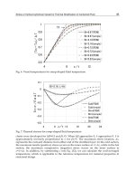
The result of the telemetry strain sensor detection in Fig. 11 shows good linearity with R
2
=
0.99. The gauge factor is calculated to be 249 up to 1000 micro-strain, with R
2
=0.96 as see in
Fig. 12. Similar gauge factor of 252 in the capacitance mode was measured in section 2. The
frequency mode also exhibits high gauge factor without any amplification.
Fig. 11. The demodulate frequency responses from the sensor transducer shows a linear
response due to bending test of the corpectomy model.
Fig. 12. The plot shows a linear gauge factor 249 in the frequency domain.
Inductively Coupled Telemetry In Spinal Fusion Application Using Capacitive Strain Sensors
71
5.1 Water tank test
For the next application in water tank test, the sensor is expected to be in a fluidic
environment. To protect from the influence of water or water vapor, the system was
hermetically sealed in a container made of nylon and painted heavily with silicone and
polyethylene. The container protected the device during corpectomy testing where it is
submerged in a water tank used to simulate the body and during spine testing where it is in
contact with tissue.
Caution has been taken for the temperature difference between water and ambient.
A temperature dependent test on the sensor shows that the frequency drift from 1752 to
1778 Hz from 0
o
C to 22.2
o
C and vice versa with no strain at the rate of about 1.8 Hz/
o
C.
The interrgating reader was not moved at about 10 cm from the sensor system, through
water, glass and air. The difference of the readings before the introduction of water and after
was unnoticeable. The corpectomy model in water test was successful in showing the
repeatability for the cyclic loads. It aslo suggestes that measurements were possible in
conditions present in-vivo. The tests of the corpectomy model in water tank were successful
in showing that measurements were possible in conditions present in-vivo.
5.2 Excise spine test
A discectomy was performed on an excised spine from a cadaver and was constrained and
loaded in a MTS system to simulate a 113.4 kg (250 lb) patient. The excised spine was potted
using a liquid lead/bismuth alloy (Cerro Products, Bellefont, PA) and attached to a MTS
Bionix mechanical testing system. A custom-built test apparatus was used to apply anatomic
loads to the spine. The intact spine was tested by applying loads ranging from 100 to 200 N
in an increasing and decrease manner with a 5-second period of pause at every increment to
accommodate the detection speed of the system. The disc between L3 and L4 was surgically
removed to simulate an unstable spine prior to fusion surgery, and the test sequence
repeated. The comparison of results before and after the disk is removed are shown in
Fig. 13.
Fig. 13. Measured strains during excised spine testing.
With the same loading steps on the spine from MTS, the spinal fusion rods experienced less
strain increment in the spine with the disk than that in the spine with the disk removed.
Modern Telemetry
72
From the wireless transmision data along with the referred metal foil strain gauge, it is
suggested that the spinal fusion rods showed that roughly one third of the load were shared
by the intact spine after the spine is fused. The telemetry system clearly shows the rigidity of
the intact spine.
6. Conclusion
A telemetry system using a capacitive strain sensor has been developed for the detection of
spinal fusion monitoring. The strain sensor was made using MEMS process with high
sensitivity and reduced size that serves the purpose for strain detection on the spinal fusion
rod. The cantilever structure of the sensor is composed of two parallel plates, galss and
silicon, respectively, with a narrow gap D
0
(3 μm) and a conjoint end. The bending strain
sensor has the characteristics of high nominal capacitance (20 pF), high sensitivity, and
compact dimensions (2 mm x 7 mm x 0.8 mm). It utilizes a variable gap configuration
comprised of silicon and glass beams that are bonded at one end and open at the opposing
end. This type of structure has been tested to withstand a strain range of 0 to 1000
με. The
inductive link between the implant circuit and the reader was sufficient for supplying
power to the implant circuit and extracting data at 10 cm distance. A specific sensor has a
linear gauge factor of 252 in the capacitive domain and 249 in the frequency domain.
Measurements made through air and water with a corpectomy model produced a linear
response consistent with a metal foil reference gauge. The strain measurement system was
also tested with the corpectomy model designed as a simplified mechanical analog of a
spine section and was then tested using a human spine. For the biomechanics application,
the sensor is expected to be in a fluidic environment. The tests of the corpectomy model
placed in water tank were successful in showing that measurements were possible in
conditions present in-vivo. The read range was limited to 10 cm due to the reader design
used and the sensor coil design constraints. Finally, a test performed using a human spine
showed the wireless implant detected strain roughly one third of the load were shared by
the intact spine after the spine is fused.
7. Acknowledgment
The authors would like to express the appreciation to Tommy Roussel, Tom Carroll, Don
Yeager, John C. Jones, Dr. Michael Voor, Dr. Rolando Puno and Robert L Burden for their
assistance with the modeling, test setups and surgery performance.
8. References
Aebersold, J.W.; Hnat, W.P.; Voor, M.J.; Puno, R.M.; Jackson, D.J.; Lin, J.T.; Walsh, K.M.&
J.F. Naber (2007). Development of a strain transferring sensor housing for a lumber
spinal fusion detection system, J. Med. Devices 1 (June 2007) 159-164.
Akar, O. ; Akin, T.& Najafi, K. (2001) A wire less batch sealed absolute capacitive pressure
sensor, Sensor and Actuator A 95 (2001) 29-38.
Arshak, K.I.; Collins, D. & Ansari, F. (1994). New high gauge-factor thick-film transducer
based on a capacitor configuration, Int. J. Electronics, 1994 vol. 77 No. 3, 387-399.
Inductively Coupled Telemetry In Spinal Fusion Application Using Capacitive Strain Sensors
73
Arshak, K.I.; McDonagh, D. & Durcan, M.A. (2000). Development of new capacitive strain
sensors based on thick film polymer and cement technologies, Sensors and
Actuators A 79 (2000) 102-114.
Baxter, L. K. (1997). Capacitive Sensors: Design and applications, IEEE press, New York,
1997 pp 17-73
Benzel, E. & Ferrara, L. ; Roy, S. & Fleischman, A. (2002) Clinical Neurosurgery, 49, 209-225
(2002).
Chatzandroulis, S.; Tsoukalas, D., & NeuKomm P. A. (2000) A miniature pressure system
with a capacitive sensor and a passive telemetry link for use in implantable
applications, Journal of Microelectromechanicalsystems Vol.9 No.1 March 2000.
DeHennis, A.& Wise, K.D. (2002), A double-sided single-chip wireless pressure sensor,
Digest IEEE conference on MEMS, (January 2002) Las Vegas. Pp 252-255. (2002) A
Passive-Telemetry-Based pressure sensing system, Digest of the Solid-state Sensor
and Actuator Workshop, (June 2002 ) Hilton Head,
DeHennis, A.& Wise, K.D. (2005). A wireless microsystem for the remote sensing of
pressure, temperature, and relative humidity, J. of MEMS Vol. 14, NO. 1 February
2005 12-22.
Vamvanij, V.; Fredrickson, B.E.; Thorpe, J.M.; Stadnick, M.E.& Yuan, H.A. (1998). Surgical
treatment of internal disc disruption: an outcome study of four fusion techniques,
Journal of Spinal Disorders, Oct.11 (5) (1998) 375-382.
Finkenzeller, K. (1999). RFID Handbook: Radio-frequency identification fundamentals and
applications, John Wiley & Sons, 1999 p 38.
Fraden, J. (1996) Hanbook of Modern Sensors (Springer-Verlag, New York, 1996)
Gibson, H. (2002). Measurement and finite element modeling of spinal rod strain, Master
thesis, Dept of Mechanical Engineering University of Louisville, May 2002.
Harpster, T.J.; Hauvespre, S.; Dokmeci, M. R. & Najafi, K. (2002). A passive humidity
monitoring system for in situ remote wireless testing of micropackages, J. of
MEMS. Vol.11 No.1 February (2002) 61-67.
Hibbeler, R.C. (1997). Mechanics of Materials, Prentice Hall, New Jersey, 3
rd
ed., 1997 pp 584
Hnat, W.; Walsh, K. & Naber J. (2008). US Patent No 7357037.
Kanayama, M.; Cunningham, B.W.; Weis, J.C.; Parker, L.M.; Kanoda, K. & McAfee, P.C.
(1997). Maturation of the posterolateral fusion and its effect on load-sharing of
spinal instrumentation, Journal Bone and Joint Surgery Am. Vol. 79 (11) (1997)
1710-1720.
Lancaster, D. (1997). CMOS cookbook, Howard W. Sams & Co. Inc. pp 226.
Lin, J T.; (2006). Development of a telemetry spinal fusion sensor system, Ph.D.
Dissertation, Electrical Engineering, University of Louisville, Louisville, KY.
Lin, J T.; Walsh, K. ; Jackson, D.; Aebersold, J.; Crain, M.; Naber, J. F. & Hnat, W. P. (2007).
Development of capacitive pure bending strain sensor for wireless spinal fusion
monitoring, Sens. Actuators,A,138 (2007) 276-28
Procter, E.& Strong, E. (1992) Capacitance strain gauges: strain gauge technology, Elsevier,
1992, PP 301-323.
Puers, R. (1993) Capacitive Sensor: When and how to use them, Sensors and Actuators A 37-
38 (1993) 93-105.
Strong, Z.A.; Wang, A.W. & C.F. Mcconagh (2002). Hydrogel-actuated capacitive transducer
for wireless biosensors, Biomed. Microdev. 4:2, (2002) 97-103.
Modern Telemetry
74
Suster, M.; Chaimanonart,; N.; Guo,J.; Ko, W. H. & Young, D. J. (January 2005). Remote-
Powered high-performance strain sensing microsystem, Technical Digest, the 18th
IEEE International Conference on Micro Electro Mechanical Systems, Miami,
Florida, January 2005, pp.255-258.
4
Ubiquitous Piezoelectric Sensor
Network (UPSN)-Based Concrete
Curing Monitoring for u-Construction
Seunghee Park and Dong-Jin Kim
Department of Civil and Environmental Engineering/u-City Design and
EngineeringSungkyunkwan University
Cheoncheon-dong Jangan-gu Suwon
Republic of Korea
1. Introduction
Recently, there has been increasing demand for high-rise buildings or wide-span bridges.
These structures are constructed with a mount of mass concrete. However, the concrete
might be susceptible to brittle fracture if the curing process is inadequate. Therefore, to
prevent this drawback, it is essential to predict the strength development of concrete during
the curing process. In addition, real-time monitoring of the curing strength is important for
reducing the construction time and cost because it can determine the appropriate curing
time to achieve sufficient strength to progress to the next phase safely. The in-situ strength
of concrete structures can be determined with a high precision by performing the strength
testing and/or material analysis on core samples removed from the structure (Irie et al.,
2008). However, this method might destroy the concrete structure. Therefore, a range of
methods based on the thermal, acoustical, electrical, magnetic, optical, radiographic, and
mechanical properties of the test materials have been developed to monitor the strength
development without damaging the host structures (ACI Committee 228, 2003; Lamind and
Pielert, 2006; Metha and Monterio, 2005). These methods typically measure certain
properties of concrete from which the strength and/or elastic constants can be estimated.
Among these techniques, several methods using a Schmidt hammer or an integrated
temperature have been normally used. However, these are unsuitable for use at construction
sites because they do not allow real-time monitoring of the curing process of concrete
structures at inaccessible places.
The recent advent of smart materials, particularly piezoelectric materials, can provide a
solution for the real-time monitoring for strength development. Electromechanical
impedance techniques that employ piezoelectric materials have emerged as a potential tool
for the implementation of a built-in monitoring system for civil infrastructures (Park G. et
al., 2000, 2003; Park S. et al. 2005, 2006, 2011). This technique utilizes high-frequency
structural excitation, which is typically > 20 kHz from surface-bonded PZT (Lead-Zirconate-
Titanate) patches, to sensitively monitor the changes in the mechanical impedance of the test
structures. Furthermore, the recent advances in online monitoring, including actuation and
sensing, on-board computing, and radio-frequency (RF) telemetry, have improved the