báo cáo hóa học: " Gait dynamics in mouse models of Parkinson''''s disease and Huntington''''s disease" doc
Bạn đang xem bản rút gọn của tài liệu. Xem và tải ngay bản đầy đủ của tài liệu tại đây (628.29 KB, 13 trang )
BioMed Central
Page 1 of 13
(page number not for citation purposes)
Journal of NeuroEngineering and
Rehabilitation
Open Access
Research
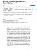
Gait dynamics in mouse models of Parkinson's disease and
Huntington's disease
Ivo Amende
†1
, Ajit Kale
†2
, Scott McCue
2
, Scott Glazier
2
, James P Morgan
1
and
Thomas G Hampton*
1,2
Address:
1
Division of Medicine, Beth Israel Deaconess Medical Center, Harvard Medical School, Boston, MA 02215 USA and
2
The CuraVita
Corporation, Boston, MA 02109 USA
Email: Ivo Amende - ; Ajit Kale - ; Scott McCue - ;
Scott Glazier - ; James P Morgan - ; Thomas G Hampton* -
* Corresponding author †Equal contributors
Gait variabilityGaitMouse modelsNeurodegenerationMovement disordersAmyotrophic Lateral SclerosisSOD1
Abstract
Background: Gait is impaired in patients with Parkinson's disease (PD) and Huntington's disease
(HD), but gait dynamics in mouse models of PD and HD have not been described. Here we
quantified temporal and spatial indices of gait dynamics in a mouse model of PD and a mouse model
of HD.
Methods: Gait indices were obtained in C57BL/6J mice treated with the dopaminergic neurotoxin
1-methyl-4-phenyl-1,2,3,6-tetrahydropyridine (MPTP, 30 mg/kg/day for 3 days) for PD, the
mitochondrial toxin 3-nitropropionic acid (3NP, 75 mg/kg cumulative dose) for HD, or saline. We
applied ventral plane videography to generate digital paw prints from which indices of gait and gait
variability were determined. Mice walked on a transparent treadmill belt at a speed of 34 cm/s after
treatments.
Results: Stride length was significantly shorter in MPTP-treated mice (6.6 ± 0.1 cm vs. 7.1 ± 0.1
cm, P < 0.05) and stride frequency was significantly increased (5.4 ± 0.1 Hz vs. 5.0 ± 0.1 Hz, P <
0.05) after 3 administrations of MPTP, compared to saline-treated mice. The inability of some mice
treated with 3NP to exhibit coordinated gait was due to hind limb failure while forelimb gait
dynamics remained intact. Stride-to-stride variability was significantly increased in MPTP-treated
and 3NP-treated mice compared to saline-treated mice. To determine if gait disturbances due to
MPTP and 3NP, drugs affecting the basal ganglia, were comparable to gait disturbances associated
with motor neuron diseases, we also studied gait dynamics in a mouse model of amyotrophic lateral
sclerosis (ALS). Gait variability was not increased in the SOD1 G93A transgenic model of ALS
compared to wild-type control mice.
Conclusion: The distinct characteristics of gait and gait variability in the MPTP model of
Parkinson's disease and the 3NP model of Huntington's disease may reflect impairment of specific
neural pathways involved.
Published: 25 July 2005
Journal of NeuroEngineering and Rehabilitation 2005, 2:20 doi:10.1186/1743-
0003-2-20
Received: 02 April 2005
Accepted: 25 July 2005
This article is available from: />© 2005 Amende et al; licensee BioMed Central Ltd.
This is an Open Access article distributed under the terms of the Creative Commons Attribution License ( />),
which permits unrestricted use, distribution, and reproduction in any medium, provided the original work is properly cited.
Journal of NeuroEngineering and Rehabilitation 2005, 2:20 />Page 2 of 13
(page number not for citation purposes)
Background
Disturbances in gait are symptomatic of Parkinson's dis-
ease (PD) and Huntington's disease (HD). Gait abnor-
malities in PD include shortened stride length [1,2], a
dyscontrol of stride frequency [3], and postural instability
[4]. Gait abnormalities in HD include reduced walking
speed [5], widened stance width [6], reduced stride length
[6,7], and sway [8]. Gait variability has also been shown
to be significantly higher in patients with PD [9-11] and
HD [7,9] compared to control subjects. Early detection of
gait disturbances may result in earlier treatment. Thera-
pies for PD and HD patients are often developed to amel-
iorate gait abnormalities [12,13]. Mouse models of PD
and HD are used to understand the pathologies of the dis-
eases and to accelerate the testing of new therapies to cor-
rect motor defects. Although spatial gait indices have been
reported [14,15], gait dynamics in mouse models of PD
and HD have not yet been described.
One common mouse model of PD is obtained by repeat-
edly administering the neurotoxin 1-methyl-4-phenyl-
1,2,3,6-tetrahydropyridine (MPTP) [16-18]. MPTP causes
damage of the nigrostriatal dopaminergic system [19],
resulting in PD symptoms, including reduced stride
length [14] and posture disturbances in mice [20]. One
common mouse model of HD is obtained by repeatedly
administering the mitochondrial toxin 3-nitropropionic
acid (3NP) [21,22]. 3NP causes striatal neurodegenera-
tion resulting in mild dystonia and bradykinesia compa-
rable to HD in people [23,24].
Motor defects in MPTP-treated mice or 3NP-treated mice
are often quantified using the rotarod test that measures
the time a subject can balance on a rotating rod [25,26].
MPTP has been shown to reduce performance on the
rotarod [27] or to have no effect on rotarod performance
[17,28]. 3NP has been shown to reduce rotarod perform-
ance [29], or to have no effect on rotarod performance
[30]. The swim test [31], balance beam test [32], and the
pole test [33] have also been used to investigate the effects
of MPTP and 3NP on motor function in mice. Results
regarding motor dysfunction in the MPTP model of PD
and the 3NP model of HD may vary due to the heteroge-
neity in protocols followed. Disparities in the degree of
motor dysfunction have suggested that large doses of
MPTP or 3NP may be required to detect motor defects
after nigrostriatal damage [18,29,34].
Several studies in mouse models of PD and HD have
described "gait" by estimating stride length [14], and
stance width [15] determined by painting the animals'
paws. Fernagut et al. reported that stride length is a relia-
ble index of motor disorders due to basal ganglia dysfunc-
tion in mice [15]. Gait dynamics in humans, however,
extend beyond the measure of stride length. Gait dynam-
ics in humans include spatial indices such as stance width
and foot placement angle. Gait dynamics in humans also
include temporal indices, such as stride frequency, stride
duration, swing duration, and stance duration.
Step-to-step gait variability in humans has also provided
important information about possible mechanisms
involved in neurodegenerative diseases, including PD and
HD [7,9-11]. In patients with PD, higher step-to-step var-
iability has been reported [9-11,35]. The stride length var-
iability increased with the progression of PD suggesting
that this index is useful in assessing the course of PD [10].
Hausdorff et al. demonstrated significantly higher varia-
bility in several gait indices, including stride duration and
swing duration, in patients with PD and HD [9], and in
subjects with amyotrophic lateral sclerosis (ALS) [36]. It
has been proposed that a matrix of gait dynamic markers
could be useful in characterizing different diseases of
motor control [36]. Comparable analyses of gait and
stride variability in mouse models of PD and HD have not
yet been reported.
We recently described ventral plane videography using a
high-speed digital camera to image the underside of mice
walking on a transparent treadmill belt [37,38]. The tech-
nology generates "digital paw prints", providing spatial
and temporal indices of gait. Here we applied ventral
plane videography to study gait dynamics in the MPTP
model of PD and the 3NP model of HD. We studied the
C57BL/6 strain, which has been shown to be sensitive to
both toxins [14,18,21,29]. Since PD, HD, and ALS share
aspects of pathogenesis and pathology of motor dysfunc-
tion, we also studied gait dynamics in the SOD1 G93A
transgenic mouse model of ALS [39] to compare gait var-
iability in mouse models of basal ganglia disease to a
mouse model of motor neuron disease.
Methods
Mice
Male C57BL/6J mice (7–8 weeks; ~22 gm) were purchased
from The Jackson Laboratory (Bar Harbor, ME). Mice
transgenic for the mutated human SOD1 G93A (TgN
[SOD1-G93A]1Gur) (SOD1 G93A) and wild-type human
SOD1 (TgN [SOD1]2Gur) wild-type controls) were pur-
chased from The Jackson Laboratory (Bar Harbor, ME)
when the mice were ~7.5 weeks old. Animals were main-
tained on a 12-hour light: 12-hour dark schedule with ad
libitum access to food and water. Handling and care of
mice were consistent with federal guidelines and
approved institutional protocols.
Experimental groups
MPTP
1-methyl-4-phenyl-1,2,3,6-tetrahydropyridine (MPTP)
(Sigma-Aldrich, St. Louis, MO) dissolved in saline was
Journal of NeuroEngineering and Rehabilitation 2005, 2:20 />Page 3 of 13
(page number not for citation purposes)
administered 30 mg/kg i.p. to 7 mice every 24 hours for 3
days (MPTP-treated mice), based on previously published
studies [40,41]. Equivolume (0.2 ml) of saline was
administered i.p. to 7 control mice every 24 hours for 3
days (saline-treated mice).
3NP
3-nitropropionic acid (3NP) (Sigma-Aldrich, St. Louis,
MO) dissolved in saline was administered 3 times to 6
mice: 25 mg/kg i.p. twice, separated by 12 hours (cumula-
tive dose of 50 mg/kg), then 25 mg/kg 24 hours later
(cumulative dose of 75 mg/kg) (3NP-treated mice).
Equivolume (0.2 ml) of saline was administered i.p.
according to the same schedule to 6 control mice. The
intoxication protocol was based on published studies
[29,42], and our own pilot observations that higher doses
resulted in high mortality rates or the inability of the mice
to walk at all on the treadmill belt.
SOD1 G93A transgenic mice
To compare gait variability in the MPTP and 3NP mouse
models of basal ganglia disease to a mouse model of
motor neuron disease, we also examined gait in a mouse
model of amyotrophic lateral sclerosis (ALS). Gait dynam-
ics in SOD1 G93A mice were measured at ages ~8 weeks
(n = 3), ~10 weeks (n = 3), ~12 weeks (n = 5), and ~13
weeks (n = 5), time points this model has been shown to
exhibit motor dysfunction [43-45], and compared to
wild-type control mice studied at ages ~8 weeks (n = 3),
~10 weeks (n = 3), ~12 weeks (n = 6), and ~13 weeks (n =
6).
Gait dynamics
Gait dynamics were recorded using ventral plane videog-
raphy, as previously described [37,38]. Briefly, we devised
a motor-driven treadmill with a transparent treadmill
belt. A high-speed digital video camera was mounted
below the transparent treadmill belt. An acrylic compart-
ment, ~5 cm wide by ~25 cm long, the length of which
was adjustable, was mounted on top of the treadmill to
maintain the mouse that was walking on the treadmill
belt within the view of the camera. Digital video images of
the underside of mice were collected at 80 frames per sec-
ond. Each image represents 12.5 ms; the paw area indi-
cates the temporal placement of the paw relative to the
treadmill belt. The color images were converted to their
binary matrix equivalents, and the areas (in pixels) of the
approaching or retreating paws relative to the belt and
camera were calculated throughout each stride. Plotting
the area of each digital paw print (paw contact area)
imaged sequentially in time provides a dynamic gait sig-
nal, representing the temporal record of paw placement
relative to the treadmill belt (Figure 1). Each gait signal for
each limb comprises a stride duration (stride time), which
includes the stance duration when the paw of a limb is in
contact with the walking surface, plus the swing duration
when the paw of the same limb is not in contact with the
walking surface. Stance duration was further subdivided
into braking duration (increasing paw contact area over
time) and propulsion duration (decreasing paw contact
area over time) (Figure 1B).
Stride frequency was calculated by counting the number
of gait signals over time. Stride length was calculated from
the equation: speed = stride frequency × stride length. To
obtain stance widths and paw placement angles at full
stance, ellipses were fitted to the paws, and the centers,
vertices, and major axes of the ellipses were determined.
Forelimb and hind limb stance widths were calculated as
the perpendicular distance between the major axes of the
left and right paw images during peak stance. Gait data
were collected and pooled from both the left and right
forelimbs, and the left and right hind limbs.
Measures of stride-to-stride variability (gait variability) for
stride length, stride time, and stance width were deter-
mined as the standard deviation and the coefficient of var-
iation (CV). The standard deviation reflects the dispersion
about the average value for a parameter. CV was calculated
from the equation: 100 × standard deviation/mean value.
Gait was recorded ~24 hours after each administration of
saline or MPTP. Gait was recorded ~12 hours after the 1
st
administration, and ~24 hours after the 2
nd
and 3
rd
administration of 3NP. Each mouse was allowed to
explore the treadmill compartment for ~1 minute with the
motor speed set to zero since our previous experience with
C57BL/6J mice [37] indicated they do not require
extended acclimatization to the treadmill. The motor
speed was then set to 34 cm/s and images were collected.
Approximately 3 seconds of videography were collected
for each walking mouse to provide more than 7 sequential
strides. Only video segments in which the mice walked
with a regularity index of 100% [46] were used for image
analyses. The treadmill belt was wiped clean between
studies if necessary.
Statistics
Data are presented as means ± SE. ANOVA was used to test
for statistical differences among saline-treated, MPTP-
treated, and 3NP-treated mice. When the F-score exceeded
F
critical
for α = 0.05, we used post hoc unpaired Student's
two-tailed t-tests to compare group means. Gait indices
between forelimbs and hind limbs within the saline-
treated mice were compared using Student's two-tailed t-
test for paired observations. Gait indices between SOD1
G93A and wild-type control mice were compared using
unpaired Student's two-tailed t-test. Differences were con-
sidered significant with P < 0.05.
Journal of NeuroEngineering and Rehabilitation 2005, 2:20 />Page 4 of 13
(page number not for citation purposes)
Results
Gait in saline-treated mice
The ventral view of a C57BL/6J mouse walking on a trans-
parent treadmill belt is shown in the upper panel of Figure
1 (and Additional file 1). Representative gait dynamics
signals for the left forelimb and right hind limb of a
saline-treated mouse walking at a speed of 34 cm/s are
shown in the lower panel of Figure 1. Walking at a speed
of 34 cm/s, C57BL/6J mice achieved ~5 steps every sec-
ond, completed one stride within ~200 ms, and traversed
~7 cm with each step. The contributions of stance and
swing durations to stride duration were ~55% (stance/
stride) and ~45% (swing/stride) respectively. Forelimb
stance width was significantly narrower than hind limb
stance width (1.7 ± 0.1 cm vs. 2.4 ± 0.2 cm, P < 0.05). The
paw placement angle of the hind limbs was significantly
more open than the paw placement angle of the forelimbs
(13.9 ± 1.6 vs. 2.6 ± 0.6, P < 0.05). Stride length variability
of hind limbs was lower than of forelimbs (0.63 ± 0.08 cm
vs. 0.78 ± 0.03 cm, P < 0.05). Likewise, stance width
Ventral view of walking saline-treated mouseFigure 1
Ventral view of walking saline-treated mouse. A. Two images depicting the ventral view of a saline-treated C57BL/6J
mouse on a transparent treadmill belt walking at a speed of 34 cm/s. The example on the left depicts full stance for the right
hind limb, and the example on the right depicts sequential full stance for the left hind limb. Cartesian coordinates are used to
determine stance width and paw placement angles for the forelimbs and hind limbs. B. Representative gait signals of the left
forelimb and right hind limb of a saline-treated C57BL/6J mouse walking at a speed of 34 cm/s. Duration of stride, stance, and
swing are indicated for the right hind limb. Duration of braking and propulsion are indicated for the left fore limb.
Journal of NeuroEngineering and Rehabilitation 2005, 2:20 />Page 5 of 13
(page number not for citation purposes)
variability of hind limbs was lower than of forelimbs
(0.14 ± 0.01 cm vs. 0.21 ± 0.02 cm, P < 0.05) in saline-
treated mice walking on a treadmill belt at 34 cm/s.
Gait in MPTP-treated mice
Gait dynamics in MPTP-treated mice after 3 administra-
tions of 30 mg/kg MPTP were significantly different than
gait dynamics in saline-treated mice (Table 1 and Figure
2). Stride length was decreased in MPTP-treated mice
compared to saline-treated mice (6.6 ± 0.1 cm vs. 7.1 ± 0.1
cm, P < 0.05) at a walking speed of 34 cm/s. Stride fre-
quency was increased in MPTP-treated mice. Stride dura-
tion was significantly shorter in MPTP-treated mice (194
± 1 ms vs. 207 ± 2 ms, P < 0.05). This was attributable to
a shorter swing duration of the hind limbs (92 ± 3 vs. 104
± 2 ms, P < 0.05), and a shorter stance duration of the
forelimbs (116 ± 2 ms vs. 126 ± 2 ms, P < 0.05). The con-
tributions of stance and swing to stride duration in MPTP-
treated mice were not different than in saline-treated
mice, despite the shorter stride duration. Forelimb stance
width and hind limb stance width were comparable in
MPTP-treated mice and saline-treated mice. The paw
placement angles of the forelimbs and hind limbs of
MPTP-treated mice were not different than in saline-
treated mice. Figure 2 illustrates the gait signal from the
right hind limb of a MPTP-treated mouse superimposed
over the gait signal from the right hind limb of a saline-
treated mouse.
Stride time dynamics for 14 sequential strides in a MPTP-
treated mouse are shown in the top panel of Figure 3. For
comparison, stride time dynamics in a 3NP-treated mouse
are illustrated in the middle panel, and in saline-treated
mouse in the bottom panel of Figure 3. Gait variability
was significantly higher in MPTP-treated mice after 3 treat-
ments compared to saline-treated mice. Stride length var-
iability of the forelimbs was higher in MPTP-treated than
in saline-treated mice (0.91 ± 0.04 cm vs. 0.78 ± 0.03 cm,
P < 0.05). Stride length variability of the hind limbs, how-
ever, was not different in MPTP-treated mice. The coeffi-
cient of variation (CV) of forelimb stride length was
significantly higher in MPTP-treated than in saline-treated
mice (13.6 ± 0.8 % vs. 11.1 ± 0.8 %, P < 0.05). The CV of
hind limb stride length was somewhat higher in MPTP-
treated than in saline-treated mice (10.0 ± 1.5 % vs. 8.0 ±
0.7 %, NS).
Stance width variability of the forelimbs was significantly
higher in MPTP-treated than in saline-treated mice (0.26
± 0.01 cm vs. 0.21 ± 0.02 cm, P < 0.05). Stance width var-
Table 1: Gait dynamics in saline-treated, MPTP-treated (90 mg/kg cumulative dose), and 3NP-treated (75 mg/kg cumulative dose)
mice walking on a treadmill belt at a speed of 34 cm/s.
Saline (n = 7) MPTP (n = 7) 3NP (n = 3)
Stride Length (cm) 7.1 ± 0.1 6.6 ± 0.1* 7.3 ± 0.1
Stride Frequency (Hz) 5.0 ± 0.1 5.4 ± 0.1* 4.9 ± 0.1
Stride Duration (ms) 207 ± 2 194 ± 1* 217 ± 5
% Stance Duration 54.3 ± 0.9 55.9 ± 1.1 59.4 ± 2.3*
% Swing Duration 45.7 ± 0.9 44.1 ± 1.1 40.6 ± 2.3*
Forelimb Stance Width (cm) 1.7 ± 0.1 1.6 ± 0.1 1.7 ± 0.1
Forelimb Paw Placement Angle (°) 2.6 ± 0.6 2.6 ± 0.4 3.5 ± 1.1
Hind limb Stance Width (cm) 2.4 ± 0.2 2.2 ± 0.1 2.8 ± 0.2
Hind limb Paw Placement Angle (°) 13.9 ± 1.6 10.8 ± 1.3 15.2 ± 1.0
Means ± SE. *P < 0.05, compared to saline-treated mice.
Gait signals in a MPTP-treated mouseFigure 2
Gait signals in a MPTP-treated mouse. Gait signal of
the right hind limb of a MPTP-treated mouse superimposed
over the gait signal of the right hind limb of a saline-treated
mouse. Stride frequency was higher in MPTP-treated mice
compared to saline treated mice. Stance duration and swing
duration were shorter in MPTP-treated mice compared to
saline-treated mice.
Journal of NeuroEngineering and Rehabilitation 2005, 2:20 />Page 6 of 13
(page number not for citation purposes)
iability of the hind limbs was higher in MPTP-treated than
in saline-treated mice (0.20 ± 0.02 cm vs. 0.14 ± 0.01 cm,
P < 0.05). The CV of forelimb stance width was higher in
MPTP-treated than in saline-treated mice (16.7 ± 1.3 % vs.
12.3 ± 1.2 %, P < 0.05). The CV of hind limb stance width
was higher in MPTP-treated than in saline-treated mice
(9.1 ± 1.1 % vs. 5.9 ± 0.5 %, P < 0.05).
Gait in 3NP-treated mice
Stride length, stride frequency, stance duration, and swing
duration were not affected by 3NP after the 1
st
and 2
nd
administrations of 25 mg/kg. The paw placement angle of
the hind limbs, however, was significantly more open in
3NP-treated mice (n = 6) compared to saline-treated mice
(16.6 ± 1.2° vs. 12.4 ± 1.5°, P < 0.05) after the 2
nd
admin-
istration of 3NP (cumulative dose of 50 mg/kg). Stance
width variability of the forelimbs, moreover, was higher
in 3NP-treated than in saline-treated mice (0.28 ± 0.01 cm
vs. 0.22 ± 0.02 cm, P < 0.05) after the 2
nd
administration
of 3NP. The CV of forelimb stance width was higher in
3NP-treated than in saline-treated mice (15.0 ± 1.2 % vs.
11.7 ± 0.6 %, P < 0.05) after the 2
nd
administration of
3NP. Neither stride length variability nor stance width
variability of the hind limbs was affected after the 2
nd
administration of 3NP (cumulative dose of 50 mg/kg).
After the 3
rd
administration of 3NP (cumulative dose of
75 mg/kg), half of the 3NP-treated mice could not walk
on the treadmill belt at a speed of 34 cm/s. Forelimb gait
indices in the three 3NP-treated mice that could walk on
Stride time dynamicsFigure 3
Stride time dynamics. Examples of stride time (gait cycle duration) in MPTP-treated, 3NP-treated, and saline-treated mice
of forelimbs (left panels) and hind limbs (right panels). In saline-treated animals, forelimb stride variability was higher than hind
limb stride variability. MPTP-treated and 3NP-treated mice exhibited significantly higher stride variability. The coefficient of
variation (CV), a measure of stride-to-stride variability, was highest in the forelimbs of 3NP-treated mice.
Journal of NeuroEngineering and Rehabilitation 2005, 2:20 />Page 7 of 13
(page number not for citation purposes)
the treadmill belt were similar to saline-treated mice.
Hind limb gait indices, however, were affected in the three
3NP-treated mice that could walk on the treadmill belt.
The hind limb stance width (2.8 ± 0.2 cm) and paw place-
ment angle (15.2 ± 1.0°) in the 3NP-treated mice that
could walk on the treadmill belt (n = 3) tended to be
greater than in saline-treated mice. The percentage of
stride spent in stance was significantly greater in 3NP-
treated mice than in saline-treated mice (59.4 ± 2.3% vs.
54.3 ± 0.9 %, P < 0.05). The percentage of stance duration
spent in propulsion (propulsion/stance) was greater of
the hind limbs in 3NP-treated mice than in saline-treated
mice (45.2 ± 2.5 % vs. 40.2 ± 0.9 %, P < 0.05). This was at
the expense of a smaller contribution of swing to stride
duration (40.6 ± 2.3 % vs. 45.7 ± 0.9 %, P < 0.05).
Stride length variability of the forelimbs, moreover, was
significantly higher in the three 3NP-treated mice that
could walk than in saline-treated mice (1.31 ± 0.09 cm vs.
0.87 ± 0.07 cm, P < 0.05). Stance width variability of the
forelimbs was also higher in 3NP-treated than in saline-
treated mice (0.31 ± 0.04 cm vs. 0.22 ± 0.01 cm, P < 0.05).
The CV of forelimb stride length was higher in 3NP-
treated than in saline-treated mice (17.9 ± 1.6 % vs. 11.8
± 0.8 %, P < 0.05) (Figure 3). The CV of forelimb stance
width was higher in 3NP-treated than in saline-treated
mice (17.3 ± 2.4 % vs. 11.7 ± 0.6 %, P < 0.05). Hind limb
stride length variability and hind limb stance width
variability were not different in the 3NP-treated mice that
could walk on the treadmill belt compared to saline-
treated mice.
Hind limb gait failure in 3NP-treated mice
Two 3NP-treated mice that could not walk on the moving
treadmill belt at a speed of 34 cm/s, however, attempted
to walk, but failed to engage the hind limbs in coordi-
nated stepping. Rather, these mice braced their hind paws
onto the base of the sidewalls of the walking compart-
ment (Figure 4, upper panel; Additional file 2), avoiding
the moving treadmill belt. The forelimbs of these 3NP-
treated mice, however, executed coordinated stepping on
the moving treadmill belt. Forelimb stride dynamics in
these 3NP-treated mice did not differ significantly from
saline-treated mice and the three 3NP-treated mice that
were able to walk on the treadmill belt at 34 cm/s (Figure
4, lower panel). Despite the limitation of these 3NP-
treated mice to only execute forelimb stepping, stride
length of forelimbs was 7.1 ± 0.1 cm, stride frequency was
5.0 ± 0.1 Hz, and stance duration was 133 ± 5 ms, all val-
ues similar to forelimb gait indices in saline-treated mice.
Gait in SOD1 G93A transgenic mice
Stride length was significantly greater in SOD1 G93A mice
(n = 5) than in wild-type mice (n = 6) at ~12 weeks and
~13 weeks of age. At ~12 weeks of age, stride length was
significantly increased in SOD1 G93A mice compared to
wild-type control mice (7.1 ± 0.1 cm vs. 6.7 ± 0.1 cm, P <
0.05). Stride frequency was lower in SOD1 G93A mice
(5.0 ± 0.1 vs. 5.4 ± 0.1 Hz, P < 0.05), and stride duration
was longer compared to wild-type control mice (210 ± 2
vs. 197 ± 3 ms, P < 0.05) at ~12 weeks of age. At ~13 weeks
of age, stride length remained significantly increased in
SOD1 G93A mice compared to wild-type control mice
(7.1 ± 0.1 cm vs. 6.8 ± 0.1 cm, P < 0.05). Stride frequency
remained lower in SOD1 G93A mice (5.0 ± 0.1 vs. 5.3 ±
0.1 Hz, P < 0.05), and stride duration remained longer
compared to wild-type control mice (209 ± 2 vs. 198 ± 3
ms, P < 0.05) at ~13 weeks of age.
Gait variability was monitored in SOD1 G93A mice at ~8
weeks, ~10 weeks, ~12 weeks, and ~13 weeks of age, coin-
ciding with the appearance of motor dysfunction reported
in this model [43-45]. Gait variability was not different in
SOD1 G93A mice compared to wild-type control mice at
age ~8 weeks, ~10 weeks, ~12 weeks, and ~13 weeks.
Stride length variability of the forelimbs and hind limbs
were comparable between SOD1 G93A mice and wild-
type control mice at all ages studied. Stance width
variability of the forelimbs and hind limbs were also com-
parable between SOD1 G93A and wild-type control mice
at age ~8 weeks, ~10 weeks, ~12 weeks, and ~13 weeks.
Discussion
Gait disturbances are characteristic of Parkinson's disease,
Huntington's disease, and amyotrophic lateral sclerosis.
Gait reflects several variables, including balance, proprio-
ception, and coordination. There are several mouse mod-
els of PD [20,47] and HD [22,48-50], and one widely
studied model of ALS [39,43-45]. Mouse models that rep-
licate PD, HD, and ALS symptoms could improve
understanding of their pathogenesis and treatment. Gait
variability indices are increasingly being recognized as
important markers of neurological diseases [4,9-11,36].
We found gait disturbances, including increased gait vari-
ability, in the MPTP-treated mouse model of PD and the
3NP-treated mouse model of HD, which may be the con-
sequence of the affected neural pathways. Gait variability
was not increased, however, in the SOD1 G93A transgenic
mouse model of ALS.
Gait in MPTP-treated mice
The MPTP-treated mouse model of PD has been exten-
sively studied for its ability to injure the nigrostriatal
dopaminergic system, damage neurons, and deplete the
brain of dopamine [16-18]. Several studies have described
motor function disturbances in MPTP-treated mice to
relate the deficits to symptoms in humans with PD. Motor
function tests in MPTP-treated mice have included grip
strength [40], the ability of the animals to balance on a
rotating rod [27,40], and swimming performance [51].
Journal of NeuroEngineering and Rehabilitation 2005, 2:20 />Page 8 of 13
(page number not for citation purposes)
MPTP significantly affects locomotor activity [17,40,52]
and motor performance [17,20,28,51], thus providing
functional readouts to test potential therapies. Shortened
stride length is one of the cardinal features of PD [1,4,11],
yet reports of reduced stride length in MPTP-treated ani-
mals are sparse. Fernagut et al., using the paw-inking
method, measured stride length in mice one week after
acute MPTP intoxication [14] and concluded that stride
length was a reliable indicator of basal ganglia dysfunc-
tion. Smaller doses of MPTP (3 mg/kg) were also found to
significantly reduce stride length in rats [53]. The difficul-
ties associated with the paw-inking method and the varia-
bility in overground walking speeds in mice [54] have
possibly limited reports of stride length in MPTP-treated
mice. Using digital paw prints obtained by ventral plane
videography, we found that stride length was significantly
decreased in MPTP-treated mice after 3 days of adminis-
tration (i.p. 30 mg/kg/day).
Gait indices, including stride duration, stance duration,
swing duration, and stride length, change with changes in
walking speed. We eliminated the confounding effects of
differences in walking speed on gait dynamics by setting
the motorized treadmill belt to 34 cm/s for all mice.
Accordingly, since stride length was decreased in MPTP-
treated mice, stride frequency was increased and stride
duration was decreased in forelimbs and hind limbs of
MPTP-treated mice. A decrease in stride duration can be
attained by decreases in stance duration and swing dura-
tion. We found that the decrease in stride duration in
Ventral view of a 3NP-treated mouse attempting to walkFigure 4
Ventral view of a 3NP-treated mouse attempting to walk. A. The ventral view of a 3NP-treated mouse attempting to
walk on the treadmill belt moving at a speed of 34 cm/s but failing to engage the hind limbs in coordinated stepping. This animal
braced its hind paws onto the base of the sidewalls of the walking compartment avoiding the moving treadmill belt. Only the
forelimbs execute coordinated stepping sequences. B. Gait signals of the left and right forelimbs of a 3NP-treated mouse dem-
onstrating coordinated stepping, despite hind limb failure of stepping. The signals of left and right hind limbs are not coordi-
nated and reflect artefacts associated with the belt contacting the braced paws.
Journal of NeuroEngineering and Rehabilitation 2005, 2:20 />Page 9 of 13
(page number not for citation purposes)
MPTP-treated mice was attained by significantly shorter
hind limb swing duration and forelimb stance duration. A
reduction of the stance duration may result in a shorter
time for limb muscles to be activated for stabilization
[55]. This may account for the significant increase in
stride-to-stride variability observed in MPTP-treated mice.
Fleming et al. studied mice overexpressing wild-type
human α-synuclein (ASO mice), a model of early onset
familial PD [47]. The authors found that although stride
length was comparable to control mice, stride frequency
and stride length variability were increased in ASO mice
[47]. ASO mice did not exhibit a loss of dopaminergic
neurons, but developed accumulation of α-synuclein in
the nigrostriatal system and show enhanced sensitivity of
nigrostriatal neurons to MPTP administration [47].
Gait in 3NP-treated mice
Gait dynamics in 3NP-treated mice were difficult to study.
Aggressive doses of 3NP resulted in high mortality or the
inability of the mice to walk at all on the treadmill belt
(data not shown). The earliest effect of 3NP (12 hours
after 1
st
dose of 25 mg/kg) on gait was an increase in fore-
limb stride length variability. Subsequent gait distur-
bances included increased gait variability of the forelimbs
and eventual failure of hind limb stepping. Our findings
of different effects of 3NP on gait dynamics of forelimbs
and hind limbs are in accordance with previous motor
behavioral assessments in 3NP-treated animals [29,56].
Fernagut et al. found no differences in stride length of
forelimbs and hind limbs after a cumulative dose of 3NP
(340 mg/kg) [29]. With a cumulative dose of 560 mg/kg
of 3NP, forelimb stride length was comparable to saline-
treated mice, but hind limb stride length was shortened
[29]. Administration of 3NP may affect hind limb gait
dynamics differently than forelimb gait dynamics via dif-
ferent effects on the neostriatum and the nucleus
accumbens [14,57]. Shimano et al. showed that hind limb
muscles in 3NP-treated rats became hypotonic with low
voltage electromyogram activity and impaired movement
[58]. Activation of the motor program required for the
two 3NP-treated mice that braced their hind limbs against
the inside walls of the walking compartment while simul-
taneously maintaining coordinated gait of the forelimbs
[59] may suggest that 3NP-induced cognitive defects [60]
did not contribute to the gait disturbances in 3NP-treated
animals.
Lin et al. reported that stride length and stance width in a
knock-in mouse model of HD did not differ from wild-
type mice [48]. Stride length variability and stance width
variability were higher, however, in the mutants [48]. In a
transgenic mouse model for HD, R6/2 mice exhibited
unevenly spaced shorter strides, staggering movements,
and an abnormal step sequence pattern [49]. No signifi-
cant abnormalities in stride length were observed in the
R6/1 HD transgenic mouse [50]. The significantly higher
gait variability of the forelimbs we observed in 3NP-
treated mice may reflect the jerky and highly variable arm
movements in HD gene carriers and patients with HD
[61]. Taken together, increases in forelimb stride
variability appear to be more characteristic of motor con-
trol deficits in early HD than decreases in stride length.
Gait in SOD1 G93A mice
Impaired performance in SOD1 G93A mice in some
motor function tests have been observed at ~8 weeks of
age [45]. Others have reported motor impairments in
SOD1 G93A mice at ~11–16 weeks of age [43,44]. It was
of interest, therefore, to find that stride length was signif-
icantly longer in SOD1 G93A mice compared to wild-type
mice at ~12 weeks and ~13 weeks of age. Increased stride
length is often associated with increased amplitude of
electromyogram activity and enhanced motor perform-
ance. Gurney et al. first described significantly shorter
stride length in SOD1 G93A mice with severe pathological
changes in the late stage of disease [39]. Puttaparthi et al.
also reported significantly shorter stride length in SOD1
G93A mice at ~24 weeks of age [44]. The reported
decrease in stride length at later stages could be due to
muscle weakness, fatigue, and motor neuron loss. The
data of Puttaparthi et al. also indicate, however, that stride
length in SOD1 G93A mice may tend to be longer at ~16
weeks of age [44]. Wooley et al., moreover, recently
reported significantly longer stride duration in SOD1
transgenic mice compared to wild-type mice walking on a
treadmill at 23 cm/s at 8 and 10 weeks of age [62], which
would mean that SOD1 transgenic mice had significantly
longer stride lengths at 8 and 10 weeks of age. It is notable
that patients with ALS who walked overground at speeds
comparable to healthy subjects also had longer stride
duration [36]. One explanation for the increased stride
length in the presymptomatic SOD1 G93A mice we
observed walking 34 cm/s could be aberrant electrical
activity of the muscles involved in treadmill walking. Kuo
et al., in fact, identified significantly elevated intrinsic
electrical excitability in cultured embryonic and neonatal
mutant SOD1 G93A spinal motor neurons [63]. Dengler
et al. surmised that new motor unit sprouting and result-
ing increases of twitch force could compensate for the loss
of motor neurons in patients with early stages of ALS [64].
To our knowledge, there are no reports regarding stride
length in patients with ALS walking on a treadmill. An
early indication of ALS could be an increase in stride
length.
Gait variability indices
The CVs of stride length and stance width in healthy
humans are ~3–6% and ~14–17%, respectively [65,66].
The CV of stride time in humans with intact neural control
is <3%, and is significantly higher in patients with PD,
Journal of NeuroEngineering and Rehabilitation 2005, 2:20 />Page 10 of 13
(page number not for citation purposes)
HD, and ALS [36]. Stride time variability was highest in
patients with HD [36]. The CV for stride length in saline-
treated C57BL/6 mice is higher than in healthy humans,
but the CV for stance width is comparable. Stride length
may be determined predominantly by gait-patterning
mechanisms, whereas stance width may be determined by
balance-control mechanisms [67]. Stride length may be
more variable in mice because of a greater number of gait
patterns [37]. Gait variability may also be high in mice
walking on a treadmill belt at a speed of 34 cm/s com-
pared to mice walking overground at preferred speeds.
We found that gait variability of the forelimbs in mice was
significantly higher than gait variability of the hind limbs.
This may be attributable to the role of the forelimbs in
balance and navigation [68,69]. We further found that the
MPTP mouse model recapitulated the higher gait variabil-
ity in patients with PD, as evidenced by a significant
increase in stride length variability of the forelimbs and a
significant increase in stance width variability of the fore-
limbs and hind limbs. We also found that the 3NP mouse
model may reflect the higher gait variability in patients
with HD, as evidenced by a significant increase in fore-
limb stride length variability and stance width variability.
We found that gait variability of the forelimbs was highest
in 3NP-treated mice, in parallel with the higher gait varia-
bility in patients with HD as compared to patients with
PD [35]. The higher forelimb stride length variability in
3NP-treated mice may reflect the jerky movements of
arms in HD patients [61]. Although pathology of PD and
HD involve different portions of the basal ganglia, pos-
tural instability is common to both diseases. Postural
instability was characteristic of MPTP-treated and 3NP-
treated mice. Increased stride length and step width varia-
bility of the hind limbs was more characteristic in the
MPTP model of PD than in the 3NP-model of HD. The
more open paw placement angle of the hind limbs in
3NP-treated mice was not accompanied by higher stance
width variability and stride length variability. Moreover,
the eventual failure of the hind limbs in 3NP-treated mice
(75 mg/kg cumulative dose) to engage in coordinated
stepping was not preceded by changes in hind limb gait
variability (50 mg/kg cumulative dose). We did not find
an increase in gait variability in transgenic SOD1 G93A
mice. Neither forelimb nor hind limb stride length varia-
bility or stance width variability in SOD1 G93A mice were
different than in wild-type controls at ~12 weeks or ~13
weeks, ages when motor function deficiencies have been
observed. In patients, gait variability was shown to be
higher with well-established ALS [36]. We do not yet
know if gait variability increases in SOD1 G93A mice as
the disease progresses. Our findings suggest, however, that
gait variability is not increased in the early stages of motor
neuron disease. Differences in gait variability among
MPTP-treated, 3NP-treated, and SOD1 G93A mice may
reflect differences in neuropathology.
Limitations
We do not know the long-term effects of extended admin-
istrations of MPTP or 3NP on gait dynamics. Different
schedules of neurotoxin administration result in differ-
ences in the mechanisms of neuronal death [34,70],
which could affect gait. We did not observe morbidity and
mortality in the MPTP-treated mice. Results in 3NP-
treated mice, however, were variable, consistent with
reports of significant inter-animal variation in response to
3NP toxicity [71]. MPTP- and 3NP-induced neuronal
damage in mice are age-dependent [72,73], and both tox-
ins have systemic effects, including the heart [42,74].
Since no postmortem analyses were performed demon-
strating neurodegeneration, the pathogenesis of the gait
disturbances is unclear. We did not measure striatal
dopamine; previous reports indicate, however, that 30
mg/kg/day MPTP for 3 days reduce striatal dopamine by
>50% [18,20]. Neither the MPTP nor the 3NP toxin mod-
els exactly replicate the pathological phenomena of PD
and HD. Future studies could compare gait dynamics in
different chemically-induced models and genetic models
of PD and HD. We did not consider effects of habituation
to treadmill walking [61] on gait indices. Gait dynamics
are strain-dependent [75], making it difficult to compare
gait dynamics in the SOD1 G93A transgenic mouse model
of ALS, which is a mix of C57BL/6 and SJL mice, to gait in
the MPTP-treated and 3NP-treated C57BL/6 mice.
Conclusion
MPTP-treated mice demonstrated significant gait distur-
bances, including shortened stride length, increased stride
frequency, and increased stride-to-stride variability, symp-
toms characteristic of patients with Parkinson's disease.
3NP-treated mice demonstrated an increased forelimb
stride-to-stride variability and a more open paw place-
ment angle of the hind limbs. Gait failure in 3NP-treated
mice resulted from an inability of the hind limbs to
engage in stepping while forelimb gait remained intact.
Gait variability was not significantly higher in SOD1
G93A mice, a model of motor neuron disease, compared
to wild-type control mice. The present study provides a
basis for additional studies of gait and gait variability in
mouse models of PD, HD, and ALS.
Competing interests
Thomas G. Hampton is owner of Mouse Specifics, Inc., a
company organized to commercialize the gait imaging
technology described in the methods.
Authors' contributions
IA participated in data collection, analyses, interpretation,
and manuscript preparation.
Journal of NeuroEngineering and Rehabilitation 2005, 2:20 />Page 11 of 13
(page number not for citation purposes)
AK assisted in the design and development of the gait
imaging system and developed the software for analyses
of gait data via ventral plane videography. AK also partic-
ipated in the collection and analyses of data. SM partici-
pated in the design of the walking compartment for mice
on the moving treadmill belt, and participated in the col-
lection of data and in manuscript preparation. SG partici-
pated in the design of the treadmill system, automation of
image acquisition and modulation of treadmill belt
speed. SG also participated in manuscript preparation.
JPM participated in study design, pharmacology and
physiology, data interpretation, and manuscript review.
TGH designed the study, and participated in the collection
and analyses of data, data interpretation, and manuscript
preparation and submission.
Additional material
Acknowledgements
I. Amende was generously supported by Förderkreis zur Verbesserung des
Gesundheitswesens e.V. We thank Walter R. Hampton and Mary K. Hamp-
ton for their valuable clinical insights. We gratefully acknowledge the excel-
lent engineering design and craftsmanship of MK Automation (Bloomfield,
CT) in the development and construction of the mouse treadmill, and
Advanced Digital Vision (Natick, MA) for expertise in image capture and
processing.
References
1. Salarian A, Russmann H, Vingerhoets FJ, Dehollain C, Blanc Y, Bur-
khard PR, Aminian K: Gait assessment in Parkinson's disease:
toward an ambulatory system for long-term monitoring.
IEEE Trans Biomed Eng 2004, 51:156-159.
2. Weller C, O'Neill CJ, Charlett A, Bowes SG, Purkiss A, Nicholson
PW, Dobbs RJ, Dobbs SM: Defining small differences in efficacy
between anti-parkinsonian agents using gait analysis: a com-
parison of two controlled release formulations of levodopa/
decarboxylase inhibitor. Br J Clin Pharmacol 1993, 35:379-385.
3. Bartolic A, Pirtosek Z, Rozman J, Ribaric S: Postural stability of
Parkinson's disease patients is improved by decreasing
rigidity. Eur J Neurol 2005, 12:156-159.
4. Nieuwboer A, Dom R, De Weerdt W, Desloovere K, Fieuws S,
Broens-Kaucsik E: Abnormalities of the spatiotemporal char-
acteristics of gait at the onset of freezing in Parkinson's
disease. Mov Disord 2001, 16:1066-1075.
5. Thaut MH, Miltner R, Lange HW, Hurt CP, Hoemberg V: Velocity
modulation and rhythmic synchronization of gait in Hunting-
ton's disease. Mov Disord 1999, 14:808-819.
6. Koller WC, Trimble J: The gait abnormality of Huntington's
disease. Neurology 1985, 35:1450-1454.
7. Bilney B, Morris ME, Churchyard A, Chiu E, Georgiou-Karistianis N:
Evidence for a disorder of locomotor timing in Huntington's
disease. Mov Disord 2005, 20:51-57.
8. Tian J, Herdman SJ, Zee DS, Folstein SE: Postural stability in
patients with Huntington's disease. Neurology 1992,
42:1232-1238.
9. Hausdorff JM, Cudkowicz ME, Firtion R, Wei JY, Goldberger AL: Gait
variability and basal ganglia disorders: stride-to-stride varia-
tions of gait cycle timing in Parkinson's disease and Hunting-
ton's disease. Mov Disord 1998, 13:428-437.
10. Blin O, Ferrandez AM, Serratrice G: Quantitative analysis of gait
in Parkinson patients: increased variability of stride length. J
Neurol Sci 1990, 98:91-97.
11. Schaafsma JD, Giladi N, Balash Y, Bartels AL, Gurevich T, Hausdorff
JM: Gait dynamics in Parkinson's disease: relationship to Par-
kinsonian features, falls and response to levodopa. J Neurol Sci
2003, 212:47-53.
12. Djaldetti R, Melamed E: New drugs in the future treatment of
Parkinson's disease. J Neurol 2002, 249(Suppl 2):II30-35.
13. Bonelli RM, Wenning GK, Kapfhammer HP: Huntington's disease:
present treatments and future therapeutic modalities. Int
Clin Psychopharmacol 2004, 19:51-62.
14. Fernagut PO, Diguet E, Labattu B, Tison F: A simple method to
measure stride length as an index of nigrostriatal dysfunc-
tion in mice. J Neurosci Methods 2002, 113:123-130.
15. Carter RJ, Lione LA, Humby T, Mangiarini L, Mahal A, Bates GP, Dun-
nett SB, Morton AJ: Characterization of progressive motor def-
icits in mice transgenic for the human Huntington's disease
mutation. J Neurosci 1999, 19:3248-3257.
16. Kopin IJ: MPTP: an industrial chemical and contaminant of
illicit narcotics stimulates a new era in research on Parkin-
son's disease. Environ Health Perspect 1987, 75:45-51.
17. Sedelis M, Hofele K, Auburger GW, Morgan S, Huston JP, Schwarting
RK: MPTP susceptibility in the mouse: behavioral, neuro-
chemical, and histological analysis of gender and strain
differences. Behav Genet 2000, 30:171-182.
18. Jakowec MW, Petzinger : GM.1-methyl-4-phenyl-1,2,3,6-tet-
rahydropyridine-lesioned model of Parkinson's disease, with
emphasis on mice and nonhuman primates. Comp Med 2004,
54:497-513.
19. Gupta M, Felten DL, Gash DM: MPTP alters central catecho-
lamine neurons in addition to the nigrostriatal system. Brain
Res Bull 1984, 13:737-742.
20. Sedelis M, Schwarting RK, Huston JP: Behavioral phenotyping of
the MPTP mouse model of Parkinson's disease. Behav Brain
Res 2001, 125:109-125.
21. Schulz JB, Matthews RT, Klockgether T, Dichgans J, Beal MF: The
role of mitochondrial dysfunction and neuronal nitric oxide
in animal models of neurodegenerative diseases. Mol Cell
Biochem 1997, 174:193-197.
22. Santamaria A, Perez-Severiano F, Rodriguez-Martinez E, Maldonado
PD, Pedraza-Chaverri J, Rios C, Segovia J: Comparative analysis of
superoxide dismutase activity between acute pharmacologi-
cal models and a transgenic mouse model of Huntington's
disease. Neurochem Res 2001, 26:419-424.
23. Guyot MC, Hantraye P, Dolan R, Palfi S, Maziere M, Brouillet E:
Quantifiable bradykinesia, gait abnormalities and Hunting-
ton's disease-like striatal lesions in rats chronically treated
with 3-nitropropionic acid. Neuroscience 1997, 7:45-56.
24. Brouillet E, Hantraye P, Ferrante RJ, Dolan R, Leroy-Willig A, Kowall
NW, Beal MF: Chronic mitochondrial energy impairment pro-
duces selective striatal degeneration and abnormal chorei-
form movements in primates. Proc Natl Acad Sci U S A 1995,
92:7105-7109.
25. Diguet E, Fernagut PO, Wei X, Du Y, Rouland R, Gross C, Bezard E,
Tison F: Deleterious effects of minocycline in animal models
Additional File 1
Movie of the ventral view of a C57BL/6J saline-treated mouse walking at
a speed of 34 cm/s. File is playable using Windows Media Player.
Click here for file
[ />0003-2-20-S1.avi]
Additional File 2
Movie of the ventral view of a 3NP-treated (cumulative dose 75 mg/kg)
C57BL/6J mouse attempting to walk at a speed of 34 cm/s, demonstrating
coordinated gait of the forelimbs but gait failure of the hind limbs. Com-
pare this to the coordinated gait of the forelimbs and hind limbs in a
saline-treated C57BL/6J mouse (Additional file 1). Files areplayable
using Windows Media Player.
Click here for file
[ />0003-2-20-S2.avi]
Journal of NeuroEngineering and Rehabilitation 2005, 2:20 />Page 12 of 13
(page number not for citation purposes)
of Parkinson's disease and Huntington's disease. Eur J Neurosci
2004, 19:3266-3276.
26. Dunham NW, Miya TS: A note on a simple apparatus for
detecting neurological deficit in rat and mice. J Am Pharm Ass
1957, 46:208-209.
27. Rozas G, Lopez-Martin E, Guerra MJ, Labandeira-Garcia JL: The
overall rod performance test in the MPTP-treated-mouse
model of Parkinsonism. J Neurosci Methods 1998, 83:165-175.
28. Willis GL, Donnan GA: Histochemical, biochemical and behav-
ioural consequences of MPTP treatment in C-57 black mice.
Brain Res 1987, 402:269-274.
29. Fernagut PO, Diguet E, Stefanova N, Biran M, Wenning GK, Canioni
P, Bioulac B, Tison F: Subacute systemic 3-nitropropionic acid
intoxication induces a distinct motor disorder in adult
C57Bl/6 mice: behavioural and histopathological
characterisation. Neuroscience 2002, 114:1005-1017.
30. Fernagut PO, Diguet E, Jaber M, Bioulac B, Tison F: Dopamine
transporter knock-out mice are hypersensitive to 3-nitro-
propionic acid-induced striatal damage. Eur J Neurosci 2002,
15:2053-2056.
31. Weihmuller FB, Hadjiconstantinou M, Bruno JP: Acute stress or
neuroleptics elicit sensorimotor deficits in MPTP-treated
mice. Neurosci Lett 1988, 85:137-42.
32. Ryu JK, Kim J, Cho SJ, Hatori K, Nagai A, Choi HB, Lee MC, McLarnon
JG, Kim SU: Proactive transplantation of human neural stem
cells prevents degeneration of striatal neurons in a rat model
of Huntington disease. Neurobiol Dis 2004, 16:68-77.
33. Ogawa N, Hirose Y, Ohara S, Ono T, Watanabe Y: A simple quan-
titative bradykinesia test in MPTP-treated mice. Res Commun
Chem Pathol Pharmacol 1988, 50:435-441.
34. Fornai F, Schluter OM, Lenzi P, Gesi M, Ruffoli R, Ferrucci M, Lazzeri
G, Busceti CL, Pontarelli F, Battaglia G, Pellegrini A, Nicoletti F, Rug-
gieri S, Paparelli A, Sudhof TC: Parkinson-like syndrome induced
by continuous MPTP infusion: convergent roles of the ubiq-
uitin-proteasome system and alpha-synuclein. Proc Natl Acad
Sci U S A 2005, 2:3413-3418.
35. Vieregge P, Stolze H, Klein C, Heberlein I: Gait quantitation in
Parkinson's disease – locomotor disability and correlation to
clinical rating scales. J Neural Transm 1997, 104:237-248.
36. Hausdorff JM, Lertratanakul A, Cudkowicz ME, Peterson AL, Kaliton
D, Goldberger AL: Dynamic markers of altered gait rhythm in
amyotrophic lateral sclerosis. J Appl Physiol 2000, 88:2045-2053.
37. Kale A, Amende I, Meyer GP, Crabbe JC, Hampton TG: Ethanol's
effects on gait dynamics in mice investigated by ventral
plane videography. Alcohol Clin Exp Res 2004, 28:1839-1848.
38. Hampton TG, Stasko MR, Kale A, Amende I, Costa AC: Gait
dynamics in trisomic mice: quantitative neurological traits of
Down syndrome. Physiol Behav 2004, 82:381-389.
39. Gurney ME, Pu H, Chiu AY, Dal Canto MC, Polchow CY, Alexander
DD, Caliendo J, Hentati A, Kwon YW, Deng HX, Chen W, Zhai P,
Sufit RL, Siddique T: Motor neuron degeneration in mice that
express a human Cu, Zn superoxide dismutase mutation. Sci-
ence 1994, 264:1772-1775.
40. Colotla VA, Flores E, Oscos A, Meneses A, Tapia R: Effects of
MPTP on locomotor activity in mice. Neurotoxicol Teratol 1990,
12:405-407.
41. Shimoji M, Zhang L, Mandir AS, Dawson VL, Dawson TM: Absence
of inclusion body formation in the MPTP mouse model of
Parkinson's disease. Brain Res Mol Brain Res 2005, 134:103-108.
42. Gabrielson KL, Hogue BA, Bohr VA, Cardounel AJ, Nakajima W,
Kofler J, Zweier JL, Rodriguez ER, Martin LJ, de Souza-Pinto NC,
Bressler J: Mitochondrial toxin 3-nitropropionic acid induces
cardiac and neurotoxicity differentially in mice. Am J Pathol
2001, 159:1507-1520.
43. Fischer LR, Culver DG, Tennant P, Davis AA, Wang M, Castellano-
Sanchez A, Khan J, Polak MA, Glass JD: Amyotrophic lateral scle-
rosis is a distal axonopathy: evidence in mice and man. Exp
Neurol 2004, 185:232-240.
44. Puttaparthi K, Gitomer WL, Krishnan U, Son M, Rajendran B, Elliott
JL: Disease progression in a transgenic model of familial
amyotrophic lateral sclerosis is dependent on both neuronal
and non-neuronal zinc binding proteins. J Neurosci 2002,
22:8790-8796.
45. Barneoud P, Lolivier J, Sanger DJ, Scatton B, Moser P: Quantitative
motor assessment in FALS mice: a longitudinal study. Neu-
roreport 1997, 8:2861-2865.
46. Hamers FP, Lankhorst AJ, van Laar TJ, Veldhuis WB, Gispen WH:
Automated quantitative gait analysis during overground
locomotion in the rat: Its application to spinal cord contu-
sion and transection injuries. J Neurotrauma 2001, 18:187-201.
47. Fleming SM, Salcedo J, Fernagut PO, Rockenstein E, Masliah E, Levine
MS, Chesselet MF: Early and progressive sensorimotor anoma-
lies in mice overexpressing wild-type human alpha-synuclein.
J Neurosci 2004, 24:9434-9440.
48. Lin CH, Tallaksen-Greene S, Chien WM, Cearley JA, Jackson WS,
Crouse AB, Ren S, Li XJ, Albin RL, Detloff PJ: Neurological abnor-
malities in a knock-in mouse model of Huntington's disease.
Hum Mol Genet 2001, 10:137-144.
49. Carter RJ, Lione LA, Humby T, Mangiarini L, Mahal A, Bates GP, Dun-
nett SB, Morton AJ: Characterization of progressive motor def-
icits in mice transgenic for the human Huntington's disease
mutation. J Neurosci 1999, 19:3248-3257.
50. Naver B, Stub C, Moller M, Fenger K, Hansen AK, Hasholt L,
Sorensen SA: Molecular and behavioral analysis of the R6/1
Huntington's disease transgenic mouse. Neuroscience 2003,
122:1049-1057.
51. Muralikrishnan D, Mohanakumar KP: Neuroprotection by bro-
mocriptine against 1-methyl-4-phenyl-1,2,3,6-tetrahydropy-
ridine-induced neurotoxicity in mice. FASEB J 1998, 12:905-912.
52. Rousselet E, Joubert C, Callebert J, Parain K, Tremblay L, Orieux G,
Launay JM, Cohen-Salmon C, Hirsch EC: Behavioral changes are
not directly related to striatal monoamine levels, number of
nigral neurons, or dose of parkinsonian toxin MPTP in mice.
Neurobiol Dis 2003, 14:218-228.
53. Tsai YF, Tsai HW, Tai MY, Lu KS: Age-related changes in loco-
motor behavior induced by MPTP in rats. Neurosci Lett 1991,
129:153-155.
54. Clarke KA, Still J: Gait analysis in the mouse. Physiol Behav 1999,
66:723-729.
55. Prochazka A, Gillard D, Bennett DJ: Positive force feedback con-
trol of muscles. J Neurophysiol 1997, 77:3226-3236.
56. Koutouzis TK, Borlongan CV, Scorcia T, Creese I, Cahill DW, Free-
man TB, Sanberg PR: Systemic 3-nitropropionic acid: long-term
effects on locomotor behavior. Brain Res 1994, 646:242-246.
57. Cools AR, Jongen-Relo AL: Role of neostriatum and nucleus
accumbens in stepping induced by apomorphine and
dexamphetamine. Brain Res Bull 1991, 26:909-917.
58. Shimano Y, Kumazaki M, Sakurai T, Hida H, Fujimoto I, Fukuda A,
Nishino H: Chronically administered 3-nitropropionic acid
produces selective lesions in the striatum and reduces mus-
cle tonus. Obes Res 1995, 3(Suppl 5):779S-784S.
59. Abernethy B, Hanna A, Plooy A: The attentional demands of pre-
ferred and non-preferred gait patterns. Gait Posture 2002,
15:256-265.
60. Shear DA, Haik KL, Dunbar GL: Creatine reduces 3-nitropropi-
onic-acid-induced cognitive and motor abnormalities in rats.
Neuroreport 2000, 11:1833-1837.
61. Smith MA, Brandt J, Shadmehr R: Motor disorder in Huntington's
disease begins as a dysfunction in error feedback control.
Nature 2000, 403:544-549.
62. Wooley CM, Sher RB, Kale A, Frankel WN, Cox GA, Seburn KL:
Gait analysis detects early changes in transgenic
SOD1(G93A) mice. Muscle Nerve 2005, 32:43-50.
63. Kuo JJ, Schonewille M, Siddique T, Schults AN, Fu R, Bar PR, Anelli R,
Heckman CJ, Kroese AB: Hyperexcitability of cultured spinal
motoneurons from presymptomatic ALS mice. J Neurophysiol
2004, 91:571-575.
64. Dengler R, Konstanzer A, Kuther G, Hesse S, Wolf W, Struppler A:
Amyotrophic lateral sclerosis: macro-EMG and twitch forces
of single motor units. Muscle Nerve 1990, 13:545-550.
65. Brach JS, Berlin JE, VanSwearingen JM, Newman AB, Studenski SA:
Too much or too little step width variability is associated
with a fall history in older persons who walk at or near nor-
mal gait speed. Journal of NeuroEngineering and Rehabilitation 2005
in press.
66. Menz HB, Latt MD, Tiedemann A, Mun San Kwan M, Lord SR: Relia-
bility of the GAITRite walkway system for the quantification
of temporo-spatial parameters of gait in young and older
people. Gait Posture 2004, 20:20-25.
67. Gabell A, Nayak US: The effect of age on variability in gait. J
Gerontol 1984, 39:662-666.
Publish with Bio Med Central and every
scientist can read your work free of charge
"BioMed Central will be the most significant development for
disseminating the results of biomedical research in our lifetime."
Sir Paul Nurse, Cancer Research UK
Your research papers will be:
available free of charge to the entire biomedical community
peer reviewed and published immediately upon acceptance
cited in PubMed and archived on PubMed Central
yours — you keep the copyright
Submit your manuscript here:
/>BioMedcentral
Journal of NeuroEngineering and Rehabilitation 2005, 2:20 />Page 13 of 13
(page number not for citation purposes)
68. Budsberg SC, Verstraete MC, Soutas-Little RW: Force plate analy-
sis of the walking gait in healthy dogs. Am J Vet Res 1987,
48:915-918.
69. Cohen AH, Gans C: Muscle activity in rat locomotion: move-
ment analysis and electromyography of the flexors and
extensors of the elbow. J Morphol 1975, 146:177-196.
70. Bezard E, Dovero S, Bioulac B, Gross C: Effects of different sched-
ules of MPTP administration on dopaminergic neurodegen-
eration in mice. Exp Neurol 1997, 148:288-292.
71. Brownell AL, Chen YI, Yu M, Wang X, Dedeoglu A, Cicchetti F,
Jenkins BG, Beal MF: 3-Nitropropionic acid-induced neurotox-
icity – assessed by ultra high resolution positron emission
tomography with comparison to magnetic resonance
spectroscopy. J Neurochem 2004, 89:1206-1214.
72. Gupta M, Gupta BK, Thomas R, Bruemmer V, Sladek JR Jr, Felten DL:
Aged mice are more sensitive to 1-methyl-4-phenyl-1,2,3,6-
tetrahydropyridine treatment than young adults. Neurosci Lett
1986, 70:326-331.
73. Brouillet E, Jenkins BG, Hyman BT, Ferrante RJ, Kowall NW, Srivas-
tava R, Roy DS, Rosen BR, Beal M: Age-dependent vulnerability
of the striatum to the mitochondrial toxin 3-nitropropionic
acid. J Neurochem 1993, 60:356-359.
74. Fuller RW, Hahn RA, Snoddy HD, Wikel JH: Depletion of cardiac
norepinephrine in rats and mice by 1-methyl-4-phenyl-
1,2,3,6-tetrahydropyridine (MPTP). Biochem Pharmacol 1984,
33:2957-2960.
75. Leblond H, L'Esperance M, Orsal D, Rossignol S: Treadmill loco-
motion in the intact and spinal mouse. J Neurosci 2003,
23:11411-11419.