báo cáo hóa học: "Effect of step-synchronized vibration stimulation of soles on gait in Parkinson''''s disease: a pilot study" doc
Bạn đang xem bản rút gọn của tài liệu. Xem và tải ngay bản đầy đủ của tài liệu tại đây (380.72 KB, 7 trang )
BioMed Central
Page 1 of 7
(page number not for citation purposes)
Journal of NeuroEngineering and
Rehabilitation
Open Access
Research
Effect of step-synchronized vibration stimulation of soles on gait in
Parkinson's disease: a pilot study
Peter Novak*
1
and Vera Novak
2
Address:
1
Department of Neurology, Boston University School of Medicine; 715 Albany Street, C315, Boston, MA 02118, USA and
2
Division of
Gerontology2, Beth Israel Deaconess Medical Center, Harvard Medical School, Boston, MA, USA
Email: Peter Novak* - ; Vera Novak -
* Corresponding author
Abstract
Background: Previous studies have suggested that impaired proprioceptive processing in the
striatum may contribute to abnormal gait in Parkinson's disease (PD).
Methods: This pilot study assessed the effects of enhanced proprioceptive feedback using step-
synchronized vibration stimulation of the soles (S-VS) on gait in PD. S-VS was used in 8 PD subjects
(3 women and 5 men, age range 44–79 years, on medication) and 8 age-matched healthy subjects
(5 women and 3 men). PD subjects had mild or moderate gait impairment associated with abnormal
balance, but they did not have gait freezing. Three vibratory devices (VDs) were embedded in
elastic insoles (one below the heel and two below the forefoot areas) inserted into the shoes. Each
VD operates independently and has a pressure switch that activates the underlying vibratory
actuator. The VD delivered the 70-Hz suprathreshold vibration pulse upon touch by the heel or
forefoot, and the vibration pulse was deactivated upon respective push-offs. Six-minute hallway
walking was studied with and without S-VS. Gait characteristics were measured using the force-
sensitive foot switches. The primary outcome was the stride variability expressed as a coefficient
of variation (CV), a measure of gait steadiness. Secondary outcome measures were walking distance
and speed, stride length and duration, cadence, stance, swing and double support duration, and
respective CVs (if applicable).
Results: The walking speed (p < 0.04) and the CV of the stride interval (p < 0.02) differed between
the groups and S-VS conditions. In the PD group, S-VS decreased stride variability (p < 0.002),
increased walking speed (p < 0.0001), stride duration (p < 0.01), stride length (p < 0.0002), and
cadence (p < 0.03). In the control group, S-VS decreased stride variability (p < 0.006) and increased
gait speed (p < 0.03), but other locomotion parameters were not significantly altered.
Conclusion: Augmented sensory feedback improves parkinsonian gait steadiness in the short-
term setting. Because the suprathreshold stimulation prevented blinding of subjects, the learning
effect and increased attention can be a confounding factor underlying results. Long-term studies
are needed to establish the clinical value of the S-VS.
Published: 04 May 2006
Journal of NeuroEngineering and Rehabilitation2006, 3:9 doi:10.1186/1743-0003-3-9
Received: 07 June 2005
Accepted: 04 May 2006
This article is available from: />© 2006Novak and Novak; licensee BioMed Central Ltd.
This is an Open Access article distributed under the terms of the Creative Commons Attribution License ( />),
which permits unrestricted use, distribution, and reproduction in any medium, provided the original work is properly cited.
Journal of NeuroEngineering and Rehabilitation 2006, 3:9 />Page 2 of 7
(page number not for citation purposes)
Background
Parkinson's disease (PD) is caused by a dopamine defi-
ciency in the basal ganglia that results in characteristic
motor abnormalities including postural instability and
gait impairment. Short shuffling steps, slow walking
speed, and increased stride variability characterize abnor-
mal gait in PD. Although PD is primarily a motor disease,
accumulating evidence suggests that abnormal proprio-
ception and kinesthesia contribute to the parkinsonian
gait. PD patients have reduced sensation on the plantar
feet [1] and impaired joint position sense [2], movement
perception [3], and movement accuracy [4-6]. It has been
proposed that an inadequate integration of sensory inputs
at the striatum and a defective proprioceptive feedback
underlie abnormal motor control movement in PD [6,7].
Sensory feedback is necessary for postural adjustments
and facilitates control of compensatory stepping reactions
evoked by postural perturbation [8-10]. Cutaneous, joint,
and muscular mechanoreceptors provide the necessary
proprioceptive inputs [11]. Mechanical stimulation of
foot mechanoreceptors can be used to perturb the propri-
oceptive feedback and to assess its role in generation of
parkinsonian gait. The foot pressure activates the plantar
mechanoreceptors that mediate postural adjustment dur-
ing the stance phases of the step [10]. Several studies
explored the effects of mechanical stimulation upon static
balance as a mean for proprioceptive feedback modula-
tion. Subsensory mechanical noise applied to the soles
has improved the quiet-standing balance in healthy con-
trols [12] and in patients with diabetes and stroke [13].
This effect was attributed to enhanced proprioceptive
feedback. The effect of the suprathreshold stimulation is
complex and depends on the frequency, amplitude, and
location of the stimulation [14,15]. For example, during
standing, the vibratory stimulation of the forefoot zones
induces early electromyographic responses in the soleus
muscle (mean latency 119 ms), followed by small forward
center of pressure (CoP) displacement (mean latency 251
ms) and backward body tilt (mean latency 434 ms).
Vibratory stimulation of rear foot zones has a similar
effect but with an opposite direction of the body tilt.
Simultaneous activation of both forefoot and rear foot
zones has no effect on body tilt but does cause CoP oscil-
lations. These results imply that characteristic postural
responses may be specific to the localization and character
of a stimulus.
We hypothesized that a vibration stimulation of foot
mechanoreceptors synchronized with the step improves
gait in PD. In this study the step-synchronized vibration
stimulation was used to enhance the proprioceptive input
during walking in healthy and PD subjects. The vibratory
stimulus was delivered to the soles during the stance
phase of the step, but not during the swing phase. Prelim-
inary results were previously published as an abstract [16].
Materials and methods
Eight subjects with a clinical diagnosis of idiopathic PD
participated in the study. Clinical and demographic char-
acteristics of the PD subjects are summarized in Table 1.
Inclusion criteria for PD subjects were history of bradyki-
nesia, rigidity, resting tremor, abnormal gait, asymmetric
onset of symptoms, and good response to dopaminergic
medication (if applicable) consistent with UK Brain Bank
criteria [17]. Eight healthy subjects (5 women and 3 men,
mean age 58.9 ± 12.3, range 45–75 years, mean weight
74.8 ± 6.4, range 67–84 kg, mean height 169.5 ± 8.5,
range 157–185 cm) were age-matched with the PD group.
Criteria for abnormal gait were mild to moderate difficul-
ties while on medication that correspond to subscore 1–2
on the Unified Parkinson Disease Rating Scale (UPDRS),
Table 1: Demographic and clinical characteristics of subjects with Parkinson's disease
PD No. Sex Age (yrs) Height
(cm)
Weight
(kg)
PD (yrs) PD Stage Unified Parkinson Disease Rating Scale LEDD
Total Motor Walk Gait PS
1 M 63 180 86 13 2.5 18.5 10.5 1 1 1 1080
2 F 45 163 57 3 2.5 23 18 1 1 1 600
3 F 59 162 61 7 2.5 47 27 2 1 1 800
4 M 79 173 72 3 2.5 32 17 1 1 1 500
5 M 72 182 81 10 2.5 32 22 2 1 1 1650
6 M 44 170 86 2 2 32 18 1 1 1 150
7 F 70 167.5 59 6 2.5 26 16 1 1 1 300
8 M 59 172 73 4 2.5 27 18 1 1 1 75
Mean 61.4 171.2 71.9 6.0 2.4 29.7 18.3 1.25 1 1 725.7
SD 12.4 7.2 11.9 3.9 0.2 8.5 4.7 0.5 0 0 510.1
No. = subject number, Stage = Hoehn and Yahr Disability Scale, walk = item 15 and gait = item 29 (Activities of daily living, Subscale II), PS (postural
stability) = item 30, (Motor Examination, Subscale III), evaluated during the on state
LEDD = the levodopa equivalent daily dose
Journal of NeuroEngineering and Rehabilitation 2006, 3:9 />Page 3 of 7
(page number not for citation purposes)
subscale II (Activities of daily living, walking subscore
item 15 and item 29) during on state. Postural stability
was evaluated using Motor Examination scale (subscale
III, item 30). Subjects with moderate gait impairment
(answer 2 in question 15) were eligible if they required no
assistance with walking. An additional inclusion criterion
was that subjects be able to walk for 6 minutes without
interruption. Exclusion criteria were history of peripheral
polyneuropathy, walking impairment due to arthritis,
pain, muscle weakness, or cardiovascular or lung disorder.
All subjects had a thorough neurological evaluation.
Subjects were included if they were able to walk for 6 min-
utes at self-paced speed without interruption. Subjects
were excluded if they had medical history of peripheral
polyneuropathy, hypertension, stroke, CNS or gait disor-
der, or diabetes or if they used walking aids. All healthy
subjects had normal gait. The Institutional Review Board
of Boston University approved the study, and all subjects
signed a written informed consent.
Vibratory Device
A wearable, battery-operated vibratory device (VD) deliv-
ers a vibration stimulus to the soles that is synchronized
with the step (Figure 1). Three VDs were embedded in
each insole: one below the heel, and two below the fore-
foot. The VD senses pressure on the sole and delivers the
vibration stimulus upon touch of the heel or forefoot. The
vibration stimulation is turned off during the swing phase
of gait. The VD delivers suprathreshold stimulation that is
perceived as a light vibration at the soles. Vibration inten-
sity is similar to that of portable devices such as cell
phones and beepers, operating in the vibration mode. VD
was mounted on shoe insoles inserted into the shoes. The
VD utilizes the miniature vibrating disk motor Optec
2890W11 (OPTEC Co., Ltd., Japan) vibrating at the fre-
quency 70 Hz and operating at 1.3 Volts. The vibratory
device consists of a vibration disk motor (diameter 18
mm) and a membrane switch glued on top of it. The
resulting thickness is ~5.0 mm, weight is ~5 grams, and
vibration range is 0.1 – 0.2 mm.
The foot sensor that provides feedback to the VD is based
on an industrial membrane switch that turns on with the
force 350 g (Nelson Nameplate, Inc., Los Angeles, CA).
The foot sensor is attached on top of the vibration motor
enclosure. The VD (e.g., vibratory motor + membrane
switch) is embedded in the elastic insoles (Dr. Scholl's
massaging gel insoles
®
, Shering-Plough, Kenilworth, NJ).
The VD is isolated from the shoe by shock-absorbing elas-
tic silicon polymer. Each VD is activated independently,
i.e., the heel switch controls the heel vibratory motor such
that heel stimulation starts with heel touch and stops
upon heel off. The forefoot switches control the underly-
ing forefoot actuators that turn on upon forefoot touch
and turn off upon toes lifting. This means that different
parts of the sole are stimulated at different sub-phases of
the step.
Study Protocol
The walking trials were done in the on medication state in
PD subjects. The insoles with VDs were inserted into the
subject's shoes. Subjects walked for 6 minutes (6-minute
walk test, [14]) at a self-paced speed in the hallway
(length 73 m, width 1.7 m) with the VD turned off, and
then they had a 5-minute sitting rest. Next, subjects
walked for 6 minutes with the VD turned on. Subjects were
not informed about the outcome measures. They were
asked to walk comfortably at their normal walking speed,
and they were specifically instructed not to walk faster or
slower than their most comfortable level. All PD subjects
were well familiar with the test place, where they had
walked several times before. Any encouragement through-
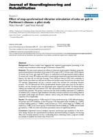
The insole with the vibratory device (A) and schematic dia-gram that shows integration of the vibratory device within the insole (B) and sequence of the vibratory stimulation dur-ing step phases (C)Figure 1
The insole with the vibratory device (A) and schematic dia-
gram that shows integration of the vibratory device within
the insole (B) and sequence of the vibratory stimulation dur-
ing step phases (C). Vibratory device consists of a vibration
disk motor (diameter 18 mm) and a membrane switch
attached to the top of the motor, with a resulting thickness
of ~5 mm and weight of ~5 grams.
Journal of NeuroEngineering and Rehabilitation 2006, 3:9 />Page 4 of 7
(page number not for citation purposes)
out the walking trials was avoided since it might affect the
gait profile [18]. To reduce expectation bias, subjects were
allowed to walk for about 1 minute with the device on and
off before the gait recordings. An investigator followed the
study subjects during walking trials as a safety measure,
and he also measured the gait distance with a "Meter-
Man" distance-measuring wheel (Winnebago, MN).
Data Acquisition and Processing
Gait signals were recorded using the Gait Logger (JAS
Research. Inc., Boston, MA) connected to the foot
switches with 4 force sensors on each foot (B&L Engineer-
ing, Inc., Tustin, CA). Gait signals were sampled at 200 Hz
per switch using a 16-bit analog/digital converter and
recorded on the portable microcontroller-based storage
device. The raw data were processed off-line using the soft-
ware written in Matlab
®
6.1 (The MathWorks, Inc., Natick,
MA). Turns were excluded from statistical analysis since
gait variability can be affected by a particular turning pat-
tern (e.g., turning in a small circle versus sudden 180
degree rotation). Stride, stance, swing, and double sup-
port duration were computed in each gait cycle (in milli-
seconds and as a percentage of the gait cycle) and averaged
over each walking trial. The primary outcome measure
was the stride variability expressed as the coefficient of
variation (CV) of the stride interval, which is a measure of
gait steadiness [19]. Secondary outcome measures were
the following gait parameters: walking distance and
speed, stride length and duration, cadence, stance, swing
duration, double support, and their respective CVs (if
applicable). Gait parameters were averaged between the
right and left legs for statistical analysis.
Statistical analysis was performed using statistical soft-
ware JPM 5.1 (SAS Institute, Cary, NC). The effects of
vibratory stimulation between the conditions (S-VS on vs.
S-VS off) and groups were compared using MANOVA
adjusting for age, sex, and height. Paired t-test was used to
compare effects of vibratory stimulation within each
group.
Results
Demographic characteristics (age, height, and weight) did
not differ between the PD and the control groups.
Walking without step-synchronized vibration stimulation
Six-minute walking trials included the straight segments
and typically 4–6 turns at 180 degrees. There were no gait-
freezing episodes or falls. PD subjects had significantly
slower walking speed and higher CV of the stride interval,
stance, and doubles support than did control subjects
(Table 2). Other locomotion parameters were not signifi-
cantly different between the groups.
Step-synchronized vibration stimulation
The vibratory device was well tolerated; none of the sub-
jects experienced gait freezing or falls. The most common
experience was an increased awareness of the foot place-
ment on the floor. The walking speed (p < 0.04) and the
CV of the stride interval (p < 0.02) differed between the
groups and between the S-VS on and S-VS off conditions
(Table 2). The walking speed increased and the CV of the
stride interval decreased during the S-VS on walking as
compared to the S-VS off walking. Other locomotor
parameters (cadence, stride, and swing duration) did not
Table 2: Gait characteristics in the Parkinson's disease and control groups during 6-minute walking with and without step-
synchronized vibration stimulation
Locomotion Parameters Parkinson's Disease Group Control Group Manova
S-VS OFF S-VS ON p
G
S-VS OFF S-VS ON p
G
p
Walking distance (m) 368 ± 73.4 402.7 ± 72.6 0.0001 453.1 ± 53.2 476.1 ± 61.6 0.03 0.02
Velocity (m/s) 1.02 ± 0.2 1.11 ± 0.2 0.0001 1.25 ± 0.2 1.32 ± 0.17 0.03 0.04
Cadence (steps/min) 104.9 ± 8.9 109.2 ± 10.2 0.03 110.9 ± 4.9 112 ± 5.7 0.11 0.21
Stride duration (ms) 1149.6 ± 90.9 1107 ± 100.9 0.01 1112.9 ± 99.0 1103.2 ± 105.4 0.11 0.25
Stride length (m) 1.17 ± 0.24 1.24 ± 0.3 0.0002 1.4 ± 0.16 1.37 ± 0.19 0.06 0.06
Stride CV (%) 5.36 ± 3.1 4.4 ± 2.7 0.002 2.8 ± 0.4 2.3 ± 0.5 0.006 0.02
Stance duration (ms) 730.8 ± 79.7 679.3 ± 90.2 0.04 653.8 ± 66.19 654.95 ± 69.9 0.8 0.04
Stance CV (%) 1.99 ± 1.0 1.6 ± 0.8 0.1 1.29 ± 0.63 0.99 ± 0.30 0.15 0.11
Swing duration (ms) 418.8 ± 54.8 427.7 ± 64.6 0.75 446.6 ± 83.4 435.8 ± 85.8 0.09 0.37
Swing CV (%) 1.86 ± 1.04 1.6 ± 0.8 0.33 0.95 ± 0.4 0.88 ± 0.45 0.09 0.12
Double support duration
(ms)
156.0 ± 51.1 134.6 ± 42.8 0.37 115.6 ± 25.7 112.1 ± 45.7 0.26 0.08
Double support CV (%) 2.78 ± 1.6 2.77 ± 1.7 0.06 0.72 ± 0.25 0.97 ± 0.87 0.43 0.05
Mean ± SD
SV OFF – walking without step-synchronized vibration stimulation, S-VS ON walking with step synchronized vibration stimulation
p = Manova comparisons between the groups and S-VS conditions, p
G
= within group comparisons using paired t-test
CV – coefficient of variation
Journal of NeuroEngineering and Rehabilitation 2006, 3:9 />Page 5 of 7
(page number not for citation purposes)
differ significantly either between groups or between S-VS
conditions. There were no significant differences between
the left and right legs in the stride interval and its corre-
sponding CV.
Parkinson's disease group
Figure 2 shows an example of the stride intervals meas-
ured during walking with and without the S-VS in a PD
subject (subject no. 2 in Table 1). Walking with the S-VS
significantly increased the walking speed (p < 0.0001),
cadence (p = 0.03), stride duration (p = 0.01), and stride
length (p = 0.0002). The CV of the stride intervals (p =
0.0002) and the stance duration (p = 0.04) decreased dur-
ing the S-VS walking. The stance percentage of the step,
double support duration, double support percentage of
the step, and coefficient of variation of the double support
were not affected. Two PD subjects with histories of falls
(subject no. 2 and no. 3 in Table 1) had the highest base-
line coefficient of variation of the stride. In these subjects
the S-VS improved the CVs of their stride intervals by
20.9% and 32%, respectively.
Control group
The walking speed increased (p = 0.03) and the CV of the
stride interval decreased (p = 0.006) during the S-VS walk-
ing. Other locomotion parameters were not significantly
altered by the S-VS.
Discussion
In this study, vibration stimulation of the foot soles syn-
chronized with the step increased the walking speed and
improved the stride variability in PD subjects. In addition,
vibration stimulation prolonged the stride interval and
the stride length. Stride variability also decreased in the
control group. Stride variability, which is an important
measure of motor performance and gait unsteadiness, is
increased in subjects with a history of falls [19-23] and is
an independent predictor of falling [19]. The step-syn-
chronized vibration may stabilize gait in PD patients by
reducing the stride variability.
The vibration stimulus was suprathreshold, a situation
that prevented blinding of the study participants. The
increased awareness of foot placement may affect gait
characteristics, as suggested by the effects of attention
strategies [24]. The subjects were instructed to walk at a
comfortable speed without any reference to gait attention
to minimize the unspecific effects of gait awareness.
Therefore, it is not likely that increased attention may
account for all S-VS effects.
In our study, subjects were walking at a comfortable pace,
without any encouragement or instructions might affect
their walking speed. The mean walking distance increased
by 9.4% in the PD group and by 5.2% in the control group
during the S-VS walking. The 6-minute walking test
(6MWT) is believed to reflect activities of daily living, but
there might be a placebo response and training effect
among repetitive walking trials [18,25]. For example, one
study found an 8% increase in walking distance on the
second trial in healthy elderly (2.5-hour break between
the trials)[26]; another found a 3% increase in patients
with fibromyalgia (1-day break between the trials) [27];
and a third found a 4.8% increase in patients with heart
failure (30 minute break between the trials) [28]. Direct
comparisons of these repetitive walking trials are prob-
lematic, as the methodology differed among them. For
example, subjects were asked to "walk a pace that was
brisk but comfortable" without encouragement [27], to
"cover as much distance as possible until exhausted" with-
out encouragement [28], or to walk at their own maximal
pace with encouragement every 30 seconds [26]. Further-
more, the stride variability in repetitive 6MWT was not
measured, and the effects of repetitive 6MWT trials on
walking distance in PD patients are unknown. Our study
differs from the above trials not only in terms of the
patient population, the much shorter inter-trial breaks,
and the lack of encouragement, but also in the fact that we
took several measures to minimize the learning effect.
These measures included walking in a familiar environ-
ment (PD group), using specific instructions to discourage
subjects to walk faster (or slower) than at their most com-
fortable speed, and having each subject walk for as long as
1 minute with the vibratory devices turned on and off
before the actual walking recordings. Nevertheless, as the
placebo and learning effect cannot be completely
excluded, only a long-term study in a larger patient popu-
lation can provide robust measures of the effects of S-VS
walking.
The effect of S-VS is likely to be related to enhanced prop-
rioceptive feedback, even upon considering other possible
confounders. Locomotor patterns are regulated through
Stride intervals of the left leg from a 45-year-old Parkinson's disease subject obtained during 6-minute walking without (left) and with (right) the vibratory stimulation of solesFigure 2
Stride intervals of the left leg from a 45-year-old Parkinson's
disease subject obtained during 6-minute walking without
(left) and with (right) the vibratory stimulation of soles.
Vibratory stimulation reduced the coefficient of variation of
stride interval from baseline value 11.49% (SD = 62.0 ms) to
9.43% (SD = 44.2 ms).
Journal of NeuroEngineering and Rehabilitation 2006, 3:9 />Page 6 of 7
(page number not for citation purposes)
the feedback loops among the proprioceptive receptors
and central motor pattern generators. Sensory feedback is
necessary for gait stability in that it provides inputs to the
central pattern generators that can instantly adapt to exter-
nal perturbations and correct programming errors in
intended movement direction, force, and execution [29-
31]. The vibration device in our study operated in a simple
closed loop mode wherein the enhanced feedback was
synchronized with the distribution of plantar pressures
during the gait cycle phase. Therefore, the synchroniza-
tion of vibration stimulation with the gait phase may
improve timing and variability of the gait cycle by
enhanced recruitment of sensorimotor pathways includ-
ing spinal circuitry and basal ganglia. Supporting this
notion are functional magnetic resonance imaging studies
that have demonstrated activation of distinct brain struc-
tures when vibration stimulus was used [32,33]. Stimula-
tion of the fingertips activated the contralateral primary
somatosensory cortex, bilateral secondary somatosensory
cortex, the precentral gyrus, the posterior insula, the pos-
terior parietal region, and the posterior cingulate [33].
Positron emission tomography studies showed that stim-
ulation of the metacarpal joints activated ipsilateral sen-
sory cortical areas and contralateral basal ganglia [32].
Results of this study, however, may be not applied to the
whole PD population given our small sample and selec-
tion of patients. The PD subjects had mild to moderate
gait impairment that was predominantly associated with
abnormal balance. None of the subjects had the gait freez-
ing episodes commonly seen in more advanced disease.
Gait freezing is a poorly understood phenomenon that
may be due to pathophysiological mechanisms different
from those causing abnormal balance [34].
Conclusion
This study indicates that the step-synchronized vibration
stimulation of the soles improves gait steadiness in Par-
kinson's disease patients with predominant balance
impairment. The suprathreshold stimulation improved
gait, presumably by enhancing the sensory feedback. Pre-
vious reports showing impaired proprioception support
this notion. In this short-term non-blinded design, possi-
ble placebo and learning effects cannot be completely
excluded. Long-term studies are needed to establish a clin-
ical value of the S-VS.
Abbreviations
PD Parkinson's disease
S-VS step-synchronized vibration stimulation
VD vibratory device
CV coefficient of variation
CoP center of pressure
UPDRS Unified Parkinson Disease Rating Scale
6MWT 6-minute walking test
LEDD levodopa equivalent scale
Competing interests
A patent for the device described in this study has been
filed with the US Patent and Trademark Office. The patent
is property of Boston Medical Center Corporation.
Authors' contributions
P.N. designed the device, the study and conducted the
experiments, data analysis, interpretation, and manu-
script preparation.
V.N. contributed to study design and participated in data
analysis, interpretation, and manuscript preparation.
Acknowledgements
This study was sponsored by The Older American Independence Center
Grant 2P60 AG08812-11.
References
1. Pratorius B, Kimmeskamp S, Milani TL: The sensitivity of the sole
of the foot in patients with Morbus Parkinson. Neurosci Lett
2003, 346:173-176.
2. Zia S, Cody F, O'Boyle D: Joint position sense is impaired by
Parkinson's disease. Ann Neurol 2000, 47:218-228.
3. Schneider JS, Diamond SG, Markham CH: Parkinson's disease:
sensory and motor problems in arms and hands. Neurology
1987, 37:951-956.
4. Dietz V: Proprioception and locomotor disorders. Nat Rev Neu-
rosci 2002, 3:781-790.
5. Zia S, Cody F, O'Boyle D: Joint position sense is impaired by
Parkinson's disease. Clin Anat 2002, 15:23-31.
6. Abbruzzese G, Berardelli A: Sensorimotor integration in move-
ment disorders. Mov Disord 2003, 18:231-240.
7. Lewis GN, Byblow WD: Altered sensorimotor integration in
Parkinson's disease. Brain 2002, 125:2089-2099.
8. Park S, Horak FB, Kuo AD: Postural feedback responses scale
with biomechanical constraints in human standing. Exp Brain
Res 2004, 154:417-427.
9. Zehr EP, Duysens J: Regulation of arm and leg movement dur-
ing human locomotion. Neuroscientist 2004, 10:347-361.
10. Maki BE, McIlroy WE: Postural control in the older adult. Clin
Geriatr Med 1996, 12:635-658.
11. Tropp H, Commentary: Functional ankle instability revisited. J
Athl Train 2002, 37:512-515.
12. Priplata AA, Niemi JB, Harry JD, Lipsitz LA, Collins JJ: Vibrating
insoles and balance control in elderly people. Lancet 2003,
362:1123-1124.
13. Priplata AA, Patritti BL, Niemi JB, Hughes R, Gravelle DC, Lipsitz LA,
Veves A, Stein J, Bonato P, Collins JJ: Noise-enhanced balance
control in patients with diabetes and patients with stroke.
Ann Neurol 2006, 59:4-12.
14. Kavounoudias A, Roll R, Roll JP: Specific whole-body shifts
induced by frequency-modulated vibrations of human
plantar soles. Neurosci Lett 1999, 266:181-184.
15. Kavounoudias A, Roll R, Roll JP: Foot sole and ankle muscle
inputs contribute jointly to human erect posture regulation.
J Physiol 2001, 532:869-878.
16. Novak P, Novak V: Short term effects of vibratory stimulation
of the soles synchronized with the step on gait in Parkinson's
disease. Movement Disorders 2004, 19:1129-1130. [Abstract]
Publish with BioMed Central and every
scientist can read your work free of charge
"BioMed Central will be the most significant development for
disseminating the results of biomedical research in our lifetime."
Sir Paul Nurse, Cancer Research UK
Your research papers will be:
available free of charge to the entire biomedical community
peer reviewed and published immediately upon acceptance
cited in PubMed and archived on PubMed Central
yours — you keep the copyright
Submit your manuscript here:
/>BioMedcentral
Journal of NeuroEngineering and Rehabilitation 2006, 3:9 />Page 7 of 7
(page number not for citation purposes)
17. Hughes AJ, Daniel SE, Kilford L, Lees AJ: Accuracy of clinical diag-
nosis of idiopathic Parkinson's disease: a clinico-pathological
study of 100 cases. J Neurol Neurosurg Psychiatry 1992, 55:181-184.
18. Olsson LG, Swedberg K, Clark AL, Witte KK, Cleland JGF: Six
minute corridor walk test as an outcome measure for the
assessment of treatment in randomized, blinded interven-
tion trials of chronic heart failure: a systematic review. Euro-
pean Heart Journal 2005, 26:778-793.
19. Maki BE: Gait changes in older adults: predictors of falls or
indictors of fear. J Am Geriatr Soc 1997, 45:313-320.
20. Nakamura T, Meguro K, Sasaki H: Relationship between falls and
stride length variability in senile dementia of the Alzheimer
type. Gerontology 1996, 42:108-113.
21. Hausdorff JM, Edelberg HK, Mitchell SL, Goldberger AL, Wei JY:
Increased gait unsteadiness in community-dwelling elderly
fallers. Arch Phys Med Rehabil 1997, 78:278-283.
22. Hausdorff JM, Nelson ME, Kaliton D, Layne JE, Bernstein MJ, Neurn-
berger A, Singh MAF: Etiology and modification of gait instabil-
ity in older adults: a randomized controlled trial of exercise.
J Appl Physiol 2001, 90:2117-2129.
23. Hausdorff JM, Rios DA, Edelberg HK: Gait variability and fall risk
in community-living older adults: a 1-year prospective study.
Arch Phys Med Rehabil 2001, 82:1050-1056.
24. Morris ME, Iansek R, Matyas TA, Summers JJ: Stride length regula-
tion in Parkinson's disease. Normalization strategies and
underlying mechanisms. Brain 1996, 119:551-568.
25. ATS Committee on Proficiency Standards for Clinical Pulmonary
Function Laboratories: ATS statement: guidelines for the six-
minute walk test. Am J Respir Crit Care Med 2002, 166:111-117.
26. Troosters T, Gosseling R, Decramer M: Six minute walking dis-
tance in healthy elderly subjects. Eur Resp 1999, 14:270-274.
27. Pankoff BA, Overend TJ, Lucy SD, White KP: Reliability of the six-
minute walk test in people with fibromyalgia. Arthritis Care Res
2000, 13:291-295.
28. Opasich C, Pinna GD, Mazza A, Febo O, Riccardi PG, Capomolla S,
Cobelli F, Tavazzi L: Reproducibility of the six-minute walking
test in patients with chronic congestive heart failure:practi-
cal implications. Am J Cardiol 1998, 15:1497-1500.
29. Ghez C, Gordon J, Ghilardi MF: Impairments of reaching move-
ments in patients without proprioception. II. Effects of visual
information on accuracy. J Neurophysiol 1995, 73:361-372.
30. Gordon J, Ghilardi MF, Ghez C: Impairments of reaching move-
ments in patients without proprioception I. Spatial errors. J
Neurophysiol 1995, 73:347-360.
31. Bard C, Fleury M, Teasdale N, Paillard J, Nougier V: Contribution
of proprioception for calibrating and updating the motor
space. Can J Physiol Pharmacol 1995, 73:246-254.
32. Boecker H, Ceballos-Baumann A, Bartenstein P, Weindl A, Siebner
HR, Fassbender T, Munz F, Schwaiger M, Conrad B: Sensory
processing in Parkinson's and Huntington's disease: investi-
gations with 3D H(2)(15)O-PET. Brain 1999, 122:1651-1665.
33. Francis ST, Kelly EF, Bowtell R, Dunseath WJ, Folger SE, McGlone F:
fMRI of the responses to vibratory stimulation of digit tips.
Neuroimage 2000, 11:188-202.
34. Giladi N, McDermott MP, Fahn S, Przedborski S, Jankovic J, Stern M,
Tanner C, Parkinson Study Group: Freezing of gait in PD: pro-
spective assessment in DATATOP cohort. Neurology 2001,
56:1712-1721.