báo cáo hóa học: " Analysis of right anterolateral impacts: the effect of trunk flexion on the cervical muscle whiplash response" potx
Bạn đang xem bản rút gọn của tài liệu. Xem và tải ngay bản đầy đủ của tài liệu tại đây (460.33 KB, 9 trang )
BioMed Central
Page 1 of 9
(page number not for citation purposes)
Journal of NeuroEngineering and
Rehabilitation
Open Access
Short report
Analysis of right anterolateral impacts: the effect of trunk flexion on
the cervical muscle whiplash response
Shrawan Kumar*
1
, Robert Ferrari
2
, Yogesh Narayan
1
and Edgar Vieira
1
Address:
1
Department of Physical Therapy, Faculty of Rehabilitation Medicine, University of Alberta, Edmonton, Alberta, T6G 2G4, Canada and
2
Department of Medicine, University of Alberta, Edmonton, Alberta, T6G 2B7, Canada
Email: Shrawan Kumar* - ; Robert Ferrari - ; Yogesh Narayan - ;
Edgar Vieira -
* Corresponding author
Abstract
Background: The cervical muscles are considered a potential site of whiplash injury, and there is
a need to understand the cervical muscle response under non-conventional whiplash impact
scenarios, including variable body position and impact direction. There is no data, however, on the
effect of occupant position on the muscle response to frontal impacts. Therefore, the objective of
the study was to measure cervical muscle response to graded right anterolateral impacts.
Methods: Twenty volunteers were subjected to right anterolateral impacts of 4.3, 7.8, 10.6, and
12.8 m/s
2
acceleration with their trunk flexed forward 45 degrees and laterally flexed right or left
by 45 degrees. Bilateral EMG of the sternocleidomastoids, trapezii, and splenii capitis and
acceleration of the sled, torso, and head were measured.
Results and discussion: With either direction of trunk flexion at impact, the trapezius EMGs
increased with increasing acceleration (p < 0.05). Time to onset of the electromyogram and time
to peak electromyogram for most muscles showed a trend towards decreasing with increasing
acceleration. With trunk flexion to the left, the left trapezius generated 38% of its maximal
voluntary contraction (MVC) EMG, while the right trapezius generated 28% of its MVC EMG. All
other muscles generated 25% or less of this measure (25% for the left splenius capitis, 8% for the
right splenius capitis, 6% for the left sternocleidomastoid, and 2% for the left sterncleidomastoid).
Conversely, with the trunk flexed to the right, the right trapezius generated 44% of its MVC EMG,
while the left trapezius generated 31% of this value, and all other muscles generated 20% or less of
their MVC EMG (20% for the left splenius capitis, 14% for the right splenius capitis, 4% for both the
left and right sternocleidomastoids).
Conclusion: When the subject sits with trunk flexed out of neutral posture at the time of
anterolateral impact, the cervical muscle response is dramatically reduced compared to frontal
impacts with the trunk in neutral posture. In the absence of bodily impact, the flexed trunk posture
appears to produce a biomechanical response that would decrease the likelihood of cervical muscle
injury in low velocity impacts.
Published: 16 May 2006
Journal of NeuroEngineering and Rehabilitation 2006, 3:10 doi:10.1186/1743-0003-3-10
Received: 31 March 2005
Accepted: 16 May 2006
This article is available from: />© 2006 Kumar et al; licensee BioMed Central Ltd.
This is an Open Access article distributed under the terms of the Creative Commons Attribution License ( />),
which permits unrestricted use, distribution, and reproduction in any medium, provided the original work is properly cited.
Journal of NeuroEngineering and Rehabilitation 2006, 3:10 />Page 2 of 9
(page number not for citation purposes)
Background
Whiplash injury is an important health problem with a
significant economic and health burden [1]. There has
been considerable research on the cervical response to
rear-end impacts using volunteers [2-18], but much less
research with volunteers in frontal impacts, most of the
early frontal impact studies being done with military per-
sonnel [19-24]. We know much less, therefore, about the
mechanism of whiplash injury in frontal collisions. This is
despite the fact that a recent large epidemiological study
has confirmed that frontal collisions are as common a
cause of whiplash claims as rear-end collisions [25].
We have applied a methodology which combines surface
EMG and extrapolations through regression based on
very-low velocity impacts to the problem of frontal
impacts. This has been done with straight-on frontal
impacts [24], and recently in this journal we also reported
on the effect of head rotation in anterolateral impacts spe-
cifically [26]. Using this approach, the regression models
are thus far in good agreement with the available data that
has been gathered in previous, small studies of higher
velocity impacts [27]. It has also been shown that if the
subject is expecting an impact, this mitigates the risk of
injury [18].
The reality is that vehicle occupants are not always posi-
tioned in this neutral position at the time of impact. Foret-
Bruno [28] has reviewed that whiplash victims may be in
the trunk-flexed position, and that, at least from dummy
experiments, this may increase the risk of injury in a fron-
tal impact, not only from impact with the vehicle interior,
but through effects of increased cervical extension when
the occupant is seated with most of the torso away from
the seat and rebounds into the seat after the impact. There
is yet, however, no volunteer data which examines the cer-
vical responses of volunteers when they are not seated in
the standard, neutral head and trunk posture.
Since we have recently reported in this journal on the
effect of head rotation in anterolateral impacts, it was of
interest to keep the impact variables constant and deter-
mine whether trunk flexion itself in anterolateral impacts
will increase or decrease the EMG activity, and how. We
thus undertook a study to assess the cervical muscle
response in right anterolateral impacts, but with the trunk
flexed to either the left or right (to mimic circumstances of
"out-of-position" vehicle occupants) at the time of
impact.
Methods
The methods for this study of frontal impacts with trunk
flexion are the same as those used previously for frontal
impact studies with the subject in either neutral posture
and/or with head rotation [24,26,29,30]. Twenty healthy,
normal subjects (10 males and 10 females) with no his-
tory of whiplash injury and no cervical spine pain during
the preceding 12 months volunteered for the study. The
20 subjects had a mean age of 23.6 ± 3.0 years, a mean
height of 172 ± 7.7 cm, and a mean weight of 69 ± 13.9
kg. The subjects were all right-hand dominant. The study
was approved by the University Research Ethics Board.
The sled device is shown in this journal in the previous
publication [26]. Subjects were then exposed to right ante-
rolateral impacts with their trunk flexed forward and to
either their left and right at accelerations of 4.3, 7.8, 10.6,
and 12.8 m/s
2
generated in a random order by a pneu-
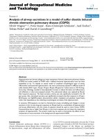
matic piston. The subjects were asked to assume a posi-
tion of trunk flexion (forward and lateral) and to look
down at their right or left foot. We positioned each of the
volunteers in 45 degrees flexion and 45 degrees rotation
either to the left or to the right (see Fig. 1). We did not use
any blocking of visual or auditory cues, which is compara-
ble to the "expected" impact data we had gathered previ-
ously [24,26], but the impact severity and posture
positions were randomly varied between the 4 levels of
acceleration. Each subject effectively underwent 4 levels of
accelerative impacts under two conditions of trunk flex-
ion, for one direction of impact (a total of 8 impacts). The
acceleration was delivered in a way that mimicked the
time course seen in motor vehicle collisions and occurred
fast enough to produce eccentric muscle contractions.
Subjects were asked to report any headache or other aches
or discomfort they experienced in the days following the
impacts for a period of up to 6 months. None were
reported.
Results and discussion
Head acceleration
As anticipated, an increase in applied acceleration resulted
in an increase in excursion of the head and accompanying
accelerations (p < 0.05). The accelerations in these
impacts were not associated with any reported symptoms
in the volunteers following the experiment and up to 6
months later.
Electromyogram amplitude
In a right anterolateral impact, with the trunk flexed 45
degrees to the right or left, the trapezius muscle ipsilateral
to the direction of trunk flexion shows the greatest EMG
response (p < 0.05). The normalized EMG for the sterno-
cleidomastoid (SCM), splenius capitis (SPL) and trape-
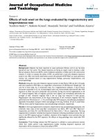
zius (TRP) muscles are shown in Figure 2. At a peak
acceleration of 12.8 m/s
2
, for example, with the trunk
flexed to the right, the right trapezius generated 44% of its
maximal voluntary contraction electromyogram, while all
other muscles generated 31% or less of this variable (31%
for the left trapezius, 20% for the left splenius capitis, 14%
for the right splenius capitis, 4% for both the left and right
Journal of NeuroEngineering and Rehabilitation 2006, 3:10 />Page 3 of 9
(page number not for citation purposes)
sternocleidomastoids). When the trunk is flexed to the
left, under these same conditions, the results are reversed
even though the impact direction remains right anterola-
teral. When flexed to the left, the left trapezius generated
38% of its maximal voluntary contraction electromyo-
gram, with 28% of the maximal voluntary contraction for
the right trapezius, and 25% or less for the remaining
muscles (25% for the left splenius capitis, 8% for the right
splenius capitis, 6% for the left sternocleidomastoid, and
4% for the left sterncleidomastoid).
As the level of applied acceleration in the impact
increased, the magnitude of the EMG recorded from the
trapezius ipsilateral to the trunk flexion increased progres-
sively and disproportionately compared to other muscles
(p < 0.05). Compared to the state of the head and trunk in
neutral posture, trunk flexion significantly reduces the tra-
pezius EMG response (p < 0.05) for all conditions of flex-
ion except for the right trapezius muscle in right trunk
flexion, where the findings are equivalent to those in neu-
tral trunk posture.
The time to onset of the sled, torso, and head acceleration
showed a trend (p > 0.05) decreased with increased
applied acceleration. Similarly, the time to onset of the
EMG shows a trend (p > 0.05) for all muscles to decrease
with increased applied acceleration. The times at which
peak EMG occurred for all the experimental conditions
showed a trend to earlier times of peak activity with
increasing acceleration, though this again did not reach
statistical significance.
The relationship between the force equivalent EMG
response of each muscle and the head acceleration are
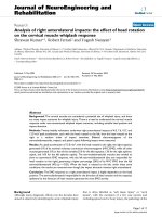
Illustration of the positioning of the subjects prior to frontal whiplash-type impactsFigure 1
Illustration of the positioning of the subjects prior to frontal whiplash-type impacts.
z
x
y
Trunk Flexion to the Right Trunk Flexion to the Left
Journal of NeuroEngineering and Rehabilitation 2006, 3:10 />Page 4 of 9
(page number not for citation purposes)
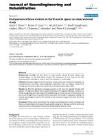
Trunk flexed to left and rightFigure 2
Trunk flexed to left and right. Normalized peak and average electromyogram (EMG) (percentage of isometric maximal volun-
tary contraction), force equivalent of EMG (N), and applied acceleration. LSCM, left sternocleidomastoid; RSCM, right sterno-
cleidomastoid; LSPL, left splenius capitis; RSPL, right splenius capitis; LTRP, left trapezius; RTRP, right trapezius.
lscm lspl ltrp rscm rspl rtrp
CHANNEL
0
20
40
60
Norm. EMG (%)
0
10
20
30
40
50
Force Equiv. EMG (N)
4.3 m/s
2
Norm. Peak EMG
Norm. Avg EMG
Force Equiv.
lscm lspl ltrp rscm rspl rtrp
0
20
40
60
Norm. EMG (%)
0
10
20
30
40
50
Force Equiv. EMG (N)
7.8 m/s
2
lscm lspl ltrp rscm rspl rtrp
0
20
40
60
Norm. EMG (%)
0
10
20
30
40
50
Force Equiv. EMG (N)
10.6 m/s
2
lscm lspl ltrp rscm rspl rtrp
0
20
40
60
Norm. EMG (%)
0
10
20
30
40
50
Force Equiv. EMG (N)
12.8 m/s
2
Left Flexion Right Flexion
lscm lspl ltrp rscm rspl rtrp
CHANNEL
0
20
40
60
Norm. EMG (%)
0
10
20
30
40
50
Force Equiv. EMG (N)
4.3 m/s
2
lscm lspl ltrp rscm rspl rtrp
0
20
40
60
Norm. EMG (%)
0
10
20
30
40
50
Force Equiv. EMG (N)
7.8 m/s
2
lscm lspl ltrp rscm rspl rtrp
0
20
40
60
Norm. EMG (%)
0
10
20
30
40
50
Force Equiv. EMG (N)
10.6 m/s
2
lscm lspl ltrp rscm rspl rtrp
0
20
40
60
Norm. EMG (%)
0
10
20
30
40
50
Force Equiv. EMG (N)
12.8 m/s
2
Journal of NeuroEngineering and Rehabilitation 2006, 3:10 />Page 5 of 9
(page number not for citation purposes)
shown in Table 1. To obtain the force equivalency of a
muscle response due to impact, we first performed a linear
regression analysis on the graded EMG data obtained in
the maximal voluntary contraction trials. This resulted
inan equation for force/emg ratio. EMG values from each
muscle as measured in this impact study were then
entered into the equation, giving us a force equivalent
value (Newtons) for each muscle as shown in Table 1. The
kinematic responses show that very-low velocity impacts
produce less force equivalent than the maximal voluntary
contraction for the same subject, and thus this experimen-
tal approach allows us to gather valuable data without
exposing subjects to any foreseeable injury. The head
accelerations were correspondingly lower than the sled
accelerations in this experiment. For very-low velocity
impacts, this is to be expected, as it is usually only when
the sled acceleration exceeds 5 g's that head acceleration
begins to exceed sled acceleration. This experiment
involved less than 2 g accelerations.
Regression analyses
The applied acceleration, and the muscles examined had
significant main effects on the peak EMG activity (p <
0.05) as shown in Table 2. We used a linear regression
model to plot the available data and extrapolate from the
experimental accelerations to accelerations on the order of
30 m/s
2
. Initially, regression analyses were performed
only up to the maximal acceleration using a linear func-
tion. The kinematic variables of head displacement, veloc-
ity, and acceleration in response to the applied
acceleration were calculated. Additionally, we also
regressed the EMG magnitudes on acceleration. The
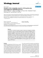
responses of the left and right muscle groups were extrap-
olated to more than twice the applied acceleration value
(see Fig. 3 and 4). It is of note that the EMG magnitudes
remain low over this range compared to previous studies
with the head and trunk in neutral posture [31].
At the time of impact, whiplash victims may be leaning
forward or leaning over as a result of watching for traffic
or speaking with other occupants, reaching for an object
on the floor, et cetera. In the current study, having kept the
impact direction constant, but varying trunk flexion to
right or left we see that the muscles likely activated by
holding this position (the ipsilateral trapezius), are most
active and differ from their counterparts. Overall, how-
ever, the EMG activity is reduced if the subjects are "out-
of-position" at the time of impact (the current study)
compared to identical impact scenarios where the head
and trunk are in neutral position. When the head was in
neutral position in a previous study of right anterolateral
impact [31], the left trapezius generated the greatest EMG,
up to 83% of the maximal voluntary contraction EMG,
and the left splenius capitis instead became more active
and reached a level of 46% of this variable. As seen in this
experiment, even the most active muscles do not exceed
44% of their maximal EMG contraction magnitude. The
sternocleidomastoid muscles, by their attachment and
action, are least likely to undergo eccentric contraction in
the presence of what we expect is much less head-torso lag
in the trunk -flexed posture. In contrast, the attachment
and action of the trapezii, cervical extension being one
action, are likely in a "pre-stretched" position in the trunk
flexed posture with the subject looking downward. Even
Table 1: Mean Force Equivalents (Newtons, N) and Mean Head Accelerations at Time of Maximal EMG in Direction of Travel for Right
Anterolateral Impact.
Force Equivalents for Muscle (N)
Sternocleidomastoid Splenius Capitis Trapezius
Sled
Acceleration
(m/s
2
)
Head
Acceleration
(m/s
2
)
Left Right Left Right Left Right
Right Trunk
flexion
4.3 1.9 (0.9) 5 (3) 4 (2) 19 (11) 14 (7) 17 (7) 19 (8)
7.8 2.7 (1.4) 7 (5) 5 (3) 25 (14) 23 (9) 20 (8) 21 (7)
10.6 3.5 (0.9) 9 (7) 6 (6) 32 (15) 26 (11) 23 (5) 24 (11)
12.8 5.5 (2.7) 11 (10) 8 (6) 35 (16) 28 (14) 26 (6) 29 (11)
Left Trunk
flexion
4.3 2.2 (0.9) 4 (4) 2 (2) 26 (10) 13 (7) 20 (7) 14 (5)
7.8 3.4 (1.4) 5 (5) 5 (2) 28 (10) 17 (8) 23 (7) 15 (3)
10.6 5.0 (1.5) 7 (6) 5 (4) 30 (10) 18 (8) 24 (10) 19 (7)
12.8 5.9 (1.6) 11 (8) 6 (3) 37 (17) 20 (10) 25 (10) 21 (6)
Values in parentheses represent one standard deviation.
Journal of NeuroEngineering and Rehabilitation 2006, 3:10 />Page 6 of 9
(page number not for citation purposes)
lower than expected head-torso lag in this posture is thus
expected to generate more response and a higher likeli-
hood of eccentric contraction in the trapezii than the ster-
nocleidomastoids.
Conclusion
It is suggested that the flexed trunk posture does not
increase the likelihood of cervical muscle injury as com-
pared to impacts with the trunk in neutral position, at
least not for low-velocity impacts. Our findings are con-
trary to previous research findings [28]. Previous research,
however, focused on dummy responses, which may
explain the difference in our findings, and also some of
the dummy experiments were of much higher velocity
impacts. Nevertheless, symptoms are reported even after
low-velocity impacts, and these lead to as many as 60% of
injury claims [16]. With low-velocity impacts, one does
not expect any significant rebounding of the subject back
into the seat, and from our extrapolations, a trunk-flexed
posture, assuming no bodily impact otherwise, does not
otherwise appear to increase the risk of cervical muscle
injury compared to occupant positioning in the neutral
posture.
Abbreviations
MVC (Maximal Voluntary Contraction); EMG (Electro-
myogram); cm (Centimetres); dB (decibels); C4 (fourth
cervical vertebra); mV/g (Millivolts per gram); Hz (Hertz);
kHz (kilohertz); g (acceleration due to gravity); m/s2
(metres per second per second); kg (kilograms); SCM
(Sternocleidomstoid); TRP (Trapezius); SPL (Splenius
capitis)
Competing interests
The author(s) declare that they have no competing inter-
ests.
Authors' contributions
SK made substantial contributions to conception and
design, to acquisition of data, and analysis and interpreta-
tion of data, was involved in drafting the article and revis-
ing it critically for important intellectual content. RF made
substantial contributions to analysis and interpretation of
data, and was involved in drafting the article and revising
it critically for important intellectual content. YN made
substantial contributions to acquisition of data, and anal-
ysis and interpretation of data. EV made substantial con-
tributions to analysis and interpretation of data. All
authors read and approved the final manuscript.
References
1. Spitzer WO, Skovron ML, Salmi LR, et al.: Scientific monograph of
the Quebec Task Force on Whiplash-Associated Disorders.
Spine 1995, 120(suppl 8):1S-73S.
2. West DH, Gough JP, Harper GTK: Low speed rear-end collision
testing using human subjects. Acc Reconstr J 1993, 5:22-26.
3. McConnell WE, Howard RP, Guzman HM, Bomar JB, Raddin JH, Ben-
edict JV, et al.: Analysis of human test subject kinematic
responses to low velocity rear end impacts. In Proceedings of the
Thirty Seventh Stapp Car Crash Conference. Paper 930889 Warrendale,
PA, Society of Automotive Engineers; 1993:21-30.
4. McConnell WE, Howard RP, Van Poppel J, Krause R, Guzman HM,
Bomar JB: Human head and neck kinematics after low velocity
rear-end impacts – understanding "whiplash". In Proceedings of
the Thirty Ninth Stapp Car Crash Conference. Paper 952724 Warrendale,
PA, Society of Automotive Engineers; 1995:215-238.
5. Scott MW, McConnell WE, Guzman HM, Howard RP, Bomar JB,
Smith HL: Comparison of human and ATD head kinematics
during low-speed rearend impacts. In Proceedings of the Thirty
Seventh Stapp Car Crash Conference. Paper 930094 Warrendale, PA,
Society of Automotive Engineers; 1993:1-8.
6. Siegmund GP, Bailey MN, King DJ: Characteristics of specific
automobile bumpers in low-velocity impacts. In Proceedings of
the Thirty Eighth Stapp Car Crash Conference Warrendale, PA, Society
of Automotive Engineers; 1994. SAE 940916
7. Siegmund GP, Williamson PB: Speed change (∆v) of amusement
park bumper cars. Proceedings of the Canadian Multidisciplinary Road
Safety Conference VIII; 1993, June 14–16; Saskatoon, Saskatchewan
1993:299-308.
8. Szabo TJ, Welcher J, Anderson RD: Human occupant kinematic
response to low speed rear-end impacts. In Proceedings of the
Thirty Eighth Stapp Car Crash Conference Warrendale, Pennsylvania:
Society of Automotive Engineers; 1994:23-35. SAE 940532
9. Szabo TJ, Welcher J: Dynamics of low speed crash tests with
energy absorbing bumpers. Volume 101. Issue 6 Warrendale,
Pennsylvania: Society of Automotive Engineers; 1992:1367-75. SAE
921573
10. Matsushita T, Sato TB, Hirabayashi K, Fujimura S, Asazuzma T, Taka-
tori T: X-Ray study of the human neck motion due to head
inertia loading. In Proceedings of the Thirty Eighth Stapp Car Crash
Conference. Paper 942208 Warrendale, PA, Society of Automotive
Engineers; 1994:55-64.
11. Rosenbluth W, Hicks L: Evaluating low-speed rear-end impact
severity and resultant occupant stress parameters. Journal of
Forensic Sciences 1994, 39:1393-1424.
12. Nielsen GP, Gough JP, Little DM, West DH, Baker VT: Human sub-
ject responses to repeated low speed impacts using utility
vehicles. In Proceedings of the Forty First Stapp Car Crash Conference.
Paper 970394 Warrendale, PA, Society of Automotive Engineers;
1997:189-212.
13. Brault JR, Wheeler JB, Siegmund GP, et al.: Clinical response of
human subjects to rear-end automobile collisions. Arch Phys
Med Rehabil 1998, 79:72-80.
14. Howard RP, Bowles AP, Guzman HM, Krenrich SW: 1998. Head,
neck, and mandible dynamics generated by "whiplash". Acc
Anal Prev 1998, 30:525-534.
Table 2: ANOVA table for Peak EMG (µV) by Muscles and Applied Acceleration.
df F Sig.
Right Trunk Flexion Accel 3 18.383 0.00
Muscle 5 23.816 0.00
Left Trunk Flexion Accel 3 12.296 0.00
Muscle 5 53.261 0.00
Journal of NeuroEngineering and Rehabilitation 2006, 3:10 />Page 7 of 9
(page number not for citation purposes)
Trunk flexed to left and rightFigure 3
Trunk flexed to left and right. Extrapolated regression plots of the effect that applied acceleration has on the left and right tra-
pezius muscles for the variables of peak electromyogram (EMG) (µV), normalized EMG (percentage of isometric maximal vol-
untary contraction), and force equivalent of EMG (N).
0 5 10 15 20 25 30 35
0
20
40
60
80
Peak EMG (
µ
V)
LTRP
0 5 10 15 20 25 30 35
0
20
40
60
80
Peak EMG (
µ
V)
RTRP
0 5 10 15 20 25 30 35
0
40
80
120
Normalized Peak EMG (%)
0 5 10 15 20 25 30 35
0
40
80
120
Normalized Peak EMG (%)
0 5 10 15 20 25 30 35
Applied Acceleration (m/s
2
)
0
20
40
60
Force Equivalent EMG (N)
0 5 10 15 20 25 30 35
Applied Acceleration (m/s
2
)
0
20
40
60
Force Equivalent EMG (N)
0 5 10 15 20 25 30 35
0
25
50
75
100
Peak EMG (µV)
LTRP
0 5 10 15 20 25 30 35
0
25
50
75
100
Peak EMG (µV)
RTRP
0 5 10 15 20 25 30 35
0
30
60
90
Normalized Peak EMG (%)
0 5 10 15 20 25 30 35
0
30
60
90
Normalized Peak EMG (%)
0 5 10 15 20 25 30 35
Applied Acceleration (m/s
2
)
0
20
40
60
Force Equivalent EMG (N)
0 5 10 15 20 25 30 35
Applied Acceleration (m/s
2
)
0
20
40
60
Force Equivalent EMG (N)
Right Flexion
Left Flexion
16.1+1.39a R
2
=0.92 12.5+0.73a R
2
=0.95
13.7+1.7a R
2
=0.85 6.7+1.5a R
2
=0.78
18.5+0.50a R
2
=0.98 9.8+0.82a R
2
=0.87
17.5+1.1a R
2
=0.98 13.4+1.7a R
2
=0.95
15.1+1.2a R
2
=0.94
7.8+2.9a R
2
=0.99
12.2+1.1a R
2
=0.98
13.2+1.2a R
2
=0.92
Journal of NeuroEngineering and Rehabilitation 2006, 3:10 />Page 8 of 9
(page number not for citation purposes)
Trunk flexed to left and rightFigure 4
Trunk flexed to left and right. Extrapolated regression plots of the effect that applied acceleration has on the left and right ster-
nocleidomastoid muscles for the variables of peak electromyogram (EMG) (µV), normalized EMG (percentage of isometric
maximal voluntary contraction), and the force equivalent of EMG (N).
0 5 10 15 20 25 30 35
0
15
30
45
60
Peak EMG (
µ
V)
LSCM
0 5 10 15 20 25 30 35
0
15
30
45
60
Peak EMG (
µ
V)
RSCM
0 5 10 15 20 25 30 35
0
6
12
18
Normalized Peak EMG (%)
0 5 10 15 20 25 30 35
0
6
12
18
Normalized Peak EMG (%)
0 5 10 15 20 25 30 35
Applied Acceleration (m/s
2
)
0
10
20
30
Force Equivalent EMG (N)
0 5 10 15 20 25 30 35
Applied Acceleration (m/s
2
)
0
10
20
30
Force Equivalent EMG (N)
0 5 10 15 20 25 30 35
0
15
30
45
60
Peak EMG (µV)
LSCM
0 5 10 15 20 25 30 35
0
15
30
45
60
Peak EMG (µV)
RSCM
0 5 10 15 20 25 30 35
0
5
10
15
Normalized Peak EMG (%)
0 5 10 15 20 25 30 35
0
5
10
15
Normalized Peak EMG (%)
0 5 10 15 20 25 30 35
Applied Acceleration (m/s
2
)
0
10
20
30
Force Equivalent EMG (N)
0 5 10 15 20 25 30 35
Applied Acceleration (m/s
2
)
0
10
20
30
Force Equivalent EMG (N)
Right Flexion
Left Flexion
4.7+1.0a R
2
=0.98 7.8+0.33a R
2
=0.94
1.5+0.3a R
2
=0.87 2.1+0.15a R
2
=0.88
-0.1+0.8a R
2
=0.84 1.0+0.38a R
2
=0.94
3.2+1.0a R
2
=0.97
4.3+0.5a R
2
=0.82
1.13+0.27a R
2
=0.94 1.8+0.19a R
2
=0.86
1.9+0.69a R
2
=0.94
1.5+0.47a R
2
=0.94
Publish with BioMed Central and every
scientist can read your work free of charge
"BioMed Central will be the most significant development for
disseminating the results of biomedical research in our lifetime."
Sir Paul Nurse, Cancer Research UK
Your research papers will be:
available free of charge to the entire biomedical community
peer reviewed and published immediately upon acceptance
cited in PubMed and archived on PubMed Central
yours — you keep the copyright
Submit your manuscript here:
/>BioMedcentral
Journal of NeuroEngineering and Rehabilitation 2006, 3:10 />Page 9 of 9
(page number not for citation purposes)
15. Magnusson ML, Pope MH, Hasselquist L, et al.: Cervical electromy-
ographic activity during low-speed rear-end impact. Euro
Spine J 1998, 8:118-125.
16. Castro WH, Schilgen M, Meyer S, et al.: Do "whiplash injuries"
occur in low-speed rear impacts? Euro Spine J 1997, 6:366-375.
17. Castro WH, Meyer SJ, Becke ME, Nentwig CG, Hein MF, Ercan BI, et
al.: No stress – no whiplash? Prevalence of "whiplash" symp-
toms following exposure to a placebo rear-end collision. Int J
Legal Med 2001, 114:316-322.
18. Kumar S, Narayan Y, Amell T: An electromyographic study of
low-velocity rear-end impacts. Spine 2002, 27:1044-1055.
19. Ewing CL, Thomas DJ: Torque versus angular displacement
response of human head to 2Gx impact acceleration. In Pro-
ceedings of the Seventeenth Stapp Car Crash Conference. Paper 730976
Warrendale, PA, Society of Automotive Engineers; 1973:309-342.
20. Ewing CL, Thomas DJ, Lustic L, et al.: The effect of the initial posi-
tion of the head and neck on the dynamic response of the
human head and neck to Gx impact acceleration. In Proceed-
ings of the Nineteenth Stapp Car Crash Conference Warrendale, PA,
Society of Automotive Engineers; 1975:487-512. SAE 751157
21. Wagner R: A 30 mph front/rear crash with human test per-
sons. In Proceedings of the Twenty Third Stapp Car Crash Conference
Warrendale, PA, Society of Automotive Engineers; 1979:827-840.
SAE 791030
22. Wismans J, Philippens M, van Oorschot E, Kallieris D, Mattern R:
Comparison of human volunteer and cadaver head-neck
response in frontal flexion. In Proceedings of the Thirty First Stapp
Car Crash Conference. Paper 872194 Warrendale, PA, Society of Auto-
motive Engineers; 1987:1-11.
23. Grunsten RC, Gilbert NS, Mawn SV: The mechanical effects of
impact acceleration on the unconstrained human head and
neck complex. Contemporary Orthopaedics 1989, 18:199-202.
24. Kumar S, Narayan Y, Amell T: Analysis of low-velocity frontal
impacts. Clin Biomech 2003, 18:694-703.
25. Cassidy JD, Carroll LJ, Cote P, et al.: Effect of eliminating com-
pensation for pain and suffering on the outcome of insurance
claims for whiplash injury. N Engl J Med 2000, 342:1179-1186.
26. Kumar S, Ferrari R, Narayan Y: An analysis of right anterolateral
impacts: the effect of head rotation on the cervical muscle
whiplash response. J Neuroeng 2005 in press.
27. Ferrari R: The Whiplash Encyclopedia. The Facts and Myths of Whiplash
Gaithersburg, Maryland: Aspen Publishers Inc; 1999:449-470.
28. Foret-Bruno JY, Tarriere C, Le Coz JY, et al.: Risk of cervical
lesions in real-world and simulated collisions. Proceedings of the
Thirty Fourth Conference of the American Association of Automotive Medi-
cine, Scottsdale, Arizona 1990:373-389.
29. Kumar S, Narayan Y, Amell T: Cervical strength of young adults
in sagittal, coronal, and intermediate planes. Clin Biomech
2001, 6:380-388.
30. Kumar S, Narayan Y, Amell T, Ferrari R: Electromyography of
superficial cervical muscles with exertions in sagittal, coro-
nal, and oblique planes. Euro Spine J 2002, 11:27-37.
31. Kumar S, Ferrari R, Narayan Y: Cervical muscle response to
whiplash-type right anterolateral impacts. Euro Spine J 2004,
13:398-407.