Fundamentals and applications in aerosol spectroscopy
Bạn đang xem bản rút gọn của tài liệu. Xem và tải ngay bản đầy đủ của tài liệu tại đây (31.49 MB, 495 trang )
Tai Lieu Chat Luong
Fundamentals and
Applications in
Aerosol Spectroscopy
Fundamentals and
Applications in
Aerosol Spectroscopy
Edited by
Ruth Signorell
■
Jonathan P. Reid
MATLAB® is a trademark of The MathWorks, Inc. and is used with permission. The MathWorks does not warrant the
accuracy of the text or exercises in this book. This book’s use or discussion of MATLAB® software or related products
does not constitute endorsement or sponsorship by The MathWorks of a particular pedagogical approach or particular
use of the MATLAB® software.
CRC Press
Taylor & Francis Group
6000 Broken Sound Parkway NW, Suite 300
Boca Raton, FL 33487-2742
© 2011 by Taylor and Francis Group, LLC
CRC Press is an imprint of Taylor & Francis Group, an Informa business
No claim to original U.S. Government works
Printed in the United States of America on acid-free paper
10 9 8 7 6 5 4 3 2 1
International Standard Book Number: 978-1-4200-8561-7 (Hardback)
This book contains information obtained from authentic and highly regarded sources. Reasonable efforts have been
made to publish reliable data and information, but the author and publisher cannot assume responsibility for the validity of all materials or the consequences of their use. The authors and publishers have attempted to trace the copyright
holders of all material reproduced in this publication and apologize to copyright holders if permission to publish in this
form has not been obtained. If any copyright material has not been acknowledged please write and let us know so we may
rectify in any future reprint.
Except as permitted under U.S. Copyright Law, no part of this book may be reprinted, reproduced, transmitted, or utilized in any form by any electronic, mechanical, or other means, now known or hereafter invented, including photocopying, microfilming, and recording, or in any information storage or retrieval system, without written permission from the
publishers.
For permission to photocopy or use material electronically from this work, please access www.copyright.com (http://
www.copyright.com/) or contact the Copyright Clearance Center, Inc. (CCC), 222 Rosewood Drive, Danvers, MA 01923,
978-750-8400. CCC is a not-for-profit organization that provides licenses and registration for a variety of users. For
organizations that have been granted a photocopy license by the CCC, a separate system of payment has been arranged.
Trademark Notice: Product or corporate names may be trademarks or registered trademarks, and are used only for
identification and explanation without intent to infringe.
Visit the Taylor & Francis Web site at
and the CRC Press Web site at
Contents
Preface...............................................................................................................................................ix
Editors............................................................................................................................................. xiii
Contributors...................................................................................................................................... xv
Section I Infrared Spectroscopy
Chapter 1 Infrared Spectroscopy of Aerosol Particles..................................................................3
Thomas Leisner and Robert Wagner
Chapter 2 Vibrational Excitons: A Molecular Model to Analyze Infrared
Spectra of Aerosols............................................................................................... 25
George Firanescu, Thomas C. Preston, Chia C. Wang, and Ruth Signorell
Chapter 3 Aerosol Nanocrystals of Water Ice: Structure, Proton Activity, Adsorbate
Effects, and H-Bond Chemistry.................................................................................. 49
J. Paul Devlin
Chapter 4 Infrared Extinction and Size Distribution Measurements
of Mineral Dust Aerosol.......................................................................................79
Paula K. Hudson, Mark A. Young, Paul D. Kleiber, and Vicki H. Grassian
Chapter 5 Infrared Spectroscopy of Dust Particles in Aerosols for Astronomical
Application........................................................................................................ 101
Akemi Tamanai and Harald Mutschke
Section II Raman Spectroscopy
Chapter 6 Linear and Nonlinear Raman Spectroscopy of Single Aerosol Particles................. 127
N.-O. A. Kwamena and Jonathan P. Reid
Chapter 7 Raman Spectroscopy of Single Particles Levitated by an Electrodynamic
Balance for Atmospheric Studies.............................................................................. 155
Alex K. Y. Lee and Chak K. Chan
v
vi
Contents
Chapter 8 Micro-Raman Spectroscopy for the Analysis of Environmental Particles............... 193
Sanja Potgieter-Vermaak, Anna Worobiec, Larysa Darchuk,
and Rene Van Grieken
Chapter 9 Raman Lidar for the Characterization of Atmospheric Particulate Pollution..........209
Detlef Müller
Section III VIS/UV Spectroscopy, Fluorescence,
and Scattering
Chapter 10 UV and Visible Light Scattering and Absorption Measurements on
Aerosols in the Laboratory........................................................................................ 243
Zbigniew Ulanowski and Martin Schnaiter
Chapter 11 Progress in the Investigation of Aerosols’ Optical Properties Using
Cavity Ring-Down Spectroscopy: Theory and Methodology................................... 269
Ali Abo Riziq and Yinon Rudich
Chapter 12 Laser-Induced Fluorescence Spectra and Angular Elastic Scattering Patterns
of Single Atmospheric Aerosol Particles.................................................................. 297
R. G. Pinnick, Y. L. Pan, S. C. Hill, K. B. Aptowicz, and R. K. Chang
Chapter 13 Femtosecond Spectroscopy and Detection of Bioaerosols........................................ 321
Luigi Bonacina and Jean-Pierre Wolf
Chapter 14 Light Scattering by Fractal Aggregates.................................................................... 341
C. M. Sorensen
Section IV UV, X-ray, and Electron Beam Studies
Chapter 15 Aerosol Photoemission.............................................................................................. 367
Kevin R. Wilson, Hendrik Bluhm, and Musahid Ahmed
Chapter 16 Elastic Scattering of Soft X-rays from Free Size-Selected Nanoparticles................ 401
Harald Bresch, Bernhard Wassermann, Burkhard Langer, Christina Graf, and
Eckart Rühl
vii
Contents
Chapter 17 Scanning Transmission X-ray Microscopy: Applications in
Atmospheric Aerosol Research................................................................................. 419
Ryan C. Moffet, Alexei V. Tivanski, and Mary K. Gilles
Chapter 18 Electron Beam Analysis and Microscopy of Individual Particles............................ 463
Alexander Laskin
Index............................................................................................................................................... 493
Preface
This book is intended to provide an introduction to aerosol spectroscopy and an overview of the
state-of-the-art of this rapidly developing field. It includes fundamental aspects of aerosol spectro
scopy as well as applications to atmospherically and astronomically relevant problems. Basic knowledge is the prerequisite for any application. However, in aerosol spectroscopy, as in many other
fields, there remain crucial gaps in our understanding of the fundamental processes. Filling this gap
can only be a first step, with the challenge then remaining to develop instruments and methods
based on those fundamental insights, instruments that can easily be used to study aerosols in planetary atmospheres as well as in space. With this in mind, this book also touches upon some of the
aspects that need further research and development. As a guideline, the chapters in this book are
arranged in the order of decreasing wavelength of light/electrons, starting with infrared spectroscopy and concluding with x-ray and electron beam studies.
Infrared spectroscopy is one of the most important aerosol characterization methods in laboratory studies, for field measurements, for remote sensing, and in space missions. It provides a wealth
of information about aerosol particles ranging from properties such as particle size and shape to
information on their composition and chemical reactivity. The analysis of spectral information,
however, is still a challenge. In Chapter 1, Leisner and Wagner provide a detailed description of the
most widely used method to analyze infrared extinction spectra, namely classical scattering theory
in combination with continuum models of the optical properties of aerosol particles. The authors
explain how information such as number concentration, size distribution, chemical composition,
and shape can be retrieved from infrared spectra, and outline where pitfalls could occur. Theoretical
considerations are illustrated with experiments performed in the large cloud chamber, aerosol interaction and dynamics in the atmosphere (AIDA).
Classical scattering theory and continuum models for optical properties are not always suitable
for a detailed analysis of particle properties. Available optical data are often not accurate enough,
and for small particles, where the molecular structure becomes important, these methods fail altogether. In Chapter 2, Firanescu, Preston, Wang, and Signorell discuss a molecular model that allows
a detailed analysis of particle properties on the basis of the band shapes observed in infrared extinction spectra. In particular, this approach explains why and when infrared spectra of molecular
aerosols are determined by particle properties such as shape, size, or architecture. After a description of the approach, the authors illustrate its application by means of a variety of examples.
Water and ice are the most important components of aerosols in our Earth’s atmosphere. They
play a crucial role in many atmospheric processes. Water ice is also ubiquitous beyond our planet
and solar system. In Chapter 3, Devlin uses infrared spectroscopy to characterize this important
type of particle and shows how the structural properties of pure and mixed ice nanocrystals can be
unraveled by this technique. Special consideration is given to the nature of the surface of these
particles, the role it plays, and how it is influenced by adsorbates. The formation and transformation
of numerous naturally occurring hydrates are discussed. These studies reveal the exceptional properties of water ice surfaces.
Chapters 4 and 5 are devoted to the infrared spectroscopy of dust particles. The infrared radiative effects of mineral dust aerosols in the Earth’s atmosphere are investigated by Hudson, Young,
Kleiber, and Grassian in Chapter 4. Remote sensing studies using infrared data from satellites provide the source of information to determine the radiative effects of these particles. Such data are
commonly analyzed using Mie theory, which treats all particles as spheres. The authors discuss
the errors associated with this assumption and demonstrate that the proper treatment of particle
ix
x
Preface
shape is crucial in retrieving reliable information about the radiative effect of mineral dust particles
from remote sensing. The properties of dust grains occurring in astrophysical environments are the
subject of Chapter 5 by Tamanai and Mutschke. Dust grains of different composition with sizes in
the micrometer range are widely distributed throughout space. Ground-based as well as satellitebased telescopes are used for infrared studies of these dust particles. Tamanai and Mutschke discuss infrared laboratory studies of astrophysically relevant dust grains and their application to the
interpretation of astronomical spectra. While the wide variety of dust properties makes spectral
analysis a difficult task, the authors demonstrate that important information can be obtained from
such measurements about the conditions under which dust grains exist and evolve in astronomical
environments.
Raman spectroscopy has proved to be a versatile tool for examining aerosol particles in controlled laboratory measurements, allowing the unambiguous identification of chemical species, the
determination of particle composition, and even the determination of particle size and temperature.
Although Raman scattering is inherently a weak process, measurements have been routinely performed on droplet trains using pulsed laser and continuous-wave laser techniques, on aerosol particles isolated in optical or electrodynamic traps, and on particles deposited on substrates. Section II
begins with a general introduction to the fundamentals of both linear and nonlinear Raman scattering from aerosol particles. In particular, Kwamena and Reid highlight the considerable accuracy
(<1 nm) that can be achieved in the determination of droplet size from the unique fingerprint of
enhanced Raman scattering that occurs at discrete wavelengths commensurate with whispering gallery modes, also referred to as morphology-dependent resonances. Before reviewing some recent
applications of Raman spectroscopy for characterizing aerosol, they introduce some of the key
experimental considerations that must be remembered when designing a Raman instrument for
aerosol studies. Lee and Chan describe the coupling of Raman spectroscopy with an electrodynamic balance in Chapter 7, outlining how information gained from Raman measurements can
complement that from other methods, including light scattering for probing particle size and morphology, or tracking evolving particle mass. In particular, they review recent studies of hygroscopicity and heterogeneous chemistry. They demonstrate that resolving Raman line shapes can provide
important insights into intermolecular interactions between solvent and solute molecules within the
condensed aerosol phase, particularly important for understanding the properties of metastable
supersaturated states accessed at high solute concentrations.
Raman analysis can provide an important tool for characterizing particulate matter of atmospheric origin as well as for probing particles in controlled laboratory measurements. PotgieterVermaak, Worobiec, Darchuk, and Van Grieken review the application of micro-Raman spectroscopy
for the analysis of environmental particles in Chapter 8. They begin by reviewing the methods available for ambient sampling and the importance of choosing suitable substrates, before discussing the
advantages and challenges of utilizing the technique on a stand-alone basis. The practicalities of
coupling micro-Raman measurements with other techniques, such as scanning electron microscopy
coupled with energy-dispersive x-ray spectrometric detection, are also described and assessed.
Key uncertainties remain in the direct and indirect impact of aerosols on climate, and coordinated monitoring of the temporal variability of global aerosol distribution is a basic requirement
of climate research. In Chapter 9, Müller describes the application of Raman LIDAR (light detection and ranging) in the characterization of atmospheric pollution. After a description of the basic
principles of Raman LIDAR, methods for deriving the optical and microphysical properties of
particulate pollution are introduced. This is followed by an illustration of the potential of modern
Raman LIDARs, particularly when measurements are made with a network of systems on a continental scale.
Elastic light scattering by particles in the visible and UV parts of the electromagnetic spectrum
provides the basis for many conventional and routine techniques for determining particle size and
concentration. More recently, it has been shown that resolving the light scattering from single
particles may lead to the development of new instruments for assessing particle size and shape.
Preface
xi
In addition, fluorescence spectroscopy is becoming an increasingly applied technique for identifying particle composition. Ulanowski and Schnaiter begin Section III with a discussion of light scattering and absorption measurements on aerosols in the laboratory. Following an introduction to key
parameters that must be typically measured, they review some of the common methods for performing extinction spectroscopy, using an optical extinction cell, and absorption spectroscopy, specifically photoacoustic spectroscopy, and applications of these instruments in laboratory and chamber
measurements. Resolving the angular dependence of light scattering has a long history in the field
of particle analysis, and recent developments have concentrated on the measurement and analysis of
complex morphologies recorded at the single-particle level, allowing the categorization of sampled
particles into distinct classes.
In Chapter 11, Riziq and Rudich describe the information that can be gained by measuring light
extinction from ensembles of accumulation mode aerosol particles using cavity ring-down spectroscopy (CRD-S). CRD-S is widely used for performing highly sensitive measurements of gas-phase
composition and is now becoming more extensively used in both field and laboratory-based aerosol
measurements. The authors introduce the underlying principles of CRD-S, before describing pulsed
and continuous-wave implementations of the technique, and the sensitivity that can be achieved.
The chapter concludes with a review of recent applications, particularly focusing on the retrieval of
aerosol optical properties.
The application of laser-induced fluorescence (LIF) spectroscopy for identifying and classifying
biological aerosol particles is described by Pinnick, Pan, Hill, Aptowicz, and Chang in Chapter 12.
Although many compounds have similar fluorescence spectra with relatively broad and indistinguishable features, unlike those that occur in Raman or IR spectra, single-particle LIF measurements can provide clear and distinguishable signatures for different classes of biological and
anthropogenic aerosol. Further classification of particle type/morphology can be achieved by twodimensional angular optical scattering (TAOS), complementing and expanding on the discussion of
this technique provided by Ulanowski and Schnaiter in Chapter 10. Bonacina and Wolf describe the
improved specificity of bioaerosol detection that can be achieved using ultrafast laser techniques,
including time-resolved pump–probe fluorescence spectroscopy, femtosecond laser-induced break
down spectroscopy, and coherent optimal control in Chapter 13. In particular, they show that the
application of an ultrafast double-pulse excitation scheme can induce strong fluorescence depletion
from biological samples such as bacteria-containing droplets, allowing discrimination from possible interferents, such as polycyclic aromatic compounds, which otherwise have similar spectroscopic properties.
In many optical studies of aerosols, particles can be assumed to be spherical in shape, allowing
the application of Mie scattering theory. In many cases, this only provides an approximate picture
and the application of more rigorous treatments that describe the nonspherical morphology of a
particle must be considered. Sorensen explores the complexity apparent in scattering measurements
from fractal aggregates in Chapter 14, concentrating on diffusion-limited cluster aggregates. The
theoretical treatment of such particles is based on the Rayleigh–Debye–Gans (RDG) approximation, which assumes that the monomeric units forming the aggregate scatter light independently.
Once the fundamental concepts describing scattering in such complex systems have been introduced, the absolute scattering and differential cross-sections are defined, and the methods used in
the analysis of data recorded from polydisperse systems are described.
Section IV deals with VUV, x-ray, and electron beam studies of aerosols. All these techniques
constitute fairly new ways of characterizing aerosols, many aspects of which have been developed
in recent years by the authors of these chapters. This book contains a unique overview of the different aspects and prospects of these methods. Photoelectron spectroscopy as applied to aerosol science is the subject of Chapter 15 by Wilson, Bluhm, and Ahmed, who provide a comprehensive
overview of the techniques, the history, and the literature in the field. The use of photoelectric
charging to probe surface composition and chemical as well as physical properties of aerosols is
demonstrated by various examples in the second part of their chapter. The third part demonstrates,
xii
Preface
with many examples, how synchrotron-based aerosol photoemission can be used to unravel chemical information on the interfaces and properties of biological nanoparticles. Bresch, Wassermann,
Langer, Graf, and Rühl demonstrate in Chapter 16 how x-ray light scattering allows them to obtain
information on aerosol properties such as surface properties or size. The use of tunable x-rays for
the aerosol scattering experiment is an exciting new approach. The authors present novel experimental results and developments for the proper analysis of the observed scattering patterns.
New approaches to characterize aerosols by scanning x-ray transmission microscopy and electron microscopy are presented in Chapter 17 by Moffet, Tivanski, and Gilles and in Chapter 18 by
Laskin. Chapter 17 provides a unique introduction to scanning transmission x-ray microscopy and
the latest developments in this field. This is the first and so far only comprehensive overview of this
promising technique to become available in the literature. The power of this technique for the characterization of atmospherically relevant aerosols is illustrated by applying the method to aerosol
samples collected from various sources in different field campaigns. The authors outline how information on aerosol morphology, surface coating, mixing state, and atmospheric processing can be
extracted from such measurements. Following this overview of scanning x-ray transmission microscopy, Laskin gives a similarly unique review of electron beam microscopy studies of aerosols and
complementary microspectroscopic methods in Chapter 18. Besides many other particle properties,
the microanalysis of aerosol particles allows one to retrieve information on the lateral distribution
of chemical species within individual particles. In one of his examples, the author shows how chemical information is extracted from studies of field-collected particles. In another, he reports on the
use of electron microscopy to study the hygroscopic properties and ice nucleation of individual
particles.
Our special thanks go to all authors who have contributed their time and expertise to this overview of the spectroscopy of aerosols. We hope that the result is as enjoyable as it is informative, not
only for aerosol scientists but also for students and other readers interested in the field.
MATLAB® is a registered trademark of The MathWorks, Inc. For product information, please
contact:
The MathWorks, Inc.
3 Apple Hill Drive
Natick, MA 01760-2098 USA
Tel: 508 647 7000
Fax: 508-647-7001
E-mail:
Web: www.mathworks.com
Ruth Signorell
Jonathan P. Reid
Editors
Ruth Signorell received undergraduate and postgraduate degrees from ETH Zürich in Switzerland
before moving to a postdoctoral fellowship at the University of Göttingen in Germany where she
became assistant professor in 2002. Since 2005, she has been professor in physical and analytical
chemistry at the University of British Columbia in Canada. She has been awarded the ETH Medal
in 1999 for her PhD thesis, the 2005 Werner Award of the Swiss Chemical Society, an A. P. Sloan
Fellowship from the United States in 2007, the 2009 Thermo Fisher Scientific Spectroscopy Award
from the Canadian Society for Analytical Sciences and Spectroscopy, and the 2010 Keith Laidler
Award from the Canadian Society for Chemistry. Her research interests focus on infrared and
extreme ultraviolet studies of aerosols.
Jonathan P. Reid received undergraduate and postgraduate degrees from the University of Oxford
(MA, DPhil) before moving to a postdoctoral fellowship at JILA, University of Colorado. In 2000,
he took up a lectureship at the University of Birmingham, United Kingdom, before moving to the
University of Bristol, United Kingdom, in 2004. He is currently professor in physical chemistry and
a Leadership Fellow of the Engineering and Physical Sciences Research Council. He was awarded
the 2001 Harrison Memorial Prize and the 2004 Marlow Medal by the Royal Society of Chemistry.
His research interests focus on developing new techniques to characterize and manipulate aerosol
particles using light.
xiii
Contributors
Musahid Ahmed
Chemical Sciences Division
Lawrence Berkeley National Laboratory
Berkeley, California
George Firanescu
Department of Chemistry
University of British Columbia
Vancouver, British Columbia, Canada
K. B. Aptowicz
Department of Physics
West Chester University
West Chester, Pennsylvania
Mary K. Gilles
Chemical Sciences Division
Lawrence Berkeley National Laboratory
Berkeley, California
Hendrik Bluhm
Chemical Sciences Division
Lawrence Berkeley National Laboratory
Berkeley, California
Christina Graf
Physikalische Chemie
Freie Universität Berlin
Berlin, Germany
Luigi Bonacina
University of Geneva—GAP-Biophotonics
Rue de l’Ecole de Medecine
Geneva, Switzerland
Vicki H. Grassian
Department of Physics and Astronomy
University of Iowa
Iowa City, Iowa
Harald Bresch
Physikalische Chemie
Freie Universität Berlin
Berlin, Germany
S. C. Hill
U.S. Army Research Laboratory
Adelphi, Maryland
Chak K. Chan
Division of Environment
Hong Kong University of Science and
Technology
Kowloon, Hong Kong, China
Paula K. Hudson
Center for Global and Regional Environmental
Research
University of Iowa
Iowa City, Iowa
R. K. Chang
Department of Applied Physics
Yale University
New Haven, Connecticut
Paul D. Kleiber
Department of Physics and Astronomy
University of Iowa
Iowa City, Iowa
Larysa Darchuk
Department of Chemistry
University of Antwerp (Campus Drie Eiken)
Universiteitsplein, Wilrijk-Antwerpen, Belgium
Nana Kwamena
School of Chemistry
University of Bristol
Bristol, United Kingdom
J. Paul Devlin
Department of Chemistry
Oklahoma State University
Stillwater, Oklahoma
Burkhard Langer
Physikalische Chemie
Freie Universität Berlin
Berlin, Germany
xv
xvi
Alexander Laskin
W. R. Wiley Environmental Molecular Science
Laboratory
Pacific Northwest National Laboratory
Richland, Washington
Alex K. Y. Lee
Department of Chemical and Biomolecular
Engineering
Hong Kong University of Science and
Technology
Kowloon, Hong Kong, China
Thomas Leisner
Karlsruhe Institute of Technology
Institute for Meteorology and Climate
Research
Hermann-von-Helmholtz-Platz
Eggenstein-Leopoldshafen, Germany
Ryan C. Moffet
Chemical Sciences Division
Lawrence Berkeley National Laboratory
Berkeley, California
Detlef Müller
Atmospheric Remote Sensing Laboratory
Gwangju Institute of Science and
Technology
Gwangju, Republic of Korea
and
Department of Physics
Leibniz Institute for Tropospheric
Research
Leipzig, Germany
Harald Mutschke
Astrophysical Institute and University
Observatory
Friedrich-Schiller-University Jena
Schillergäßchen, Jena, Germany
Y. L. Pan
U.S. Army Research Laboratory
Adelphi, Maryland
R. G. Pinnick
U.S. Army Research Laboratory
Adelphi, Maryland
Contributors
Sanja Potgieter-Vermaak
Department of Chemistry
University of Antwerp
(Campus Drie Eiken),
Universiteitsplein, Wilrijk-Antwerpen,
Belgium
Thomas C. Preston
Department of Chemistry
University of British Columbia
Vancouver, British Columbia, Canada
Jonathan P. Reid
School of Chemistry
University of Bristol
Bristol, United Kingdom
Ali Abo Riziq
Department of Environmental Sciences
Weizmann Institute
Rehovot, Israel
Yinon Rudich
Department of Environmental
Sciences
Weizmann Institute
Rehovot, Israel
Eckart Rühl
Physikalische Chemie
Freie Universität Berlin
Berlin, Germany
Martin Schnaiter
Karlsruhe Institute of Technology
Institute for Meteorology and Climate
Research
Hermann-von-Helmholtz-Platz
Eggenstein-Leopoldshafen,
Germany
C. M. Sorensen
Department of Physics
Kansas State University
Manhattan, Kansas
Akemi Tamanai
Astrophysical Institute and University
Observatory
Friedrich-Schiller-University Jena
Schillergäßchen, Jena, Germany
xvii
Contributors
Alexei V. Tivanski
Chemical Sciences Division
Lawrence Berkeley National Laboratory
Berkeley, California
Bernhard Wassermann
Physikalische Chemie
Freie Universität Berlin
Berlin, Germany
Zbigniew Ulanowski
Centre for Atmospheric and Instrumentation
Research
University of Hertfordshire, Hatfield
Herts, United Kingdom
Kevin R. Wilson
Chemical Sciences Division
Lawrence Berkeley National Laboratory
Berkeley, California
Rene Van Grieken
Department of Chemistry
University of Antwerp (Campus Drie Eiken)
Universiteitsplein, Wilrijk-Antwerpen,
Belgium
Jean-Pierre Wolf
University of Geneva—GAPBiophotonics
Rue de l’Ecole de Medecine
Geneva, Switzerland
Robert Wagner
Karlsruhe Institute of Technology
Institute for Meteorology and Climate Research
Hermann-von-Helmholtz-Platz
Eggenstein-Leopoldshafen, Germany
Anna Worobiec
Department of Chemistry
University of Antwerp
(Campus Drie Eiken)
Universiteitsplein, Wilrijk-Antwerpen,
Belgium
Chia C. Wang
Department of Chemistry
University of British Columbia
Vancouver, British Columbia, Canada
Mark A. Young
Department of Chemistry
University of Iowa
Iowa City, Iowa
Section I
Infrared Spectroscopy
Spectroscopy
1 Infrared
of Aerosol Particles
Thomas Leisner and Robert Wagner
Contents
1.1 Introduction...............................................................................................................................3
1.2 Theory........................................................................................................................................5
1.2.1 Particle Sizes Small Compared to the Wavelength (Rayleigh Regime)........................5
1.2.1.1 General Equations and Comparison with
Bulk Absorption Measurements.....................................................................5
1.2.1.2 Influence of Particle Shape.............................................................................7
1.2.1.3 Derivation of Optical Constants from the Absorption Spectra of
Small Particles................................................................................................8
1.2.2 Infrared Extinction Spectra of Wavelength-Sized Particles (Mie Regime)................ 10
1.2.2.1 Dependence of the Spectral Habitus on the Particle Size............................. 10
1.2.2.2 Influence of Particle Shape........................................................................... 13
1.2.2.3 Size Distribution Retrieval............................................................................ 16
1.3 Examples.................................................................................................................................. 17
1.3.1 Typical Infrared Spectral Habitus of Large Cloud Particles....................................... 17
1.3.2 Solution Ambiguity of the Size Distribution Retrieval for
Aspherical Ice Particles............................................................................................... 19
1.4 Concluding Remarks...............................................................................................................20
References......................................................................................................................................... 22
1.1 Introduction
Mid-infrared extinction spectroscopy has been established as an important tool to derive
m icrophysical properties such as size, shape, and phase of aerosols and individual aerosol particles and to monitor multiphase processes, both in laboratory measurements as well as in remote
sensing applications.1,2 The extinction of an incident infrared beam is the sum of light absorption
in the particles and light scattering by the particles. Absorption is the dominant contribution
for particle sizes small compared to the wavelength of the incident light. At mid-infrared wavelengths, this holds for particle diameters below approximately 200 nm. In its absorption contribution, the infrared spectrum is susceptible to the distinctive bands of organic and inorganic
functional groups inherent in molecularly structured aerosol particles and can thus be a powerful
tool for chemical characterization. Recent examples include the analysis of the chemical evolution of secondary organic aerosol in a smog chamber and the unique discrimination between
different types of polar stratospheric cloud particles in satellite infrared measurements.3,4 Moreover,
infrared spectroscopy is ideally suited to investigate the deliquescent and efflorescent behavior of
aerosol particles, identifying the phase transition by the appearance and disappearance of the
broad liquid water absorption band at around 3300 cm−1.5−9 Exploiting the different spectral habitus of the absorption bands of liquid water droplets and ice crystals, infrared measurements are
3
4
Fundamentals and Applications in Aerosol Spectroscopy
also a common experimental tool in studies on the ice-freezing behavior of supercooled aqueous
solution droplets.10−14
In the limit of small particles whose interaction with light can be described by Rayleigh theory,15
the absorption spectrum only depends on the volume of the particles but not on the details of the
aerosol size distribution. Only for particle diameters below approximately 20 nm, pronounced
size-dependent phenomena might appear in the absorption spectra, in particular for particles composed of equivalent molecules and vibrational bands with a strong molecular transition dipole.16
The size-dependent spectral habitus of such transitions can be modeled quite accurately with the
quantum–mechanical vibrational exciton model by taking into account the resonant transition
dipole coupling between the molecules in the aerosol particle. As shown, for example, with the
asymmetric stretching mode of CO2 (see Figure 3 in Sigurbjörnsson et al.16), the modulation of
these intermolecular interactions by the particle boundaries becomes important for particle diameters below 20 nm, provoking that each particle size below this threshold exhibits a unique fine
structure in the absorption spectrum.
For particle diameters larger than 200 nm, the scattering contribution to the infrared extinction
spectra begins to manifest itself in slanted baselines in nonabsorbing spectral regimes and in dispersion features superimposing and distorting the absorption bands.17 In contrast to the Rayleigh limit,
scattering is sensitive to the particle size and in principle allows a retrieval of the aerosol size distribution. This involves a least-squares minimization procedure between a measured and a calculated
infrared spectrum, using the size-distribution vector of the aerosol sample as the optimization
parameter. Most frequently, the classical scattering theory is used to compute the extinction spectrum of aerosol particles, including Mie theory for spheres15 and the T-matrix approach18 or the
discrete dipole approximation (DDA)19 for nonspherical particles. The quantitative applicability of
this approach relies on accurate frequency-dependent optical constants, that is, the real (n) and
imaginary (k) parts of the complex refractive index N (N = n + ik) that are used as input values in the
calculations. Over the past decade, a significant portion of the publications on aerosol infrared spectroscopy has been devoted to improving the database of optical constants of atmospherically relevant aerosol particles, see also the discussion on the indices of refraction tabulated in the recent
HITRAN 2008 database of spectroscopic parameters.20 In particular, the pronounced temperature
dependence of the infrared refractive indices, which is apparent for many substances, has been systematically investigated for the first time, including, for example, the H2SO4/H2O/HNO3 system,21−24
supercooled water,25,26 and ice.25
For spherical particles, it was shown that even bi- or multimodal aerosol size distributions can
be retrieved with good accuracy from measured infrared extinction spectra.1 Important aerosol
constituents such as solid sodium chloride and ammonium sulfate crystals, mineral dust particles,
and ice crystals, however, partly reveal highly irregular morphologies. In such cases, the spectral
analysis might be affected by severe size/shape ambiguities: different sets of shape–size distributions might satisfy the same optical data, thereby impeding the unique retrieval of both the size
and the shape of the aerosol particles.27 In such cases, an a priori information or an independent
reference measurement of either the size distribution or the particle morphology is indispensable.
Note that particle shape does not only influence the magnitude of the scattering contribution for
larger aerosol particles, but might also strongly affect the spectral habitus of the absorption bands
in the Rayleigh limit. For certain values of the optical constants n and k (e.g., n ≈ 0 and k ≈ 2 for
a sphere), shape–dependent resonances (surface modes) might provoke that a small-particle spectrum strongly deviates from the corresponding bulk absorption spectrum and shows a high sensitivity to the particle shape.15
In the present contribution, we want to give a concise survey of size and shape effects on the
infrared extinction spectra of aerosol particles within the framework of classical continuum models
(Section 1.2). We will briefly address the strategies for the derivation of optical constants from the
extinction spectra of airborne particles and point to the uncertainties associated with the size distribution retrieval for nonspherical particles. Selected applications of aerosol infrared spectroscopy to
5
Infrared Spectroscopy of Aerosol Particles
retrieve particle properties and to analyze aerosol multiphase processes are shown in Section 1.3 for
measurements on particle ensembles in the large aerosol and cloud chamber AIDA of the Karlsruhe
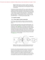
Institute of Technology.
1.2 Theory
1.2.1 Particle Sizes Small Compared to the Wavelength (Rayleigh Regime)
1.2.1.1 General Equations and Comparison with Bulk Absorption Measurements
For infrared optical depth measurements on airborne particles, the Lambert–Beer equation can be
written under the single scattering criterion in discrete form as
τ(ν j ) = −log
I (ν j )
l
=
I 0 (ν j ) ln 10
N
∑ n( D )C
i
ext
( Di , ν j )
j = 1… M .
i =1
(1.1)
It relates the measured optical depth τ( ν j ) at a specific wave number ν j to the optical path length
l, the number concentration n(Di ) of particles in a particular size bin Di of width ΔD, and the sizebin averaged extinction cross-section Cext (Di , ν j ), given by
1
Cext ( Di , ν j ) =
∆D
Di +
∆D
2
∫
Cext ( D, ν j ) dD.
∆D
Di −
2
(1.2)
The extinction cross-section Cext is the sum of the absorption cross-section Cabs and the scattering
cross-section Csca:
Cext ( D, ν j ) = Cabs ( D, ν j ) + Csca ( D, ν j ).
(1.3)
In the Rayleigh approximation, the absorption cross-section of a small sphere for transmission
measurements in air (refractive index of the medium ≈ 1) is written as
N 2 ( ν j ) − 1
Cabs ( D, ν j ) = 6 πν j V ( D)Im 2
,
N ( ν j ) + 2
(1.4)
with V(D) denoting the volume of the sphere of diameter D and N( ν j ) symbolizing the complex
refractive index of the particle with N ( ν j ) = n( ν j ) + ik ( ν j ). Note that Equation 1.4 is derived under
the assumption that x = πDν j 1 and x | N (ν j ) | 1 .15 Further assuming that the scattering contribution to extinction can be neglected, Equation 1.1 reduces to
τ( ν j ) =
=
6 πν j lVtot
N 2 ( ν j ) − 1
Im 2
ln 10
N ( ν j ) + 2
6 πν j lVtot
6 n( ν j )k ( ν j )
ln 10 n( ν )2 − k ( ν )2 + 2 2 + 2 n( ν )k ( ν )
j
j
j
j
(
) (
.
2
)
(1.5)
6
Fundamentals and Applications in Aerosol Spectroscopy
In this expression, the recorded optical depth only depends on the total particle-volume concentration Vtot and not on the details of the aerosol number size distribution n(Di ), that is, different size
distributions with the same overall particle-volume concentration give rise to identical infrared
absorption spectra. It is interesting to compare Equation 1.5 with the absorption spectrum of the
same substance in the bulk phase. For transmission measurements of a small film of thickness d, the
optical depth is directly proportional to the imaginary part of the complex refractive index:
τ(ν j ) =
4 πν j d
k (ν j ).
ln 10
(1.6)
The ratio of the optical depths measured for a small-particle and a thin-film spectrum thereby
becomes proportional to n( ν j )/((n( ν j )2 − k ( ν j )2 + 2)2 + (2 n( ν j )k ( ν j ))2 ) . For spectral regimes with
less intense absorption bands (k < 0.3), which only provoke a small amplitude of the anomalous
dispersion feature in the corresponding n spectrum, the proportionality factor reduces to
n(ν j )/(n(ν j )2 + 2)2 when assuming n( ν j ) k ( ν j ) over the considered wave number region. With
n( ν j ) only revealing small-amplitude dispersion features, the small-particle absorption spectrum
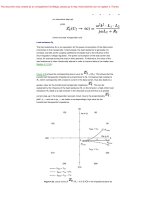
will not be considerably different from that of the bulk phase. This is demonstrated in panels a and
b of Figure 1.1 with aqueous sulfuric acid (25 wt% H 2 SO28
4 ) as an example. On the other hand,
Optical depth
(a)
0.04
0.03
25 wt% H2SO4
Particles
Bulk
0.02
(c)
0.12
0.10
0.08
(NH4)2SO4
Particles
Bulk
0.06
0.04
0.01
0
4000 3500 3000 2500 2000 1500 1000
n and k
(b)
1.6
1.4 n
1.2
1.0
0.8
0.6
0.4
0.2
k
0
4000 3500 3000 2500 2000 1500 1000
Wave number (cm–1)
0.02
0
4000 3500 3000 2500 2000 1500 1000
(d)
2.5
2.0
1.5
n
1.0
0.5
0
k
4000 3500 3000 2500 2000 1500 1000
Wave number (cm–1)
Figure 1.1 Panel a: Small-particle absorption spectrum of aqueous sulfuric acid with 25 wt% H2SO4
(black line), as computed from Equation 1.5 with Vtot = 1000 μm3/cm3 and l = 100 m based on the optical constants from Palmer and Williams28 (shown in panel b). The same refractive index data set was used to compute
the corresponding thin-film absorption spectrum (gray line) from Equation 1.6 with d = 0.085 μm. Panel c:
Small-particle absorption spectrum of crystalline ammonium sulfate spheres (black line), as computed from
Equation 1.5 with Vtot = 1000 μm3/cm3 and l = 100 m based on the optical constants from Earle et al.29 (shown
in panel d, data set for T = 298 K). Comparison with a calculated thin-film absorption spectrum (gray line) for
d = 0.080 μm (Equation 1.6).
Infrared Spectroscopy of Aerosol Particles
7
strong absorption bands with k > 1 and concomitantly high-amplitude anomalous dispersions in the
n spectrum might provoke that certain spectral regimes approximately fulfill the resonance condition that is inherent in Equation 1.5. For wave numbers with n ≈ 0 and k ≈ 2 , there will be an
enhanced cross-section in the absorption spectra of small spheres, provoking that the spectral habitus of an absorption band (including band intensity and peak position) might strongly differ from the
corresponding bulk absorption feature. As an example, panel c of Figure 1.1 compares the smallparticle and bulk absorption spectrum of crystalline ammonium sulfate spheres.29 Just in the regime
of the intense v3 (SO24−) absorption band at 1100 cm−1, the small-particle absorption band is shifted
to higher wave numbers and shows an increased intensity. Thus, the absorption maximum is shifted
kmax ) , that is, the bulk peak wave number, to a position where the corresponding optical
from ν(
constants n and k (Figure 1.1d) better fulfill the resonance condition of Equation 1.5.
1.2.1.2 Influence of Particle Shape
As already indicated in the introduction, the spectral habitus of intense small-particle absorption
bands might also strongly depend on the particle shape. As a simple case study, we want to summarize the results for needle- and disk-like spheroids, representing two subgroups of a general
ellipsoidal particle. For an exhaustive discussion of the shape effects, the reader is referred to the
textbook of Bohren and Huffman.15 For ellipsoidal particles, the geometrical factor L has to be
introduced in the expression for the absorption cross-section:
N 2 (ν j ) − 1
.
Cabs ( DV , ν j , L ) = 6 πν jV ( DV )Im
3L N 2 (ν j ) − 1 + 3
(1.7)
The particle diameter D V may now be interpreted as the diameter of the sphere with the same
volume as the nonspherical particle. For each of the three principal axes of an ellipsoid, there is a
distinct value for the geometrical factor L (L1, L2, and L3) and the average absorption cross-section
for randomly oriented ellipsoids can be obtained from the arithmetic mean of the three principal
cross-sections. For a sphere with L1 = L2 = L3 = ¹∕³, Equation 1.7 just reduces to Equation 1.5. For
needle- and disk-like spheroids, there are two distinct geometrical factors with L1 = 0, L2 = L3 = 0.5
(needle) and L1 = L2 = 0, L3 = 1 (disk). Therefore, two distinct resonances might be observed for
these particle shapes instead of the single absorption band for a sphere. The intensity of these bands
and their spectral splitting, however, depends on whether the actual spectral variation of the n and k
values over the considered wave number range is sufficient to cover both resonance conditions.30 As
an example, Figure 1.2a compares the small-particle absorption spectra of randomly oriented crystalline ammonium sulfate needles and disks with the corresponding sphere computation from
Figure 1.1c in the regime of the v3 (SO24− ) vibration. And indeed, both spheroidal shapes reveal a
band splitting, featuring one common mode at 1090 cm−1 from the principal component with L = 0.
This band gains a higher intensity for the disk-like shape due to its duplicate contribution to the
averaged cross-section, see Figure 1.2b, c. On the other hand, the 1120 cm−1 needle absorption band
and the shoulder at 1140 cm−1 for ammonium sulfate disks are due to the resonances for L = 0.5 and
L = 1, respectively. For these two bands, the needle-like shape gives rise to a higher intensity, both
due to the doubled weight of the L = 0.5 principal cross-section and a better match of the resonance
condition compared to the geometrical factor L = 1.
In a set of recent publications (see Sigurbjörnsson et al.16 and references therein), also the quantum–
mechanical vibrational exciton model was successfully applied to reproduce the shape effects in the
infrared absorption spectra of Rayleigh-sized particles. In these analyses, the calculations were
explicitly done for particle radii from 10 to 100 nm, that is, size and shape effects that occur in
nanosized particles, as addressed in the introduction, were excluded. From the observation that
strong shape effects are only evident for intense vibrational bands with a high molecular transition
8
Fundamentals and Applications in Aerosol Spectroscopy
Optical depth
(a) 0.15
Sphere
Disk
Needle
0.12
0.09
0.06
0.03
0
1250
1200
1150
1100
Optical depth
(b) 0.15
Disk
0.12
0.09
0.06
1000
950
Total
Contribution
from L1 = L2 = 0
Contribution
from L3=1
0.03
0
1250
1200
(c) 0.08
Optical depth
1050
1150
1100
1050
1000
950
Needle
Total
Contribution
from L1 = 0
Contribution
from L2 = L3 = 0.5
0.06
0.04
0.02
0
1250
1200
1150
1100
1050
Wave number (cm–1)
1000
950
Figure 1.2 Panel a: Small-particle absorption spectra of crystalline ammonium sulfate spheres, needles,
and disks, as computed from Equation 1.7 with Vtot = 1000 μm3/cm3 and l = 100 m based on the optical constants from Earle et al.29 Panels b and c elucidate the contributions from the two distinct geometrical factors
for the disk- and needle-like particles. See text for details.
dipole, it was concluded that the strong resonant intermolecular transition dipole coupling provides
the microscopic explanation of the shape effects in small-particle absorption spectra. The exciton
coupling leads to a delocalization of the excitation energy over the whole particle, which in turn
gives rise to the shape sensitivity of the absorption bands.
Apart from crystalline ammonium sulfate, other important aerosol constituents, which feature
pronounced shape-dependent infrared absorption bands in certain spectral regimes include mineral
dust (which, if containing silicates, exhibits a prominent Si– O stretch resonance at around
1050 cm−1),31 nitric acid dihydrate (in the nitrate absorption regime between 1500 and 1000 cm−1),32
and ammonia aerosols (ν2 N–H bending mode at 1060 cm−1).33
1.2.1.3 Derivation of Optical Constants from the Absorption Spectra of Small Particles
Two different approaches are usually applied to determine the frequency-dependent complex refractive indices from infrared optical measurements. On the one hand, the spectra of the optical constants n and k can be approximated by a set of Lorentz damped harmonic oscillators, with each
oscillator characterized by its peak wave number, band width (damping constant), and intensity.34
Starting from an a priori guess for the band parameters, their values can be optimized in an inversion
scheme by minimizing the summed-squared residuals between measured and calculated infrared
9
Infrared Spectroscopy of Aerosol Particles
spectra. The other approach exploits the Kramers–Kronig relation between the real and imaginary
parts of the complex refractive index,15
∞
n(ν 0 ) − 1 =
2
k (ν )ν
dν ,
P 2
π
ν − ν 20
∫
(1.8)
from which the value for n at a specific wave number ν0 can be computed from the spectrum of k
over the entire frequency range. Equation 1.8 can be directly applied to the analysis of thin-film
absorption spectra,23 given that k( ν ) is directly obtained from the transmission measurements, see
Equation 1.6. The experimental data, however, are often limited to mid-infrared wavelengths and
thus do not cover the complete wave-number range to evaluate the integral in Equation 1.8. Therefore,
suitable extensions of the k( ν ) spectrum beyond the experimentally accessible wave-number range
(e.g., at UV–VIS and far-IR wavelengths) have to be introduced to avoid potentially severe truncation
errors in the Kramers–Kronig transformation.35 It is sometimes proposed to employ the so-called
subtractive Kramers–Kronig integration to minimize the effect of truncation errors.36,37 In this
approach, the real part of the refractive index has to be known at some specific wave-number position ν x, preferentially located within the measured frequency range. Using n( ν x ) as a so-called
anchor point in the evaluation of the Kramers–Kronig integral, that is,
0
∞
n(ν 0 ) = n(ν x ) +
k (ν )ν
2(ν 20 − ν 2x )
P
dν ,
2
π
(ν − ν 20 )(ν 2 − ν 2x )
∫
0
(1.9)
may reduce the weight of the unknown frequency behavior of k( ν ) for wave numbers far above or
below the anchor point by introducing the additional factor ( ν 2 − ν 2x ) in the denominator of the
Kramers–Kronig integral. A Kramers–Kronig relation also exists between the reflectivity and phase
shift for reflection and can be used to obtain complex refractive indices from infrared reflection
spectra of bulk materials.38,39
Concerning infrared transmission measurements of airborne particles, Rouleau and Martin40
have emphasized that the Kramers–Kronig relation not only holds for N( ν j ), but also for the composite function f = ( N 2 (ν j ) − 1)/( N 2 (ν j ) + 2) :
Re{ f }(ν 0 ) =
∞
Im { f }( ν ) ν
2
P
dν .
π
ν 2 − ν 20
∫
0
(1.10)
Equation 1.10 thereby offers the most direct approach to deduce the optical constants from transmission spectroscopy of particles, provided that, (1), the particle sizes are small enough to fulfill the
requirements for Equation 1.5, (2), the particles are of (near) spherical shape, and (3), the overall
particle volume concentration can be measured with high accuracy by supplementary methods (e.g.,
analyses of filter samples or size distribution measurements). Then, the imaginary part of f can be
directly obtained from the measured optical depth, and, together with its real part, computed from
the Kramers–Kronig integral (Equation 1.10) with proper extension for the unmeasured spectral
range, allows the calculation of n and k (see Segal–Rosenheimer et al.41 for a recent example). This
procedure may also be applied with sufficient accuracy to the infrared spectra of particles with a
small scattering contribution, manifesting itself in a slightly slanted baseline at nonabsorbing wave
numbers. Then, the scattering part can be subtracted from the extinction spectrum by assuming a
Rayleigh-like Csca (ν ) ∝ ν 4 behavior (see, e.g., Figure 4 in Norman et al.22) and the residuum
absorption contribution can be treated with Equation 1.5. If it is not possible to experimentally prepare particle sizes which fall into the regime of Equation 1.5, the retrieval of the optical constants
becomes much more laborious. The inversion schemes are then based on Mie theory and usually
10
Fundamentals and Applications in Aerosol Spectroscopy
involve an iterative adjustment of n and k together with the parameters of the underlying particle
size distribution.42,43 Small differences between the data sets of optical constants obtained from different studies (see, e.g., a comparison between two recently derived n and k data sets for supercooled
water25,26) reflect the less stringent and approximate nature of these iterative inversion strategies.
A significant part of the atmospheric aerosol is composed of inhomogeneous particles, ranging
from comparatively simple core–shell structures (e.g., a particle containing a solid nucleus like soot
and a liquid organic or inorganic coating layer) to complex aggregates such as mineral dust, featuring a mixture of various minerals whose infrared refractive indices might strongly vary from mineral to mineral.44 In the case of dust samples, only the so-called effective or average optical constants
can be deduced from (infrared) optical measurements when performing the spectra analysis as if
the particles were homogeneous.15 Clearly, such data sets have a limited range of applicability, given
that each individual sample features a diverse mineralogical composition and aggregate structure.
On the modeling part, different mixing rules are proposed to calculate the refractive indices of
inhomogeneous, multicomponent particles such as mineral dust aggregates from the data sets of the
individual components. In the Maxwell–Garnet approximation, the composite particle is treated as
a homogeneous matrix with embedded inclusions, implying that a clear distinction between the
inclusions and the host matrix can be made. On the contrary, the Bruggeman theory applies to a
random inhomogeneous medium where the distinction between inclusion and host becomes unnecessary and both components can be treated symmetrically.15,44
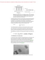
1.2.2 Infrared Extinction Spectra of Wavelength-Sized Particles (Mie Regime)
1.2.2.1 Dependence of the Spectral Habitus on the Particle Size
In the Rayleigh limit, the cross-sections for extinction and scattering are obtained by imposing that
the particles at each instant are exposed to an electromagnetic field that is uniform over their
dimension. The scattered field is then described by the electric dipole radiation of an oscillating
dipole. For particle sizes comparable to the wavelength of the infrared light, that is, in the framework of Mie theory for spheres, the scattered electromagnetic field is written as an infinite sum of
normal modes of the spherical particles weighted by the scattering coefficients an ( ν , D, N ) and
bn ( ν , D, N ), yielding the following expression for the extinction cross-section:
Cext (ν , D, N ) =
1
2 πν 2
∞
∑ (2n + 1)Re[a (ν , D, N ) + b (ν , D, N )].
n
n =1
(1.11)
n
Guidelines for the computation of the scattering coefficients an and bn, including the number of
terms that are required to obtain convergence in the series of Equation 1.11, are given for example,
in the textbook by Bohren and Huffman.15
In the following, we give a brief overview about extinction features in the framework of Mie
theory, taking ice as an example. Based on these results for spherical ice particles, Section 1.2.2.2
addresses the influence of particle asphericity on the absorption and scattering cross-sections. Panel
a of Figure 1.3 illustrates the evolution of the spectral habitus of the infrared extinction spectrum of
ice spheres when going from submicron to wavelength-sized particles. The Mie calculations were
done for a log-normal number size distribution with a common mode width of σg = 1.5 and count
median diameters (CMD) ranging from 0.1 to 13 μm; the employed optical constants are shown in
panel b. The lowermost extinction spectrum for the 0.1 μm-sized ice spheres is solely governed by
the absorption contribution, with the most prominent absorption bands located at around 3250 cm−1
(O –H molecular stretching mode) and 800 cm−1 (intermolecular vibration). The increasing scattering contribution for larger particle sizes first manifests itself in slightly slanted baselines at non
absorbing wave numbers greater than 3600 cm−1, without provoking in the first part a significant
distortion of the spectral habitus of the extinction bands at 3250 and 800 cm−1 (spectra for CMD of
11
Infrared Spectroscopy of Aerosol Particles
0.3 and 0.5 μm). For particle sizes between 1 and 4 μm, the prominent extinction bands gradually
adopt the spectral shape of the anomalous dispersion feature that is inherent in the spectrum of the
real part of the complex refractive index. Toward even larger sphere diameters, the infrared spectra
are characterized by a quite constant optical depth over the entire wave number range, except for
two pronounced extinction minima at around 3500 and 950 cm−1. These minima are caused by the
Christiansen effect and reflect the reduced scattering cross-sections of the ice spheres in these wave
number regimes, because the value for the real part of the refractive index approaches unity, that is,
corresponds to the value of the surrounding medium.45−47 A detailed view of the Christiansen band
at 950 cm−1 is shown in panel c of Figure 1.3. It becomes obvious that the wavelength position of
minimal optical depth does not exactly correspond to the minimum of the n spectrum (panel d),
because the minimized scattering contribution is counterbalanced by a large absorption contribution due to the high value for the imaginary index k. Instead, the extinction minimum is shifted to
larger wave numbers where absorption by the ice spheres is reduced. For smaller particle diameters
(a)
(c)
13 μm
13 μm
11 μm
11 μm
9 μm
Optical depth (arb. units)
7 μm
9 μm
6 μm
5 μm
7 μm
4 μm
3 μm
1 μm
6 μm
2 μm
5 μm
0.5 μm
0.3 μm
0.1 μm
n and k
(b)
6000
5000
4000
3000
2000
1000
(d)
1200
1.6
1.6
1.4 n
1.4
1.2
1.2
1.0
1.0
0.8
0.8
0.6
0.6
0.4
0.4
0.2
0.2
k
0
6000
5000
4000
3000
2000
Wave number (cm–1)
1000
1100
1000
900
800
1100
1000
900
Wave number (cm–1)
800
n
k
0
1200
Figure 1.3 Panel a: Infrared extinction spectra of ice spheres computed with Mie theory for a log-normal
number distribution of particle sizes with a mode width of σg = 1.5 and varying values for the count medium
diameter, as indicated in the figure panel. All spectra are normalized to unity and are offset for clarity. The
employed complex refractive indices for ice were taken from Zasetsky et al.25 (data set for 210 K) and are
shown in panel b. Panel c: Subset of the computed extinction spectra for the larger particle diameters in
the wave number regime of the Christiansen minimum at 950 cm−1. The corresponding part of the spectrum
of the optical constants n and k is shown in panel d. See text for details.