Photodiodes World Activities in 2011 Part 4 pptx
Bạn đang xem bản rút gọn của tài liệu. Xem và tải ngay bản đầy đủ của tài liệu tại đây (3.3 MB, 30 trang )
CMOS Photodetectors
81
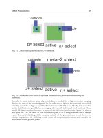
Fig. 11. CMOS-based photodiode: n+/p-substrate.
Fig. 12. Photodiode with metal-2 layer as a shield to block photons from reaching the
substrate.
In order to create a dense array of photodiodes, as needed for a high-resolution imaging
device, the ratio of the area designated for the collection of light to the area used for control
circuitry should be as high as possible. This is known as the fill-factor. Ideally, this would be
unity, but this is not possible for an imaging device with individual pixel read-out. Thus,
actual fill-factors are less than one. A layout of the APS pixel as shown in Figure 5 is shown
in Figure 13 . The fill factor of this 3 transistor pixel is 41% using scalable CMOS design
rules. The metal shielding of the circuitry outside of the photodetector is not shown for
clarity; in practice, this shielding would cover all non-photoactive areas and can also be
used as the circuit ground plane.
n+ select
p+ select
cathode
anode
active
n+ select
p+ select
cathode
anode
active
metal-2 shield
Photodiodes - World Activities in 2011
82
In order to create an array of imaging pixels, the layout not only requires maximizing the
active photodetector area, but also requires that the power (VDD), control and readout wires
be routed so that when a pixel is put into an array, these wires are aligned. An example of
this is shown in Figure 14.
Fig. 13. Layout of the T3-APS pixel as shown in Figure 5.
A slightly more complex structure is the buried double junction, or BDJ, photodiode [66].
The BDJ is formed from two vertically stacked standard p-n junctions, shown in Figure 15.
The shallow junction is formed by the p-base and N-well, and the deep junction is formed
by the N-well and P-substrate. As discussed previously, the depth of each junction is
determined by the thickness of the p-base and n-well. Incident light will be absorbed at
different depths, so the two junctions will produce currents based on the wavelength of the
incident light. The current flow through two junctions is proportional to the light intensity
at the junction depth. An example layout of the structure is shown in Figure 16.
PD
Reset
V
DD
Select
Read
Bus
SF
CMOS Photodetectors
83
Fig. 14. Simple 3x3 arrays of pixels shown in Figure 13. Notice that the wires align vertically
and horizontally.
Fig. 15. Cross-sectional view of the BDJ (not to scale).
Photodiodes - World Activities in 2011
84
Fig. 16. Layout of a 24μm x 24μm BDJ photodiode in the AMI 1.5μm process.
The final structure we will discuss is the phototransistor. Typically, a phototransistor can
produce a current output which is several times larger than a same area size photodiode due
to the high gain of the transistor. However, a major drawback of these phototransistors is
their low bandwidth, which is typically limited to hundreds of kHz. Additionally, the
current-irradiance relationship of the phototransistor is nonlinear, which makes it less than
ideal to use in many applications. Like the photodiode, there are a number of possible
configurations for the phototransistor in a standard CMOS process, such as the vertical p-n-
p phototransistor and lateral p-n-p phototransistor [67-71].
A cross-section of a vertical p-n-p phototransistor is shown in Figure 17 and an example
layout is provided in Figure 18.
CMOS Photodetectors
85
P-Active
N-well
P-Substrate
Emitter Contact
Poly contact
Collector Contact
Poly Grid
Fig. 17. Cross-sectional view of a vertical p-n-p phototransistor (not to scale).
Fig. 18. Layout of a 60 x 60 um vertical p-n-p phototransistor.
10. Current trends in performance optimization
10.1 Tune device responsivity
In a standard CMOS technology, a photodiode can be formed using different available
active layers, including n-active/p-substrate, p-active/n-well and n-well/p-substrate, to
form a p-n junction. In a photodiode, the photo-conversion mostly takes place in the
depletion region where an incident photon creates an electron and hole pair with the
Photodiodes - World Activities in 2011
86
electron passing to the n-region and hole to the p-region. Hence, varying the depth at which
the depletion region forms in the silicon wafer would control the performance of the
photodiodes in terms of responsivity and quantum efficiency. Also, varying the width of the
depletion region by appropriately applying a reverse bias to the photodiode, one could
control the response time of the detector. A wider depletion region reduces the junction
capacitance of the p-n-junction and improves the response time of the detector.
Here, we will aim to understand the effect on responsivity and external quantum efficiency
on the design of photodiode structures. Given that all materials in a standard CMOS process
are set by the manufacturer, the external quantum efficiency, which takes into account only
the photon-generated carriers collected as a result of the light absorption or, in other words,
the useful portion of signal generated by interaction of light and photodetector, is more
relevant. The external quantum efficiency depends on the absorption coefficient of the
material, a (units: cm
-1
) and thickness of the absorbing material. Assuming that the entire
incident light is absorbed by the detector, if the photon flux density incident at the surface is
Φ
o
, then the photon flux at depth, x, is given by Beer’s law (Equation 1) [72].
The external quantum efficiency is also a function of wavelength of the incident light. Thus
in a CMOS photodiode, one can strategically chose the depth of the location of the depletion
region to which photons are likely to penetrate and thereby optimize the photodetector to
provide high absorption for particular spectrum of wavelengths. In practical optoelectronic
systems development, responsivity, that is defined as the output current divided by the
incident light power, may be a more relevant performance metric. Responsivity is related to
quantum efficiency by a factor of hµ/q, where, q is the electron charge, h is Planck’s constant,
and µ is the frequency of the incident photon. The spectral response curve is a plot of
responsivity as a function of wavelength.
Thus, to optimize a silicon photodiode structure for detecting blue wavelengths, the
depletion region should be near to the silicon surface. For red wavelengths, the depletion
region should be placed deeper in the silicon substrate. Based on this idea, Yotter et al. [73]
have compared photodiode structures (p-active/n-well and n-well/p-substrate) to develop
photodiodes better suited for blue or green wavelengths for specific biosensing applications.
The blue-enhanced structure used interdigitated p+-diffusion fingers to increase the
depletion region area near the surface of the detector, while the green-enhanced structure
used n-well fingers to increase the depletion region slightly deeper within the substrate.
Bolten et al. [74] provided a thorough treatment of the photodiode types and their
properties. They reported that in a standard CMOS process n-well/p-substrate structure
provides relatively better quantum efficiency for biosensors operating in visible
electromagnetic spectrum.
Using the properties that external quantum efficiency varies as a function of wavelength of
the incident light and Beer’s law, many research groups reported the use of buried double p-
n junction (BDJ) and buried triple p-n junction structures, which can be implemented with a
standard CMOS process, for monochromatic color detection [75, 76]. The BDJ structure has
two standard p-n junctions (p-base/n-well/p-substrate) are stacked vertically in the CMOS
chip. For the BDJ detector, we obtain I
top
(only from top p-n junction) and I
bottom
(sum of
currents from top and bottom p-n junctions) from the detector. The current ratio, I
top
/I
top-
I
bottom
can be used for the color/ wavelength measurements. The CMOS BDJ detector has
been used for fluorescence detection in microarrays [77], and for the detection and
measurement of ambient light sources [78]. The BDJ color detectors have been used in many
chemical and biological sensors such as seawater pH measurement [79] and volatile organic
compounds detection [80].
CMOS Photodetectors
87
10.2 Monolithic integration of photonic devices on photodetectors
10.2.1 Microlens and microfilters
Most CMOS image sensors are monochrome devices that record the intensity of light. A
layer of color filters or color filter array (CFA) is fabricated over the silicon integrated circuit
using a photolithography process to add color detection to the digital camera. CFA is
prepared by using color pigments mixed with photosensitive polymer or resist carriers.
Many recent digital color imaging systems use three separate sensors to record red, green,
and blue scene information, but single-sensor systems are also common [81]. Typically,
single-sensor color imaging systems have a color filter array (CFA) in a Bayer pattern as
shown in Figure 19. The Bayer pattern was invented at Eastman Kodak Company by Bryce
Bayer in 1976 [82]. This CFA pattern has twice as many green filtered pixels as red or blue
filtered pixels. The spatial configuration of the Bayer pattern is tailored to match the
optimum sensitivity of human vision perception. Imager sensors also include microlenses
placed over the CFA to improve the photosensitivity of the detection system and improve
the efficiency of light collection by proper focusing of the incident optical signal over the
photodetectors [83]. A microlens is usually a single element with one plane surface facing
the photodiode and one spherical convex surface to collect and focus the light. Thus, as
photons pass through the microlens and through the CFA filter, thus passing only
wavelengths of red, green, or blue color and finally reach the photodetectors. The
photodetectors are integrated as part of an active pixel sensor to convert the incident optical
signal into electrical output [84]. The analog electrical data from the photopixels are then
digitized by an analog-to-digital converter. To produce a full color image, a spatial color
interpolation operation known as demosaicing is used. The image data is then further
processed to perform color correction and calibration, white balancing, infrared rejection,
and reducing the negative effects of faulty pixels [85, 86].
Fig. 19. Bayer film pattern and microlenses integrated onto a device.
One of the first example of a monolithic microlens array fabricated on the MOS color imager
was done using photolithography of a polymethacrylate type transparent photoresist [87].
Photodiodes - World Activities in 2011
88
In commercial camera production, glass substrates are typically used as carrier and spacer
wafers for the lenses and are filled with an optical polymer material which is
photolithographically patterned to form the microlenses. A fairly straightforward method
used in many microlens implementations is to photolithographically pattern small cylinders
of a suitable resin on a substrate. The small cylinders are then melted in carefully controlled
heating conditions. Hence, after melting they tend to form into small hemispheres due to
surface tension forces. However, molten resin had a tendency to spread such that lens size
and spatial location is difficult to control. A well-defined spherical surface for the microlens
is required to achieve high numerical aperture which improves the image sensor efficiency.
Different techniques are used to control the spherical shape and spatial location of the
microlens including pre-treatment of the substrate to adjust the surface tension to control
the reflow of the microlens [88] and use of microstructures such as pedestals to control the
surface contact angle [83]. In more recent processes the glass substrates are eliminated and
instead microlenses are made with polymer materials that are molded using master stamps.
The molded polymer microlenses are cured with ultra violet exposure or heat treatment. By
replacing the glass substrates, wafer-level system manufacturers face fewer constraints on
the integration optics and imager integrated circuit enabling the production of compact and
efficient imager sensors.
10.3 Waveguides, gratings, and couplers
In this section, we will concentrate on understanding device architectures that deal with
monolithic integration of photonic waveguides, gratings and couplers with CMOS
photodetectors for applications in optoelectronics to improve quantum efficiency, spectral
response selectivity, and planar coupling and guiding of light signals to on-chip
photodetectors. CMOS photodetectors operate only in visible and near infra-red region
between 400nm and 1.1µm of the electromagnetic spectrum. There are applications in
sensing and optical communications in this wavelength region where silicon or CMOS
photodetectors can offer low-cost and miniaturized systems. Monolithic integration of
photonic components with silicon/CMOS photodetectors started as a major research area
since early 1980’s [89-92]. It is advantageous that a monolithic integrated optoelectronic
system on silicon use materials typically employed in CMOS-technology. The dielectrics
available in CMOS are favorable as the light guiding layer for wavelengths in the visible
and near infrared region. The available materials in CMOS processing technology to
develop the photonic devices include layers such as silicon nitride [3], Phospho-Silicate
Glass (PSG) —SiO
2
doped with P
2
O
5
[4] or silicon oxynitride layers deposited as
insulating and passivation layers. Confinement of light is achieved by an increased
refractive index in the light guiding film, compared to silicon oxide. The first proposed
CMOS compatible devices were based on using silicon oxynitride waveguides
sandwiched with silicon dioxide (SiO
2
) layers [93-95].
System-level integration is commonly used in compact spectrometers with Lysaght et al.
[96] [97] first proposing a spectrometer system in the year 1991 that would integrate silicon
photodiode array with microfabricated grating structures for diffraction of the incident light
signals and subsequent detection of the optical spectrum components by the silicon
photodiode array. More recent and commercial available compact spectrometers use a
CMOS line array. Csutak et al. [98] provided an excellent background for related work done
CMOS Photodetectors
89
prior to their research article. After considering the absorption length of silicon and required
bandwidth for high-speed optical communications, the improvement of quantum efficiency
of the photodetectors remains an important challenge.
10.4 Biosensors on CMOS detectors
Many research groups are working on the idea of contact imaging systems for imaging or
detection of a biological specimens coupled directly to the chip surface which was first
proposed by Lamture et al. [99] using a CCD camera. As the photodetector components in
biosensors, CMOS imagers are preferable to convert the optical signals into electrical signals
because of monolithic integration of photodetection elements and signal processing circuitry
leading to low cost miniaturized systems [100, 101]. In 1998, a system termed as
bioluminescent-bioreporter integrated circuit (BBIC) was introduced that described placing
genetically engineered whole cell bioreporters on integrated CMOS microluminometers
[102]. In a more recent implementation of BBIC system includes sensing low concentrations
of a wide range of toxic substances such as salicylate and naphthalene in both gas and liquid
environments using genetically altered bacteria, Pseudomonas fluorescens 5RL, as the
bioreporter [103]. BBIC system operates on the basis of using a large CMOS photodiode
(1.47 mm
2
area using n-well/p-substrate structure) for detection of low levels of
luminescence signals by integration of the photocurrent generated by the photodiode over
time and a current-to-frequency converter as signal processing circuit to provide a digital
output proportional to the photocurrent.
Recent implementations of contact imaging include using custom-designed CMOS imagers
as platform for imaging of cell cultures [104] and DNA sequencing [105, 106]. Now
researchers are working on the integration of molded and photolithographically patterned
polymer filters and microlenses with CMOS photodetectors and imagers towards complete
development of miniaturized luminescence sensors. Typically, luminescence sensors require
an optical excitation source for exciting the sensor materials with electromagnetic radiation
and a photodetector component for monitoring the excited state emission response from the
sensor materials at a higher wavelength electromagnetic spectrum that is filtered from the
excitation input. The next step towards convenient monolithic integration of filters,
biological support substrates, and microfluidic interfaces create interesting challenges for
engineers and scientists. A recent report discusses the approach of using poly(acrylic acid)
filters integrated with custom-designed CMOS imager ICs to detect fluorescent micro-
spheres [107]. Polydimethylsiloxane (PDMS) could offer a more versatile material to
fabricate lenses, filters, diffusers and other components for optical sensors [108]. PDMS is a
silicone-based organic polymer that is soft, flexible, biocompatible and optically transparent
and well amenable to various microfabrication techniques. PDMS can be doped with apolar
hydrophobic color dyes such as Sudan-I, -II or -III to form optical filters that work in
different regions of visible electromagnetic spectrum [109]. The Authors group recently
proposed a prototype compact optical gaseous O
2
sensor microsystem using xerogel based
sensor elements that are contact printed on top of trapezoidal lens-like microstructures
molded into PDMS that is doped with Sudan-II dye as shown in Figure 20 [110]. The
molded PDMS structure serves triple purpose acting as immobilization platform, filtering of
excitation radiation and focusing of emission radiation onto the detectors. The PDMS
structure is then integrated on top of a custom design CMOS imager to create a contact
Photodiodes - World Activities in 2011
90
imaging sensor system. The low-cost polymer based filters is best suited for LED excitation
and may not be able to provide optimum excitation rejection performance when laser
radiation is used for excitation. As a more traditional alternative, Singh et al. [111] proposed
micromachining a commercially available thin-film interference filter and gluing it to the
CMOS imager die.
Fig. 20. Fabricated microlenses and xerogel sensors.
11. Optical sensor chip with Color Change-Intensity Change Disambiguation
(CCICD)
In this section we present a CMOS-sensor chip that can detect irradiance and color
information simultaneously. Compared with other BDJ-based systems [112-114], this system
includes an irradiance detection pathway that can be used in combination with the color
information to provide color change -intensity change disambiguation (CCICD). The
irradiance detection pathway is based on the work by Delbruck and Mead [23] with a single
standard CMOS photodiode (see Figure 21). Thus, this pathway can function ambient light
conditions without an additional light source, is more robust to background light changes,
has a higher bandwidth for time-varying signals, and has the ability to emulate adaptation
to background light levels, which is an important phenomena found in biological visual
systems. The color detection pathway consists of a BDJ photodetector and subsequent
processing circuitry to produce a single voltage as the chip output without additional
external circuitry. The BDJ produces two currents which are used as inputs to individual
logarithmic current to voltage converter circuits whose outputs are converted to a voltage
Colored and
molded PDMS
microlens array
Xerogel based
oxygen sensors
CMOS Photodetectors
91
difference using a differential amplifier. The output of this pathway is a single voltage that
represents the color of the input signal, with better than 50nm resolution. Both pathways are
integrated on the same IC.
Fig. 21. Block diagram of the sensor chip pathways (from [115]).
The irradiance detection pathway out is shown in Figure 21. The response of the irradiance
detection pathway circuit is logarithmic over the measured irradiance range spanning
nearly 3 orders of magnitude.
Photodiodes - World Activities in 2011
92
Fig. 22. Output of the irradiance detection pathway (from [115]).
Fig. 23. Output of the color detection pathway as a function of incident light wavelength
(from [115]).
Incident Light Wavelength (nm)
400 500 600 700 800 900
Color Pathway Output (V)
1.0
1.2
1.4
1.6
1.8
2.0
2.2
2.4
2.6
2.8
CMOS Photodetectors
93
Fig. 24. Output of the color detection pathway as a function of incident power (from [115]).
From the experimental results, we can see that the output voltage is larger for longer
incident light wavelengths (see Figure 23). So, for a practical implementation based on this
chip, a look-up table can be used to map the output voltage to the incident wavelength.
Moreover, from Figure 24, the changes in the output voltage caused by irradiance change
will not cause confusion between which color (primary wavelength) is detected; the R, G
and B curves will not overlap for a normal operating range of irradiance. The reason for this
performance is because the I2/I1 ratio from the BDJ is (ideally) independent of light
intensity.
12. References
[1] A. R. Thryft. (2011, February 1) CCD vs. CMOS image sensors. Test & Measurement
World. Available:
image_sensors.php
[2] J. W. Horton, et al., "The scanistor-A solid-state image scanner," Proceedings of the IEEE,
vol. 52, pp. 1513-1528, 1964.
[3] S. R. Morrison, "A new type of photosensitive junction device," Solid-State Electronics, vol.
6, pp. 485-494.
[4] M. A. Schuster and G. Strull, "A monolithic mosaic of photon sensors for solid state
imaging applications," in Electron Devices Meeting, 1965 International, 1965, pp. 20-
21.
Photodiodes - World Activities in 2011
94
[5] M. A. Schuster and G. Strull, "A monolithic mosaic of photon sensors for solid-state
imaging applications," Electron Devices, IEEE Transactions on, vol. 13, pp. 907-912,
1966.
[6] G. P. Weckler, "Operation of p-n Junction Photodetectors in a Phton Flux Integrating
Mode," IEEE Journal of Solid State Circuits, vol. SC-2, pp. 65-73, Sept 1967.
[7] R. Melen, "The tradeoffs in monolithic image sensors; MOS vs CCD," Electronics, vol. 46,
pp. 106-11, 1973.
[8] S. Mendis, et al., "CMOS active pixel image sensor," IEEE Trans. Electron Devices, vol. 41,
pp. 452-453, 1994.
[9] S. K. Mendis, et al., "CMOS Active Pixel Image Sensors for Highly Integrated Imaging
Systems," IEEE J. Solid-State Circuits, vol. 32, pp. 187-196, 1997.
[10] E. R. Fossum, "CMOS Image Sensors: electronic camera-on-a-chip," IEEE Transactions
on Electron Devices, vol. 44, pp. 1689-1697, 1997.
[11] B. Dierickx, et al., "Random addressable active pixel image sensors," in Proceedings of the
SPIE vol. 2950, ed. Berlin, Germany: SPIE, 1996, pp. 2-7.
[12] D. X. D. Yang and A. El Gamal, "Comparative analysis of SNR for image sensors with
enhanced dynamic range," in Proceedings of the SPIE vol. 3649, ed. San Jose, CA,
USA: SPIE, 1999, pp. 197-211.
[13] O. Yadid-Pecht, "Wide-dynamic-range sensors," Optical Engineering, vol. 38, pp. 1650-
60, 1999.
[14] S. G. Chamberlain and J. P. Y. Lee, "A novel wide dynamic range silicon photodetector
and linear imaging array," Electron Devices, IEEE Transactions on, vol. 31, pp. 175-
182, 1984.
[15] N. Ricquier and B. Dierickx, "Pixel structure with logarithmic response for intelligent
and flexible imager architectures," Microelectronic Engineering, vol. 19, pp. 631-634,
1992.
[16] N. Ricquier and B. Dierickx, "Random addressable CMOS image sensor for industrial
applications," Sensors and Actuators A (Physical), vol. A44, pp. 29-35, 1994.
[17] M. A. Pace and J. J. Zarnowski, "Complementary metal oxide semiconductor imaging
device," USA Patent 6 084 229, 2000.
[18] S. Hanson, et al., "A 0.5 V Sub-Microwatt CMOS Image Sensor With Pulse-Width
Modulation Read-Out," Solid-State Circuits, IEEE Journal of, vol. 45, pp. 759-767,
2010.
[19] M. Furuta, et al., "A high-speed, high-sensitivity digital CMOS image sensor with a
global shutter and 12-bit column-parallel cyclic A/D converters," IEEE Journal of
Solid-State Circuits, vol. 42, pp. 766-74, 2007.
[20] S. Kleinfelder, et al., "A 10000 frames/s CMOS digital pixel sensor," Solid-State Circuits,
IEEE Journal of, vol. 36, pp. 2049-2059, 2001.
[21] C. A. Mead and M. A. Mahowald, "A Silicon Model of Early Visual Processing," Neural
Networks, vol. 1, pp. 91-97, 1988.
[22] T. Delbruck, "Silicon retina with correlation-based, velocity-tuned pixels," Neural
Networks, IEEE Transactions on, vol. 4, p. 529, 1993.
CMOS Photodetectors
95
[23] T. Delbruck and C. A. Mead, "Adaptive Photoreceptor with Wide Dynamic Range," in
1994 IEEE International Symposium on Circuits and Systems, London, England, 1994,
pp. 339-342.
[24] R. A. Deutschmann and C. Koch, "An analog VLSI velocity sensor using the gradient
method," Proceedings of the 1998 IEEE International Symposium on Circuits and
Systems,, vol. 6, p. 649, 1998.
[25] T. J. Drabik, et al., "2D Silicon/Ferroelectric Liquid Crystal Spatial Light Modulators,"
IEEE Micro, vol. 15, pp. 67-76, 1995.
[26] R. Etienne-Cummings, et al., "Hardware implementation of a visual-motion pixel using
oriented spatiotemporal neural filters," Circuits and Systems II: Analog and Digital
Signal Processing, IEEE Transactions on [see also Circuits and Systems II: Express Briefs,
IEEE Transactions on], vol. 46, pp. 1121-1136, 1999.
[27] R. Etienne-Cummings, et al., "A foveated silicon retina for two-dimensional tracking,"
IEEE Transactions on Circuits and Systems II: Analog and Digital Signal Processing, vol.
47, pp. 504-517, 2000.
[28] C. M. Higgins, et al., "Pulse-based 2-D motion sensors," Circuits and Systems II: Analog
and Digital Signal Processing, IEEE Transactions on [see also Circuits and Systems II:
Express Briefs, IEEE Transactions on], vol. 46, pp. 677-687, 1999.
[29] R. M. Philipp and R. A. Etienne-Cummings, "A 1V current-mode CMOS active pixel
sensor," in Proc. IEEE Int.Symp. Circ. Syst., Kobe, Japan, 2005, pp. 4771-1774.
[30] A. Sartori, et al., "A 2D photosensor array with integrated charge amplifier," Sensors and
Actuators A: Physical, vol. 46, pp. 247-250, 1995.
[31] B. E. Shi, "A One-Dimensional CMOS Focal Plane Array for Gabor-Type Image
Filtering," IEEE Transactions on Circuits and Systems I: Fundamental Theory and
Applications, vol. 46, pp. 323-327, 1999.
[32] A. H. Titus and T. J. Drabik, "An Improved Silicon Retina Chip with Optical Input and
Optical Output," in Proceedings: Tenth Annual IEEE International ASIC Conference,
Portland, OR, USA, 1997, pp. 88-91.
[33] H. G. Graf, et al., "High Dynamic Range CMOS Imager Technologies for Biomedical
Applications," Solid-State Circuits, IEEE Journal of, vol. 44, pp. 281-289, 2009.
[34] A. H. Titus, et al., "Autonomous low-power glare sensing chip," Electronics Letters, vol.
47, pp. 508-509, 2011.
[35] K. Murari, et al., "A CMOS In-Pixel CTIA High-Sensitivity Fluorescence Imager,"
Biomedical Circuits and Systems, IEEE Transactions on, vol. PP, pp. 1-10, 2011.
[36] Y. Lei, et al., "CMOS Imaging of Pin-Printed Xerogel-Based Luminescent Sensor
Microarrays," Sensors Journal, IEEE, vol. 10, pp. 1824-1832, 2010.
[37] K. Kwang Hyun and K. Young Soo, "Scintillator and CMOS APS Imager for
Radiography Conditions," Nuclear Science, IEEE Transactions on, vol. 55, pp. 1327-
1332, 2008.
[38] K. Murari, et al., "Which Photodiode to Use: A Comparison of CMOS-Compatible
Structures," Sensors Journal, IEEE, vol. 9, pp. 752-760, 2009.
[39] A. Rogalski, et al., Narrow-gap semiconductor photodiodes. Bellingham, Wash.: SPIE Press,
2000.
Photodiodes - World Activities in 2011
96
[40] G. Ghione, Semiconductor devices for high-speed optoelectronics. Cambridge; New York:
Cambridge University Press, 2009.
[41] K. K. Ng, Complete guide to semiconductor devices. Hoboken: Wiley-Interscience, 2002.
[42] G. P. Weckler, "Operation of p-n Junction Photodetectors in a Photon Flux Integrating
Mode," IEEE Journal of Solid State Circuits, vol. SC-2, pp. 65-73, Sept 1967.
[43] J. Ohta, Smart CMOS image sensors and applications. Boca Raton, Fla.; London: CRC ;
Taylor & Francis [distributor], 2006.
[44] A. Hoffman, et al., "CMOS Detector Technology," Experimental Astronomy, vol. 19, pp.
111-134, Feb 01 2005.
[45] P. Magnan, "Detection of visible photons in CCD and CMOS: A comparative view,"
Nuclear Instruments and Methods in Physics Research Section A: Accelerators,
Spectrometers, Detectors and Associated Equipment, vol. 504, pp. 199-212, Jun 21 2003.
[46] A. Gamal, et al., "CMOS Sensors for Optical Molecular Imaging," in CMOS
Biotechnology, H. Lee, et al., Eds., ed: Springer US, 2007, pp. 331-379.
[47] B. Sapoval and C. Hermann, Physics of semiconductors. New York: Springer-Verlag,
1995.
[48] K. G. Mckay and K. B. Mcafee, "Electron Multiplication in Silicon and Germanium,"
Physical Review, vol. 91, pp. 1079-1084, 1953.
[49] S. Miller, "Ionization Rates for Holes and Electrons in Silicon," Physical Review, vol. 105,
pp. 1246-1249, Mar 1957.
[50] H. Melochior and W. Lynch, "Signal and Noise Response of High Speed Germanium
Avalanche Photodiodes," Ieee Transactions On Electron Devices, vol. ED13, pp. 829-
&, 1966.
[51] D. Stoppa, et al., "Single-Photon Avalanche Diode CMOS Sensor for Time-Resolved
Fluorescence Measurements," IEEE Sensors Journal, vol. 9, pp. 1084-1090, 2009.
[52] A. Biber, et al., "Avalanche photodiode image sensor in standard BiCMOS technology,"
Ieee Transactions On Electron Devices, vol. 47, pp. 2241-2243, Nov 01 2000.
[53] A. Biber, et al., "Avalanche photodiode image sensor in standard silicon BiCMOS
technology," Sensors And Actuators A-Physical, vol. 90, pp. 82-88, 2001.
[54] A. Rochas, et al., "Low-noise silicon avalanche photodiodes fabricated in conventional
CMOS technologies," Ieee Transactions On Electron Devices, vol. 49, pp. 387-394, Apr
01 2002.
[55] G. F. Marshall, et al., "Avalanche photodiode-based active pixel imager," Ieee
Transactions On Electron Devices, vol. 51, pp. 509- 511, Apr 01 2004.
[56] L. Pancheri, et al., "Low-Noise Avalanche Photodiode in Standard 0.35-μm CMOS
Technology," Ieee Transactions On Electron Devices, vol. 55, pp. 457-461, 2008.
[57] C. Niclass, et al., "Design and characterization of a CMOS 3-D image sensor based on
single photon avalanche diodes," presented at the Ieee Journal of Solid-State
Circuits, 2005.
[58] C. Niclass
, et al., "A 4 μs integration time imager based on CMOS single photon
avalanche diode technology," presented at the Sensors And Actuators A-Physical,
2006.
CMOS Photodetectors
97
[59] C. Niclass, et al., "A single photon avalanche diode implemented in 130-nm CMOS
technology," Ieee Journal Of Selected Topics In Quantum Electronics, vol. 13, pp. 863-
869, 2007.
[60] D. Stoppa, et al., "A CMOS 3-D imager based on single photon avalanche diode,"
presented at the Ieee Transactions on Circuits and Systems I-Regular Papers, 2007.
[61] N. Faramarzpour, et al., "Fully integrated single photon avalanche diode detector in
standard CMOS 0.18-mu m technology," Ieee Transactions On Electron Devices, vol.
55, pp. 760-767, 2008.
[62] D. E. Schwartz, et al., "A Single-Photon Avalanche Diode Array for Fluorescence
Lifetime Imaging Microscopy," presented at the Ieee Journal of Solid-State Circuits,
2008.
[63] B. R. Rae, et al., "A Vertically Integrated CMOS Microsystem for Time-Resolved
Fluorescence Analysis," presented at the Ieee Transactions on Biomedical Circuits
and Systems, 2010.
[64] C. J. Stapels, et al., "CMOS-based avalanche photodiodes for direct particle detection,"
presented at the Nuclear Instruments & Methods in Physics Research Section
a-Accelerators Spectrometers Detectors and Associated Equipment, 2007.
[65] R. J. Baker, CMOS Circuit Design, Layout, and Simulation, 3rd ed. Hoboken, N.J.: IEEE
Press/Wiley, 2010.
[66] G. de Graaf and R. F. Wolffenbuttel, "Smart optical sensor systems in CMOS for
measuring light intensity and colour," Sensors & Actuators A-Physical, vol. A67, pp.
115-119, 1998.
[67] H. Zimmermann, "Silicon Optoelectronic Integrated Circuits," Berlin, Germany:
Springer-Verlag,, 2004.
[68] M. Davenport, "Detection and Processing Strategies for Integrated Xerogel-based
Sensor Systems," in Ph.D. Dissertation, Department of Electrical Engineering:
University at Buffalo, The State University of New York,, 2004.
[69] M.P. Vidal, et al., "A bipolar photodetector compatible with standard CMOS
technology," Solid-State Electronics, vol. 34, pp. 809-814, 1991.
[70] R. W. Sandage and J. A. Connelly, "Producing phototransistors in a standard digital
CMOS technology," in Circuits and Systems, 1996. ISCAS '96., 'Connecting the
World'., 1996 IEEE International Symposium on, 1996, pp. 369-372 vol.1.
[71] R. W. Sandage and J. A. Connelly, "A 128x128 imaging array using lateral bipolar
phototransistors in a standard CMOS process [fingerprint detection]," in Circuits
and Systems, 1998. ISCAS '98. Proceedings of the 1998 IEEE International Symposium
on, 1998, pp. 641-644 vol.6.
[72] B. E. A. Saleh and M. C. Teich, Fundamentals of Photonics. New York: Wiley, 1991.
[73] R. A. Yotter, et al., "Optimized CMOS photodetector structures for the detection of
green luminescent probes in biological applications," Sensors and Actuators B-
Chemical, vol. 103, pp. 43-49, Sep 29 2004.
[74] E. K. Bolton, et al., "Integrated CMOS photodetectors and signal processing for very
low-level chemical sensing with the bioluminescent bioreporter integrated circuit,"
Sensors and Actuators B-Chemical, vol. 85, pp. 179-185, Jun 20 2002.
Photodiodes - World Activities in 2011
98
[75] M. Ben Chouikha, et al., "Colour detection using buried triple pn junction structure
implemented in BiCMOS process," Electronics Letters, vol. 34, pp. 120-122, Jan 8
1998.
[76] G. N. Lu, et al., "Colour detection using a buried double p-n junction structure
implemented in the CMOS process," Electronics Letters, vol. 32, pp. 594-6, 1996.
[77] G. N. Lu, et al., "Investigation of CMOS BDJ detector for fluorescence detection in
microarray analysis," 1st Annual International Ieee-Embs Special Topic Conference on
Microtechnologies in Medicine & Biology, Proceedings, pp. 381-386, 2000.
[78] G. de Graaf and R. Wolffenbuttel, "Optical CMOS sensor system for detection of light
sources," Sensors and Actuators a-Physical, vol. 110, pp. 77-81, Feb 1 2004.
[79] M. Sedjil, et al., "A colorimetric method with the use of BDJ detector for seawater pH
measurement," Analytica Chimica Acta, vol. 377, pp. 179-184, December 31 1998.
[80] X. Y. Fang, et al., "Colorimetric porous photonic bandgap sensors with integrated
CMOS color detectors," Ieee Sensors Journal, vol. 6, pp. 661-667, Jun 2006.
[81] R. Palum, "Image sampling with the Bayer color filter array," Pics 2001: Image
Processing, Image Quality, Image Capture, Systems Conference, Proceedings, pp. 239-245,
2001.
[82] P. L. P. Dillon, et al., "Color Imaging-System Using a Single Ccd Area Array," Ieee
Journal of Solid-State Circuits, vol. 13, pp. 28-33, 1978.
[83] Z. D. Popovic, et al., "Technique for Monolithic Fabrication of Microlens Arrays,"
Applied Optics, vol. 27, pp. 1281-1284, Apr 1 1988.
[84] A. El Gamal and H. Eltoukhy, "CMOS image sensors," Ieee Circuits & Devices, vol. 21,
pp. 6-20, May-Jun 2005.
[85] E. R. Fossum, "CMOS image sensors: Electronic camera-on-a-chip," Ieee Transactions on
Electron Devices, vol. 44, pp. 1689-1698, Oct 1997.
[86] J. Adams, et al., "Color processing in digital cameras," Ieee Micro, vol. 18, pp. 20-30,
Nov-Dec 1998.
[87] Y. Ishihara and K. Tanigaki, "A High Photosensitivity IL-CCD Image Sensor with
Monolithic Resin Lens Array," in Proceedings of International Electron Devices
Meeting, Washington, DC, 1983, pp. 497-500.
[88] A. Schilling, et al., "Surface profiles of reflow microlenses under the influence of surface
tension and gravity," Optical Engineering, vol. 39, pp. 2171-2176, Aug 2000.
[89] D. G. Hall, "Survey of Silicon-Based Integrated-Optics," Computer, vol. 20, pp. 25-32,
Dec 1987.
[90] S. Valette, et al., "Si-Based Integrated-Optics Technologies," Solid State Technology, vol.
32, pp. 69-75, Feb 1989.
[91] U. Hilleringmann and K. Goser, "Optoelectronic System Integration on Silicon - Wave-
Guides, Photodetectors, and Vlsi Cmos Circuits on One-Chip," Ieee Transactions on
Electron Devices, vol. 42, pp. 841-846, May 1995.
[92] R. A. Soref, "Silicon-Based Optoelectronics," Proceedings of the Ieee, vol. 81, pp. 1687-
1706, Dec 1993.
[93] S. Wunderlich, et al., "Integration of Sion Wave-Guides and Photodiodes on Silicon
Substrates," Applied Optics, vol. 31, pp. 4186-4189, Jul 20 1992.
CMOS Photodetectors
99
[94] D. Peters, et al., "Integrated-Optics Based on Silicon Oxynitride Thin-Films Deposited
on Silicon Substrates for Sensor Applications," Sensors and Actuators a-Physical, vol.
26, pp. 425-431, Mar 1991.
[95] C. H. Henry, et al., "Low-Loss Si3n4-Sio2 Optical Wave-Guides on Si," Applied Optics,
vol. 26, pp. 2621-2624, Jul 1 1987.
[96] M. J. Lysaght, et al., "Laptop Chemistry - a Fiberoptic, Field-Portable, near-Infrared
Spectrometer," Review of Scientific Instruments, vol. 62, pp. 507-515, Feb 1991.
[97] H. Bezzaoui and E. Voges, "Integrated-Optics Combined with Micromechanics on
Silicon," Sensors and Actuators a-Physical, vol. 29, pp. 219-223, Dec 1991.
[98] S. M. Csutak, et al., "CMOS-compatible planar silicon waveguide-grating-coupler
photodetectors fabricated on silicon-on-insulator (SOI) substrates," Ieee Journal of
Quantum Electronics, vol. 38, pp. 477-480, May 2002.
[99] J. B. Lamture, et al., "Direct-Detection of Nucleic-Acid Hybridization on the Surface
of a Charge-Coupled-Device," Nucleic Acids Research, vol. 22, pp. 2121-2125, Jun 11
1994.
[100] J. P. Golden and F. S. Ligler, "A comparison of imaging methods for use in an array
biosensor," Biosensors & Bioelectronics, vol. 17, pp. 719-725, Sep 2002.
[101] L. Yao, et al., "CMOS Imaging of Pin-Printed Xerogel based Luminescent Sensor
Microarrays," Ieee Sensors Journal, vol. 10, pp. 1824-1832, 2010.
[102] M. L. Simpson, et al., "Bioluminescent-bioreporter integrated circuits form novel
whole-cell biosensors," Trends in Biotechnology, vol. 16, pp. 332-338, Aug 1998.
[103] S. K. Islam, et al., "A bioreporter bioluminescent integrated circuit for very low-level
chemical sensing in both gas and liquid environments," Sensors and Actuators B-
Chemical, vol. 123, pp. 922-928, May 21 2007.
[104] J. Honghao, et al., "CMOS contact imager for monitoring cultured cells," in Circuits and
Systems, 2005. ISCAS 2005. IEEE International Symposium on, 2005, pp. 3491-3494
Vol. 4.
[105] H. Eltoukhy, et al., "A 0.18-/mu m CMOS bioluminescence detection lab-on-chip," Ieee
Journal of Solid-State Circuits, vol. 41, pp. 651-662, Mar 2006.
[106] Y. J. Wang, et al., "A CMOS image sensor utilizing opacity of nanometallic particles for
DNA detection," Ieee Transactions on Electron Devices, vol. 54, pp. 1549-1554, Jun
2007.
[107] Y. Dattner and O. Yadid-Pecht, "Low Light CMOS Contact Imager with an Integrated
Poly-Acrylic Emission Filter for Fluorescence Detection," Sensors, vol. 10, pp. 5014-
5027, 2010.
[108] T. K. Shih, et al., "Fabrication of PDMS (polydimethylsiloxane) microlens and diffuser
using replica molding," Microelectronic Engineering, vol. 83, pp. 2499-2503, Nov-Dec
2006.
[109] O. Hofmann
, et al., "Monolithically integrated dye-doped PDMS long-pass filters for
disposable on-chip fluorescence detection," Lab on a Chip, vol. 6, pp. 981-987, 2006.
[110] D. S. Daivasagaya, et al., "Contact CMOS Imaging of Gaseous Oxygen Sensor Array,"
Sensors and Actuators: B Chemical, vol 157, pp. 408-416, 2011
Photodiodes - World Activities in 2011
100
[111] R. R. Singh, et al., "A CMOS/Thin-Film Fluorescence Contact Imaging Microsystem for
DNA Analysis," Ieee Transactions on Circuits and Systems I-Regular Papers, vol. 57, pp.
1029-1038, May 2010.
[112] G. D. Graaf and R. F. Wolffenbuttel, "Optical CMOS sensor system for detection of
light sources," Sensors & Actuators A-Physical, vol. 110, pp. 77-81, 2004.
[113] X. Fang, et al., "Colorimetric porous photonic bandgap sensors with integrated CMOS
color detectors," IEEE Sensors Journal, vol. 6, pp. 661 - 667, 2006.
[114] G. N. Lu, et al., "Design and testing of a CMOS BDJ detector for integrated micro-
analysis systems," Microelectronics journal, vol. 32, pp. 227-234, 2001.
[115] F. ZhenHong and A. H. Titus, "CMOS neuromorphic optical sensor chip with color
change-intensity change disambiguation (CCICD)," IEEE Sensors Journal, vol. 9, pp.
689-96, 2009.
5
Image Artifacts by Charge Pocket
in Floating Diffusion Region on
CMOS Image Sensors
Sang-Gi Lee
1
, Jong-Min Kim
1
, Sang-Hoon Bae
2
,
Jin-Won Park
1
and Yoon-Jong Lee
1
1
Division of Mixed Signal Process
Development, Dongbu HiTek
2
Foveon, Inc. Santa Clara, California
1
Korea
2
USA
1. Introduction
Complementary metal-oxide-semiconductor (CMOS) image sensors (CIS) are widely used in
applications such as mobile equipment, PC cameras, and portable digital cameras due to
their low power consumption and low cost. Today, CIS pixel sizes are being continuously
reduced, down to the 1.0 μm level in 90-nm CMOS, as industrial applications require
higher pixel density. The industry requests, however, continued good performance even
when the pixel dimensions are further reduced. In particular, dark current in CIS is an
important parameter that determines the image performance in low light. The dark current
component can change the charge capacity in photo diodes (PD) and hence the output signal
as a function of location and time (J. P. Albert, 2001 and H S. Philip, 1998). An increase in
dark current can also affect the dynamic range due to an increase in shot noise (H.Y.Cheng,
2003). By suppressing the dark current, the fixed pattern noise of the imager and the white
pixel-defects can be reduced (K. A. Parulski, 1985). Photo diodes and pixel architecture of
image sensors have been optimized to reduce image artifacts and hence dark current. (N. V.
Loukianova, 2003; H.I.Kwon, 2004).
The key technology feature for high image-quality CMOS image sensor is the formation of a
low-leakage buried photodiode with a transfer gate (TG). The buried PD is a promising
concept to reduce the dark current. Many researchers reported that the vicinity of the
shallow-trench isolation (STI) to the PD is the main source of dark leakage. By designing
and characterizing special diode test structures, H. I. Kwon et al. (2004) demonstrated that
the dark leakage decreases as the distance between STI and PD increases. Takashi Watanabe
et al. (2010) showed that dark current can be improvefd by avoiding charge diffusion
injection from the PD to the substrate using an optimized well structure under the PD. The
effects of area, perimeter, and corner on lekage were, however, not separately investigated.
The buried floating diffusion (FD), on the other side of the pass transistor, was also studied
by H.I.Kwon et al. (2004) and K. Mabuchi, et al. (2004). H.I.Kwon et al. (2004) explained that
Photodiodes - World Activities in 2011
102
the FD can be the source of increased dark currents in the single frame capture mode, even
though dark signal in the normal operation mode is negligible because the signal processing
time in the FD is very short compared to the integration time of the transfer gate. K.
Mabuchi, et al.(2004) explained that zero lag and zero noise can be achieved when the signal
electrons in the buried FD are completely transferred. There was no analysis done, however,
on image artifacts, called dark spots, originating from the buried FD in CMOS image
sensors.
The following two sections of this chapter deal with technologies to reduce dark leakage in
buried photo diodes (Richard Merrill), and to reduce image artifacts in floating diffusions.
The discussion begins with the design of new diodes with surface and buried configurations
for photo diode and floating diffusion region. In the first section, the mechanism
accountable for leakage in diode structures and the dependence of leakage current on
electric field are discussed, explaining the importance of introducing a buried floating
diffusion to improve dark leakage. In the second section, the mechanism for image artifact
in floating diffusions is explained and a new method to avoid artifacts is suggested.
In this paper, leakage current was measured at diode structures with and without boron in
Si surface to analyze the difference between surface and buried PD by comparing activation
energy. Pinch off voltage and the charge pocket as a function of buried FD types are
examined. Simulation and image characterization are done to find out the source of the dark
spot. Finally generation mechanism for dark spot in CMOS Active Pixel Sensor (APS) is
explained.
2. Diode design and fabrication
The design and fabrication of photo diodes and floating diffusion are discussed
separately.
2.1 Design of surface and buried photo diodes
New test structures are designed to investigate potential sources of leakage at the area,
perimeter, and corner of surface and buried photo diodes.
A schematic cross-section of the surface and buried test structures is shown in Figure. 1(a).
The buried structure minimizes the surface leakage component as opposed to the surface
diode where interface-state generation increases leakage. Fig. 1(b) shows the top view of the
test structure with island cell, block, periphery (peri), and island-types. Here, island cell
includes the transfer gate and island type does not. The structures are designed to have
same area but different perimeter and number of corners to analyze edge and corner leakage
components and compare them with standard n+/pwell diodes of logic devices (Fig. 2). To
analyze the contribution from the area, perimeter, and corners, leakage current is measured
using test patterns with large area. Large area block diodes (area 60000 μm
2
, peri. : 910 μm,
corner : 4 ea) are designed to monitor the leakage current due to bulk and surface of the PD,
and perimeter finger type diodes (area : 60,000 μm
2
, peri., : 12,750 μm, corner : 300 ea) are
designed to measure the leakage current by the sidewall junctions. Corner intensive type
diodes (area : 60,000 μm
2
, peri.: 24,000 μm, corner : 4,800 ea) also are designed to monitor the
corner effect on photo diode. To compare the image quality with and without Si surface
effects, CMOS image sensors with 3.3μm x 3.3μm pixel size are fabricated using the
standard 0.18μm CMOS logic process.
Image Artifacts by Charge Pocket in Floating Diffusion Region on CMOS Image Sensors
103
Surface PD
Buried PD
STI
PD
FD
P-sub
n-type
n-type
p-layer
STI
metal
P-sub
n-type
p+
n+
contact
Block Peri Island
Island
cell
TG
(a) (b) (c)
Fig. 1. (a) Schematic cross-section of CMOS image sensor for surface and buried diode. (b)
(b) Top view for buried diode structure with island cell, block, peri, and island-types. (c)
Concept for test pattern to measure the leakage of diode.
N+ imp
Pwell
P- Substrate
N+pwell
STI
Block Island
(a) (b) (c)
Fig. 2. (a) Shows the cross sectional view for standard n+/pwell diode structure. Fig. 2(b)
and 2(c) show top view of standard diode structure for block and island types, respectively.
Standard n+/pwell diodes constitute a reference for junction leakage characteristics.
2.2 Diode designs of floating diffusion
To evaluate the leakage characteristics of floating diffusion, two different structures are
designed, one with a top p-layer (Fig. 3a), and the other without a top p-layer (Fig. 3b). Both
floating diffusions are placed at a safe distance from the STI boundaries. The purpose of
adding a top p-layer to one of the buried diffusions is to suppress the surface leakage
component by reducing the spread of the junction depletion region into the surface.
Photodiodes - World Activities in 2011
104
STI
N-type
P-layer
P-sub
STI
N-type
P-sub
(a) (b)
Fig. 3. Schematic cross-section of floating diffusion test structures. (a) with top p-layer. (b)
without top p-layer. The p-layer reduces the spread of junction depletion into the surface
and hence suppresses the surface leakage component.
2.3 Fabrication
The surface and buried photo diodes in Fig. 1 are fabricated in a typical 0.18um CMOS logic
technology. This includes shallow trench isolation, retrograde channel doping, dual gate
oxide (70Å/32Å for 3.3 V/1.8 V) and self-aligned gate and source/drain. The diode is
formed by implanting an n-region at a phosphorus dose of 5.0E12 cm
-2
with 75keV while the
standard N+ source/drain is implanted with arsenic at a dose of 5E15 cm
-2
with 50keV.
After completion of the process, micro lenses are formed to evaluate the image quality by
focusing more light into pixels. Fig.4(a) shows the process sequence for photo diode and
floating diffusion region. Photo diode is fabricated after gate patterning. Floating diffusion
region as a buried type is applied prosperous implant after photo diode formation and then
processed boron implant before and after sidewall etching. In order to control doping
profile under the overlap region of transfer gate, two different kinds of concept are
processed as shown in Fig. 4(b). Process-1 means floating diffusion applying phosphorus
and boron in FDN and boron in FDP to control capacitance in FD region. This concept is
applied a large amount of Ph dose under the overlap region. But, in process-2, phosphorus
is applied separately in FDN and FDP step. Ph dose is applied smaller dose in FDN step to
control doping profile under the sidewall spacer region as a lower dose than process-1 and
is added at FDP step for the remaining dose to make same amount with process-1 which is
to control capacitance as the same target with process-1 at the non-overlap region. Thus,
boron dose is applied same dose on process-1 and process-2 at FDP step. P-layer dose has to
be decided to minimize the leakage and control total capacitance in floating diffusion on
both processes. Only difference between process-1 and process-2 is Ph dose under gate
overlap in FD side, which is to manage potential profile.
Image Artifacts by Charge Pocket in Floating Diffusion Region on CMOS Image Sensors
105
PD
FD n-type
FD p-type
p-layer
n-type
PD
STI
FD
TG
p-type
PR
implant
Process-1
FDN (Ph+BF
2
a)
Sidewall Etch
FDP (BF
2
b)
Process-2
FDN (Ph_a+BF
2
a)
Sidewall Etch
FDP(Ph_b+BF
2
b)
PD (Ph+BF
2
) PD (Ph+BF
2
)
(a) (b)
Fig. 4. (a) Process sequence for photo diode and floating diffusion region. Floating diffusion
region was fabricated after photo diode process and p-layer region on FD was applied to
reduce leakage current. (b) Process condition with process schemes was compared. Total
dose for Ph_a and Ph_b in process-2 is the same amount with Ph in process-1
3. Measurements and results
This section discusses the electrical characteristics with test pattern types and temperature
dependence that have become critical elements affecting image performance with PD and
FD structures in scaled CIS technology.
3.1 Photo diode
The new diode structures to monitor silicon surface effects and STI edge effects on PD are
evaluated. Characteristics such as dark leakage and activation energy are analyzed. Also,
the PD image performance is characterized to confirm the surface effects.
3.1.1 Junction leakage characteristics
The reverse leakage current was measured at room temperature 25℃. The reverse dark
current characteristics are shown for different diode configurations in Fig. 5(a). Good
leakage characteristic is found for standard n+/pwell diodes of block type (Fig. 1). Surface
diodes exhibit the highest leakage current. For a given reverse applied bias of 2.0V, the
standard n+/pwell diode of block type shows around one order of magnitude lower
leakage current than the buried diode even though the buried diode was formed at a lower
dose than the standard n+/pwell junction. Fig. 5(b) shows the reverse dark current
characteristics measured in photo diodes of different area, perimeter, and number of
contacts. The block type without the STI edge-effect shows the lowest leakage compared to
other structures. As shown in Fig. 5(b), current density of PD for perimeter and corner is
higher than that of the area type. Diode like pixel array shows highest leakage due to the
more corners when it compares with normal diode types.
