báo cáo hóa học: " Using animal models to determine the significance of complement activation in Alzheimer''''s disease" pptx
Bạn đang xem bản rút gọn của tài liệu. Xem và tải ngay bản đầy đủ của tài liệu tại đây (465.47 KB, 12 trang )
BioMed Central
Page 1 of 12
(page number not for citation purposes)
Journal of Neuroinflammation
Open Access
Review
Using animal models to determine the significance of complement
activation in Alzheimer's disease
David A Loeffler*
Address: Department of Neurology, William Beaumont Hospital Research Institute, Royal Oak, MI 48073, USA
Email: David A Loeffler* -
* Corresponding author
Alzheimer's diseaseanimal modelscomplement activationtransgenic mice
Abstract
Complement inflammation is a major inflammatory mechanism whose function is to promote the
removal of microorganisms and the processing of immune complexes. Numerous studies have
provided evidence for an increase in this process in areas of pathology in the Alzheimer's disease
(AD) brain. Because complement activation proteins have been demonstrated in vitro to exert both
neuroprotective and neurotoxic effects, the significance of this process in the development and
progression of AD is unclear. Studies in animal models of AD, in which brain complement activation
can be experimentally altered, should be of value for clarifying this issue. However, surprisingly little
is known about complement activation in the transgenic animal models that are popular for
studying this disorder. An optimal animal model for studying the significance of complement
activation on Alzheimer's – related neuropathology should have complete complement activation
associated with senile plaques, neurofibrillary tangles (if present), and dystrophic neurites. Other
desirable features include both classical and alternative pathway activation, increased neuronal
synthesis of native complement proteins, and evidence for an increase in complement activation
prior to the development of extensive pathology. In order to determine the suitability of different
animal models for studying the role of complement activation in AD, the extent of complement
activation and its association with neuropathology in these models must be understood.
Background
Alzheimer's disease and complement activation
A variety of inflammatory processes are increased in
regions of pathology in the Alzheimer's disease (AD)
brain [1-4]. There is a reciprocal relationship between this
local inflammation and senile plaques (SPs) and neurofi-
brillary tangles (NFTs); both SPs and NFTs, as well as
damaged neurons and neurites, stimulate inflammatory
responses [5], and inflammatory processes exert multiple
effects, some of which promote neuropathology [6-8].
Numerous retrospective studies have shown that long-
term administration of nonsteroidal anti-inflammatory
drugs (NSAIDs) to individuals with arthritis significantly
reduces the risk for these individuals for developing AD
[9]. These findings, together with the demonstration of
elevated glial cell activation [10-12], complement activa-
tion [13-15], and increased acute phase reactant produc-
tion [16-19] at sites of pathology in the AD brain, support
the hypothesis that local inflammation may contribute to
the development of this disorder [20]. Although a short-
Published: 12 October 2004
Journal of Neuroinflammation 2004, 1:18 doi:10.1186/1742-2094-1-18
Received: 05 August 2004
Accepted: 12 October 2004
This article is available from: />© 2004 Loeffler; licensee BioMed Central Ltd.
This is an open-access article distributed under the terms of the Creative Commons Attribution License ( />),
which permits unrestricted use, distribution, and reproduction in any medium, provided the original work is properly cited.
Journal of Neuroinflammation 2004, 1:18 />Page 2 of 12
(page number not for citation purposes)
term trial of AD patients with the NSAID indomethacin
suggested protection from cognitive decline [21], subse-
quent trials with other anti-inflammatory drugs have
found no evidence for slowing of the dementing process
[22-25]. These findings underscore the current perception
of CNS inflammation as a "double edged sword" [26,27],
with neuroprotective roles for some inflammatory com-
ponents and neurotoxic effects for others [28-30].
The significance of complement activation, a major
inflammatory mechanism, in AD is particularly problem-
atic. The complement system is composed of more than
30 plasma and membrane-associated proteins which
function as an inflammatory cascade. Complement acti-
vation promotes the removal of microorganisms and the
processing of immune complexes. The liver is the main
source of these proteins in peripheral blood, but they are
also synthesized in other organs including the brain [31].
Protein fragments generated during activation of the sys-
tem enzymatically cleave the next protein in the sequence,
generating a variety of "activation proteins" with diverse
activities (Table 1). Three complement pathways, the clas-
sical, alternative, and lectin-mediated cascades, have been
identified (Fig. 1). Full activation results in the generation
of C5b-9, the "membrane attack complex" (MAC), which
penetrates the surface membrane of susceptible cells on
which it is deposited and may result in cell death if present
in sufficient concentration. The presence of early comple-
ment activation proteins [32-37] and of the MAC [38-42]
has been demonstrated by immunocytochemical staining
in the AD brain. Subsequent studies found that comple-
ment activation increases Aβ aggregation [43,44] and
potentiates its neurotoxicity [45], attracts microglia
[46,47], promotes microglial and macrophage secretion
of inflammatory cytokines [48,49], and induces neuronal
injury, and sometimes neuronal death, via the MAC [50].
These findings suggested that complement activation
might contribute to the neurodegenerative process in AD.
However, recent studies have also revealed neuroprotec-
tive functions for some complement activation proteins,
including in vitro protection against excitotoxicity [51,52]
and Aβ-induced neurotoxicity [53], as well as anti-apop-
totic effects [54,55]. Further, C1q, the first complement
protein to be deposited on cell membranes during activa-
tion of the classical complement sequence, may facilitate
the clearance of Aβ by microglia [56], although this is con-
troversial [57]. Understanding the role of complement
activation in AD is of clinical relevance because some
complement-inhibiting drugs are available, and others are
being developed (see reviews by Sahu and Lambris [58],
and Morgan and Harris [59]). Conditions for which these
agents are currently being investigated include stroke [60],
organ transplantation [61], glomerulonephritis [62],
ischemic cardiomyopathy [63], and hereditary
angioedema [64]. Modulation of CNS complement acti-
vation in experimental animal models of AD, both by
treatment with complement-inhibiting drugs and by gen-
eration of AD-type pathology in complement-deficient
animals, should be useful for obtaining a greater under-
standing of the role of this process in the development of
AD-type pathology. Unfortunately, knowledge of the
extent of complement activation in animal models is lack-
ing. This paper will review (a) criteria for an optimal ani-
mal model to study this issue, (b) present knowledge
about complement activation in animal models of AD,
and (c) additional animal models which offer alternatives
for addressing this question.
Criteria for an optimal animal model for studying AD-
related complement activation
While animal models of human disease generally have
similar pathological findings to the human disorders, dis-
tinct differences remain. These models may be appropri-
ate for studying some aspects of a disease process, while
less suitable for others. To determine the significance of
complement activation in the development of AD-type
pathology, for example, some animal models may be of
value primarily for investigating the relationship between
early complement activation and SP and NFT formation,
whereas others may be more relevant for studying the role
of the MAC in neuronal loss.
Table 1: Biological activities of complement activation proteins, with relevance to AD.
Name Biological activity
C1q Enhances Aβ aggregation [43,44]; may facilitate Aβ clearance [56]; enhances Aβ-induced cytokine secretion by microglia [49]
C3a Anaphylatoxin (increases capillary permeability) [155] ; protects neurons vs. excitotoxicity [52]
C3b Immune adherence and opsonization [89] (may facilitate Aβ clearance by phagocytic microglia)
C4a Anaphylatoxin (weak) [156]
C5a Anaphylatoxin; protects neurons vs. excitotoxicity [51]; chemotaxic attraction of microglia [46,47]; inhibits apoptosis 54; increases
cytokine release from Aβ-primed monocytes [48]
C5b-9 Neurotoxicity [50]; sublytic concentrations may have both pro- and anti- inflammatory activities [157]
Journal of Neuroinflammation 2004, 1:18 />Page 3 of 12
(page number not for citation purposes)
1. Complete activation of complement
Investigators at the Academic Hospital Free University in
Amsterdam first reported the presence of early activation
proteins in the classical complement cascade in the AD
brain [32-34,36,37]. The MAC was not detected. How-
ever, further studies by other laboratories convincingly
demonstrated the MAC, by a variety of techniques, in AD
specimens [38-42]. The Dutch group has more recently
reported detection of the MAC in brain specimens from
subjects with dementia with Lewy bodies who met
CERAD neuropathological criteria for AD [65]. The MAC
has similarly been reported in SPs from subjects with
Down's syndrome [66] and with familial British dementia
[67], disorders in which typical AD-type neuropathology
is present. An optimal animal model for studying AD-
related complement activation should therefore have
complete complement activation.
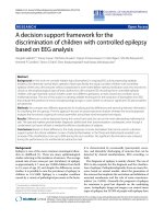
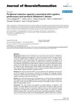
Schematic diagram of classical, alternative, and lectin complement activation pathwaysFigure 1
Schematic diagram of classical, alternative, and lectin complement activation pathways. There is evidence for activation of the
classical and alternative pathways in the AD brain. (Adapted from Sahu and Lambris, 2000 [58]).
Classical Pathway
Ab-Ag complexes, AE,
or phosphorylated tau
Alternative Pathway
+ Lectin Pathway
C3b C1q C1r C1s Mannose binding lectin
+ (MBL), MBL-associated
Factor B proteases (MASPs),
+ microbial surfaces
Properdin (P)
_ _
C1qC1rC1s
+C4
C4a, C4b C4c, C4d
C3bBbP +C2
+ polysaccharides, C2b
+C3 microbial cells, or AE +C3
C3a
C3a C4b2a3b
C3b + C5
+
C3bBbP
C5a
(C3b)
2
BbP C5b
+C6
+C7
+C8
+C9
C5b678(9)
n
(“membrane attack complex”)
Journal of Neuroinflammation 2004, 1:18 />Page 4 of 12
(page number not for citation purposes)
2. Association of complement activation proteins with
neuropathology
Complement proteins are detectable on or closely associ-
ated with SPs, NFTs, and dystrophic neurites in the AD
brain. These findings are in agreement with in vitro studies
indicating that Aβ and tau protein, the major components
in SPs and NFTs, can fully activate human complement
[42,68-71]. Although the above studies suggested that
complement is activated principally by the aggregated
forms of Aβ and tau, soluble, non-fibrillar Aβ may also be
capable of activating complement [72]. In contrast to the
robust staining of complement proteins in mature
plaques, immunoreactivity to these proteins in diffuse
plaques has generally been below the level of detection,
though it has been reported in some studies [36,73,74].
Complement activation in the AD brain is increased pri-
marily in regions containing extensive pathology (e.g., the
hippocampus and cortex), and whether early complement
components are also present in the diffuse plaques that
develop in the AD cerebellum is controversial [74,75].
The above findings suggest that complement activation in
an optimal animal model of AD should be associated with
SPs and, in those models in which neurofibrillary pathol-
ogy occurs, with NFTs.
3. Initiation of complement activation early in development of
pathology
How the increased complement activation in AD relates
to the development of SPs and NFTs, and to neuronal loss,
is unclear. Immunocytochemical staining for
complement activation proteins in the aged normal
human brain is generally faint, and may be below the
level of detection [42,69,73]; of relevance is a recent
report describing extensive neuron-associated C1q reac-
tivity in a cognitively normal subject with neuropatholog-
ical findings limited to diffuse cortical plaques [76].
Elderly "high pathology controls," lacking dementia but
with increased numbers of entorhinal NFTs and neocorti-
cal Aβ deposits, have a slight increase in the percentage of
C5b-9-immunoreactive plaques in comparison with aged
normal subjects, though this percentage is far lower than
in the AD brain [39]. A recent study in our laboratory [77]
used enzyme-linked immunosorbent assay (ELISA) to
measure the concentrations of two early complement acti-
vation proteins, C4d and iC3b, in brain specimens from
AD and normal subjects. ELISA is more sensitive than
immunocytochemical staining, though it provides no
information regarding the cellular association of comple-
ment immunoreactivity. Increased concentrations of
these early complement activation proteins were present
in some aged normal specimens. These reports suggest
that early complement activation may increase prior to
the development of plaques and NFTs. Similar findings
are desirable in an optimal animal model for studying
AD-related complement activation.
4. Increased CNS production of native complement proteins
Both mRNA expression and protein synthesis of native
complement proteins are increased in the AD brain [78-
80]. (Note: the distinction between detection of native
complement proteins, vs. detection of complement acti-
vation proteins, has frequently been blurred. In some
studies in which immunoreactivity to complement activa-
tion proteins (C3c, C4c, C4d) has been reported, the
antisera used were also capable of detecting the respective
native complement proteins (C3 or C4) [40,80]. Only
when antisera are used whose immunoreactivity is limited
to activation-specific neo-epitopes can complement acti-
vation be confirmed. The paucity of antisera which can
detect complement activation proteins in experimental
animal models is a significant obstacle to determining the
extent of complement activation in these models.) In
addition to neurons, complement proteins are synthe-
sized by other cells in the CNS including microglia, astro-
cytes, oligodendrocytes, and endothelial cells [31]. The
biological effects of these activation proteins are mediated
by numerous regulatory proteins including CD59, clus-
terin, vitronectin, C1-inhibitor, C4-binding protein,
decay-activating factor, and Factor H, which inhibit differ-
ent steps in the complement cascade. All of these regula-
tory proteins are produced in the human brain, but less is
known about their CNS synthesis in other species [31].
The status of some of these regulatory proteins in AD is
unclear; for example, there are conflicting reports regard-
ing the up-regulation of C1-inhibitor [81,82] and CD59
[41,82,83]. Thus, while an optimal animal model for
studying AD-related complement activation should have
up-regulated CNS synthesis of complement proteins, the
alterations that should be present in complement regula-
tory proteins are less clear.
5. Alternative as well as classical complement activation
Complement activation in the AD brain was initially
thought to be limited to the classical pathway, but recent
reports have also indicated increased concentrations of
the alternative activation factors Bb and Ba, and Factor H,
a regulatory factor for the alternative pathway, in the AD
brain [84,85]. Alternative complement activation has also
been reported in other familial dementias with patholo-
gies similar to AD [67]. Therefore, while activation of the
classical pathway is an absolute requirement for an opti-
mal animal model of AD-related complement activation,
an increase in the alternative pathway is also desirable.
Complement activation in animal models of AD: present
knowledge
The examination of complement activation in experimen-
tal models of AD has been limited to mice and rats. The
extent of complement activation and its relationship to
the development of AD-type neuropathology have gener-
ally not been determined in these studies.
Journal of Neuroinflammation 2004, 1:18 />Page 5 of 12
(page number not for citation purposes)
APP/sCrry mouse
Increased complement activation was induced by over-
production of transforming growth factor beta1 (TGF-β1)
in transgenic mice expressing mutations in the human
amyloid precursor protein (hAPP) gene. The APP muta-
tions expressed in these mice have been associated with
early-onset, familial AD [86]. The TGF-β1 overproduction
resulted in a 50% reduction in Aβ accumulation in the
hippocampus and cerebral cortex [87]. Because the pro-
duction of soluble Aβ was unchanged, these results sug-
gested that reduction in Aβ may have been due to its
increased clearance by microglia. A subsequent study by
the same investigators [88] found that the mRNA level of
C3 in the cerebral cortex was 5-fold higher in APP/TGF-β1
mice than in APP mice at 2 months of age (prior to depo-
sition of Aβ) and 2-fold higher at 12–15 months, when
senile plaques are present. Thus, in this model, increased
CNS synthesis of C3 precedes senile plaque formation.
Because C3b, an activation protein produced by cleavage
of C3, functions as an opsonin [89], the increased C3 lev-
els together with the reduced Aβ deposition in the APP/
TGF-β1 mice suggested a neuroprotective role for comple-
ment in this model. To investigate this possibility, the APP
mice were crossed with mice expressing soluble comple-
ment receptor-related protein y (sCrry), a rodent-specific
inhibitor of early complement activation [90]. APP/sCrry
mice had a 2- to 3- fold increase in Aβ deposition in the
neocortex and hippocampus at 10–12 months of age,
together with a 50% loss of pyramidal neurons in hippoc-
ampal region CA3. The authors concluded that comple-
ment activation may protect against Aβ-induced toxicity,
and may reduce the accumulation or promote the clear-
ance of amyloid and degenerating neurons [88]. Neuro-
protective functions (protection against excitotoxicity)
have been demonstrated in vitro for C3a [52], and the
increased neuronal loss in the APP/sCrry mouse may be
due to decreased production of C3a as well as the
opsonin, C3b. However, whether inhibition of comple-
ment activation in the AD brain would similarly result in
increased neuropathology is unclear, because comple-
ment activation in AD is likely to be more extensive than
in the APP mouse. Although no peer-reviewed articles
have appeared in which the extent of complement activa-
tion in the APP mouse has been examined, two abstracts
have dealt with this issue. Yu et al. [91] reported C3, C5,
and C6 immunoreactivity to thioflavin-S-reactive plaques,
whereas McGeer et al. [92] found only weak complement
staining of plaques and slight upregulation of comple-
ment proteins. Significantly, neither study reported detec-
tion of the MAC. At least two factors, in addition to the
lack of NFTs, mitigate against complement activation in
the APP mouse being equivalent to that in AD: (a) the
mouse complement system is functionally deficient, as
mouse C4 lacks C5 convertase activity [93] and many
mouse strains have low complement levels relative to
other mammals [94], and (b) mouse C1q binds less effi-
ciently to human Aβ than does human C1q, resulting in
less activation of mouse complement than of human
complement in the presence of human Aβ [95].
PS/APP mouse
In addition to APP, mutations in the gene encoding for
presenilin-1 (PS-1) have also been associated with famil-
ial AD [96]. The PS/APP mouse carries both of these trans-
genes and has been extensively used as a model for
studying processes relating to the formation of SPs. Aβ
deposition occurs more rapidly in these mice than in the
single transgenic APP mouse [97]. In neither model does
NFT formation occur. Aβ deposition in PS/APP mice is
initially detected at 3 months of age, and increases with
age; total Aβ burden peaks at one year of age, although the
percentage of Aβ that is fibrillar (thioflavin-S reactive)
increases up to 2 years of age. Matsuoka et al. [98]
described the CNS inflammatory response to Aβ in these
animals. Activated astrocytes and microglia increased in
parallel with total Aβ and were closely associated with
both diffuse and fibrillar plaques. C1q immunoreactivity
was detected at both 7 and 12 months of age, co-localiz-
ing with activated microglia and fibrillar Aβ. These find-
ings were similar to those in the AD brain in that
complement activation was associated with SP formation.
The extent of complement activation was not addressed in
this study.
APP (Tg2576)/C1q-deficient mouse
Fonseca et al. [99] investigated the role of C1q in AD by
crossing Tg2576 (APP) mice [100] and APP/PS1 mice
with C1q knockout mice [101]. C1q immunoreactivity
was associated with plaque formation in the APP Tg2576
animals, as previously reported by Matsuoka et al. [98]. In
both the Tg2576/C1q
-
and APP/PS1/C1q
-
animals, lack of
C1q did not alter either plaque density or the time course
of plaque deposition. Neuronal cell numbers (NeuN
+
cells), assessed only in the Tg2576 (APP) mouse, were not
changed by the absence of C1q; however, immunoreactiv-
ity to MAP-2 (a marker for neuronal dendrites and cell
bodies) and synaptophysin (a marker for presynaptic ter-
minals) in the hippocampus (region CA3) was increased
2-fold in the APP/C1q
-
animals, compared with APP mice.
Microglial and astrocytic activation was significantly
reduced in the APP/C1q
-
animals. These results were inter-
preted to suggest that in these animal models of AD, (1)
early complement activation (as indicated by C1q deposi-
tion) in response to fibrillar Aβ deposition might be
responsible for the chemotactic attraction of activated
glial cells, and (2) the activated microglia, while unable to
clear fibrillar Aβ, may have contributed to the loss of neu-
ronal integrity indicated by reduced MAP-2 and synapto-
physin staining in the APP mice. By recruiting activated
microglia, complement activation could potentially con-
Journal of Neuroinflammation 2004, 1:18 />Page 6 of 12
(page number not for citation purposes)
tribute to neuronal injury even if full activation (MAC for-
mation) does not occur.
Postischemic hyperthermic rat model
Coimbra and colleagues [102] described progressive neu-
ronal loss in the hippocampus and cerebral cortex in rats
subjected to common carotid artery occlusion to produce
transient forebrain ischemia, as an animal model for
stroke. The post-surgical hyperthermia which occurs
spontaneously in these animals was suggested to promote
the infiltration of microglia, whose secretory products
increased the subsequent neuronal loss. A later study by
the same group [103] found that subjecting the rats to
post-surgical hyperthermia (38.5 – 40°C) increased
microglial and astrocytic infiltration and accompanying
neuronal loss, and resulted in the formation of AD-type
pathology. Aβ-reactive diffuse plaques were detected in
the cerebral cortex at 2 months post-surgery, with more
compact plaques in the hippocampus and cortex by 6
months. Increased ubiquitin and phosphorylated tau
immunoreactivity was observed at both time points,
together with staining for C5b-9 in the somatosensory
cortex. The MAC immunoreactivity co-localized with acid
fuchsin staining, a marker for neuronal death [104]. Other
complement proteins were not evaluated in these studies.
This is apparently the only animal model of AD in which
full complement activation has been reported. It is note-
worthy that while both SPs and neurofibrillary pathology
were present in these animals, the MAC apparently did
not co-localize with these structures, unlike in AD.
Acute lesioning
Alterations in native complement mRNA and protein lev-
els have been evaluated in the rat hippocampus following
experimental induction of acute neuronal injury. These
surgical and pharmacological procedures result in neuro-
nal loss in the entorhinal cortex, and deafferentation of
hippocampal neurons, similar to that which occurs in AD
[105]. Selective damage to the rat hippocampus has been
induced by surgical transection of the perforant pathway,
which runs between the entorhinal cortex and the molec-
ular layer of the dentate gyrus [106,107], systemic admin-
istration of the excitotoxin kainic acid [108,109], or
injection of the neurotoxin colchicine into the dorsal hip-
pocampus [109]. Surgical transection of the perforant
pathway increased C1qB mRNA in the entorhinal cortex
and hippocampus [106] and C9 immunoreactivity in the
hippocampus [107]. Injection of kainic acid similarly
increased C1qB and C4 mRNA expression and C1q
immunoreactivity in the hippocampus [108,109]. Colch-
icine infusion into the dorsal hippocampus, which selec-
tively damages granule cells of the dentate gyrus,
produced elevated mRNA expression of hippocampal
C1qB and C4 [109]. Though the acute neuronal damage
in these studies differs from the chronic, progressive neu-
rodegenerative process that occurs in AD, these results
demonstrated that the neuronal response to injury
includes upregulation of native complement protein syn-
thesis. The significance of this upregulation, i.e. whether it
promotes neuroprotection or neurotoxicity, was not
addressed.
Infusion of A
β
and C1q into rats
Frautschy et al. [56] examined the effects of infusion of
human C1q and oral administration of rosmarinic acid
on glial cell proliferation (microgliosis and astrocytosis),
plaque load, and memory (Morris water maze) in Aβ-
infused rats. Rosmarinic acid inhibits both the classical
and the alternative complement cascades, by covalent
binding to newly formed C3b [110]; it also possesses anti-
inflammatory [111,112], anti-oxidative [113], and anti-
amyloidogenic properties [114]. Gliosis was greater with
C1q and Aβ infusion than with Aβ alone. Plaque density
was decreased by C1q infusion (note: this result differs
from the in vitro study of Webster et al. [57], in which C1q
was found to inhibit microglial phagocytosis of Aβ, and
also from the recent study of Fonseca et al. [99] in which
C1q deficiency had no effect on plaque density in APP
mice), but, curiously, performance in the water maze
worsened. Treatment with rosmarinic acid had the oppo-
site effect; though plaque load increased, memory was
improved. These findings were interpreted as suggesting
that C1q and/or complement activation may, by promot-
ing microglial activation, worsen memory independent of
the clearance of Aβ.
Additional animal models for studying AD-related
complement activation
TAPP and 3xTg-AD mice
Mutations in the gene encoding for human tau protein
have been linked to the development of frontotemporal
dementia with parkinsonism [115]. By combining this
mutation with the human APP and PS1 mutations associ-
ated with familial AD, animal models of AD have been
produced in which NFTs as well as SPs are formed. Lewis
et al. [116] crossed human APP
swe
mice (Tg2576) with
mice expressing the transgene for a human tau mutation
(JNPL3 mice) to generate a double mutant tau/APP
mouse (the "TAPP mouse"). These mice develop SPs sim-
ilar to APP mice (high numbers of plaques are present in
older [8.5–15 months of age] mice, in the olfactory cortex,
cingulate gyrus, amygdala, entorhinal cortex, and
hippocampus), and older TAPP mice have NFTs, in asso-
ciation with increased astrocyte proliferation, in limbic
areas. The plaques contain both Aβ
40
and Aβ
42
. Oddo et
al. [117] injected the human transgenes for APP and
mutated tau into embryos of PS1 "knock-in" mice, gener-
ating the "3xTg-AD" mouse which develops both SPs and
NFTs in an age-related, region-specific manner. Aβ depo-
sition in these animals precedes NFT formation, with
Journal of Neuroinflammation 2004, 1:18 />Page 7 of 12
(page number not for citation purposes)
extracellular Aβ (primarily Aβ
42
) detected in the frontal
cortex by 6 months of age, and in other cortical regions
and hippocampus by 12 months. Many of the extracellu-
lar Aβ deposits are thioflavin-S-positive and are associated
with reactive astrocytes. Phosphorylated tau initially
appears in the hippocampus and subsequently in cortical
regions; it is detected within neurons by 12–15 months
and within dystrophic neurites at 18 months. Though Aβ
immunoreactivity precedes that of tau, these proteins co-
localize to the same neurons. The presence of NFTs as well
as SPs suggests that the 3xTg-AD and TAPP models may be
more relevant than APP or APP/PS-1 mice for studying the
significance of complement activation in the develop-
ment of AD-type pathology. Potential drawbacks for using
these models for complement-related studies include, as
discussed earlier, functional deficiencies in activation of
mouse complement [93], decreased complement levels in
common laboratory mouse strains [94], and the
decreased efficiency of binding of mouse C1q by the
human Aβ within the SPs in these animals [95]. It is not
known whether a similar decrease in the efficiency of acti-
vation of mouse complement occurs when mouse C1q
binds to human, rather than murine, tau protein.
AD11 (anti-NGF) mouse
Ruberti et al. [118] developed a mouse transgenic model,
the AD11 mouse, in which neutralizing antibody to nerve
growth factor (NGF) is secreted by neurons and glial cells.
NGF exerts trophic effects on basal forebrain cholinergic
neurons and is widely distributed in these neurons [119];
the local secretion of anti-NGF antibody in these mice
results in marked loss of basal forebrain cholinergic neu-
rons. Aβ-containing plaques, tau hyperphosphorylation,
and NFTs are present at 15–18 months of age. CNS pro-
duction of anti-NGF antibody increases with age in these
animals, therefore pathology develops only in adult mice.
Extracellular deposition of APP is widespread in the brain,
including the cortex and hippocampus. Phosphorylated
tau immunoreactivity is present in neurons and glia in the
cortex and hippocampus, and intracellular NFTs, extracel-
lular neurofibrillary deposits, neuropil threads, and dys-
trophic neurites are observed in the cortex. Behavioral
abnormalities, including impaired object recognition and
spatial learning, are associated with this neuropathology
[120]. The Aβ-containing plaques in the AD11 mouse are
of murine, rather than human, origin, allowing the prob-
lem of the poor efficiency of activation of mouse comple-
ment by human Aβ [95] to be overcome. However, it is
unclear whether plaques in these animals contain Aβ in
the β-pleated sheet conformation, which is thought to be
the most effective conformation for activating comple-
ment [71]. The distribution of SPs and NFTs in this model
is less similar to AD than for 3xTg-AD and TAPP mice,
because in addition to the cortex and hippocampus, large
numbers of APP-reactive structures are present in the
neostriatum (where, in AD, plaques are primarily diffuse
[121]), and in other areas of the brain. Despite these con-
cerns, the AD11 mouse is attractive as a potential model
for studying the significance of AD-related complement
activation.
Chlamydia pneumoniae-infected mouse
C. pneumoniae is an intracellular, gram-negative or gram-
variable bacterium long identified as a respiratory patho-
gen. It has more recently been demonstrated to be a caus-
ative agent in reactive arthritis [122] and to be associated
with autoimmune disorders including multiple sclerosis
[123] and atherosclerosis [124]. Some laboratories have
also reported an association of this agent with AD [125-
127], although this has not been confirmed by others
[128-131]. A recent study by Little et al. [132] examined
the hypothesis that experimental C. pneumoniae infection
in BALB/c mice could produce AD-like pathology. Intra-
nasal inoculation with C. pneumoniae resulted in deposi-
tion of Aβ
1–42
in the hippocampus, amygdala, entorhinal
cortex, perirhinal cortex, and thalamus by 3 months post-
inoculation. The majority of these Aβ deposits appeared
similar to diffuse plaques, though a small number of them
were thioflavin-S-reactive. NFTs were not detected. The
authors suggested that soluble factors such as lipopolysac-
charides, which are present in the cell wall of all Chlamy-
diae [133], may have been responsible for the altered
amyloid processing which resulted in Aβ deposition.
Because the Aβ within the SPs in these animals is of
endogenous origin, and because other chlamydial species
have been shown to activate complement [134,135], the
C. pneumoniae-infected mouse may offer a novel infec-
tious model for studying the relationship of complement
activation to the development of Aβ-containing plaques.
Aged dogs
Old dogs, in particular the beagle, have been extensively
investigated as a model for CNS Aβ deposition and asso-
ciated age-related cognitive dysfunction. Aβ deposits are
detectable in the brains of most older dogs [136]. The
regional distribution of Aβ in the dog brain resembles that
in humans, found initially in the prefrontal cortex, subse-
quently in entorhinal and parietal cortices, and lastly in
occipital cortex [137]. Aβ
42
is the predominant type of Aβ
deposited in plaques [138]. Canine plaques are nonfibril-
lar and do not contain neuritic elements; thus, they resem-
ble diffuse Aβ deposits in the human brain, but not the
mature plaques predominating in AD. The neuropatho-
logical findings in old dogs also differ from AD in that
activated glial cells are rarely associated with Aβ deposits,
and NFTs are not detected [136,139]. Age-related cogni-
tive impairment, termed "canine cognitive dysfunction
syndrome," occurs in some older dogs and correlates with
Aβ deposition in the hippocampus and frontal cortex
[140,141]. The endogenous nature of the deposited Aβ in
Journal of Neuroinflammation 2004, 1:18 />Page 8 of 12
(page number not for citation purposes)
old dog brain, and similarities between canine and
human Aβ in their patterns of regional deposition, suggest
that this model may be useful for studying the relation-
ship between complement activation and plaque
formation.
Non-human primates
Age-related formation of SPs has been reported in a vari-
ety of non-human primates including the cynomolgus
monkey [142], rhesus monkey [143], chimpanzee [144],
and marmoset [145]. Aβ within these plaques is predom-
inantly Aβ
40
[146]. NFTs apparently do not form in the
brains of most aged primates, with a few exceptions. The
brain of the aged baboon contains phosphorylated tau
protein [147,148], and an age-related accumulation of tau
also occurs in the neocortex of the mouse lemur [149-
151]. In this latter species, Aβ deposition occurs in the cer-
ebral cortex and amygdala but is not age-dependent [151].
The mouse lemur appears to be the most promising pri-
mate species to date for studying the significance of AD-
related complement activation because of the presence of
NFTs as well as plaques.
Other animal species
Scattered reports of AD-type pathology in other species
have also appeared. Adding trace amounts of copper to
the water supply of cholesterol-fed rabbits results in Aβ
deposition within SP-like structures in the hippocampus
and temporal cortex, with associated learning deficits
[152]. The neuropathology in the aged cat is similar to
that in the old dog in that Aβ is deposited only as diffuse,
Aβ
42
-containing plaques, and NFTs are not detected [138].
A report of AD-type pathology in an aged wolverine [153]
described neuritic as well as diffuse plaques in the cortex
and hippocampus, and intracellular NFTs containing
phosphorylated tau protein in cortical and hippocampal
neurons. Finally, the aged polar bear brain also contains
both diffuse plaques and NFTs [154]. While the neu-
ropathological findings in the aged wolverine and polar
bear resemble AD more closely than in most species
examined to date, their inaccessibility to laboratory
researchers limits the usefulness of these species for stud-
ies of AD-related complement activation.
Conclusions
1. Complement activation has been extensively studied in
the AD brain. There is convincing evidence for activation
of both the classical and alternative pathways, resulting in
full activation as indicated by the presence of the MAC.
Both aggregated Aβ (in SPs) and phosphorylated tau (in
NFTs) are likely to be responsible for this activation.
2. Because complement activation generates both both
neuroprotective and neurotoxic effects, the significance of
increased complement activation in the development and
progression of AD is unclear.
3. An optimal animal model for studying the significance
of complement activation in the development of AD-type
pathology would have complete activation of this process,
with co-localization of complement activation proteins
with SPs and with NFTs (if present). Other desirable fea-
tures include early complement activation prior to the
development of extensive neuropathology, increased CNS
production of native complement proteins, and both clas-
sical and alternative pathway activation.
4. Surprisingly little is known about the extent of comple-
ment activation in animal models of AD. The pos-
tischemic hyperthermic rat [103] is the only animal
model of AD in which full complement activation has
been reported. The few studies with APP-transgenic mice
have yielded conflicting results, with one investigation
suggesting a neuroprotective role for complement activa-
tion [88], while another found that early complement
activation (as indicated by C1q deposition) was associ-
ated with a loss of neuronal integrity [99]. Transgenic
mouse models may be problematic for studies of AD-
related complement activation because of inherent defi-
ciencies in mouse complement activation and inefficient
activation of mouse complement by the human Aβ
present in the SPs in these animals. Other animal models
in which SPs (and NFTs, if present) are of endogenous,
rather than human, origin offer alternatives to transgenic
mice for studying this issue.
5. The extent of complement activation and its association
with neuropathology must be determined in animal mod-
els of AD to clarify the relevance of these models for inves-
tigating the significance of complement activation in the
development of AD-type pathology.
Abbreviations used
Aβ, amyloid beta; AD, Alzheimer's disease; APP, amyloid
precursor protein; CNS, central nervous system; MAC,
membrane attack complex; mRNA, messenger ribonucleic
acid; NFTs, neurofibrillary tangles; NGF, nerve growth fac-
tor; PS-1, presenilin-1; sCrry, soluble complement recep-
tor-related protein y; SPs, senile plaque; TGF-β1,
transforming growth factor beta1.
Competing interests
The author declares that he has no competing interests.
Acknowledgements
Thanks are expressed to Elizabeth Head, Ph.D, Dianne Camp, Ph.D., Steph-
anie Conant, Ph.D., and Peter LeWitt, M.D., for reviewing the manuscript.
This work was supported by a donation from Mrs. Martha Loeffler in mem-
ory of Erwin S. Loeffler, Ph.D., and Harold J. Loeffler, Ph.D.
Journal of Neuroinflammation 2004, 1:18 />Page 9 of 12
(page number not for citation purposes)
References
1. Bamberger ME, Landreth GE: Inflammation, apoptosis, and
Alzheimer's disease. Neuroscientist 2002, 8:276-283.
2. Gupta A, Pansari K: Inflammation and Alzheimer's disease. Int J
Clin Pract 2003, 57:36-39.
3. Hoozemans JJ, Veerhuis R, Rozemuller AJ, Eikelenboom P: The path-
ological cascade of Alzheimer's disease: the role of inflam-
mation and its therapeutic implications. Drugs Today (Barc)
2002, 38:429-443.
4. McGeer EG, McGeer PL: Inflammatory processes in Alzhe-
imer's disease. Prog Neuropsychopharmacol Biol Psychiatry 2003,
27:741-749.
5. Akiyama H, Barger S, Barnum S, Bradt B, Bauer J, Cole GM, Cooper
NR, Eikelenboom P, Emmerling M, Fiebich BL, Finch CE, Frautschy S,
Griffin WS, Hampel H, Hull M, Landreth G, Lue L, Mrak R, Mackenzie
IR, McGeer PL, O'Banion MK, Pachter J, Pasinetti G, Plata-Salaman C,
Rogers J, Rydel R, Shen Y, Streit W, Strohmeyer R, Tooyoma I, Van
Muiswinkel FL, Veerhuis R, Walker D, Webster S, Wegrzyniak B,
Wenk G, Wyss-Coray T: Inflammation and Alzheimer's
disease. Neurobiol Aging 2000, 21:383-421.
6. Combs CK, Karlo JC, Kao SC, Landreth GE: beta-Amyloid stimu-
lation of microglia and monocytes results in TNFalpha-
dependent expression of inducible nitric oxide synthase and
neuronal apoptosis. J Neurosci 2001, 21:1179-1188.
7. Griffin WS, Mrak RE: Interleukin-1 in the genesis and progres-
sion of and risk for development of neuronal degeneration in
Alzheimer's disease. J Leukoc Biol 2002, 72:233-238.
8. Griffin WS, Sheng JG, Royston MC, Gentleman SM, McKenzie JE, Gra-
ham DI, Roberts GW, Mrak RE: Glial-neuronal interactions in
Alzheimer's disease: the potential role of a 'cytokine cycle' in
disease progression. Brain Pathol 1998, 8:65-72.
9. McGeer PL, Schulzer M, McGeer EG: Arthritis and anti-inflam-
matory agents as possible protective factors for Alzheimer's
disease: a review of 17 epidemiologic studies. Neurology 1996,
47:425-432.
10. Benveniste EN, Nguyen VT, O'Keefe GM: Immunological aspects
of microglia: relevance to Alzheimer's disease. Neurochem Int
2001, 39:381-391.
11. Meda L, Baron P, Scarlato G: Glial activation in Alzheimer's dis-
ease: the role of Abeta and its associated proteins. Neurobiol
Aging 2001, 22:885-893.
12. Mrak RE, Griffin WS: The role of activated astrocytes and of the
neurotrophic cytokine S100B in the pathogenesis of Alzhe-
imer's disease. Neurobiol Aging 2001, 22:915-922.
13. Emmerling MR, Watson MD, Raby CA, Spiegel K: The role of com-
plement in Alzheimer's disease pathology. Biochim Biophys Acta
2000, 1502:158-171.
14. McGeer PL, McGeer EG: The possible role of complement acti-
vation in Alzheimer disease. Trends Mol Med 2002, 8:519-523.
15. Tenner AJ: Complement in Alzheimer's disease: opportuni-
ties for modulating protective and pathogenic events. Neuro-
biol Aging 2001, 22:849-861.
16. Abraham CR: Reactive astrocytes and alpha1-antichymot-
rypsin in Alzheimer's disease. Neurobiol Aging 2001, 22:931-936.
17. Kovacs DM: alpha2-macroglobulin in late-onset Alzheimer's
disease. Exp Gerontol 2000, 35:473-479.
18. Loeffler DA, Sima AA, LeWitt PA: Ceruloplasmin immunoreac-
tivity in neurodegenerative disorders. Free Radic Res 2001,
35:111-118.
19. Wood JA, Wood PL, Ryan R, Graff-Radford NR, Pilapil C, Robitaille
Y, Quirion R: Cytokine indices in Alzheimer's temporal cor-
tex: no changes in mature IL-1 beta or IL-1RA but increases
in the associated acute phase proteins IL-6, alpha 2-mac-
roglobulin and C-reactive protein. Brain Res 1993, 629:245-252.
20. McGeer PL, Rogers J: Anti-inflammatory agents as a therapeu-
tic approach to Alzheimer's disease. Neurology 1992,
42:447-449.
21. Rogers J, Kirby LC, Hempelman SR, Berry DL, McGeer PL, Kaszniak
AW, Zalinski J, Cofield M, Mansukhani L, Willson P, Kogan F: Clinical
trial of indomethacin in Alzheimer's disease. Neurology 1993,
43:1609-1611.
22. Aisen PS, Davis KL, Berg JD, Schafer K, Campbell K, Thomas RG,
Weiner MF, Farlow MR, Sano M, Grundman M, Thal LJ: A rand-
omized controlled trial of prednisone in Alzheimer's disease.
Alzheimer's Disease Cooperative Study. Neurology 2000,
54:588-593.
23. Aisen PS, Schafer KA, Grundman M, Pfeiffer E, Sano M, Davis KL, Far-
low MR, Jin S, Thomas RG, Thal LJ: Alzheimer's Disease Cooper-
ative Study. Effects of rofecoxib or naproxen vs placebo on
Alzheimer disease progression: a randomized controlled
trial. JAMA 2003, 289:2819-2826.
24. Sainetti SM, Ingram DM, Talwalker S, Geis GS: Results of a double-
blind, randomized, placebo-controlled study of celecoxib in
the treatment of progression of Alzheimer's disease
[abstract]. Sixth International Stockholm/Springfield Symposium on
Advances in Alzheimer Therapy 2000:180.
25. Van Gool WA, Weinstein HC, Scheltens P, Walstra GJ, Scheltens PK:
Effect of hydroxychloroquine on progression of dementia in
early Alzheimer's disease: an 18-month randomised, double-
blind, placebo-controlled study. Lancet 2001, 358:455-460.
26. Shen Y, Meri S: Yin and Yang: complement activation and reg-
ulation in Alzheimer's disease. Prog Neurobiol 2003, 70:463-472.
27. Wyss-Coray T, Mucke L: Inflammation in neurodegenerative
disease–a double-edged sword. Neuron 2002, 35:419-432.
28. Neumann H: The immunological microenvironment in the
CNS: implications on neuronal cell death and survival. J Neu-
ral Transm Suppl 2000, 59:59-68.
29. Polazzi E, Contestabile A: Reciprocal interactions between
microglia and neurons: from survival to neuropathology. Rev
Neurosci 2002, 13:221-242.
30. van Beek J, Elward K, Gasque P: Activation of complement in the
central nervous system: roles in neurodegeneration and
neuroprotection. Ann N Y Acad Sci 2003, 992:56-71.
31. Barnum SR: Complement biosynthesis in the central nervous
system. Crit Rev Oral Biol Med 1995, 6:132-146.
32. Eikelenboom P, Hack CE, Rozemuller JM, Stam FC: Complement
activation in amyloid plaques in Alzheimer's dementia. Vir-
chows Arch B Cell Pathol Incl Mol Pathol 1989, 56:259-262.
33. Eikelenboom P, Stam FC: Immunoglobulins and complement
factors in senile plaques. An immunoperoxidase study. Acta
Neuropathol (Berl) 1982, 57:239-42.
34. Eikelenboom P, Stam FC: An immunohistochemical study on
cerebral vascular and senile plaque amyloid in Alzheimer's
dementia. Virchows Arch B Cell Pathol Incl Mol Pathol 1984, 47:17-25.
35. Ishii T, Haga S: Immuno-electron-microscopic localization of
complements in amyloid fibrils of senile plaques. Acta Neu-
ropathol (Berl) 1984, 63:296-300.
36. Veerhuis R, Janssen I, Hack CE, Eikelenboom P: Early complement
components in Alzheimer's disease brains. Acta Neuropathol
(Berl) 1996, 91:53-60.
37. Veerhuis R, van der Valk P, Janssen I, Zhan SS, Van Nostrand WE,
Eikelenboom P: Complement activation in amyloid plaques in
Alzheimer's disease brains does not proceed further than
C3. Virchows Arch 1995, 426:603-610.
38. Itagaki S, Akiyama H, Saito H, McGeer PL: Ultrastructural locali-
zation of complement membrane attack complex (MAC)-
like immunoreactivity in brains of patients with Alzheimer's
disease. Brain Res 1994, 645:78-84.
39. Lue LF, Brachova L, Civin WH, Rogers J: Inflammation, A beta
deposition, and neurofibrillary tangle formation as corre-
lates of Alzheimer's disease neurodegeneration. J Neuropathol
Exp Neurol 1996, 55:1083-1088.
40. McGeer PL, Akiyama H, Itagaki S, McGeer EG: Activation of the
classical complement pathway in brain tissue of Alzheimer
patients. Neurosci Lett 1989, 107:341-346.
41. McGeer PL, Walker DG, Akiyama H, Kawamata T, Guan AL, Parker
CJ, Okada N, McGeer EG: Detection of the membrane inhibitor
of reactive lysis (CD59) in diseased neurons of Alzheimer
brain. Brain Res 1991, 544:315-319.
42. Webster S, Lue L-F, Brachova L, Tenner AJ, McGeer PL, Terai K,
Walker DG, Bradt B, Cooper NR, Rogers J: Molecular and cellular
characterization of the membrane attack complex, C5b-9, in
Alzheimer's disease. Neurobiol Aging 1997, 18:415-421.
43. Webster S, Glabe C, Rogers J: Multivalent binding of comple-
ment protein C1q to the amyloid beta-peptide (A beta)
promotes the nucleation phase of A beta aggregation. Bio-
chem Biophys Res Commun 1995, 217:869-875.
44. Webster S, O'Barr S, Rogers J: Enhanced aggregation and beta
structure of amyloid beta peptide after coincubation with
C1q. J Neurosci Res 1994, 39:448-456.
Journal of Neuroinflammation 2004, 1:18 />Page 10 of 12
(page number not for citation purposes)
45. Schultz J, Schaller J, McKinley M, Bradt B, Cooper N, May P, Rogers J:
Enhanced cytotoxicity of amyloid beta-peptide by a comple-
ment dependent mechanism. Neurosci Lett 1994, 175:99-102.
46. Nolte C, Moller T, Walter T, Kettenmann H: Complement 5a con-
trols motility of murine microglial cells in vitro via activation
of an inhibitory G-protein and the rearrangement of the
actin cytoskeleton. Neuroscience 1996, 73:1091-1107.
47. Yao J, Harvath L, Gilert DL, Colton CA: Chemotaxis by a CNS
macrophage, the microglia. J Neurosci Res 1990, 27:36-42.
48. O'Barr S, Cooper NR: The C5a complement activation peptide
increases IL-1beta and IL-6 release from amyloid-beta
primed human monocytes: implications for Alzheimer's
disease. J Neuroimmunol 2000, 109:87-94.
49. Veerhuis R, Van Breemen MJ, Hoozemans JM, Morbin M, Ouladhadj J,
Tagliavini F, Eikelenboom P: Amyloid beta plaque-associated
proteins C1q and SAP enhance the Abeta1–42 peptide-
induced cytokine secretion by adult human microglia in
vitro. Acta Neuropathol (Berl) 2003, 105:135-144.
50. Shen Y, Halperin JA, Lee CM: Complement-mediated neurotox-
icity is regulated by homologous restriction. Brain Res 1995,
671:282-292.
51. Osaka H, Mukherjee P, Aisen PS, Pasinetti GM: Complement-
derived anaphylatoxin C5a protects against glutamate-
mediated neurotoxicity. J Cell Biochem 1999, 73:303-311.
52. van Beek J, Nicole O, Ali C, Ischenko A, MacKenzie ET, Buisson A,
Fontaine M: Complement anaphylatoxin C3a is selectively
protective against NMDA-induced neuronal cell death. Neu-
roreport 2001, 12:289-293.
53. O'Barr SA, Caguioa J, Gruol D, Perkins G, Ember JA, Hugli T, Cooper
NR: Neuronal expression of a functional receptor for the C5a
complement activation fragment. J Immunol 2001,
166:4154-4162.
54. Mukherjee P, Pasinetti GM: Complement anaphylatoxin C5a
neuroprotects through mitogen-activated protein kinase-
dependent inhibition of caspase 3. J Neurochem 2001, 77:43-49.
55. Soane L, Cho HJ, Niculescu F, Rus H, Shin ML: C5b-9 terminal
complement complex protects oligodendrocytes from
death by regulating Bad through phosphatidylinositol 3-
kinase/Akt pathway. J Immunol 2001, 167:2305-2311.
56. Frautschy SA, Hammer H, Hu S, Hu W, Oh M, Miller SA, Lim GP, Har-
ris-White ME, Tenner AJ: C1q stimulates Abeta clearance but
worsens memory in Alzheimer model: too much C1q may
be worse than too little. In Program No. 667.12. Abstract Viewer/Itin-
erary Planner Washington, DC: Society for Neuroscience; 2003.
57. Webster SD, Yang AJ, Margol L, Garzon-Rodriguez W, Glabe CG,
Tenner AJ: Complement component C1q modulates the
phagocytosis of Abeta by microglia. Exp Neurol 2000,
161:127-138.
58. Sahu A, Lambris JD: Complement inhibitors: a resurgent con-
cept in anti-inflammatory therapeutics. Immunopharmacol 2000,
49:133-148.
59. Morgan BP, Harris CL: Complement therapeutics; history and
current progress. Mol Immunol 2003, 40:159-170.
60. De Simoni MG, Storini C, Barba M, Catapano L, Arabia AM, Rossi E,
Bergamaschini L: Neuroprotection by complement (C1) inhib-
itor in mouse transient brain ischemia. J Cereb Blood Flow Metab
2003, 23:232-239.
61. Kirschfink M: C1-inhibitor and transplantation. Immunobiology
2002, 205:534-541.
62. Quigg RJ: Role of complement and complement regulatory
proteins in glomerulonephritis. Springer Semin Immunopathol
2003, 24:395-410.
63. de Zwaan C, van Dieijen-Visser MP, Hermens WT: Prevention of
cardiac cell injury during acute myocardial infarction: possi-
ble role for complement inhibition. Am J Cardiovasc Drugs 2003,
3:245-251.
64. Farkas H, Harmat G, Fust G, Varga L, Visy B: Clinical management
of hereditary angio-oedema in children. Pediatr Allergy Immunol
2002, 13:153-161.
65. Rozemuller AJ, Eikelenboom P, Theeuwes JW, Jansen Steur EN, de
Vos RA: Activated microglial cells and complement factors
are unrelated to cortical Lewy bodies. Acta Neuropathol (Berl)
2000, 100:701-708.
66. Stoltzner SE, Grenfell TJ, Mori C, Wisniewski KE, Wisniewski TM,
Selkoe DJ, Lemere CA: Temporal accrual of complement pro-
teins in amyloid plaques in Down's syndrome with Alzhe-
imer's disease. Am J Pathol 2000, 156:489-499.
67. Rostagno A, Revesz T, Lashley T, Tomidokoro Y, Magnotti L, Braend-
gaard H, Plant G, Bojsen-Moller M, Holton J, Frangione B, Ghiso J:
Complement activation in chromosome 13 dementias. Sim-
ilarities with Alzheimer's disease. J Biol Chem 2002,
277:49782-49790.
68. Bradt BM, Kolb WP, Cooper NR: Complement-dependent
proinflammatory properties of the Alzheimer's disease
beta-peptide. J Exp Med 1998, 188:431-438.
69. Rogers J, Cooper NR, Webster S, Schultz J, McGeer PL, Styren SD,
Civin WH, Brachova L, Bradt B, Ward P, Lieberburg I: Complement
activation by β-amyloid in Alzheimer disease. Proc Natl Acad Sci
USA 1992, 89:10016-10020.
70. Shen Y, Lue L, Yang L, Roher A, Kuo Y, Strohmeyer R, Goux WJ, Lee
V, Johnson GV, Webster SD, Cooper NR, Bradt B, Rogers J: Com-
plement activation by neurofibrillary tangles in Alzheimer's
disease. Neurosci Lett 2001, 305:165-168.
71. Webster S, Bradt B, Rogers J, Cooper N: Aggregation state-
dependent activation of the classical complement pathway
by the amyloid beta peptide. J Neurochem 1997, 69:388-398.
72. Bergamaschini L, Canziani S, Bottasso B, Cugno M, Braidotti P, Agos-
toni A: Alzheimer's beta-amyloid peptides can activate the
early components of complement classical pathway in a
C1q-independent manner. Clin Exp Immunol 1999, 115:526-533.
73. Akiyama H, Mori H, Saido T, Kondo H, Ikeda K, McGeer PL: Occur-
rence of the diffuse amyloid beta-protein (Abeta) deposits
with numerous Abeta-containing glial cells in the cerebral
cortex of patients with Alzheimer's disease. Glia 1999,
25:324-331.
74. Rozemuller JM, van der Valk P, Eikelenboom P: Activated microglia
and cerebral amyloid deposits in Alzheimer's disease. Res
Immunol 1992, 143:646-649.
75. Lue LH, Rogers J: Full complement activation fails in diffuse
plaques of the Alzheimer's disease cerebellum. Dementia 1992,
3:308-313.
76. Fonseca MI, Kawas CH, Troncoso JC, Tenner AJ: Neuronal locali-
zation of C1q in preclinical Alzheimer's disease. Neurobiol Dis
2004, 15:40-46.
77. Loeffler DA, Camp DM, Schonberger M, Singer DJ, LeWitt PA:
ELISA measurement of C4d and iC3b in Alzheimer's disease
and normal brain specimens. Neurobiol Aging 2004,
25:1001-1007.
78. Shen Y, Li R, McGeer EG, McGeer PL: Neuronal expression of
mRNAs for complement proteins of the classical pathway in
Alzheimer brain. Brain Res 1997, 769:391-395.
79. Walker DG, McGeer PL: Complement gene expression in
human brain: comparison between normal and Alzheimer
disease cases. Brain Res Mol Brain Res 1992, 14:109-116.
80. Yasojima K, Schwab C, McGeer EG, McGeer PL: Up-regulated pro-
duction and activation of the complement system in Alzhe-
imer's disease brain. Am J Pathol 1999, 154:927-936.
81. Walker DG, Yasuhara O, Patston PA, McGeer EG, McGeer PL:
Complement C1 inhibitor is produced by brain tissue and is
cleaved in Alzheimer disease. Brain Res 1995, 675:75-82.
82. Yasojima K, McGeer EG, McGeer PL: Complement regulators C1
inhibitor and CD59 do not significantly inhibit complement
activation in Alzheimer disease. Brain Res 1999, 833:297-301.
83. Yang L-B, Li R, Meri S, Rogers J, Shen Y: Deficiency of comple-
ment defense protein CD59 may contribute to neurodegen-
eration in Alzheimer's disease. J Neurosci 2000, 20:7505-7509.
84. Strohmeyer R, Ramirez M, Cole GJ, Mueller K, Rogers J: Association
of factor H of the alternative pathway of complement with
agrin and complement receptor 3 in the Alzheimer's disease
brain. J Neuroimmunol 2002, 131:135-146.
85. Strohmeyer R, Shen Y, Rogers J: Detection of complement alter-
native pathway mRNA and proteins in the Alzheimer's dis-
ease brain. Brain Res Mol Brain Res 2000, 81:7-18.
86. Janus C, Phinney AL, Chishti MA, Westaway D: New develop-
ments in animal models of Alzheimer's disease. Curr Neurol
Neurosci Rep 2001, 1:451-457.
87. Wyss-Coray T, Lin C, Yan F, Yu GQ, Rohde M, McConlogue L,
Masliah E, Mucke L: TGF-beta1 promotes microglial amyloid-
beta clearance and reduces plaque burden in transgenic
mice. Nat Med 2001, 7:612-618.
Journal of Neuroinflammation 2004, 1:18 />Page 11 of 12
(page number not for citation purposes)
88. Wyss-Coray T, Yan F, Lin AH, Lambris JD, Alexander JJ, Quigg RJ,
Masliah E: Prominent neurodegeneration and increased
plaque formation in complement-inhibited Alzheimer's
mice. Proc Natl Acad Sci USA 2002, 99:10837-10842.
89. Lindorfer MA, Hahn CS, Foley PL, Taylor RP: Heteropolymer-
mediated clearance of immune complexes via erythrocyte
CR1: mechanisms and applications. Immunol Rev 2001,
183:10-24.
90. Molina H, Wong W, Kinoshita T, Brenner C, Foley S, Holers VM: Dis-
tinct receptor and regulatory properties of recombinant
mouse complement receptor 1 (CR1) and Crry, the two
genetic homologues of human CR1. J Exp Med 1992,
175:121-129.
91. Yu JX, Bradt BM, Hsiao K, Carrol MC, Cooper NR: Amyloid
plaques in a transgenic mouse model of Alzheimer's disease
contain complement components and pro-inflammatory
cytokines. In Program No. 491.1. 2000 Abstract Viewer/Itinerary Planner
Washington, DC: Society for Neuroscience; 2000. Online
92. McGeer PL, Schwab C, Staufenbiel M, Hosokawa M, McGeer EG:
Amyloid transgenic mice: an incomplete model of Alzhe-
imer disease. In Program No. 295.2. 2002 Abstract Viewer/Itinerary
Planner Washington, DC: Society for Neuroscience; 2002. Online
93. Ebanks RO, Isenman DE: Mouse complement component C4 is
devoid of classical pathway C5 convertase subunit activity.
Mol Immunol 1996, 33:297-309.
94. Ong GL, Mattes MJ: Mouse strains with typical mammalian lev-
els of complement activity. J Immunol Methods 1989,
125:147-158.
95. Webster SD, Tenner AJ, Poulos TL, Cribbs DH: The mouse C1q
A-chain sequence alters beta-amyloid-induced complement
activation. Neurobiol Aging 1999, 20:297-304.
96. Cruts M, Van Broeckhoven C: Presenilin mutations in Alzhe-
imer's disease. Hum Mutat 1998, 11:183-190.
97. Holcomb L, Gordon MN, McGowan E, Yu X, Benkovic S, Jantzen P,
Wright K, Saad I, Mueller R, Morgan D, Sanders S, Zehr C, O'Campo
K, Hardy J, Prada CM, Eckman C, Younkin S, Hsiao K, Duff K: Accel-
erated Alzheimer-type phenotype in transgenic mice carry-
ing both mutant amyloid precursor protein and presenilin 1
transgenes. Nat Med 1998, 4:97-100.
98. Matsuoka Y, Picciano M, Malester B, LaFrancois J, Zehr C, Daeschner
JM, Olschowka JA, Fonseca MI, O'Banion MK, Tenner AJ, Lemere CA,
Duff K: Inflammatory responses to amyloidosis in a trans-
genic mouse model of Alzheimer's disease. Am J Pathol 2001,
158:1345-1354.
99. Fonseca MI, Zhou J, Botto M, Tenner AJ: Absence of C1q leads to
less neuropathology in transgenic mouse models of Alzhe-
imer's disease. J Neurosci 2004, 24:6457-6465.
100. Hsiao K, Chapman P, Nilsen S, Eckman C, Harigaya Y, Younkin S, Yang
F, Cole G: Correlative memory deficits, Abeta elevation, and
amyloid plaques in transgenic mice. Science 1996, 274:99-102.
101. Botto M, Dell'Agnola C, Bygrave AE, Thompson EM, Cook HT, Petry
F, Loos M, Pandolfi PP, Walport MJ: Homozygous C1q deficiency
causes glomerulonephritis associated with multiple apop-
totic bodies. Nat Genet 1998, 19:56-59.
102. Coimbra C, Drake M, Boris-Moller F, Wieloch T: Long-lasting neu-
roprotective effect of postischemic hypothermia and treat-
ment with an anti-inflammatory/antipyretic drug. Evidence
for chronic encephalopathic processes following ischemia.
Stroke 1996, 27:1578-1585.
103. Sinigaglia-Coimbra R, Cavalheiro EA, Coimbra CG: Postischemic
hyperthermia induces Alzheimer-like pathology in the rat
brain. Acta Neuropathol (Berl) 2002, 103:444-452.
104. Lee JH, Kim SR, Bae CS, Kim D, Hong H, Nah S: Protective effect
of ginsenosides, active ingredients of Panax ginseng, on kai-
nic acid-induced neurotoxicity in rat hippocampus. Neurosci
Lett 2002, 325:129-133.
105. Hyman BT, Van Horsen GW, Damasio AR, Barnes CL: Alzheimer's
disease: cell-specific pathology isolates the hippocampal
formation. Science 1984, 225:1168-1170.
106. Johnson SA, Lampert-Etchells M, Pasinetti GM, Rozovsky I, Finch CE:
Complement mRNA in the mammalian brain: responses to
Alzheimer's disease and experimental brain lesioning. Neuro-
biol Aging 1992, 13:641-648.
107. Johnson S, Young-Chan CS, Laping NJ, Finch CE: Perforant path
transection induces complement C9 deposition in
hippocampus. Exp Neurol 1996, 138:198-205.
108. Pasinetti GM, Johnson SA, Rozovsky I, Lampert-Etchells M, Morgan
DG, Gordon MN, Morgan TE, Willoughby D, Finch CE: Comple-
ment C1qB and C4 mRNAs responses to lesioning in rat
brain. Exp Neurol 1992, 118:117-125.
109. Rozovsky I, Morgan TE, Willoughby DA, Dugichi-Djordjevich MM,
Pasinetti GM, Johnson SA, Finch CE: Selective expression of clus-
terin (SGP-2) and complement C1qB and C4 during
responses to neurotoxins in vivo and in vitro. Neuroscience
1994, 62:741-758.
110. Sahu A, Rawal N, Pangburn MK: Inhibition of complement by
covalent attachment of rosmarinic acid to activated C3b. Bio-
chem Pharmacol 1999, 57:1439-1446.
111. Osakabe N, Yasuda A, Natsume M, Yoshikawa T: Rosmarinic acid
inhibits epidermal inflammatory responses: anticarcinogenic
effect of Perilla frutescens extract in the murine two-stage
skin model. Carcinogenesis 2004, 25:549-557.
112. Youn J, Lee KH, Won J, Huh SJ, Yun HS, Cho WG, Paik DJ: Benefi-
cial effects of rosmarinic acid on suppression of collagen
induced arthritis. J Rheumatol 2003, 30:1203-1207.
113. Parejo I, Viladomat F, Bastida J, Schmeda-Hirschmann G, Burillo J,
Codina C: Bioguided isolation and identification of the nonvol-
atile antioxidant compounds from fennel (Foeniculum vul-
gare Mill.) waste. J Agric Food Chem 2004, 52:1890-1897.
114. Ono K, Hasegawa K, Naiki H, Yamada M: Curcumin has potent
anti-amyloidogenic effects for Alzheimer's beta-amyloid
fibrils in vitro. J Neurosci Res 2004, 75:742-750.
115. Spillantini MG, Goedert M: Tau protein pathology in neurode-
generative diseases. Trends Neurosci 1998, 21:428-433.
116. Lewis J, Dickson DW, Lin WL, Chisholm L, Corral A, Jones G, Yen
SH, Sahara N, Skipper L, Yager D, Eckman C, Hardy J, Hutton M,
McGowan E: Enhanced neurofibrillary degeneration in trans-
genic mice expressing mutant tau and APP. Science 2001,
293:1487-1491.
117. Oddo S, Caccamo A, Shepherd JD, Murphy MP, Golde TE, Kayed R,
Metherate R, Mattson MP, Akbari Y, LaFerla FM: Triple-transgenic
model of Alzheimer's disease with plaques and tangles:
intracellular Abeta and synaptic dysfunction. Neuron 2003,
39:409-421.
118. Ruberti F, Capsoni S, Comparini A, Di Daniel E, Franzot J, Gonfloni S,
Rossi G, Berardi N, Cattaneo A: Phenotypic knockout of nerve
growth factor in adult transgenic mice reveals severe deficits
in basal forebrain cholinergic neurons, cell death in the
spleen, and skeletal muscle dystrophy. J Neurosci 2000,
20:2589-2601.
119. Lauterborn JC, Isackson PJ, Gall CM: Nerve growth factor
mRNA-containing cells are distributed within regions of
cholinergic neurons in the rat basal forebrain. J Comp Neurol
1991, 306:439-446.
120. Capsoni S, Ugolini G, Comparini A, Ruberti F, Berardi N, Cattaneo A:
Alzheimer-like neurodegeneration in aged antinerve growth
factor transgenic mice. Proc Natl Acad Sci USA 2000,
97:6826-6831.
121. Gearing M, Levey AI, Mirra SS: Diffuse plaques in the striatum in
Alzheimer disease (AD): relationship to the striatal mosaic
and selected neuropeptide markers. J Neuropathol Exp Neurol
1997, 56:1363-1370.
122. Braun J, Laitko S, Treharne J, Eggens U, Wu P, Distler A, Sieper J:
Chlamydia pneumoniae –a new causative agent of reactive
arthritis and undifferentiated oligoarthritis. Ann Rheum Dis
1994, 53:100-105.
123. Sriram S, Stratton CW, Yao S, Tharp A, Ding L, Bannan JD, Mitchell
WM: Chlamydia pneumoniae infection of the central nervous
system in multiple sclerosis. Ann Neurol 1999, 46:6-14.
124. Campbell LA, Kuo CC: Chlamydia pneumoniae –an infectious
risk factor for atherosclerosis? Nat Rev Microbiol 2004, 2:23-32.
125. Balin BJ, Gerard HC, Arking EJ, Appelt DM, Branigan PJ, Abrams JT,
Whittum-Hudson JA, Hudson AP: Identification and localization
of Chlamydia pneumoniae in the Alzheimer's brain. Med Micro-
biol Immunol (Berl) 1998, 187:23-42.
126. Mahony JB, Woulfe J, Munoz D, Browning D, Chong S, Smieja M:
Identification of Chlamydiae pneumoniae in the Alzheimer's
brain. World Alzheimer Congress 2000, 7:1120.
127. Ossewaarde JM, Gielis-Proper SK, Meijer A, Roholl PJM: Chlamydia
pneumoniae antigens are present in the brains of Alzheimer
patients, but not in the brains of patients with other demen-
Publish with Bio Med Central and every
scientist can read your work free of charge
"BioMed Central will be the most significant development for
disseminating the results of biomedical research in our lifetime."
Sir Paul Nurse, Cancer Research UK
Your research papers will be:
available free of charge to the entire biomedical community
peer reviewed and published immediately upon acceptance
cited in PubMed and archived on PubMed Central
yours — you keep the copyright
Submit your manuscript here:
/>BioMedcentral
Journal of Neuroinflammation 2004, 1:18 />Page 12 of 12
(page number not for citation purposes)
tias. In Proceedings of the 4th Meeting of European Society for Chlamydia
Research: 20–23 August 2000; Helsinki, Finland :284.
128. Gieffers J, Reusche E, Solbach W, Maass M: Failure to detect
Chlamydia pneumoniae in brain sections of Alzheimer's dis-
ease patients. J Clin Microbiol 2000, 38:881-882.
129. Nochlin D, Shaw CM, Campbell LA, Kuo CC: Failure to detect
Chlamydia pneumoniae in brain tissues of Alzheimer's
disease. Neurology 1999, 53:1888.
130. Ring RH, Lyons JM: Failure to detect Chlamydia pneumoniae in
the late-onset Alzheimer's brain. J Clin Microbiol 2000,
38:2591-2594.
131. Wozniak MA, Cookson A, Wilcock GK, Itzhaki RF: Absence of
Chlamydia pneumoniae in brain of vascular dementia
patients. Neurobiol Aging 2003, 24:761-765.
132. Little CS, Hammond CJ, MacIntyre A, Balin BJ, Appelt DM: Chlamy-
dia pneumoniae induces Alzheimer-like amyloid plaques in
brains of BALB/c mice. Neurobiol Aging 2004, 25:419-429.
133. Jawetz E, Melnick JL, Adelberg EA: Review of Medical Microbiology Los
Altos: Lange Medical Publications; 1972:272.
134. Hall RT, Strugnell T, Wu X, Devine DV, Stiver HG: Characteriza-
tion of kinetics and target proteins for binding of human
complement component C3 to the surface-exposed outer
membrane of Chlamydia trachomatis serovar L2. Infect Immun
1993, 61:1829-1834.
135. Megran DW, Stiver HG, Bowie WR: Complement activation and
stimulation of chemotaxis by Chlamydia trachomatis. Infect
Immun 1985, 49:670-673.
136. Satou T, Cummings BJ, Head E, Nielson KA, Hahn FF, Milgram NW,
Velazquez P, Cribbs DH, Tenner AJ, Cotman CW: The progression
of beta-amyloid deposition in the frontal cortex of the aged
canine. Brain Res 1997, 774:35-43.
137. Head E, McCleary R, Hahn FF, Milgram NW, Cotman CW: Region-
specific age at onset of beta-amyloid in dogs. Neurobiol Aging
2000, 21:89-96.
138. Cummings BJ, Satou T, Head E, Milgram NW, Cole GM, Savage MJ,
Podlisny MB, Selkoe DJ, Siman R, Greenberg BD, Cotman CW: Dif-
fuse plaques contain C-terminal A beta 42 and not A beta 40:
evidence from cats and dogs. Neurobiol Aging 1996, 17:653-659.
139. Cummings BJ, Su JH, Cotman CW, White R, Russell MJ: Beta-amy-
loid accumulation in aged canine brain: a model of early
plaque formation in Alzheimer's disease. Neurobiol Aging 1993,
14:547-560.
140. Cummings BJ, Head E, Afagh AJ, Milgram NW, Cotman CW: Beta-
amyloid accumulation correlates with cognitive dysfunction
in the aged canine. Neurobiol Learn Mem 1996, 66:11-23.
141. Cummings BJ, Head E, Ruehl W, Milgram NW, Cotman CW: The
canine as an animal model of human aging and dementia.
Neurobiol Aging 1996, 17:259-28.
142. Kimura N, Tanemura K, Nakamura S, Takashima A, Ono F, Sakakibara
I, Ishii Y, Kyuwa S, Yoshikawa Y: Age-related changes of Alzhe-
imer's disease-associated proteins in cynomolgus monkey
brains. Biochem Biophys Res Commun 2003, 310:303-311.
143. Sloane JA, Pietropaolo MF, Rosene DL, Moss MB, Peters A, Kemper
T, Abraham CR: Lack of correlation between plaque burden
and cognition in the aged monkey. Acta Neuropathol (Berl) 1997,
94:471-478.
144. Gearing M, Rebeck GW, Hyman BT, Tigges J, Mirra SS: Neuropa-
thology and apolipoprotein E profile of aged chimpanzees:
implications for Alzheimer disease. Proc Natl Acad Sci USA 1994,
91:9382-9386.
145. Geula C, Nagykery N, Wu CK: Amyloid-beta deposits in the cer-
ebral cortex of the aged common marmoset (Callithrix jac-
chus): incidence and chemical composition. Acta Neuropathol
(Berl) 2002, 103:48-58.
146. Gearing M, Tigges J, Mori H, Mirra SS: A beta40 is a major form
of beta-amyloid in nonhuman primates. Neurobiol Aging 1996,
17:903-908.
147. Schultz C, Hubbard GB, Rub U, Braak E, Braak H: Age-related pro-
gression of tau pathology in brains of baboons. Neurobiol Aging
2000, 21:905-912.
148. Schultz C, Hubbard GB, Tredici KD, Braak E, Braak H: Tau pathol-
ogy in neurons and glial cells of aged baboons. Adv Exp Med Biol
2001, 487:59-69.
149. Bons N, Jallageas V, Silhol S, Mestre-Frances N, Petter A, Delacourte
A: Immunocytochemical characterization of Tau proteins
during cerebral aging of the lemurian primate Microcebus
murinus. C R Acad Sci III 1995, 318:77-83.
150. Bons N, Mestre N, Petter A: Senile plaques and neurofibrillary
changes in the brain of an aged lemurian primate, Microce-
bus murinus. Neurobiol Aging 1992, 13:99-105.
151. Giannakopoulos P, Silhol S, Jallageas V, Mallet J, Bons N, Bouras C,
Delaere P: Quantitative analysis of tau protein-immunoreac-
tive accumulations and beta amyloid protein deposits in the
cerebral cortex of the mouse lemur, Microcebus murinus. Acta
Neuropathol (Berl) 1997, 94:131-139.
152. Sparks DL, Schreurs BG: Trace amounts of copper in water
induce beta-amyloid plaques and learning deficits in a rabbit
model of Alzheimer's disease. Proc Natl Acad Sci USA 2003,
100:11065-11069.
153. Roertgen KE, Parisi JE, Clark HB, Barnes DL, O'Brien TD, Johnson
KH: Abeta-associated cerebral angiopathy and senile plaques
with neurofibrillary tangles and cerebral hemorrhage in an
aged wolverine (Gulo gulo). Neurobiol Aging 1996, 17:243-247.
154. Tekirian TL, Saido TC, Markesbery WR, Russell MJ, Wekstein DR,
Patel E, Geddes JW: N-terminal heterogeneity of parenchymal
and cerebrovascular Abeta deposits. J Neuropathol Exp Neurol
1998, 57:76-94.
155. Bokisch VA, Muller-Eberhard HJ, Cochrane CG: Isolation of a frag-
ment (C3a) of the third component of human complement
containing anaphylatoxin and chemotactic activity and
description of an anaphylatoxin inactivator of human serum.
J Exp Med 1969, 129:1109-1130.
156. Gorski JP, Hugli TE, Muller-Eberhard HJ: C4a: the third anaphyla-
toxin of the human complement system. Proc Natl Acad Sci U
S A 1979, 76:5299-5302.
157. Gasque P, Dean YD, McGreal EP, VanBeek J, Morgan BP: Comple-
ment components of the innate immune system in health
and disease in the CNS. Immunopharmacology 2000, 49:171-186.