báo cáo hóa học: " Inflammatory cytokine levels correlate with amyloid load in transgenic mouse models of Alzheimer''''s disease" potx
Bạn đang xem bản rút gọn của tài liệu. Xem và tải ngay bản đầy đủ của tài liệu tại đây (355.02 KB, 10 trang )
BioMed Central
Page 1 of 10
(page number not for citation purposes)
Journal of Neuroinflammation
Open Access
Research
Inflammatory cytokine levels correlate with amyloid load in
transgenic mouse models of Alzheimer's disease
Nikunj S Patel*, Daniel Paris, Venkatarajan Mathura, Amita N Quadros,
Fiona C Crawford and Michael J Mullan
Address: Roskamp Institute, 2040 Whitfield Avenue, Sarasota, FL34243, USA
Email: Nikunj S Patel* - ; Daniel Paris - ; Venkatarajan Mathura - ;
Amita N Quadros - ; Fiona C Crawford - ; Michael J Mullan -
* Corresponding author
Abstract
Background: Inflammation is believed to play an important role in the pathology of Alzheimer's
disease (AD) and cytokine production is a key pathologic event in the progression of inflammatory
cascades. The current study characterizes the cytokine expression profile in the brain of two
transgenic mouse models of AD (TgAPPsw and PS1/APPsw) and explores the correlations between
cytokine production and the level of soluble and insoluble forms of Aβ.
Methods: Organotypic brain slice cultures from 15-month-old mice (TgAPPsw, PS1/APPsw and
control littermates) were established and multiple cytokine levels were analyzed using the Bio-plex
multiple cytokine assay system. Soluble and insoluble forms of Aβ were quantified and Aβ-cytokine
relationships were analyzed.
Results: Compared to control littermates, transgenic mice showed a significant increase in the
following pro-inflammatory cytokines: TNF-α, IL-6, IL-12p40, IL-1β, IL-1α and GM-CSF. TNF-α, IL-
6, IL-1α and GM-CSF showed a sequential increase from control to TgAPPsw to PS1/APPsw
suggesting that the amplitude of this cytokine response is dependent on brain Aβ levels, since PS1/
APPsw mouse brains accumulate more Aβ than TgAPPsw mouse brains. Quantification of Aβ levels
in the same slices showed a wide range of Aβ soluble:insoluble ratio values across TgAPPsw and
PS1/APPsw brain slices. Aβ-cytokine correlations revealed significant relationships between Aβ1–
40, 1–42 (both soluble and insoluble) and all the above cytokines that changed in the brain slices.
Conclusion: Our data confirm that the brains of transgenic APPsw and PS1/APPsw mice are under
an active inflammatory stress, and that the levels of particular cytokines may be directly related to
the amount of soluble and insoluble Aβ present in the brain suggesting that pathological
accumulation of Aβ is a key driver of the neuroinflammatory response.
Background
Alzheimer's disease is a progressive neurodegenerative
disorder characterized by intra-cellular abnormally phos-
phorylated tau protein and extra-cellular beta amyloid
plaques. It has been suggested that inflammation may be
a key player in the pathophysiology of AD as evidenced by
epidemiological studies which have revealed that the long
term use of non-steroidal anti-inflammatory drugs
Published: 11 March 2005
Journal of Neuroinflammation 2005, 2:9 doi:10.1186/1742-2094-2-9
Received: 17 January 2005
Accepted: 11 March 2005
This article is available from: />© 2005 Patel et al; licensee BioMed Central Ltd.
This is an Open Access article distributed under the terms of the Creative Commons Attribution License ( />),
which permits unrestricted use, distribution, and reproduction in any medium, provided the original work is properly cited.
Journal of Neuroinflammation 2005, 2:9 />Page 2 of 10
(page number not for citation purposes)
reduces the risk of developing AD [1-3]. Transgenic mouse
models of Alzheimer's disease that over-express β-amy-
loid (Aβ) exhibit significant cerebrovascular inflamma-
tion and microgliosis around areas of plaque deposition
[4-7]. Chronic administration of ibuprofen can reduce
plaque pathology and brain Aβ levels in these animal
models of AD [8,9].
There are numerous reports of increased levels of
cytokines in the brains of Alzheimer's disease patients,
and in transgenic mouse models of Alzheimer's disease
[10-12]. However, all these reports have focused on a
small number of cytokines within the same sample. It is
not clear which cytokines are key in promoting and main-
taining the inflammatory environment in the AD brain.
Furthermore, it is unclear which Aβ species (1–40, 1–42,
soluble or insoluble) are most closely related to cytokine
levels. Multiplex technology enables the simultaneous
quantification of many cytokines within a single sample.
By examining different mouse models of AD using multi-
plex technology, it is possible to more clearly characterize
the particular cytokines which maintain the inflammatory
environment and to relate them to particular forms of Aβ
(1–40, 1–42, soluble or insoluble).
There is considerable debate over which length of Aβ and
which conformations are most potently toxic. Recently,
specific oligomeric forms have been shown to be most
toxic to neurons. These soluble species of Aβ differ from
the higher-molecular-weight aggregated insoluble forms
that are found precipitated in the AD patient and mouse
brain. This study sought to determine whether soluble or
insoluble Aβ fractions were most closely related to
cytokine levels.
Materials and methods
Organotypic brain slice cultures
Mouse brain slice cultures were prepared as previously
described [29]. Briefly, 15-month-old PS1 (M146L),
TgAPPsw (K670M / N671L), PS1/APPsw and wildtype lit-
termates were humanely euthanized and the brains
extracted under sterile conditions. One-mm-thick brain
slices were sectioned from co-ordinates 1 to -4 from
bregma using a mouse brain slicer. Sections were cultured
in neurobasal medium with 5% B27 supplement (Gibco-
Invitrogen, CA) and Penicillin-Streptomycin-Fungizone
mixture (Cambrex Corp., NJ). After 40 hours, media was
collected for quantification of cytokine levels.
Multi-plex cytokine array analysis was performed using
the Bio-plex protein multi-array system, which utilizes
Luminex-based technology [13]. For the current experi-
ments, a mouse 12-plex assay was used according to the
recommendations of the manufacturer (BioRad, CA).
Measurement of A
β
levels in brain slices
Brain slices were washed with PBS (BioSource, CA), and
300 µl of lysis buffer was added. Lysis buffer consisted of
mammalian protein extraction reagent (Pierce-Endogen,
IL) with 1X protease inhibitor cocktail XI (Calbiochem,
CA), 100 µM Sodium Orthovanadate, and 1 µM Phenyl-
methylsulfonyl Fluoride (PMSF) (Sigma-Aldrich, MO).
The resulting mixture was sonicated using a sonic dis-
membrator (Fisher Scientific, PA)
Protein content in each slice was determined using the
bicinchoninic acid (BCA) protein reagent kit (Pierce-
Endogen, IL), as per the manufacturers protocol. Insolu-
ble Aβ was extracted using 70% formic acid as previously
published [14].
Aβ content in brain slices was determined using human
Aβ 1–40 and Aβ 1–42 ELISA detection kits (Biosource,
CA), as per the manufacturers protocol.
Statistical analyses
For statistical analyses, ANOVA and t-tests were per-
formed where appropriate using SPSS for Windows
release 10.1. Hierarchical cluster analysis of Aβ-cytokine
data from brain slices were performed with the R program
/>. A correlation matrix was con-
structed using the raw data and subsequently converted to
a distance matrix by subtracting each element in the cor-
relation matrix from 1. The distance matrix was used as
the dissimilarity matrix for building an hierarchical cluster
using the averaging method. The resulting dendrogram
consists of closely related members under the same node.
The farther one needs to traverse across the tree to reach
another member, the higher the dissimilarity represented.
The distance from the base in the y-axis represents dissim-
ilarity or 1-r, where r is the correlation co-efficient.
Results
Cytokine production by organotypic brain slice cultures
Cytokine production was evaluated by multi-plex
cytokine array analysis using the cell culture supernatant
of organotypic brain slice cultures from control, PS1
(Presenilin 1 mutant heterozygotes), TgAPPsw, and
TgPS1/APPsw mice at 15 months of age. We chose non-
transgenic littermates as controls for the TgAPPsw mice
and the PS1 animals as controls for the PS1/APPsw mice
as the PS1 animals were the littermates of the PS1/APPsw
mice. There were no significant differences in cytokine
production between control slices and PS1 slices showing
that PS1 over-expression does not directly induce inflam-
matory events. Compared to control slices, production of
IL-1α, TNF-α, GM-CSF and IL-6 was increased in TgAPPsw
slices (figs. 1, 2). Compared to TgAPPsw slices, PS1/
APPsw brain slices produced significantly more IL-12p40,
IL-1β, IL-1α, TNF-α, GM-CSF and IL-6. Across control,
Journal of Neuroinflammation 2005, 2:9 />Page 3 of 10
(page number not for citation purposes)
TgAPPsw, and PS1/APP transgenic brain slices, there was
a graduated increase in IL-1α, TNF-α, GM-CSF and IL-6.
Correlation between A
β
level and cytokine production by
transgenic mouse brain slices
Quantification of amyloid levels in brain mouse slices
revealed that PS1/APPsw mice produce significantly more
total Aβ as compared to TgAPPsw mice at the same age,
and levels of insoluble and soluble Aβ (both 1–40 and 1–
42) correlated well with each other (Table 1). Analysis of
the ratio of soluble:insoluble Aβ revealed a wide range of
values across the TgAPPsw and PS1/APPsw mouse brain
slices, with a 15.3-fold variance for Aβ 1–40 and a 5.4-fold
variance for Aβ 1–42 (for Aβ 1–40, comparison of solu-
ble:insoluble ratios revealed an average difference of 3.9
fold, and an average 1.7-fold difference for Aβ 1–42).
Although all the cytokines that changed in the transgenic
brain slices were correlated with increases in Aβ levels,
some showed a closer relationship than others to Aβ levels
(Figs. 3, 4, and 5). A table of r-correlation values is given
in Additional file 1. It is important to note that the den-
drograms depict the closeness of a correlation between a
particular cytokine and Aβ levels, and that all the mem-
bers in the dendrograms are in fact highly correlated with
Aβ levels (1% significance was considered as r >= 0.496,
and 5% significance was considered as r >= 0.388). IL-4
and IL-5 were not produced in detectable amounts, were
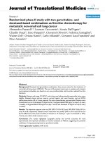
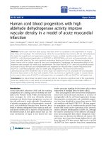
Cytokine production by brain slices from transgenic mouse models of AD at 15 months of ageFigure 1
Cytokine production by brain slices from transgenic mouse models of AD at 15 months of age. Freshly harvested
brain slices were incubated in neurobasal medium with B27 supplement. Media was collected after 24 hours, and cytokine lev-
els measured. Mean concentrations (N = 15) +/- standard error are expressed in picograms per milligram of protein. P < 0.05
was considered statistically significant.
0
20
40
60
80
100
120
140
160
180
200
IL-12p40 IL-10 IL-5 IL-4 IL-3 IL-2 IL-1β IL-1α TNF-α IFN-y GM-CSF
Cytokines (pg/m g protein)
Control
TgAPPs w
PS/A PPsw
*
*
*
*
*
Journal of Neuroinflammation 2005, 2:9 />Page 4 of 10
(page number not for citation purposes)
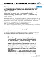
Cytokine production by brain slices from transgenic mouse models of AD at 15 months of ageFigure 2
Cytokine production by brain slices from transgenic mouse models of AD at 15 months of age. Freshly harvested
brain slices were incubated in neurobasal medium with B27 supplement. Media was collected after 24 hours, and cytokine lev-
els measured. Mean concentrations (N = 15) +/- standard error are expressed in picograms per milligram of protein. P < 0.05
was considered statistically significant.
Table 1: Quantification of Aβ levels in TgAPPsw and PS1/APPsw mouse brain slices. Data expressed as picograms/mg protein, mean ±
S.E.M. for 13 determinations.
TgAPPsw PS1/APPsw
Soluble Aβ1–40 331.15 ± 35.36 4957.79 ± 322.30
Soluble Aβ1–42 68.11 ± 6.82 1644.29 ± 90.30
Insoluble Aβ1–40 67619.38 ± 7089.61 4095442 ± 409212.3
Insoluble Aβ1–42 6837.22 ± 2741.70 286463.3 ± 31395.63
0
1000
2000
3000
4000
5000
6000
7000
8000
IL-12p40 IL-10 IL-6 IL-5 IL-4 IL-3 IL-2 IL-1b IL-1a TN F-a IFN-y GM-CSF
Cytokines (pg/mg protein)
Control
TgAPPsw
PS/APP sw
*
*
Journal of Neuroinflammation 2005, 2:9 />Page 5 of 10
(page number not for citation purposes)
therefore omitted from the dendrograms. Of all the
cytokines, IL-12p40 showed the strongest correlation with
levels of both Aβ1–40 and 42 (soluble or insoluble). IL-
1α and IL-1β were also highly correlated with Aβ1–40 and
42 (soluble or insoluble).
Discussion
Levels of both peripheral and local CNS cytokines are ele-
vated in AD patients, indicating that there is cellular acti-
vation occurring in response to inflammatory stimuli [15-
20]. However, there is still considerable debate over
exactly what is triggering this inflammation. Studies using
mouse models of AD have shown that ibuprofen is effec-
tive in reducing plaque pathology and also in improving
behavioral deficits characteristic of these transgenic mod-
els [8,21]. The transgenic mouse models used to study AD
exhibit some of the pathological features seen in the AD
patient brain and show an increased production of
inflammatory markers such as COX-2, PGE
2
and also
increased levels of the pro-inflammatory cytokines IFN-γ
and IL-12, TNF-α, IL-1α, IL-1β and IL-6 [12,22]. Patholog-
ical analysis of tissue from AD patients and from mouse
models of AD shows that there is extensive astrocytic and
microglial activation around areas of Aβ plaque deposi-
tion [6,7]. In addition, the chronic use of non-steroidal
anti-inflammatory drugs (NSAIDs) has been associated
with a reduced risk of developing AD [23,24], suggesting
that inflammation is an important contributor to the
pathophysiology of AD.
One aim of this study was to create a cytokine expression
profile for organotypic brain slice cultures from transgenic
mouse models of Alzheimer's disease, and to further
relate this increase to the level of Aβ present in the brain.
Another purpose of our study was to determine whether
inflammatory events may be correlated with the accumu-
lation of particular forms of Aβ; either soluble or
insoluble.
In the current study, we used the organotypic brain slice
culture model to assess multiple cytokine production in
the culture medium surrounding brain slices from trans-
genic mice that are engineered to over-produce Aβ.
Cytokine production from 15-month-old control, PS1,
TgAPPsw and PS1/APPsw mouse brain slices was assessed
using the Bioplex cytokine multi-array system. Cytokine
levels were not significantly elevated in PS1 brain slices
compared to control slices, indicating that the PS1
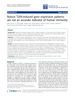
Dendrogram correlations of Aβ1–40 and Aβ1–42-cytokine relationshipsFigure 3
Dendrogram correlations of Aβ1–40 and Aβ1–42-cytokine relationships. Closely related members appear under the
same node. The farther one needs to travel across the tree to reach another member, the greater the dissimilarity.
Journal of Neuroinflammation 2005, 2:9 />Page 6 of 10
(page number not for citation purposes)
(M146L) mutation does not have a significant impact on
cytokine production. No significant change in the
production of IL-4 and IL-10 was observed in the brains of
these transgenic mice compared to their respective con-
trols, indicating the absence of an anti-inflammatory
response. All of the cytokines that were increased in the
TgAPPsw brain slices (IL-1α, TNF-α, GM-CSF and IL-6)
were further increased in the PS1/APP brain slices. This
Dendrogram correlations of Total Aβ (Aβ1–40+Aβ1–42)-cytokine relationshipsFigure 4
Dendrogram correlations of Total Aβ (Aβ1–40+Aβ1–42)-cytokine relationships. Closely related members appear
under the same node. Total Aβ levels were calculated by adding soluble and formic acid extracted Aβ. The farther one needs
to travel across the tree to reach another member, the greater the dissimilarity.
Total Aβ
Journal of Neuroinflammation 2005, 2:9 />Page 7 of 10
(page number not for citation purposes)
suggests that the presence of these inflammatory mole-
cules is related to the amount of β-amyloid protein
present, in agreement with a pro-inflammatory effect of
Aβ [25-29]. A recent report has also shown increases in IL-
1β, IL-6 and TNFα in-vivo after intra-cerebral administra-
tion of fibrillar Aβ into rat brain [30].
In order to further understand the correlation between the
amount of Aβ and cytokine levels in the brains of trans-
genic mice, levels of both soluble and insoluble (formic
acid-extracted) Aβ1–40 and 1–42 were quantified in the
same slices from which cytokine production was meas-
ured, allowing a direct correlation of Aβ-cytokine levels.
Levels of soluble and insoluble Aβ1–40 correlated well
with each other, and the same was observed for Aβ1–42.
As expected, quantification of Aβ levels generally revealed
significantly higher amyloid levels in the PS1/APPsw
mouse brain slices compared to TgAPPsw (for soluble Aβ,
approximately 15 fold more Aβ1–40, and 20 fold more 1–
42) but there was considerable slice-to-slice variation in
soluble and insoluble Aβ levels within and between geno-
types. The TgAPPsw and PS1/APPsw mice express equal
levels of the APPsw molecule, but the PS1/APPsw model
produces greater levels of Aβ and develops plaques at an
earlier age (10 weeks) [31-33]. This increased deposition
of Aβ in the PS1/APPsw mouse is due to a PS1 mutation,
resulting in increased production of Aβ1–42 [34-36].
The Aβ data in the current report found a significant range
of values for soluble:insoluble Aβ ratios between brain
slices. This broad spread of values allowed correlation
Dendrogram correlations of (Aβ1–42:40 ratio)-cytokine relationshipsFigure 5
Dendrogram correlations of (Aβ1–42:40 ratio)-cytokine relationships. Total Aβ1–42:40 ratio's were calculated for
both soluble and formic acid extracted Aβ. Closely related members appear under the same node. The farther one needs to
travel across the tree to reach another member, the greater the dissimilarity.
Aβ 1-42:40 ratio
Journal of Neuroinflammation 2005, 2:9 />Page 8 of 10
(page number not for citation purposes)
with equally wide ranges of cytokine production. This
approach of examining Aβ-cytokine correlations within
the same slices in the same aged animals eliminated the
confounding factor of age related changes in cytokine pro-
duction. Both Aβ1–40 and 1–42 correlated closely with
all the cytokines that changed in the brain slices, but the
correlation was particularly striking with IL-12p40. IL-12
is a hetero-dimeric cytokine which can comprise two sub-
units; IL-12p40 and IL-12p35. It is produced mainly by
monocytes and macrophages and is a crucial factor in
directing the T-cell response to infection, by inducing a
Th1-type cytokine response. Our data agrees with that of
previous reports showing that IL-12p40 is strongly up-reg-
ulated in-vitro (in response to an inflammatory stimulus)
and in-vivo in the cerebral cortex of TgAPPsw mice
[12,37,38].
IL-1, which was increased in the transgenic brain slices, is
a major immune-response molecule functioning in the
periphery and brain. The family comprises three related
proteins (IL-1α, IL-1β and IL-1 receptor antagonist (IL-
1ra)). IL-1α and IL-1β are two different isoforms of IL-1
that have similar affinities for their receptor IL-1R, and
therefore have similar activities. Both are capable of
inducing inflammatory cascades in-vivo and in-vitro, and
it has been shown that they are capable of up-regulating
expression of astrocyte-derived S100B and APP [39,40]. It
has been shown that IL-1β can promote β-secretase cleav-
age of APP in human astrocytes and thereby increase pro-
duction of Aβ1–40 and 1–42 [41,42]. It is also known that
accumulation of plaques and the formation of neurofi-
brillary tangles are correlated with increased IL-1 levels in
the AD brain [43-45]. Certain polymorphisms of IL-1A
(the gene for IL-1α) are associated with late onset AD,
although there is controversy as to whether all IL-1 gene
polymorphisms represent risk factors for AD [46-50].
Microglia, in particular, have been shown to locally up
regulate IL-1α at both the protein and mRNA level when
inflamed, a situation that occurs in chronic disease states
such as AD [51]. Both IL-1α and IL-1β can enhance the
translation of APP mRNA in human astrocytes [52]; an
up-regulation of IL-1α/β production in-vivo could there-
fore increase Aβ production, and an inflammatory cycle
with increased Aβ levels may further increase IL-1α/β
production.
The Aβ 1–42:40 ratio is also of considerable interest in
relation to cytokine levels and although there are cur-
rently no studies correlating Aβ 1–42:40 ratio with
cytokine levels in-vivo, certain reports have suggested that
cytokines can modulate Aβ production [53-55]. PS1
mutations are known to cause a shift in the production of
Aβ species, favoring the production of Aβ1–42 over 1–40
and causing an increase in the Aβ1–42:40 ratio [56]. Since
TNF-α correlated better with the level of Aβ1–42 than
with that of Aβ 1–40, and correlated particularly well with
the Aβ1–42:40 ratio in our study, TNF-α levels may be
partly determined by this ratio. Higher levels of Aβ1–42
can promote the formation of toxic oligomers [57-59],
and it therefore seems possible that the increased level of
Aβ oligomers in PS1/APP mice (compared to APPsw) and
the level of oligomeric forms present in the brains of our
transgenic mice may be related to the amount of TNF-α
being produced.
It is important to consider the nature of the exact form of
Aβ that may be most responsible for the inflammatory
events seen in AD brains. Aβ can exist in various forms
(monomeric, dimeric, oligomeric and fibrillar), but it is
not yet clear which of these forms are most potent in
inducing inflammatory cellular responses [57,60,61].
This is of interest because the oligomeric forms of Aβ
which are thought to be the most toxic are produced more
readily by Aβ1–42 (for review see [62]). Future studies
will assess the relative proportions of monomers/dimers,
oligomers or fibrils occurring in these mice brains and
their relationship with the cytokine increases observed.
List of abbreviations
AD: Alzheimer's disease
APP: Amyloid precursor protein
APPsw: Amyloid precursor protein Swedish mutation
PS1: Presenilin 1
Aβ: Beta-amyloid
Tg: Transgenic
TNF: Tumor necrosis factor
IL-x: Interleukin-x
IL-1ra: Interleukin-1 receptor antagonist
GM-CSF: Granulocyte macrophage colony stimulating
factor
PBS: Phosphate buffered saline
COX-2: Cyclo-oxygenase-2
PGE2: Prostaglandin E2
IFN: Interferon
NSAID: Non-steroidal anti-inflammatory drug
Journal of Neuroinflammation 2005, 2:9 />Page 9 of 10
(page number not for citation purposes)
Competing interests
The author(s) declare that they have no competing
interests.
Authors' contributions
NP carried out the in-vitro brain slice assays, processed
brain tissues, performed the Bio-plex assay, ELISAs and
drafted the manuscript. DP conceived the design of the
study, carried out Bio-plex assays, performed statistical
analyses and aided in manuscript preparation. VM ana-
lyzed data and constructed dendrograms. AQ aided in
ELISA and Bio-plex assays and collected mouse brain tis-
sues. FC oversees management of the mouse colonies.
MM aided in manuscript preparation and gave critical
analysis of the manuscript.
Additional material
Acknowledgements
The authors would like to thank Bob and Diane Roskamp for their gener-
ous support.
References
1. Anthony JC, Breitner JC, Zandi PP, Meyer MR, Jurasova I, Norton
MC, Stone SV: Reduced prevalence of AD in users of NSAIDs
and H2 receptor antagonists: the Cache County study. Neu-
rology 2000, 54:2066-71.
2. Etminan M, Gill S, Samii A: Effect of non-steroidal anti-inflamma-
tory drugs on risk of Alzheimer's disease: systematic review
and meta-analysis of observational studies. BMJ 2003, 327:128.
3. Szekely CA, Thorne JE, Zandi PP, Ek M, Messias E, Breitner JC, Good-
man SN: Nonsteroidal anti-inflammatory drugs for the pre-
vention of Alzheimer's disease: a systematic review.
Neuroepidemiology 2004, 23:159-69.
4. Frautschy SA, Yang F, Irrizarry M, Hyman B, Saido TC, Hsiao K, Cole
GM: Microglial response to amyloid plaques in APPsw trans-
genic mice. Am J Pathol 1998, 152:307-17.
5. Stalder M, Phinney A, Probst A, Sommer B, Staufenbiel M, Jucker M:
Association of microglia with amyloid plaques in brains of
APP23 transgenic mice. Am J Pathol 1999, 154:1673-84.
6. Wegiel J, Wang KC, Imaki H, Rubenstein R, Wronska A, Osuchowski
M, Lipinski WJ, Walker LC, LeVine H: The role of microglial cells
and astrocytes in fibrillar plaque evolution in transgenic
APP(SW) mice. Neurobiol Aging 2001, 22:49-61.
7. Vehmas AK, Kawas CH, Stewart WF, Troncoso JC: Immune reac-
tive cells in senile plaques and cognitive decline in Alzhe-
imer's disease. Neurobiol Aging 2003, 24:321-31.
8. Lim GP, Yang F, Chu T, Chen P, Beech W, Teter B, Tran T, Ubeda O,
Ashe KH, Frautschy SA, Cole GM: Ibuprofen suppresses plaque
pathology and inflammation in a mouse model for Alzhe-
imer's disease. J Neurosci 2000, 20:5709-14.
9. Yan Q, Zhang J, Liu H, Babu-Khan S, Vassar R, Biere AL, Citron M,
Landreth G: Anti-inflammatory drug therapy alters beta-amy-
loid processing and deposition in an animal model of Alzhe-
imer's disease. J Neurosci 2003, 23:7504-9.
10. Mehlhorn G, Hollborn M, Schliebs R: Induction of cytokines in
glial cells surrounding cortical beta-amyloid plaques in trans-
genic Tg2576 mice with Alzheimer pathology. Int J Dev
Neurosci 2000, 18:423-31.
11. Apelt J, Schliebs R: Beta-amyloid-induced glial expression of
both pro- and anti-inflammatory cytokines in cerebral cor-
tex of aged transgenic Tg2576 mice with Alzheimer plaque
pathology. Brain Res 2001, 891:21-30.
12. Abbas N, Bednar I, Mix E, Marie S, Paterson D, Ljungberg A, Morris
C, Winblad B, Nordberg A, Zhu J: Up-regulation of the inflam-
matory cytokines IFN-gamma and IL-12 and down-regula-
tion of IL-4 in cerebral cortex regions of APP(SWE)
transgenic mice. J Neuroimmunol 2002, 126:50-7.
13. Prabhakar U, Eirikis E, Davis HM: Simultaneous quantification of
proinflammatory cytokines in human plasma using the Lab-
MAP assay. J Immunol Methods 2002, 260:207-18.
14. Kawarabayashi T, Younkin LH, Saido TC, Shoji M, Ashe KH, Younkin
SG: Age-dependent changes in brain, CSF, and plasma amy-
loid (beta) protein in the Tg2576 transgenic mouse model of
Alzheimer's disease. J Neurosci 2001, 21:372-81.
15. Araujo DM, Lapchak PA: Induction of immune system media-
tors in the hippocampal formation in Alzheimer's and Par-
kinson's diseases: selective effects on specific interleukins
and interleukin receptors. Neuroscience 1994, 61:745-54.
16. Cacabelos R, Alvarez XA, Fernandez-Novoa L, Franco A, Mangues R,
Pellicer A, Nishimura T: Brain interleukin-1 beta in Alzheimer's
disease and vascular dementia. Methods Find Exp Clin Pharmacol
1994, 16:141-51.
17. Blum-Degen D, Muller T, Kuhn W, Gerlach M, Przuntek H, Riederer
P: Interleukin-1 beta and interleukin-6 are elevated in the
cerebrospinal fluid of Alzheimer's and de novo Parkinson's
disease patients. Neurosci Lett 1995, 202:17-20.
18. Griffin WS, Sheng JG, Roberts GW, Mrak RE: Interleukin-1 expres-
sion in different plaque types in Alzheimer's disease: signifi-
cance in plaque evolution. J Neuropathol Exp Neurol 1995,
54:276-81.
19. Singh VK, Guthikonda P: Circulating cytokines in Alzheimer's
disease. J Psychiatr Res 1997, 31:657-60.
20. Tarkowski E, Wallin A, Regland B, Blennow K, Tarkowski A: Local
and systemic GM-CSF increase in Alzheimer's disease and
vascular dementia. Acta Neurol Scand 2001, 103:166-74.
21. Lim GP, Yang F, Chu T, Gahtan E, Ubeda O, Beech W, Overmier JB,
Hsiao-Ashec K, Frautschy SA, Cole GM: Ibuprofen effects on
Alzheimer pathology and open field activity in APPsw trans-
genic mice. Neurobiol Aging 2001, 22:983-91.
22. Benzing WC, Wujek JR, Ward EK, Shaffer D, Ashe KH, Younkin SG,
Brunden KR: Evidence for glial-mediated inflammation in
aged APP(SW) transgenic mice. Neurobiol Aging 1999, 20:581-9.
23. Andersen K, Launer LJ, Ott A, Hoes AW, Breteler MM, Hofman A:
Do nonsteroidal anti-inflammatory drugs decrease the risk
for Alzheimer's disease? The Rotterdam Study. Neurology
1995, 45:1441-5.
24. Stewart WF, Kawas C, Corrada M, Metter EJ: Risk of Alzheimer's
disease and duration of NSAID use. Neurology 1997, 48:626-32.
25. Gitter BD, Boggs LN, May PC, Czilli DL, Carlson CD: Regulation of
cytokine secretion and amyloid precursor protein process-
ing by proinflammatory amyloid beta (A beta). Ann N Y Acad
Sci 2000, 917:154-64.
26. Rah JC, Kim HS, Kim SS, Bach JH, Kim YS, Park CH, Seo JH, Jeong SJ,
Suh YH: Effects of carboxyl-terminal fragment of Alzheimer's
amyloid precursor protein and amyloid beta-peptide on the
production of cytokines and nitric oxide in glial cells. FASEB J
2001, 15:1463-5.
27. Paris D, Townsend KP, Obregon DF, Humphrey J, Mullan M: Pro-
inflammatory effect of freshly solubilized beta-amyloid pep-
tides in the brain. Prostaglandins Other Lipid Mediat 2002, 70:1-12.
28. Giovannini MG, Scali C, Prosperi C, Bellucci A, Vannucchi MG, Rosi
S, Pepeu G, Casamenti F: Beta-amyloid-induced inflammation
and cholinergic hypofunction in the rat brain in vivo: involve-
ment of the p38MAPK pathway. Neurobiol Dis 2002, 11:257-74.
29. Quadros A, Patel N, Crescentini R, Crawford F, Paris D, Mullan M:
Increased TNFalpha production and Cox-2 activity in organ-
otypic brain slice cultures from APPsw transgenic mice. Neu-
rosci Lett 2003, 353:66-8.
30. Rosales-Corral S, Tan DX, Reiter RJ, Valdivia-Velazquez M, Acosta-
Martinez JP, Ortiz GG: Kinetics of the neuroinflammation-oxi-
Additional File 1
Correlation table of levels of different
β
-amyloid species with cytokines in
transgenic mouse models of Alzheimer's disease.
Click here for file
[ />2094-2-9-S1.htm]
Journal of Neuroinflammation 2005, 2:9 />Page 10 of 10
(page number not for citation purposes)
dative stress correlation in rat brain following the injection
of fibrillar amyloid-beta onto the hippocampus in vivo. J
Neuroimmunol 2004, 150:20-8.
31. McGowan E, Sanders S, Iwatsubo T, Takeuchi A, Saido T, Zehr C, Yu
X, Uljon S, Wang R, Mann D, Dickson D, Duff K: Amyloid pheno-
type characterization of transgenic mice overexpressing
both mutant amyloid precursor protein and mutant preseni-
lin 1 transgenes. Neurobiol Dis 1999, 6:231-44.
32. Takeuchi A, Irizarry MC, Duff K, Saido TC, Hsiao Ashe K, Hasegawa
M, Mann DM, Hyman BT, Iwatsubo T: Age-related amyloid beta
deposition in transgenic mice overexpressing both Alzhe-
imer mutant presenilin 1 and amyloid beta precursor pro-
tein Swedish mutant is not associated with global neuronal
loss. Am J Pathol 2000, 157:331-9.
33. Kurt MA, Davies DC, Kidd M, Duff K, Rolph SC, Jennings KH,
Howlett DR: Neurodegenerative changes associated with
beta-amyloid deposition in the brains of mice carrying
mutant amyloid precursor protein and mutant presenilin-1
transgenes. Exp Neurol 2001, 171:59-71.
34. Borchelt DR, Thinakaran G, Eckman CB, Lee MK, Davenport F, Rato-
vitsky T, Prada CM, Kim G, Seekins S, Yager D, Slunt HH, Wang R,
Seeger M, Levey AI, Gandy SE, Copeland NG, Jenkins NA, Price DL,
Younkin SG, Sisodia SS: Familial Alzheimer's disease-linked
presenilin 1 variants elevate Abeta1–42/1–40 ratio in vitro
and in vivo. Neuron 1996, 17:1005-13.
35. Citron M, Westaway D, Xia W, Carlson G, Diehl T, Levesque G,
Johnson-Wood K, Lee M, Seubert P, Davis A, Kholodenko D, Motter
R, Sherrington R, Perry B, Yao H, Strome R, Lieberburg I, Rommens
J, Kim S, Schenk D, Fraser P, St George Hyslop P, Selkoe DJ: Mutant
presenilins of Alzheimer's disease increase production of 42-
residue amyloid beta-protein in both transfected cells and
transgenic mice. Nat Med 1997, 3:67-72.
36. Holcomb L, Gordon MN, McGowan E, Yu X, Benkovic S, Jantzen P,
Wright K, Saad I, Mueller R, Morgan D, Sanders S, Zehr C, O'Campo
K, Hardy J, Prada CM, Eckman C, Younkin S, Hsiao K, Duff K: Accel-
erated Alzheimer-type phenotype in transgenic mice carry-
ing both mutant amyloid precursor protein and presenilin 1
transgenes. Nat Med 1998, 4:97-100.
37. Yang Y, Han SH, Kim H, Kim C, Kim KY, Shin SM, Choi I, Pyun KH:
Interleukin-12 p40 gene expression is induced in lipopolysac-
charide-activated pituitary glands in vivo. Neuroendocrinology
2002, 75:347-57.
38. Ichikawa D, Matsui A, Imai M, Sonoda Y, Kasahara T: Effect of vari-
ous catechins on the IL-12p40 production by murine perito-
neal macrophages and a macrophage cell line, J774.1. Biol
Pharm Bull 2004, 9:1353-8.
39. Sheng JG, Ito K, Skinner RD, Mrak RE, Rovnaghi CR, Van Eldik LJ, Grif-
fin WS: In vivo and in vitro evidence supporting a role for the
inflammatory cytokine interleukin-1 as a driving force in
Alzheimer pathogenesis. Neurobiol Aging 1996, 17:761-6.
40. Mrak RE, Griffin WS: The role of activated astrocytes and of the
neurotrophic cytokine S100B in the pathogenesis of Alzhe-
imer's disease. Neurobiol Aging 2001, 22:915-22.
41. Schmitt TL, Steiner E, Klinger P, Sztankay A, Grubeck-Loebenstein B:
The production of an amyloidogenic metabolite of the
Alzheimer amyloid beta precursor protein (APP) in thyroid
cells is stimulated by interleukin 1 beta, but inhibited by
interferon gamma. J Clin Endocrinol Metab 1996, 81:1666-9.
42. Blasko I, Veerhuis R, Stampfer-Kountchev M, Saurwein-Teissl M, Eike-
lenboom P, Grubeck-Loebenstein B: Costimulatory effects of
interferon-gamma and interleukin-1beta or tumor necrosis
factor alpha on the synthesis of Abeta1–40 and Abeta1–42 by
human astrocytes. Neurobiol Dis 2000, 7:682-9.
43. Griffin WS, Stanley LC, Ling C, White L, MacLeod V, Perrot LJ, White
CL 3rd, Araoz C: Brain interleukin 1 and S-100 immunoreac-
tivity are elevated in Down syndrome and Alzheimer
disease. Proc Natl Acad Sci U S A 1989, 86:7611-5.
44. Sheng JG, Mrak RE, Griffin WS: Glial-neuronal interactions in
Alzheimer disease: progressive association of IL-1alpha+
microglia and S100beta+ astrocytes with neurofibrillary tan-
gle stages. J Neuropathol Exp Neurol 1997, 56:285-90.
45. Griffin WS, Mrak RE: Interleukin-1 in the genesis and progres-
sion of and risk for development of neuronal degeneration in
Alzheimer's disease. J Leukoc Biol 2002, 72:233-8.
46. Du Y, Dodel RC, Eastwood BJ, Bales KR, Gao F, Lohmuller F, Muller
U, Kurz A, Zimmer R, Evans RM, Hake A, Gasser T, Oertel WH, Grif-
fin WS, Paul SM, Farlow MR: Association of an interleukin 1
alpha polymorphism with Alzheimer's disease. Neurology 2000,
55:480-3.
47. Nicoll JA, Mrak RE, Graham DI, Stewart J, Wilcock G, MacGowan S,
Esiri MM, Murray LS, Dewar D, Love S, Moss T, Griffin WS: Associ-
ation of interleukin-1 gene polymorphisms with Alzheimer's
disease. Ann Neurol 2000, 47:365-8.
48. Grimaldi LM, Casadei VM, Ferri C, Veglia F, Licastro F, Annoni G,
Biunno I, De Bellis G, Sorbi S, Mariani C, Canal N, Griffin WS, Franc-
eschi M: Association of early-onset Alzheimer's disease with
an interleukin-1alpha gene polymorphism. Ann Neurol 2000,
47:361-5.
49. Fidani L, Goulas A, Mirtsou V, Petersen RC, Tangalos E, Crook R,
Hardy J: Interleukin-1A polymorphism is not associated with
late onset Alzheimer's disease. Neurosci Lett 2002, 323:81-3.
50. Sciacca FL, Ferri C, Licastro F, Veglia F, Biunno I, Gavazzi A, Calabrese
E, Martinelli Boneschi F, Sorbi S, Mariani C, Franceschi M, Grimaldi
LM: Interleukin-1B polymorphism is associated with age at
onset of Alzheimer's disease. Neurobiol Aging 2003, 24:927-31.
51. Hetier E, Ayala J, Denefle P, Bousseau A, Rouget P, Mallat M, Prochi-
antz A: Brain macrophages synthesize interleukin-1 and inter-
leukin-1 mRNAs in vitro. J Neurosci Res 1988, 21:391-7.
52. Rogers JT, Leiter LM, McPhee J, Cahill CM, Zhan SS, Potter H, Nilsson
LN: Translation of the alzheimer amyloid precursor protein
mRNA is up-regulated by interleukin-1 through 5'-untrans-
lated region sequences. J Biol Chem 1999, 274:6421-31.
53. Del Bo R, Angeretti N, Lucca E, De Simoni MG, Forloni G: Recipro-
cal control of inflammatory cytokines, IL-1 and IL-6, and
beta-amyloid production in cultures. Neurosci Lett 1995,
188:70-4.
54. Brugg B, Dubreuil YL, Huber G, Wollman EE, Delhaye-Bouchaud N,
Mariani J: Inflammatory processes induce beta-amyloid pre-
cursor protein changes in mouse brain. Proc Natl Acad Sci U S A
1995, 92:3032-5.
55. Liao YF, Wang BJ, Cheng HT, Kuo LH, Wolfe MS: Tumor necrosis
factor-alpha, interleukin-1beta, and interferon-gamma stim-
ulate gamma-secretase-mediated cleavage of amyloid pre-
cursor protein through a JNK-dependent MAPK pathway. J
Biol Chem 2004, 279:49523-32.
56. Borchelt DR: Metabolism of presenilin 1: influence of preseni-
lin 1 on amyloid precursor protein processing. Neurobiol Aging
1998, 19:S15-8.
57. Chromy BA, Nowak RJ, Lambert MP, Viola KL, Chang L, Velasco PT,
Jones BW, Fernandez SJ, Lacor PN, Horowitz P, Finch CE, Krafft GA,
Klein WL: Self-assembly of Abeta(1–42) into globular
neurotoxins. Biochemistry 2003, 42:12749-60.
58. Lambert MP, Barlow AK, Chromy BA, Edwards C, Freed R, Liosatos
M, Morgan TE, Rozovsky I, Trommer B, Viola KL, Wals P, Zhang C,
Finch CE, Krafft GA, Klein WL: Diffusible, nonfibrillar ligands
derived from Abeta1–42 are potent central nervous system
neurotoxins. Proc Natl Acad Sci U S A 1998, 95:6448-53.
59. Bitan G, Kirkitadze MD, Lomakin A, Vollers SS, Benedek GB, Teplow
DB: Amyloid beta-protein (Abeta) assembly: Abeta 40 and
Abeta 42 oligomerize through distinct pathways. Proc Natl
Acad Sci U S A 2003, 100:330-5.
60. Grace EA, Rabiner CA, Busciglio J: Characterization of neuronal
dystrophy induced by fibrillar amyloid beta: implications for
Alzheimer's disease. Neuroscience 2002, 114:265-73.
61. Kayed R, Head E, Thompson JL, McIntire TM, Milton SC, Cotman
CW, Glabe CG: Common structure of soluble amyloid oli-
gomers implies common mechanism of pathogenesis. Science
2003, 300:486-9.
62. Ross CA, Poirier MA: Protein aggregation and neurodegenera-
tive disease. Nat Med 2004, 10:S10-7.