báo cáo hóa học: " Microglial inflammation in the parkinsonian substantia nigra: relationship to alpha-synuclein deposition" doc
Bạn đang xem bản rút gọn của tài liệu. Xem và tải ngay bản đầy đủ của tài liệu tại đây (549.55 KB, 8 trang )
BioMed Central
Page 1 of 8
(page number not for citation purposes)
Journal of Neuroinflammation
Open Access
Research
Microglial inflammation in the parkinsonian substantia nigra:
relationship to alpha-synuclein deposition
Emilie Croisier
1
, LindaBMoran
1
, David T Dexter
2
, Ronald KB Pearce
1
and
Manuel B Graeber*
1
Address:
1
Department of Neuropathology, Division of Neuroscience and Mental Health, Imperial College London, and Hammersmith Hospitals
Trust, London, UK and
2
Department of Cellular and Molecular Neuroscience, Division of Neuroscience and Mental Health, Imperial College
London, London, UK
Email: Emilie Croisier - ; Linda B Moran - ; David T Dexter - ;
Ronald KB Pearce - ; Manuel B Graeber* -
* Corresponding author
Abstract
Background: The role of both microglial activation and alpha-synuclein deposition in Parkinson's
disease remain unclear. We have tested the hypothesis that if microglia play a primary role in
Parkinson's disease pathogenesis, the microglial "activated" phenotype should be associated with
histopathological and/or clinical features of the disease.
Methods: We have examined microglial MHC class II expression, a widely used marker of
microglial activation, the occurrence of CD68-positive phagocytes and alpha-synuclein
immunoreactivity in post-mortem human substantia nigra affected by idiopathic Parkinson's disease
(PD). Using semi-quantitative severity ratings, we have examined the relationship between
microglial activation, alpha-synuclein deposition, classical neuropathological criteria for PD, subtype
of the disease and clinical course.
Results: While we did not observe an association between microglial MHC class II expression and
clinical parameters, we did find a correlation between disease duration and the macrophage marker
CD68 which is expressed by phagocytic microglia. In addition, we observed a significant correlation
between the degree of MHC class II expression and alpha-synuclein deposition in the substantia
nigra in PD.
Conclusion: While microglia appeared to respond to alpha-synuclein deposition, MHC class II
antigen expression by microglia in the substantia nigra cannot be used as an indicator of clinical PD
severity or disease progression. In addition, a contributory or even causative role for microglia in
the neuronal loss associated with PD as suggested by some authors seems unlikely. Our data
further suggest that an assessment of microglial activation in the aged brain on the basis of
immunohistochemistry for MHC class II antigens alone should be done with caution.
Introduction
Parkinson's Disease (PD) is a common neurodegenerative
disorder with the cardinal clinical features of tremor,
rigidity, bradykinesia and loss of postural reflexes.
Published: 03 June 2005
Journal of Neuroinflammation 2005, 2:14 doi:10.1186/1742-2094-2-14
Received: 26 April 2005
Accepted: 03 June 2005
This article is available from: />© 2005 Croisier et al; licensee BioMed Central Ltd.
This is an Open Access article distributed under the terms of the Creative Commons Attribution License ( />),
which permits unrestricted use, distribution, and reproduction in any medium, provided the original work is properly cited.
Journal of Neuroinflammation 2005, 2:14 />Page 2 of 8
(page number not for citation purposes)
Neuropathologically, the disease is characterized by a
marked loss of dopaminergic neurons in the substantia
nigra pars compacta (SN) and the presence of alpha-synu-
clein (aSN)-positive Lewy bodies (LBs) in neurons of this
and other brain areas also affected by nerve cell death. An
international consensus definition of Lewy body diseases
on the basis of molecular as well as morphological crite-
ria, which takes into account aSN status of the brain, has
been published recently
[1].
The discovery of aSN mutations and gene amplification in
some familial forms of PD [2-6]] and the identification of
this protein as a major component of Lewy bodies (LBs)
in common sporadic PD [7], has spurred interest in the
role of aSN in the pathophysiology of PD and other synu-
cleinopathies. However, no direct causal relationship has
yet been established between aSN aggregation and the
selective neuronal cell death characteristic of PD. LBs are
also found incidentally in aged brain in the absence of
other pathological features and without a clinical history
of parkinsonism or dementia [8]. Attempts have been
made to link the clinical progression of PD to the presence
of aSN inclusions and an anatomical staging model has
been proposed [9], but the latter has been questioned by
subsequent studies in which clinical data were also taken
into account [10].
Apart from well established morphological criteria, acti-
vated microglia can be defined in tissue sections on the
basis of the expression of several immune function-
related proteins, notably complement receptors and MHC
class II antigens (MHCII). Phagocytic activity and cyto-
toxic properties are usually considered end stages of
microglial activation, at which point they are phenotypi-
cally indistinguishable from blood-borne macrophages.
Activated microglia are associated with a large range of
neurological insults from trauma and infection to autoim-
mune conditions, and their presence represents a com-
mon finding also in neurodegenerative disorders [11].
However there is little knowledge about the molecular
processes that mediate microglial activation and exactly
which biological consequences may result from their
enhanced state of "immune alertness" within affected
CNS tissue. A transcriptome signature of interferon-
gamma activated microglia has been provided recently
[12]. Microglial phenotypic changes have also been
observed in normal aged individuals [13]. Thus, "micro-
glial senescence" confounds the problem of a definition
of microglial activation in disease states, and in neurode-
generative diseases in particular which are often age-
related, as no specific causative stimulus has been identi-
fied in the process.
While microglia clearly show changes in their phenotypic
profile in neurodegeneration, it is by no means clear
whether they are actively involved in the progression of
PD. Microglia-derived macrophages can be found in the
PD SN, and neuromelanin pigment taken up from degen-
erated dopaminergic nerve cells is characteristically
observed in SN phagocytes. In animal models of nigrostri-
atal degeneration using 6-hydroxydopamine and 1-
methyl-4-phenyl-1,2,3,6-tetrahydropyridine (MPTP),
inhibition or attenuation of the microglial immune
response increases neuronal survival. However, those
results have so far not been replicated in clinico-patholog-
ical studies, and the simple chemical lesions currently
employed in animal studies by all likelihood do not fully
reflect the chronic neurodegenerative disease process in
humans [14].
In the present study, we independently evaluate the sever-
ity of alpha-synuclein deposition and microglial activa-
tion identified by immunohistochemical staining in the
SN in a large cohort of clinically and pathologically con-
firmed PD cases. We have studied the microglial response
in PD on two levels, by observing MHCII -immunoreac-
tive cells (putatively activated microglia but possibly only
senescent cells) and CD68-immunopositive macrophages
(corresponding to either phagocytic microglia or cells
derived from invading blood-borne macrophages).
Materials and methods
Parkinson's disease cases
37 PD nigrae were evaluated immunohistochemically. 20
cases were provided by the UK Parkinson's Disease Society
Tissue Bank at Imperial College London (PDSTB). Addi-
tional tissue sections from 17 other cases came from a pre-
vious study originally performed at the Institute of
Neuropathology, University of Munich, Germany. These
Parkinson's cases had been previously diagnosed, neu-
ropathologically screened for confounding pathology,
and examined in a study of apoptosis and microglial acti-
vation [15]. Archival sections were immunolabelled for
alpha-synuclein (see below) and used as a control group
to ensure that variation within our PDSTB cohort was
within an established range.
Clinical and neuropathological assessment of cases
For the PDSTB cohort, clinical reports were evaluated in
detail by an experienced neurologist with a special interest
in Parkinson's disease (RKBP). Neuropathological assess-
ment was based on slides provided by the PDSTB for
alpha-synuclein, tau and beta-amyloid immunohisto-
chemistry of superior frontal gyrus, the hippocampal
region and midbrain as minimum data sets, and screening
of the cases for confounding pathology was based on
hematoxylin and eosin examination of a standard series
of 18 tissue blocks following a standardised dissection
procedure [16]. Nine cases showed varying degrees of con-
current Alzheimer's disease (AD)-type pathology (tau-
Journal of Neuroinflammation 2005, 2:14 />Page 3 of 8
(page number not for citation purposes)
immunopositive tangles and/or beta-amyloid-immunop-
ositive plaques) of isocortical and/or entorhinal type
ranging from grades 1–3
. Three
cases were excluded based on a final neuropathological
diagnosis of AD, progressive supranuclear palsy (PSP),
and young-onset familial PD, respectively, leaving a
cohort of 17 cases in each the Munich and PDSTB groups
(Table 1).
Immunohistochemical evaluation of protein levels
Immunohistochemical reactions were performed using
the avidin-biotin complex (ABC)/peroxidase method
with mouse monoclonal antibodies anti-human HLA-DP,
DQ, DR (clone CR3/43, Dako, dilution 1/100) and anti-
alpha-synuclein (Becton-Dickinson, dilution 1/300). For
the PDSTB group, additional immunohistochemistry was
carried out with anti-CD68 (clone PGM1, Dako, dilution
1/200). Sections were dewaxed in xylene, rehydrated, and
endogenous peroxidase activity was blocked by 30 min
exposure to 1% hydrogen peroxide in methanol. Antigen
unmasking consisted of boiling in 0.01 M EDTA (20 min.
at 350 W in microwave) and 100% formic acid treatment
(3 min.) prior to incubation with anti-HLA-DP, DQ, DR
and anti-alpha-synuclein, respectively. No antigen
unmasking was used with anti-CD68. Slides were then
incubated in primary antibody diluted in phosphate-buff-
ered saline (PBS) overnight at 4°C. The following day,
after washing in PBS, they were incubated in horse-anti-
mouse secondary antibody (Vector, dilution 1/200) and
finally in ABC complex (Vector, dilution 1/200) each for
1 hour at room temperature. Immunoreactivity was visu-
alised with 3,3'-diaminobenzidine (Vector kit).
After immunohistochemical staining, sections were given
semi-quantitative severity ratings for aSN, MHCII, and
CD68 immunoreactivity by two investigators (EC and
MBG) blinded to case number. The SN was defined as the
area extending laterally from the exit of the third nerve,
superior to the cerebral peduncle and inferior to the
medial lemniscus, ideally at the height of the red nucleus
with the presence of melanised neurons or their remnants
indicating the main region of interest. The severity ratings
were determined across the entire region of SN, based on
the density of immunopositive structures, with 0 (none),
1 (mild), 2 (moderate) and 3 (high). For aSN, both intra-
and extra-cellular inclusions were considered provided
they fell within the immediate area of the substantia nigra.
This was particularly relevant in areas of severe neuronal
loss, often encountered more laterally, where significant
alpha-synuclein pathology could still be observed. The
morphological variation in aSN deposition was not
assessed, simply the frequency of events. All clearly iden-
tifiable aSN-immunoreactive structures, including LBs,
neurites, fibrils, and smaller, punctate formations, were
considered. For microglial response, severity was judged
primarily by immunoreactivity, however morphology was
taken into account in that perivascular immunoreactivity
was excluded. The thickening of microglial processes
increased the apparent density of microglial staining, such
that cases undergoing a more intense microglial response
were clearly differentiated on the basis of
Table 1: PDSTB cases examined.
CASE SEX DIAGNOSIS AAO AAD DD MHCII AVE aSN AVE CD68 AVE
1 m PD, H-T 63 72 9 2 1 1.5
2 fPDD H-T678114 2 1 1
3 m PD, A-R 57 71 14 2.75 3 2.25
4 f PD, H-T 67 85 18 2.5 2.25 0.5
5 m PD, H-T 57 75 18 2 1.5 2
6 m PD, A-R 78 83 5 1.75 1 2.75
7 f PD, H-T 55 73 18 1.75 2 2.25
8 m PD, H-T 49 77 28 2.5 2.5 0.5
9 f PD, H-T 65 75 10 2 2 2.75
10 mPD, A-R7582 7 11.752
11 m PDD H-T 72 81 9 2 2 1.25
12 m PD, A-R 69 75 6 2 2.5 1.75
13 mPD, A-R6583182.51.51.5
14 m PD, H-T 70 77 7 2 1 1.5
15 m PD, A-R 86 89 3 2.75 2.5 2
16 fPD, H-T728311 21.51.25
17 mPD, H-T6676102.52.53
Abbreviations: PD, Parkinson's disease; PDD, Parkinson's disease with dementia; H-T, hemi-tremulous; A-R, akinetic-rigid; AAO, age at disease
onset; AAD, age at death; DD, disease duration; AVE, semi-quantitative severity rating, averaged across two observers.
Journal of Neuroinflammation 2005, 2:14 />Page 4 of 8
(page number not for citation purposes)
immunoreactivity alone. Morphological features of acti-
vated microglia were always noted, however there were no
cases for which the severity rating would have changed
substantially on the basis of morphological features, ie.
cases with low MHCII immunoreactivity but most micro-
glia adopting an amoeboid morphology or cases with
high MHCII immunoreactivity but most microglia
appearing ramified.
Regression analysis revealed the two sets of ratings from
independent observers were highly correlated (p <
0.0001). Ratings were then averaged to generate a severity
score for each immunohistochemical stain.
Results
All of the cases examined showed the severe dopaminergic
neuronal loss and extra-cellular (free-lying) neuromela-
nin typical of advanced PD. All were also positive for
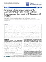
alpha-synuclein, MHCII, and CD68 (Figure 1). Semi-
quantitative ratings for the PDSTB cohort are shown in
Table 1. MHCII immunoreactivity was confined to micro-
glial or macrophage-like cells. Most of the CD68-positive
cells were of a brain macrophage phenotype, i.e. cells with
an enlarged immunoreactive cytoplasm containing lyso-
somal structures and/or neuromelanin degradation prod-
ucts, shortened and less ramified, stout (compared with
typical microglia) cell processes but rarely of the appear-
ance of the full-blown macrophages commonly found in
brain infarcts or multiple sclerosis lesions. aSN inclusions
were observed in neurons, white matter, and occasionally
glial cells and their fine processes. MHCII immunolabel-
ling and the presence of macrophages showed significant
variation between cases. Semi-quantitative ratings
revealed that despite this inconsistency across the group,
aSN deposition and MHCII immunoreactivity were found
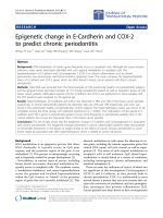
to correlate within individual cases (p < 0.001) (Figure 2).
A General Linear Model test (SPSS) revealed no difference
between PDSTB and University of Munich cases (p =
0.01), and the relationship remained statistically signifi-
cant when we considered only the PDSTB group. We did
not find a similar relationship between aSN and CD68
immunoreactivity. Regression analysis revealed no signif-
icant statistical link between the two stains.
In our cohort of PDSTB cases (n = 17), for which clinical
information was available, we then assessed how immu-
noreactivity may relate to clinical history. Cases were
assessed based on clinical subtype (tremor or akinetic-
rigid), gender, disease duration (DD), and ages of onset
(AAO) and death (AAD). In addition, absence or presence
of AD-related pathology was determined following
ICDNS criteria
. Cases were evalu-
ated as individual data points and in groups. No correla-
tions were found with aSN or MHCII as histological
reference points, and no clinical classification seemed to
reflect the relationship between aSN deposition and
MHCII immunoreactivity in individual cases. A signifi-
cant difference between CD68 immunoreactivity in PD
cases with a DD of 10 years or less (n = 9, mean 7.3 ± 2.4
years) compared to those with a DD greater than 10 years
(n = 8, mean 17 ± 5.0 years) was detected when using Stu-
dent's t-test, with CD68 immunoreactivity significantly
higher in cases with a shorter DD (p < 0.05). DD was
highly inversely correlated with AAO (p < 0.0005). AAO
was also significantly different in the two DD groups
according to Student's t-test (p < 0.005) while AAD
remained consistent across cases in both the shorter- (n =
9, mean 78.9 ± 5.3) and longer DD groups (n = 8, mean
78.5 ± 5.2). MHCII and aSN immunoreactivity did not
show any significant variation across DD, or any other
clinically defined PD groups.
Discussion
Our finding of an overall correlation between aSN depo-
sition and MHCII-expressing microglia in the substantia
nigra is in line with the finding that both phenotypic
changes are associated with neurodegeneration in PD, but
it remains unclear whether there is any pathogenetic link.
It is perhaps more noteworthy than the correlation
between alpha-synuclein deposition and microglial acti-
vation that we failed to find any correlation between these
parameters and clinical indicators of disease progression.
Studies of multiple system atrophy (MSA), PSP and corti-
cobasal degeneration (CBD) have previously detected a
stochastic link between the presence of activated micro-
glia and protein deposition in the neuroanatomic systems
specifically affected by the disease and hypothesize that
microglial activation may in part be induced by the accu-
mulation of pathological protein in tissue [17,18]. aSN
could be one of the pathological substrates that initiate
microglial activation. However, there is no evidence that
LBs can directly provoke this response [19,20]. LBs may
contain complement proteins and chromogranin A [21],
which can induce microglial activation in vitro [22], but in
post-mortem tissue microglia have not been observed to
interact preferentially with these particular LBs [19].
Much has been speculated about the potentially deleteri-
ous effects of activated microglia on neuronal survival in
PD. Specifically, emphasis has been placed on the produc-
tion of pro-inflammatory cytokines and reactive oxygen
species potentially increasing oxidative stress on sur-
rounding neurons. In various animal and cell culture
models, the inhibition of microglial activation has been
demonstrated to be neuroprotective in some circum-
stances [23,24]. However, there is no evidence that micro-
glia initiate neurodegeneration, and their response does
not always correlate to active cell death occurring in their
microenvironment. The presence of MHCII-immunoreac-
Journal of Neuroinflammation 2005, 2:14 />Page 5 of 8
(page number not for citation purposes)
tive microglia in the SN of monkeys one year following
chronic administration of MPTP has been interpreted as
evidence that the neurodegenerative process was still
active and associated with the glial response [25]. How-
ever, a comparable experiment demonstrated that
although microglia do indeed remain present in the SN,
they are absent from the striatum where active neurode-
generation could still be detected [26].
In contrast to the focal activation observed in animal
models and acute CNS insults, microglial activation in PD
is widespread and not limited to areas of marked cell
death [27]. This phenomenon, and the persistence of acti-
vation in the SN long after most dopaminergic neurons
have been lost, may in part be attributable to the differ-
ences in microglial responsiveness between young and
aged individuals. The number of MHCII-expressing
microglia in the human CNS increases steadily with age
independent of disease or trauma [13,28]. In addition,
microglia and astrocytes in culture harvested from older
rats are more inclined to proliferation and MHCII expres-
sion and are less sensitive to transforming growth factor-
beta than glia from younger donors [29]. This is in line
with the findings of a study on MPTP-treated mice show-
ing that this toxin acts in an age-dependent manner, with
protracted microglial activation in older animals [30].
Our failure to find any correlation between MHCII in the
parkinsonian nigra and DD, AAO, AAD, gender or pre-
dominant motor symptoms raises questions about the
significance of microglial MHCII expression as a marker
of their involvement in PD and in other chronic CNS dis-
eases especially in the aged brain. Are microglia desig-
nated 'active' by the presence of certain proteins
necessarily functionally active? Or does microglial MHCII
expression serve as a "firewall" against T-cell invasion of
already compromised CNS tissue since co-stimulators
such as B7 may not be expressed at a sufficient level [31]?
The use of MHCII immunoreactivity as a marker of micro-
glial "activation" should be re-evaluated in the light of
studies suggesting very long-lasting microglial involve-
ment in chronic and late onset neurological conditions.
Microglial "activation" under such conditions may have
the quality of a "microglial scar" which would have differ-
ent functional relevance, and this should be taken into
account in the interpretation of neuroimaging studies of
activated microglia [32].
Furthermore, it may also be that the presence of activated
microglia is a reflection of agonal state rather than indica-
tive of a chronic disease state, and that our microglia
results are in part attributable to factors such as hypoxia or
infection prior to patient death. This could account for
our failure to identify clinical correlates to MHCII expres-
sion; however the strong correlation of this expression
Immunohistochemical staining of (A) aSN deposition in and around melanised dopaminergic neurons of the SN, (B) microglial expression of MHCII, and (C) macrophage expres-sion of CD68Figure 1
Immunohistochemical staining of (A) aSN deposition in and
around melanised dopaminergic neurons of the SN, (B)
microglial expression of MHCII, and (C) macrophage expres-
sion of CD68. All images taken at 40X primary magnification.
Journal of Neuroinflammation 2005, 2:14 />Page 6 of 8
(page number not for citation purposes)
with the presence of aSN deposition remains unex-
plained. Another possibility is that the inflammatory
response in PD peaks early in the course of the disease, at
which time a pathogenetic link between MHCII and clin-
ical progression could be detected. However, by the time
of post-mortem evaluation most relevant microglial activ-
ity may have stopped. Yet, high levels of CD68 in some
cases indicate that microglial phagocytosis can still occur
at the time of death. As the presence of tissue macro-
phages may be considered a sign of ongoing tissue
destruction, and macrophages are known to be crucial
players in the cytotoxic phase of an inflammatory
response, it is of particular interest that they were found to
be more prevalent in PD cases with shorter DD. This sug-
gests that microglial phagocytosis may not persist when it
is no longer functionally relevant.
Increased extraneuronal neuromelanin and decreased
aSN pathology in the SN have been associated with the
progression of PD as defined by a staging model based on
pathological, rather than clinical criteria [9]. It is possible
that as PD progresses from one pathological tier to
another, increasing extracellular neuromelanin deposi-
tion causes more microglia to adopt a phagocytic pheno-
type. Neuromelanin has been demonstrated to induce
microglial activation in vivo [33], and, since one of the pri-
mary functions of activated microglia is the removal of
debris produced by necrotic cells and neuromelanin is a
readily identifiable component of this debris, the presence
of this compound in the SN may very well have a relation-
ship with the presence of phagocytic microglia in the
region. However, this pathological staging is not reflective
of our disease cohort. All of the cases evaluated for this
Case-by-case semi-quantitative severity ratings for MHC class II and alpha-synuclein immunopositivityFigure 2
Case-by-case semi-quantitative severity ratings for MHC class II and alpha-synuclein immunopositivity.
0
0.5
1
1.5
2
2.5
3
Semi-quantitative severity ratings
MHC clas s II alpha-s ynuclein
Journal of Neuroinflammation 2005, 2:14 />Page 7 of 8
(page number not for citation purposes)
study were in both clinically and pathologically advanced
stages of PD, and would have fallen within the later, more
severe proposed tiers. Because the model does not address
clinical information regarding disease progression, the
distribution of DD across our cases would not affect their
pathological staging. Cases with shorter DD may have
progressed more rapidly, or they may have remained pre-
symptomatic for a longer period. Our failure to find any
relationship between aSN deposition and DD does not
contradict observations that LBs decrease as the disease
progresses, it merely supports the assertion that our
cohort was entirely situated in the most severe stages of
the disease. Extraneuronal neuromelanin would be
expected, and was observed, throughout our cohort. Our
observation that an increase in CD68 immunoreactive,
putatively phagocytic microglia is correlated with a
shorter DD provides a clinical refinement beyond the
scope of a model based exclusively upon pathological
observations. Whether microglial phenotype is a direct
result of increased neuromelanin deposition does not
affect the significance of our finding that it is related to
DD.
Regardless of how it is induced, microglial phagocytosis
of neural debris is not a rapid process [34,35] and this
peculiarity of the brain's intrinsic phagocytes may provide
the most straightforward explanation for the persistence
of some CD68-positive cells in the SN even after a long
disease course. Alternatively, there may be a difference
between early- and late-onset cases of PD with respect to
their formal pathogenesis, with earlier onset cases having
a lower level of phagocytosis throughout the degenerative
process. Parkinsonian-type, age-related neurodegenera-
tion, diagnosed as idiopathic PD, observed in late-onset
cases may share clinical symptoms with true idiopathic
PD in spite of causative, prognostic, or pathogenic
differences.
In conclusion, this study demonstrates that throughout
the SN, PD cases with relatively high levels of aSN deposi-
tion can be expected to contain higher numbers of MHCII
positive microglia but there is no correlation with specific
clinical subtypes or symptoms. We also report that, unlike
CD68-expressing macrophages, neither aSN deposition
nor microglial MHCII is indicative of the duration of the
disease course. Both aSN deposition and microglial
MHCII expression are likely to hold some as yet unknown
functional significance in the progression of PD, and their
careful localization and characterization throughout the
brain will help to shed light on their specific role in the
disease process. However, attempts to link alpha-synu-
clein deposition or microglial activation with the clinical
course of PD should be made with caution. Our finding
that CD68 immunoreactivity correlates negatively with
disease duration suggests that there may be a pathogenic
difference between earlier and later-onset PD. Follow-up
studies addressing genomic, transcriptomic, and
proteomic differences, possible drug interactions and spe-
cific clinical correlates are needed.
List of abbreviations
AAD, age at death; AAO, age at onset; AD, Alzheimer's dis-
ease; A-R, akinetic-rigid; aSN, alpha-synuclein; DD, dis-
ease duration; H-T, hemi-tremulous; LB, Lewy bodies;
MHCII, major histocompatibility complex class II; MPTP,
1-methyl-4-phenyl-1,2,3,6-tetrahydropyridine; MSA,
multiple systems atrophy; PD, Parkinson's disease; PDD,
Parkinson's disease with dementia; PDSTB. Parkinson's
Disease Society Tissue Bank; PSP, progressive supranu-
clear palsy; SN, substantia nigra.
Competing interests
The author(s) declare that they have no competing
interests
Authors' contributions
MG and EC designed this study. EC did most of the lab
work and wrote major parts of the paper. The data analysis
was done jointly by EC, MG, RP and LM where indicated.
DD played a crucial role in the provision of the necessary
case material and contributed to the writing.
Acknowledgements
We would like to thank Dr. Kirsten Goldring, manager of the UK Parkin-
son's Disease Society Tissue Bank, and Miss Helen C. Cairns and Miss Lou-
isa Djerbib, laboratory of the tissue bank. We are indebted to the brain
donors and their families and their support is most gratefully acknowledged.
This work was funded in part by a programme grant from the UK Parkin-
son's Disease Society.
References
1. Achim C, Auer R, Bergeron C, Cardozo A, Deprez M, de Vos R, Duy-
ckaerts C, Egensperger R, Esiri M, Frosch MP, Giannini C, Goebel HH,
Graeber MB, Graham DI, Gray F, Haltia M, Hashizume Y, Ikeda K,
Ironside JW, Kreutzberg GW, Lantos P, Lowe J, Ludwin S, Matsumoto
Y, Olsson Y, Sasaki A, Scheithauer BW, Takahashi H, Tolnay M, Tro-
janowski JQ, Troost D, de F, Webster H: Global democratic con-
sensus on neuropathological disease criteria. Lancet Neurol
2002, 1:340.
2. Polymeropoulos MH, Lavedan C, Leroy E, Ide SE, Dehejia A, Dutra A,
Pike B, Root H, Rubenstein J, Boyer R, Stenroos ES, Chandrasekhar-
appa S, Athanassiadou A, Papapetropoulos T, Johnson WG, Lazzarini
AM, Duvoisin RC, Di Iorio G, Golbe LI, Nussbaum RL: Mutation in
the alpha-synuclein gene identified in families with Parkin-
son's disease. Science 1997, 276:2045-2047.
3. Chartier-Harlin MC, Kachergus J, Roumier C, Mouroux V, Douay X,
Lincoln S, Levecque C, Larvor L, Andrieux J, Hulihan M, Waucquier
N, Defebvre L, Amouyel P, Farrer M, Destee A: Alpha-synuclein
locus duplication as a cause of familial Parkinson's disease.
Lancet 2004, 364:1167-1169.
4. Kruger R, Kuhn W, Muller T, Woitalla D, Graeber M, Kosel S,
Przuntek H, Epplen JT, Schols L, Riess O: Ala30Pro mutation in
the gene encoding alpha-synuclein in Parkinson's disease.
Nat Genet 1998, 18:106-108.
5. Singleton AB, Farrer M, Johnson J, Singleton A, Hague S, Kachergus J,
Hulihan M, Peuralinna T, Dutra A, Nussbaum R, Lincoln S, Crawley A,
Hanson M, Maraganore D, Adler C, Cookson MR, Muenter M,
Baptista M, Miller D, Blancato J, Hardy J, Gwinn-Hardy K: alpha-
Publish with Bio Med Central and every
scientist can read your work free of charge
"BioMed Central will be the most significant development for
disseminating the results of biomedical research in our lifetime."
Sir Paul Nurse, Cancer Research UK
Your research papers will be:
available free of charge to the entire biomedical community
peer reviewed and published immediately upon acceptance
cited in PubMed and archived on PubMed Central
yours — you keep the copyright
Submit your manuscript here:
/>BioMedcentral
Journal of Neuroinflammation 2005, 2:14 />Page 8 of 8
(page number not for citation purposes)
Synuclein locus triplication causes Parkinson's disease. Sci-
ence 2003, 302:841.
6. Zarranz JJ, Alegre J, Gomez-Esteban JC, Lezcano E, Ros R, Ampuero
I, Vidal L, Hoenicka J, Rodriguez O, Atares B, Llorens V, Gomez Tor-
tosa E, del Ser T, Munoz DG, de Yebenes JG: The new mutation,
E46K, of alpha-synuclein causes Parkinson and Lewy body
dementia. Ann Neurol 2004, 55:164-173.
7. Spillantini MG, Schmidt ML, Lee VM, Trojanowski JQ, Jakes R, Goed-
ert M: Alpha-synuclein in Lewy bodies. Nature 1997,
388:839-840.
8. Parkkinen L, Soininen H, Laakso M, Alafuzoff I: Alpha-synuclein
pathology is highly dependent on the case selection. Neu-
ropathol Appl Neurobiol 2001, 27:314-325.
9. Braak H, Del Tredici K, Rub U, de Vos RAI, Jansen Steur ENH, Braak
E: Staging of brain pathology related to sporadic Parkinson's
disease. Neurobiol Aging 2003, 24:197-211.
10. Parkkinen L, Kauppinen T, Pirttila T, Autere JM, Alafuzoff I: Alpha-
synuclein pathology does not predict extrapyramidal symp-
toms or dementia. Ann Neurol 2005, 57:82-91.
11. McGeer PL, Itagaki S, Boyes BE, McGeer EG: Reactive microglia
are positive for HLA-DR in the substantia nigra of Parkin-
son's and Alzheimer's disease brains. Neurology 1988,
38:1285-1291.
12. Moran LB, Duke DC, Turkheimer FE, Banati RB, Graeber MB:
Towards a transcriptome definition of microglial cells. Neu-
regenetics 2004, 5:95-108.
13. Streit WJ, Sammons NW, Kuhns AJ, Sparks DL: Dystrophic micro-
glia in the aging human brain. Glia 2004, 45:208-212.
14. Grunblatt E, Mandel S, Youdim MB: Neuroprotective strategies
in Parkinson's disease using the models of 6-hydroxy-
dopamine and MPTP. Ann N Y Acad Sci 2000, 899:262-273.
15. Kosel S, Egensperger R, von EU, Mehraein P, Graeber MB: On the
question of apoptosis in the parkinsonian substantia nigra.
Acta Neuropathol (Berl) 1997, 93:105-108.
16. Vonsattel JP, Aizawa H, Ge P, DiFiglia M, McKee AC, MacDonald M,
Gusella JF, Landwehrmeyer GB, Bird ED, Richardson EP, et al.: An
improved approach to prepare human brains for research. J
Neuropathol Exp Neurol 1995, 54:42-56.
17. Ishizawa K, Komori T, Sasaki S, Arai N, Mizutani T, Hirose T: Micro-
glial activation parallels system degeneration in multiple sys-
tem atrophy. J Neuropathol Exp Neurol 2004, 63:43-52.
18. Ishizawa K, Dickson DW: Microglial activation parallels system
degeneration in progressive supranuclear palsy and cortico-
basal degeneration. J Neuropathol Exp Neurol 2001, 60:647-657.
19. Togo T, Iseki E, Marui W, Akiyama H, Ueda K, Kosaka K: Glial
involvement in the degeneration process of Lewy body-bear-
ing neurons and the degradation process of Lewy bodies in
brains of dementia with Lewy bodies. J Neurol Sci 2001,
184:71-75.
20. Rozemuller AJ, Eikelenboom P, Theeuwes JW, Jansen Steur EN, de
Vos RA: Activated microglial cells and complement factors
are unrelated to cortical Lewy bodies. Acta Neuropathol (Berl)
2000, 100:701-708.
21. Yasuhara O, Kawamata T, Aimi Y, McGeer EG, McGeer PL: Expres-
sion of chromogranin A in lesions in the central nervous sys-
tem from patients with neurological diseases. Neurosci Lett
1994, 170:13-16.
22. Taupenot L, Ciesielski-Treska J, Ulrich G, Chasserot-Golaz S, Aunis
D, Bader MF: Chromogranin A triggers a phenotypic transfor-
mation and the generation of nitric oxide in brain microglial
cells. Neuroscience 1996, 72:377-389.
23. Orr CF, Rowe DB, Halliday GM: An inflammatory review of Par-
kinson's disease. Prog Neurobiol 2002, 68:325-340.
24. Liu B, Hong JS: Role of microglia in inflammation-mediated
neurodegenerative diseases: mechanisms and strategies for
therapeutic intervention. J Pharmacol Exp Ther 2003, 304:1-7.
25. Barcia C, Sanchez Bahillo A, Fernandez-Villalba E, Bautista V, Poza Y,
Poza M, Fernandez-Barreiro A, Hirsch EC, Herrero MT: Evidence of
active microglia in substantia nigra pars compacta of parkin-
sonian monkeys 1 year after MPTP exposure. Glia 2004,
46:402-409.
26. Hurley SD, O'Banion MK, Song DD, Arana FS, Olschowka JA, Haber
SN: Microglial response is poorly correlated with neurode-
generation following chronic, low-dose MPTP administra-
tion in monkeys. Exp Neurol 2003, 184:659-668.
27. Imamura K, Hishikawa N, Sawada M, Nagatsu T, Yoshida M, Hashi-
zume Y: Distribution of major histocompatibility complex
class II-positive microglia and cytokine profile of Parkinson's
disease brains. Acta Neuropathol (Berl) 2003, 106:518-526.
28. Mrak RE, Griffin ST, Graham DI: Aging-associated changes in
human brain. J Neuropathol Exp Neurol 1997, 56:1269-1275.
29. Rozovsky I, Finch CE, Morgan TE: Age-related activation of
microglia and astrocytes: in vitro studies show persistent
phenotypes of aging, increased proliferation, and resistance
to down-regulation. Neurobiol Aging 1998, 19:97-103.
30. Sugama S, Yang L, Cho BP, DeGiorgio LA, Lorenzl S, Albers DS, Beal
MF, Volpe BT, Joh TH: Age-related microglial activation in 1-
methyl-4-phenyl-1,2,3,6-tetrahydropyridine (MPTP)-
induced dopaminergic neurodegeneration in C57BL/6 mice.
Brain Res 2003, 964:288-294.
31. Graeber MB: Glial inflammation in neurodegenerative dis-
eases. In 8th Annual Congress of the British Society for Immunology, Har-
rogate, 5–8 December 2000, Immunology Issue Supplement Oxford:
Blackwells; 2000. Abstract
32. Cagnin A, Gerhard A, Banati RB: In vivo imaging of
neuroinflammation. Eur Neuropsychopharmacol 2002, 12:581-586.
33. Wilms H, Rosenstiel P, Sievers J, Deuschl G, Zecca L, Lucius R: Acti-
vation of microglia by human neuromelanin is NF-kappaB
dependent and involves p38 mitogen-activated protein
kinase: implications for Parkinson's disease. FASEB J 2003,
17:500-2.
34. Reichert F, Rotshenker S: Deficient activation of microglia dur-
ing optic nerve degeneration. J Neuroimmunol 1996, 70:153-161.
35. Kosel S, Egensperger R, Bise K, Arbogast S, Mehraein P, Graeber MB:
Long-lasting perivascular accumulation of major histocom-
patibility complex class II-positive lipophages in the spinal
cord of stroke patients: possible relevance for the immune
privilege of the brain. Acta Neuropathol (Berl) 1997, 94:532-538.