báo cáo hóa học: " Signaling pathways mediating a selective induction of nitric oxide synthase II by tumor necrosis factor alpha in nerve growth factor-responsive cells" pptx
Bạn đang xem bản rút gọn của tài liệu. Xem và tải ngay bản đầy đủ của tài liệu tại đây (919.12 KB, 13 trang )
BioMed Central
Page 1 of 13
(page number not for citation purposes)
Journal of Neuroinflammation
Open Access
Research
Signaling pathways mediating a selective induction of nitric oxide
synthase II by tumor necrosis factor alpha in nerve growth
factor-responsive cells
Michael S Thomas
1
, WenRu Zhang
1
, Paivi M Jordan
1
, H Uri Saragovi
2
and
Giulio Taglialatela*
1
Address:
1
Department of Neuroscience and Cell Biology, the University of Texas Medical Branch at Galveston, Texas - USA and
2
Department of
Pharmacology and Therapeutics, McGill University, Montreal, QC, Canada
Email: Michael S Thomas - ; WenRu Zhang - ; Paivi M Jordan - ; H
Uri Saragovi - ; Giulio Taglialatela* -
* Corresponding author
Abstract
Background: Inflammation and oxidative stress play a critical role in neurodegeneration associated with
acute and chronic insults of the nervous system. Notably, affected neurons are often responsive to and
dependent on trophic factors such as nerve growth factor (NGF). We previously showed in NGF-
responsive PC12 cells that tumor necrosis factor alpha (TNFα) and NGF synergistically induce the
expression of the free-radical producing enzyme inducible nitric oxide synthase (iNOS). We proposed that
NGF-responsive neurons might be selectively exposed to iNOS-mediated oxidative damage as a
consequence of elevated TNFα levels. With the aim of identifying possible therapeutic targets, in the
present study we investigated the signaling pathways involved in NGF/TNFα-promoted iNOS induction.
Methods: Western blotting, RT-PCR, transcription factor-specific reporter gene systems, mutant cells
lacking the low affinity p75NTR NGF receptor and transfections of TNFα/NGF chimeric receptors were
used to investigate signalling events associated with NGF/TNFα-promoted iNOS induction in PC12 cells.
Results: Our results show that iNOS expression resulting from NGF/TNFα combined treatment can be
elicited in PC12 cells. Mutant PC12 cells lacking p75NTR did not respond, suggesting that p75NTR is
required to mediate iNOS expression. Furthermore, cells transfected with chimeric TNFα/NGF receptors
demonstrated that the simultaneous presence of both p75NTR and TrkA signaling is necessary to
synergize with TNFα to mediate iNOS expression. Lastly, our data show that NGF/TNFα-promoted
iNOS induction requires activation of the transcription factor nuclear factor kappa B (NF-κB).
Conclusion: Collectively, our in vitro model suggests that cells bearing both the high and low affinity NGF
receptors may display increased sensitivity to TNFα in terms of iNOS expression and therefore be
selectively at risk during acute (e.g. neurotrauma) or chronic (e.g. neurodegenerative diseases) conditions
where high levels of pro-inflammatory cytokines in the nervous system occur pathologically. Our results
also suggest that modulation of NFκB-promoted transcription of selective genes could serve as a potential
therapeutic target to prevent neuroinflammation-induced neuronal damage.
Published: 06 September 2005
Journal of Neuroinflammation 2005, 2:19 doi:10.1186/1742-2094-2-19
Received: 10 March 2005
Accepted: 06 September 2005
This article is available from: />© 2005 Thomas et al; licensee BioMed Central Ltd.
This is an Open Access article distributed under the terms of the Creative Commons Attribution License ( />),
which permits unrestricted use, distribution, and reproduction in any medium, provided the original work is properly cited.
Journal of Neuroinflammation 2005, 2:19 />Page 2 of 13
(page number not for citation purposes)
Background
Neuroinflammation is thought to play a prominent role
in neurodegeneration associated with a variety of acute
and chronic insults in both the central (CNS) and periph-
eral (PNS) nervous system [1,2]. Examples of neurotrau-
matic or neurodegenerative conditions where the
occurrence or role of neuroinflammation has been docu-
mented include peripheral nerve injury [3-6], acute and
chronic spinal cord injury [7-11], traumatic brain injury
[12-14], stroke [15-17], amyotrophic lateral sclerosis
(ALS, [18-20] and Alzheimer Disease (AD, [21-24].
Neurons susceptible to neuroinflammatory insults are
often dependent for their survival on target derived neuro-
trophic factors such as nerve growth factor (NGF), brain-
derived neurotrophic factor (BDNF) or glia-derived neu-
rotrophic factor (GDNF). The same neurodegenerative
conditions have also been associated with the presence of
damaging high levels of free radical species leading to
pathological oxidative stress [25]. For example, inflamma-
tory involvement in AD pathogenesis has been proposed
partly based on observations of increased levels of the
pro-inflammatory cytokines tumor necrosis factor alpha
(TNFα) and interleukin-1 beta (IL-1β) in cerebrospinal
fluid and brain cortex of AD patients [26,27]. Addition-
ally, among the most affected neurons in AD are the basal
forebrain cholinergic neurons (BFCN, [28-30]), which
rely upon trophic support by target-derived NGF [31,32].
Furthermore, there is strong evidence for the presence of
oxidative damage in the AD brain [33-36]. Similarly, neu-
ronal damage following acute spinal cord injury or
peripheral nerve injury has been shown to involve a neu-
roinflammatory as well as oxidative stress component
[1,8,10,11,37-39], and traumatic head injury is also
known to be associated with increased circulating concen-
trations of inflammatory cytokines and reduced numbers
of basal forebrain cholinergic neurons [13,40-42].
Thus, there seems to be an intimate relationship between
pro-inflammatory cytokines, oxidative stress and trophic
factors that underscores the neuropathological conse-
quences of extrinsic (e.g. traumatic) or intrinsic (e.g. dis-
ease-related) injury to the nervous system. Our previous
work has shown that in NGF-responsive rat pheochromo-
cytoma (PC12) cells TNFα induces expression of the free
radical nitric oxide (NO) synthesizing enzyme NOS II
(iNOS) only in the presence of NGF acting through its
high affinity receptor TrkA [43]. Indeed, perturbed levels
of NOS and NO-derived oxidative damage have been
reported in both acute and chronic neurodegenerative
conditions [25], including spinal cord injury [44-46],
stroke [47,48] and AD [49-53]. However, TNFα alone has
not been shown to be an effective inducer of human iNOS
promoter activity [54] or of rat cortical iNOS expression
when administered intracerebroventricularly [55]. None-
theless, TNFα has been shown to contribute to the death
of NGF-dependent neurons in vitro [56] and in vivo
[57,58]. Therefore, our previous results suggest the attrac-
tive idea that one mechanism through which increased
levels of TNFα affect certain trophic factor-responsive
neurons may involve NO-derived oxidative damage
brought about by a synergistic induction of iNOS. Under-
standing the molecular mechanisms mediating the syner-
gistic NGF/TNFα-promoted induction of iNOS may thus
provide novel therapeutic targets for the prevention of cer-
tain neurodegenerative events associated with acute or
chronic injury of the nervous system.
Here we report that a reversible expression of iNOS, pro-
duced in PC12 cells by simultaneous exposure to NGF
and TNFα, requires the simultaneous presence of both the
low-affinity p75NTR and the high-affinity TrkA NGF
receptors. Furthermore, using specific inhibitors and a
reporter gene assay, we show that such synergistic effect of
the combined NGF/TNFα treatment is mediated by the
transcription factor nuclear factor kappa B (NF-κB).
Methods
Materials
All routine reagents and chemicals were obtained from
Sigma-Aldrich (St Louis, MO, USA), except where noted
otherwise. Recombinant human and rat TNF and rat IGF
were obtained from R&D Systems, Minneapolis, MN,
USA, purified mouse NGF from Harlan Bioproducts, Indi-
anapolis, IN, USA, and pyrrolidine dithiocarmbamate
(PDTC), the octapeptide proteasome inhibitor (PSI),
PD98059, K252a and 1400 W from Calbiochem, San
Diego, CA, USA.
Clonal cell lines
Stock cultures of rat pheochromocytoma cells (PC12; a
kind gift of Dr. Lloyd Greene, Columbia University, New
York, NY, USA) and PC12 cells lacking the low affinity
p75NTR NGF receptor were maintained in 75 cm
2
tissue
culture flasks in 10 ml RPMI-1640 culture medium sup-
plemented with 5% heat inactivated fetal bovine serum in
a humidified cell incubator at 37°C kept at a 5% CO
2
atmosphere. Half of the medium was replaced every other
day and the cells were split once a week to maintain cell
viability.
Expression vectors
Transient transfection of cells was performed by a lipo-
somal packaging system. Briefly, 1.2 pmol of expression
vector were mixed with DMRIE-C (Life Technologies,
Carlsbad, CA, USA) in a 1:3 DNA to liposome ratio. The
DNA/liposomes were diluted in 400 µl serum free trans-
fection medium (Optimem) and then added to approxi-
mately 100,000 cells in a 12 well cell culture plate. The
cells were allowed to take up the liposomal DNA for 3
α
.
Journal of Neuroinflammation 2005, 2:19 />Page 3 of 13
(page number not for citation purposes)
hours before being washed and returned to cell culture
medium. Cells were allowed to recover for 24 hours
before any treatments. The cDNA coding for chimeric pro-
teins bearing the extracellular domain of the TNFR1 recep-
tor and the transmembrane and cytosolic domains of the
NGF receptors (either p75NTR or TrkA) was a kind gift
from Dr. Eric Shooter and prepared as described [77],
(Stanford University, Palo Alto, Ca, USA). The p-SEAP
expression vector, containing the SEAP gene under NF-kB,
AP1 or CRE enhancer control, was purchased from Clon-
tech (Palo Alto, CA, USA). Conditioned medium from
cells transfected with the SEAP reporter vectors was
assayed for alkaline phosphatase by sampling the
medium and using the chemiluminescent Great EscAPe
SEAP assay (Clontech, Palo Alto, CA, USA), according to
manufacturer's instructions.
Western blot analysis
Cells were lysed using an SDS-based lysis buffer (2% SDS,
5 mM EDTA, 50 mM Tris, 1 mM each of DTT, PMSF and
protease inhibitor cocktail). Following an ice-cold PBS
wash, cells were lysed with SDS lysis buffer and the soni-
cated briefly before clarifying by centrifugation at 20,000
g for 20 minutes at 4°C. After centrifugation the superna-
tant was collected and protein content was measured
using the standard BCA protein assay (Pierce, Rockford,
IL, USA). Protein extracts (40 µg) were diluted in 6X sam-
ple buffer and loaded onto a 6% SDS-polyacrylamide gel.
Gels were run for one hour at 100 V and then were trans-
ferred to a nitrocellulose membrane overnight at 25 V. All
incubations were at room temperature in 0.5% Tween in
Tris buffered saline (TTBS). The membranes were blocked
for one hour in 5% milk in TTBS. Primary monoclonal
anti-iNOS (Signal Transduction Laboratories, San Diego,
CA, USA) or polyclonal anti-TNFR1 (Santa Cruz Biotech-
nology, Santa Cruz, CA, USA) were diluted in 2.5% milk
in TTBS at 1:1000 and membranes were incubated with
the antibody for one hour at room temperature. Mem-
branes were washed three times for ten minutes each in
TTBS before incubating for one hour with a horseradish-
peroxidase secondary antibody (BioRad, Hercules, CA,
USA) at 1:7500 in 2.5% milk in TTBS. Finally, membranes
were washed again in TTBS three times for ten minutes
each. Immunoreactive bands were visualized by a chemi-
luminescent western blot detection kit (Amersham Bio-
sciences, Piscatay, NJ, USA) according to manufacturer's
instructions. Images were captured using a 12 bit mono-
chrome camera (UVP, Upland, CA, USA).
Reverse transcriptase polymerase chain reaction assay
Total RNA was extracted with Trizol Extraction Kit (Gibco
BRL, San Diego, CA, USA) according to manufacturer's
instructions. One µg of total RNA from each sample was
applied to Ready-to-go RT-PCR Beads (Amersham Bio-
sciences, Piscatay, NJ, USA) and used to complete the
amplification protocol according to manufacturer's
instructions. Primer sequences for rat iNOS were as fol-
lows; forward 5'-CAC GGA GAA CAG AGT TGG-3' and
reverse 5'-GGA ACA CAG TAA TGG CCG ACC-3'. Ampli-
fied samples were run on agarose gels and stained with
ethidium bromide. Images were captured using a 12 bit
monochrome camera (UVP, Upland, CA, USA).
Flow cytometry
One µg of antibody against TrkA or p75
NTR
(Santa Cruz
Biotechnology, Santa Cruz, CA, USA) was labeled with
Zenon Rabbit IgG labeling kit from Molecular Probes
(Eugene, OR) according to manufacturer's instructions
and incubated for 1 hr with the cells in suspension. After
incubation, labeled cells were visualized and quantified
using a Becton Dickinson FACS Vantage Flow Cytometer
set at appropriate instrument parameters.
Statistical analysis
Where appropriate, data were expressed as mean +/-
standard error of the mean (S.E.M.), and analyzed by stu-
dent unpaired two-tailed t test with significance set at p <
0.05.
Results
Combined NGF and TNF
α
induce iNOS message and
protein
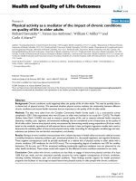
The upper panel of figure 1 shows a western blot detecting
iNOS in PC12 cells treated simultaneously with 10 ng/ml
NGF and 10 ng/ml TNFα in the presence or absence of 50
nM K252a, an inhibitor of phosphorylative events associ-
ated with tyrosine kinase receptor activation that has been
shown to block the function of the high affinity NGF
receptor TrkA [61]. There was a marked induction of iNOS
expression only in cells simultaneously treated with NGF
and TNFα, while neither treatment alone elicited any
effect. Furthermore, K252a completely abolished NGF/
TNFα-promoted iNOS induction, suggesting that TrkA
function is essential to mediate it. As shown in the lower
panel of figure 1, along with increased protein levels there
was also an induction of iNOS mRNA in PC12 cells
treated with NGF and TNFα but not in cells treated with
either factor alone.
NGF and TNF
α
are both required for sustained iNOS
expression
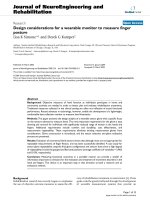
Figure 2A shows western blots detecting iNOS in cells
treated with increasing concentrations of NGF (top panel)
or TNFα (bottom panel), in the presence or absence of a
fixed amount of TNFα or NGF, respectively. Either factor
was ineffective when added alone at any of the concentra-
tions tested. However, there was a marked dose-response
increase in iNOS expression when increasing concentra-
tions of NGF or TNFα were added in the presence of a
fixed amount of TNFα or NGF, respectively. Figure 2B
Journal of Neuroinflammation 2005, 2:19 />Page 4 of 13
(page number not for citation purposes)
shows a representative western blot detecting iNOS
expression in cells continuously treated with NGF and
TNFα as compared to cells in which the combined treat-
ment was withdrawn after 24 hr. The expression of iNOS
returned to basal, undetectable, levels between 24 and 48
hr after withdrawal of both TNFα and NGF. Furthermore,
as shown in figure 2C, withdrawal of either NGF or TNFα
alone was sufficient to abolish iNOS expression induced
by the combined treatment, both at the protein (top
panel) and mRNA level (bottom panel). To exclude the
involvement of unknown serum factors, NGF/TNFα-pro-
moted induction of iNOS was determined in cells cul-
tured for 24 hr in serum free or in defined medium N2
(Figure 2D). There was a detectable iNOS induction in
both serum free- and defined medium-cultured cells,
although much reduced in serum free conditions, which
is predictable as PC12 cells do not survive for longer peri-
ods of time (24–48 hrs) in the absence of serum or N2
supplements. Since insulin is present in both serum and
the N2 supplement, and can activate the insulin-like
growth factor (IGF) receptor, we asked whether TNFα may
synergize with IGF, which is also present in serum, to
induce iNOS expression. The results shown in Figure 2E
indicate that this is not the case.
TNF
α
/NGF-mediated iNOS expression is independent of
NOS enzymatic activity
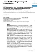
In order to determine whether the enzymatic activity of
iNOS may play a role in sustaining TNFα/NGF-promoted
signaling we pretreated PC12 cells with two NOS inhibi-
tors prior to TNFα/NGF tretament. Pretreatment with
N(G)-nitro-L-arginine methyl ester (L-NAME) did not
affect expression of iNOS induced by the NGF/ TNFα
combined treatment (Figure 3A). The same result was
observed if a more specific inhibitor of iNOS (1400 W)
was used instead of L-NAME (Figure 3B). Concentrations
of 1400 W used here have been previously shown to be
effective in inhibiting selectively iNOS activity in PC12
cells by others [78]. These results suggest that sustained
iNOS expression in response of the combined NGF/TNFα
treatment is independent of NOS enzymatic activity.
NGF/TNF
α
promoted iNOS induction requires the
transcription factor NF-
κ
B
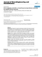
Figure 4 shows results from PC12 cells transiently trans-
fected with a secreted alkaline phosphatase reporter gene
construct (SEAP) promoted by enhancer sequences spe-
cific for nuclear factor kappa B (NF-κB), activator protein
1 (AP-1), cAMP-responsive element (CRE) or Tal (non-
inducible control). Twenty-four hr after transfection cells
were treated with 10 ng/ml each of TNFα and NGF (alone
or combined) and SEAP released in the culture medium
(an index of endogenous transcription factor activation)
was assayed 3 hr and 12 hr later. At 3 hr, cells treated with
TNFα showed a significant increase in NF-κB activity but
not AP-1 or CRE. Cells treated with NGF alone showed at
3 hr no significant increase in NF-κB, AP1 or CRE activity.
When cells were exposed to the combined NGF/ TNFα
treatment, there was a robust increase in NF-κB activity
that was significantly higher than the response induced by
the individual treatment with TNFα. On the other hand,
neither AP-1 nor CRE activity were significantly affected
by the combined NGF/ TNFα treatment. At 12 hr, both
TNFα and NGF/TNFα combined treatments significantly
increased NF-κB activity, but were not statistically signifi-
cantly different. NGF-treated cells showed a significant
increase in AP-1 and CRE activity at 12 hr, while NF-κB
activity was not affected. As a result, there was also a sig-
nificant increase in AP-1 and CRE activity elicited by the
NGF/TNFα combined treatment at 12 hr. Neither NGF
nor TNFα (alone or combined) elicited any effect on the
control reporter construct Tal, either at 3 or 12 hr.
Involvement of NF-κB was further explored by determin-
ing the extent to which pharmacological inhibition of NF-
κB would block NGF/TNFα-promoted iNOS induction in
PC12 cells. As shown in figure 5A, treatment of PC12 cells
with either pyrrolidine di-thio-carbamate (PDTC) or the
octapeptide proteasome inhibitor PSI (two effective NF-
κB inhibitors that have distinct mechanisms of action
A: (Top) Western blot analysis detecting the presence of iNOS in 40 µg total protein extracts from PC12 cells treated for 24 hr with 10 ng/ml NGF and 10 ng/ml TNF, individually or combined (Both), in the presence of 50 nM of the recep-tor tyrosine kinase inhibitor K252aFigure 1
A: (Top) Western blot analysis detecting the presence of
iNOS in 40 µg total protein extracts from PC12 cells treated
for 24 hr with 10 ng/ml NGF and 10 ng/ml TNF , individually
or combined (Both), in the presence of 50 nM of the recep-
tor tyrosine kinase inhibitor K252a. Positive control (Pos) is
4 µg of total protein extracts from mouse macrophages.
(Bottom) RT-PCR detecting iNOS mRNA in PC12 cells
treated for 24 hr with 10 ng/ml NGF and 10 ng/ml TNF ,
individually or combined (Both) compared to untreated cells
(Cont). Internal PCR controls lacking reverse transcriptase
(RT-) were performed on each sample as shown. Results
shown are representative of 3 replicate experiments.
α
.
α
.
Journal of Neuroinflammation 2005, 2:19 />Page 5 of 13
(page number not for citation purposes)
A: Western blots detecting iNOS in total protein extracts from PC12 cells treated for 24 hr with increasing concentrations of NGF in the presence or absence of 10 ng/ml TNFα (Top) or treated with increasing concentrations of TNFα in the presence or absence of 25 ng/ml NGF (Bottom)Figure 2
A: Western blots detecting iNOS in total protein extracts from PC12 cells treated for 24 hr with increasing concentrations of
NGF in the presence or absence of 10 ng/ml TNFα (Top) or treated with increasing concentrations of TNFα in the presence
or absence of 25 ng/ml NGF (Bottom). Positive control (Pos) is 4 µg of total protein extracts from mouse macrophages.
Results shown are representative of 2 replicate experiments. B: Western blot analysis detecting iNOS in total protein extracts
from PC12 cells simultaneously pre-treated with 10 ng/ml NGF and 10 ng/ml TNFα. At 24 hr treatment was withdrawn and
the presence of iNOS was determined 24, 48, and 72 hr thereafter. Results shown are representative of 3 replicate experi-
ments. C: Western blot analysis (Top) and RT-PCR (Bottom) detecting iNOS protein and mRNA in total protein extracts
and total RNA from PC12 cells simultaneously pre-treated for 24 hr with 10 ng/ml NGF and 10 ng/ml TNFα (Both). After 24
hr, treatment was withdrawn and replaced with either NGF or TNFα alone or with both and iNOS expression determined 24
hr thereafter. Results shown are representative of 2 replicate experiments. D: Western blot detecting iNOS in total protein
extracts from PC12 cells simultaneously treated for 24 hr with 10 ng/ml NGF and 10 ng/ml TNFα in medium containing serum,
in serum free medium (SF) or in defined medium (N2). Results shown are representative of 3 replicate experiments. E: West-
ern blot analysis detecting the presence of iNOS in total protein extracts from PC12 cells treated for 72 hr with 100 ng/ml IGF
and 10 ng/ml TNF , individually or combined, as compared to cells simultaneously treated with 10 ng/ml NGF and 10 ng/ml
TNFα or untreated controls (Cont). Results shown are representative of 4 replicate experiments.
α
.
Journal of Neuroinflammation 2005, 2:19 />Page 6 of 13
(page number not for citation purposes)
[8,63-65], completely abolished NGF/ TNFα-promoted
iNOS induction. In this experiment, PD98059, a selective
MAPK inhibitor, was used as a negative control. Both NF-
κB inhibitors effectively blocked NF-κB-mediated tran-
scriptional activity as determined by SEAP reporter gene
assay (Figure 5B), whereas PD98059 had no effect. How-
ever, PD98059 completely blocked NGF-promoted neur-
ite outgrowth (Figure 5C), an event that in PC12 cells is
dependent on MAPK activation [66]. Furthermore, con-
sistent with the results reported in Figure 4, inhibition of
NOS activity by L-NAME did not affect NFκB activation by
NGF/TNFα combined treatment (Figure 5D).
NGF/TNF
α
-promoted iNOS induction requires the
simultaneous presence of both the p75NTR and TrkA NGF
receptors
Next, we subcloned a PC12 mutant cell line (PC12
p75NTR
(-)
) that lacks p75NTR expression while retaining TrkA at
levels comparable with wild type PC12 cells (Figure 6A).
NF-κB activity was not significantly increased by the NGF/
TNFα combined treatment over the levels induced by
TNFα alone in PC12
p75NTR (-)
(Figure 6B). Consistent with
this finding, PC12
p75NTR (-)
cells exposed to the combined
NGF/TNFα treatment did not show any induction of
iNOS expression as compared to the parent cell line (Fig-
ure 6C). It is important to note that the PC12
p75NTR (-)
cells
used here express TNFα receptor type 1 (TNFR1) at levels
comparable (or even higher) than wild type PC12 cells
(Figure 6D). Therefore lack of iNOS induction by the
A: Western blot detecting iNOS in total protein extracts from PC12 cells treated for 24 hr with 10 ng/ml NGF and 10 ng/ml TNFα, either individually or simultaneously (Both)Figure 3
A: Western blot detecting iNOS in total protein extracts
from PC12 cells treated for 24 hr with 10 ng/ml NGF and 10
ng/ml TNFα, either individually or simultaneously (Both).
Cells were pretreated with vehicle or 0.5 µM of the generic
NOS inhibitor L-NAME. Positive control (Pos) is 4 µg of
total protein extracts from mouse macrophages. Results
shown are representative of 3 replicate experiments. B:
Western blot detecting iNOS in total protein extracts from
PC12 cells simultaneously treated with 10 ng/ml NGF and 10
ng/ml TNFα (Both), in the presence or absence of a pre-
treatment with varying concentrations of the iNOS-specific
inhibitor 1400 W. Results shown are representative of 4 rep-
licate experiments.
Detection of SEAP in the culture medium of PC12 cells transfected with a SEAP reporter gene construct under the transcriptional control of enhancers specific for NF-κB, AP-1 or CREFigure 4
Detection of SEAP in the culture medium of PC12 cells
transfected with a SEAP reporter gene construct under the
transcriptional control of enhancers specific for NF-κB, AP-1
or CRE. pTal is the non-enhanced control SEAP reporter
vector. Twenty-four hr after transfection, cells were treated
with vehicle (Control), 10 ng/ml NGF, 10 ng/ml TNFα or
NGF plus TNFα (Both) and the presence of SEAP in the cul-
ture medium assayed 3 hr (Top) or 12 hr (Bottom) there-
after. Results are normalized to control cells in each
transfection group (N = 3). * and #: p < 0.05 vs. control and
TNFα-alone, respectively (two-tailed unpaired Student's t-
test). Results shown are representative of 3 replicate
experiments.
Journal of Neuroinflammation 2005, 2:19 />Page 7 of 13
(page number not for citation purposes)
A: Western blot detecting iNOS in PC12 cells simultaneously treated with 10 ng/ml NGF and 10 ng/ml TNFα for 24 hrFigure 5
A: Western blot detecting iNOS in PC12 cells simultaneously treated with 10 ng/ml NGF and 10 ng/ml TNFα for 24 hr. Thirty
minutes before NGF/ TNFα treatment cells were pre-treated with 10 µM pyrrolidinedithyocarbamate (PDTC), 2 µM of a oli-
gopeptide proteosome inhibitor (PSI) or 10 µM of a MAPK inhibitor (PD98059). Results shown are representative of 2 repli-
cate experiments. B: SEAP release in the culture medium of PC12 cells transfected for 24 hr with an NF-κB-sensitive SEAP
reporter gene construct and treated for 12 hr with vehicle (Control), 10 ng/ml NGF, 10 ng/ml TNFα or NGF plus TNFα in the
presence of 10 µM PD98059, 10 µM PDTC or 2 µM PSI. Data are shown as mean ± S.E.M. from 3 independent replicate exper-
iments. * and #: p < 0.05 vs. control or TNFα-alone cells, respectively (two-tailed unpaired Student's t-test). C: Representative
photomicrographs of PC12 cells treated for 48 hr with 10 ng/ml NGF in the presence or absence of 10 µM PD98059 or 2 µM
PDTC. D: NFκB transcriptional activity (as measured by a transiently transfected SEAP reporter vector) in PC12 cells treated
for 24 hr with 10 ng/ml NGF, 10 ng/ml TNFα or NGF plus TNFα (Both) in the presence of 0.5 µM L-NAME. Data are shown
as mean ± S.E.M. from 3 independent replicate experiments. * and #: p < 0.05 vs. control or TNFα-alone cells, respectively
(two-tailed unpaired Student's t-test).
Journal of Neuroinflammation 2005, 2:19 />Page 8 of 13
(page number not for citation purposes)
NGF/TNFα combined treatment in these cells cannot be
ascribed to lack of TNFα responsiveness (as can also be
appreciated by the NFκB response induced by TNFα alone
shown in figure 6B).
The results obtained in PC12
p75NTR(-)
would suggest that
p75NTR is essential to mediate iNOS induction by the
combined TNFα/NGF treatment while the results
obtained using K252a (Figure 1) would suggest a promi-
nent role for TrkA. In order to ultimately ascertain the rel-
ative role of the two NGF receptors in mediating TNFα/
NGF-promoted iNOS induction we made use of PC12
cells transiently transfected with expression vectors coding
for chimeric TNFα/NGF receptors constructed as
described by Rovelli et al. [77]. These constructs bear the
ligand binding domain from the human TNFR1 and the
signal transduction domain from rat NGF receptors, either
TrkA or p75NTR. Previously, it has been shown that trans-
fection with these chimeras allows for TNF-promoted
NGF signaling [77]. Figure 7 shows a western blot detect-
ing iNOS in PC12 cells individually or simultaneously
transfected with chimeric TNFα receptors bearing the
intracellular domain of p75NTR (p55/p75NTR) or TrkA
(p55/TrkA). Transfected cells were then treated either with
TNFα and NGF alone, or with both TNFα and NGF. As
expected, the combined TNFα/NGF treatment induced a
robust expression of iNOS in these PC12 cells, regardless
of the presence of any transfected expression vector. As
also expected, NGF alone did not elicit iNOS expression
in any of the transfected cells. Similarly, TNFα alone did
not induce iNOS in cells transfected with either p55/
p75NTR or p55/TrkA chimeric receptors. However, TNFα
promptly induced iNOS expression in cells transfected
with both p55/p75NTR and p55/TrkA chimeric receptors.
Discussion
The work presented here stems from our original observa-
tion that iNOS expression and subsequent NO produc-
tion can be synergistically induced by NGF and TNFα in a
TrkA-dependent manner in PC12 cells [43]. Our present
results investigated the signalling pathways involved.
Since we consistently observed a higher iNOS expression
if NGF is added simultaneously to TNFα, we propose that
iNOS expression was induced selectively in NGF-respon-
sive cells. These results do not allow us to rule out the
possibility that intermediate factors induced by TNFα or
NGF may play a role in sensitizing indirectly cells to NGF
or TNFα, respectively. However, the results shown in Fig-
ure 2 seem to exclude such a possibility. Indeed, while
withdrawal of NGF and/or TNFα allows for a prompt
ablation of iNOS expression (Figure 2B), neither NGF nor
TNFα alone is sufficient to sustain iNOS expression fol-
lowing withdrawal of TNFα or NGF (Figure 2C). These
observations suggest that the simultaneous and continu-
ous presence of both factors is required to sustain iNOS
induction/expression and that cell sensitization through a
priming mechanism seems unlikely. Nonetheless, other
researchers have attributed increased TNFα toxicity in
PC12 cells to NGF-induced differentiation [67]. However,
our results seem to exclude that differentiation of PC12
cells may have played a role. First, in our experimental
conditions iNOS expression occurs as early as 3 hr after
the exposure to the combined NGF/TNFα treatment [43],
earlier than any morphological differentiation induced by
NGF. Second, while blockade of NGF-induced differenti-
ation by the MAPK inhibitor PD98059 (Figure 5C, [68])
had no effect on NGF/TNFα-promoted iNOS expression
(Figure 5A), blockade of NFκB did not affect NGF-
induced differentiation (Figure 5C) but completely inhib-
ited iNOS expression.
In the present study we also report that induction and
maintenance of iNOS expression by the combined NGF/
TNFα treatment requires continuous de novo iNOS mRNA
synthesis, presumably due to transcription factor regula-
tion. Indeed, abolishing iNOS enzymatic activity had no
effect on NGF/TNFα-promoted iNOS induction (Figure
4A,B). Therefore, the involvement of positive feedback
due to NO seems unlikely. On the other hand, analysis of
transcriptional activity of NF-κB, AP-1 and CRE revealed
that NF-κB most likely mediates synergistic iNOS induc-
tion by TNFα and NGF. Since iNOS induction can be
observed as early as 3 hr after NGF/TNFα combined treat-
ment in PC12 cells [43], the results shown in figure 5 sug-
gest that NF-κB is the only transcription factor among
those tested here that is responsive to the simultaneous
treatment with TNFα and NGF in a fashion consistent
with induction of iNOS expression. In fact, while TNFα
alone induced NFκB at 3 hr, this induction was signifi-
cantly lower than the one promoted by the combined
NGF/TNFα treatment. Whether the extent to which NFκB
is activated or whether qualitative differences in NFκB
subunit composition in response to TNFα as compared to
NGF/TNFα treatment may play a role in inducing iNOS
expression remains to be established. Nonetheless, inhibi-
tion of NF-κB completely inhibited iNOS induction while
inhibition of MAPK was ineffective (Figure 5A). Lastly,
inhibition of NOS activity failed to block NGF/TNFα-pro-
moted NFκB activation, thus further supporting the idea
that targeting NO may acutely ameliorate associated oxi-
dative stress, but could not represent the most
comprehensive approach to achieve a long term correc-
tion of these events.
Previous studies indicated that NGF can induce NF-κB by
acting through the low affinity p75
NTR
receptor [70]. Thus,
involvement of NF-κB in mediating NGF/TNFα combined
effects would suggest a role for p75NTR. Indeed, we found
that mutant PC12 cells that lack expression of the p75NTR
receptor failed to respond in terms of iNOS expression
Journal of Neuroinflammation 2005, 2:19 />Page 9 of 13
(page number not for citation purposes)
A: Graph depicting the percentage of TrkA- or p75NTR- immunopositive cells in wild type (wt)PC12 cells and PC12 cell mutants lacking the low affinity NGF receptor (PC12
p75NTR(-)
) from flow cytometry dataFigure 6
A: Graph depicting the percentage of TrkA- or p75NTR- immunopositive cells in wild type (wt)PC12 cells and PC12 cell
mutants lacking the low affinity NGF receptor (PC12
p75NTR(-)
) from flow cytometry data. Results shown are representative of 3
replicate flow cytometry experiments on the same cell line. B: SEAP release in the culture medium of PC12
p75NTR (-)
cells trans-
fected for 24 hr with an NF-κB-sensitive SEAP reporter gene construct and treated for 12 hr with vehicle (Cont), 10 ng/ml
NGF, 10 ng/ml TNFα or NGF plus TNFα (Both). Data are shown as mean ± S.E.M. from 3 independent replicate experiments.
* : p < 0.05 vs. control or NGF-alone cells (two-tailed unpaired Student's t-test). C: Western blot detecting the presence of
iNOS in wtPC12 cells and PC12
p75NTR (-)
cells treated for 24 hr with vehicle (Cont), 10 ng/ml NGF, 10 ng/ml TNFα or NGF
plus TNFα (Both). Membrane was re-probed for β-actin (lower panel) to control for equal protein loading. Positive control
(Pos) is 4 µg of total protein extracts from mouse macrophages. Results shown are representative of 4 replicate experiments.
D: Western blot detecting the presence of TNFR-I in total protein extracts from wtPC12 cells and PC12
p75NTR (-)
cells. Twenty
µg of total protein extracts from rat dorsal root ganglia (DRG) were used as a positive control.
Journal of Neuroinflammation 2005, 2:19 />Page 10 of 13
(page number not for citation purposes)
when simultaneously treated with NGF and TNFα. Con-
sistent with this finding, in PC12 cell mutants lacking
p75NTR expression NF-κB activity was not induced by the
combined NGF/TNFα treatment above the levels
observed in cells treated with TNFα alone (Figure 6B).
That PC12 cells bearing only the TrkA receptor failed to
respond the combined NGF/TNFα treatment suggests that
signaling from p75NTR in combination with TNFα is nec-
essary to induce iNOS expression. On the other hand, our
previous work illustrated the importance of TrkA-associ-
ated signaling in mediating NGF/TNFα-promoted
induction of iNOS [43] (see also figure 1). These results
are only apparently in contrast. Indeed, in an admittedly
artificial system making use of chimeric constructs we
observed that only in the presence of both TNFα-respon-
sive NGF receptor signaling can TNFα promote iNOS
expression when added alone. Whether this is a conse-
quence of simultaneous but independent signaling of
both types of NGF receptors [79] or recruitment of intrac-
ellular signalling elements uniquely driven by the simul-
taneous activation of both NGF receptors' signaling
domains remains to be investigated. On the other hand,
these results exclude the possibility that the combined
action of TNFα and NGF may derive from yet undescribed
interaction(s) of the extracellular domains of their respec-
tive receptors following ligand binding.
Thus, our combined results would indicate that there
exists a specific pathway involving NF-κB and requiring
the simultaneous expression or both types of NGF recep-
tors that is synergistically induced by TNFα and NGF to
promote expression of iNOS. This is of particular interest
given that neuron types expressing both TrkA and
p75NTR receptors are limited and known to be affected in
neurodegenerative conditions where neuroinflammation
and pro-inflammatory cytokines have been shown to play
a significant role. Notably, simultaneous expression of
TrkA and p75NTR in the CNS is mostly restricted to the
BFCN that are known to be particularly affected in AD.
Indeed, others have also described signaling pathways
that require the simultaneous expression of both TrkA and
p75NTR [71,72] as well as the convergence of TrkA and
p75NTR-mediated signaling impinging upon NF-κB [73].
Western blot detecting iNOS in 40 µg total protein extracts from PC12 cells treated for 24 hr with 10 ng/ml human TNFα, 10 ng/ml NGF, or bothFigure 7
Western blot detecting iNOS in 40 µg total protein extracts from PC12 cells treated for 24 hr with 10 ng/ml human TNFα, 10
ng/ml NGF, or both. Twenty-four hr before treatment, cells were transfected with either an empty vector or expression vec-
tors for chimeric receptor proteins bearing the human TNFR1 ligand binding domains and the intracellular domain of either rat
p75
NTR
or TrkA NGF receptors (p75
NTR
, TrkA or p75NTR+TrkA). Positive control (Pos) is 40 µg of total protein extract
from wild type PC12 cells treated with both rat TNFα and NGF. Membrane was re-probed for β-actin (lower panel) to control
for equal protein loading and is representative from 3 independent transfections and treatments.
Journal of Neuroinflammation 2005, 2:19 />Page 11 of 13
(page number not for citation purposes)
Recent reports in neurons of TNF-promoted signaling
occurring selectively in the presence of the glutamate
agonist NMDA [4] illustrate the importance of
considering the signaling "context" when studying the
effects of cytokine treatment.
Overall, our data indicate the possibility that a conver-
gence between NGF-promoted trophic signaling and
TNFα could selectively endanger NGF-responsive neurons
under conditions of neuroinflammation because of a
synergistic action between TNFα and NGF to induce iNOS
expression. For example, TNFα overexpressing transgenic
mice show selective neurodegeneration of NGF-respon-
sive basal forebrain cholinergic neurons [57] and direct
TNFα administration in the brain of mice results in an
impairment of basal forebrain cholinergic function [58].
However, whether induction of iNOS and subsequent oxi-
dative damage may play a role in these two models
remains to be determined [80].
Conclusion
TNFα and NGF, via concerted signaling events involving
NFκB transcriptional activity and targeting NGF-respon-
sive cells bearing both the high and low affinity NGF
receptors, converge to stimulate de novo transcription of
iNOS. Our present results are relevant to neurodegenera-
tive conditions such as AD [22,74], stroke [17,75], ALS
[20,76] and spinal chord injury [8,10] where neuroin-
flammation and high levels of pro-inflammatory
cytokines have been shown to play a significant role and
proposed as therapeutic targets.
List of Abbreviations
AraC, cytosine β-D-arabinofuranoside; AD, Alzheimers
disease; BDNF, brain derived neurotrophic factor; BFCN,
basal forebrain cholinergic neurons; CNS, central nervous
system; CRE, cyclic-AMP response element; GDNF, glial
derived neurotrophic factor; IGF, insulin-like growth fac-
tor; IL-1β, interleukin-1beta; iNOS, inducible nitric oxide
synthase; MAPK, mitogen activated protein kinase; NF-κB,
nuclear factor kappa B; NGF, nerve growth factor; NO,
nitric oxide; nNOS, neuronal nitric oxide synthase; NTR,
neurotrophin receptor; PC12, pheochromocytoma; PCN,
penicillin; PDTC, pyrrolidinedithyocarbamate; PSI, prote-
osome inhibitor; SDS, sodium dodecylsulfate; SEAP,
secreted alkaline phosphatase; S.E.M, standard error of the
mean; Strep, streptomycin; TNFα, tumor necrosis factor
alpha; TrkA, troponin-like receptor kinase A; TTBS, tris-
buffered saline with tween 20;
Competing interests
The author(s) declare they have no competing interests.
Authors' contributions
MST participated in the conception and design of the
study, carried out the bulk of experiments, performed data
analysis, and drafted the manuscript. PMJ participated in
study design especially with regards to the IGF
experiments. WZ participated in study design and coordi-
nation and provided the expertise for RTPCR and with-
drawal experiments. HUS sub-cloned the PC12
p75NTR(-)
cells and participated in study design and result interpre-
tation of experiments involving these cells. GT partici-
pated in conception, study design, coordination and
helped to draft and review the manuscript. All authors
read and approved the final manuscript.
Acknowledgements
This work was supported in part by a research development grant by the
UTMB Sealy Endowed Fund for Biomedical Research. Michael Thomas is
supported by an NIEHS training grant pre-doctoral fellowship from T32
ES007254 and the UTMB Sealy Center for Aging pre-doctoral fellowship.
References
1. Floyd RA: Neuroinflammatory processes are important in
neurodegenerative diseases: an hypothesis to explain the
increased formation of reactive oxygen and nitrogen species
as major factors involved in neurodegenerative disease
development. Free Radic Biol Med 1999, 26:1346-55.
2. McGeer PL, McGeer EG: Innate immunity, local inflammation,
and degenerative disease. Sci Aging Knowledge Environ 2002,
2002:re3.
3. Creange A, Lefaucheur JP, Authier FJ, Gherardi RK: Cytokines and
peripheral neuropathies. Revue Neurologique 1998, 154:208-216.
4. Sung YJ, Ambron RT: Pathways that elicit long-term changes in
gene expression in nociceptive neurons following nerve
injury: contributions to neuropathic pain. Neurol Res 2004,
26:195-203.
5. Chandross KJ: Nerve injury and inflammatory cytokines mod-
ulate gap junctions in the peripheral nervous system. Glia
1998, 24:21-31.
6. Stoll G, Jander S, Myers RR: Degeneration and regeneration of
the peripheral nervous system: from Augustus Waller's
observations to neuroinflammation. J Peripher Nerv Syst 2002,
7:13-27.
7. Norenberg MD, Smith J, Marcillo A: The pathology of human spi-
nal cord injury: defining the problems. J Neurotrauma 2004,
21:429-40.
8. La Rosa G, Cardali S, Genovese T, Conti A, Di Paola R, La Torre D,
Cacciola F, Cuzzocrea S: Inhibition of the nuclear factor-kappaB
activation with pyrrolidine dithiocarbamate attenuating
inflammation and oxidative stress after experimental spinal
cord trauma in rats. J Neurosurg Spine 2004, 1:311-21.
9. Popovich PG, Jones TB: Manipulating neuroinflammatory reac-
tions in the injured spinal cord: back to basics. Trends Pharma-
col Sci 2003, 24:13-7.
10. Hausmann ON: Post-traumatic inflammation following spinal
cord injury. Spinal Cord 2003, 41:369-78.
11. Bareyre FM, Schwab ME: Inflammation, degeneration and
regeneration in the injured spinal cord: insights from DNA
microarrays. Trends Neurosci 2003, 26:555-63.
12. Bayir H, Kochanek PM, Clark RS: Traumatic brain injury in
infants and children: mechanisms of secondary damage and
treatment in the intensive care unit. Crit Care Clin 2003,
19:529-49.
13. Morganti-Kossmann MC, Rancan M, Stahel PF, Kossmann T: Inflam-
matory response in acute traumatic brain injury: a double-
edged sword. Curr Opin Crit Care 2002, 8:101-5.
14. Lenzlinger PM, Morganti-Kossmann MC, Laurer HL, McIntosh TK:
The duality of the inflammatory response to traumatic brain
injury. Mol Neurobiol 2001, 24:169-81.
Journal of Neuroinflammation 2005, 2:19 />Page 12 of 13
(page number not for citation purposes)
15. Sundararajan S, Landreth GE: Antiinflammatory properties of
PPARgamma agonists following ischemia. Drug News Perspect
2004, 17:229-36.
16. Dirnagl U: Inflammation in stroke: the good, the bad, and the
unknown. Ernst Schering Res Found Workshop 2004:87-99.
17. Danton GH, Dietrich WD: Inflammatory mechanisms after
ischemia and stroke. J Neuropathol Exp Neurol 2003, 62:127-36.
18. Consilvio C, Vincent AM, Feldman EL: Neuroinflammation, COX-
2, and ALS – a dual role? Exp Neurol 2004, 187:1-10.
19. Pompl PN, Ho L, Bianchi M, McManus T, Qin W, Pasinetti GM: A
therapeutic role for cyclooxygenase-2 inhibitors in a trans-
genic mouse model of amyotrophic lateral sclerosis. Faseb J
2003, 17:725-7.
20. McGeer PL, McGeer EG: Inflammatory processes in amyo-
trophic lateral sclerosis. Muscle Nerve 2002, 26:459-70.
21. Cacquevel M, Lebeurrier N, Cheenne S, Vivien D: Cytokines in
neuroinflammation and Alzheimer's disease. Curr Drug Targets
2004, 5:529-34.
22. McGeer EG, McGeer PL: Inflammatory processes in Alzhe-
imer's disease. Prog Neuropsychopharmacol Biol Psychiatry 2003,
27:741-9.
23. Gupta A, Pansari K: Inflammation and Alzheimer's disease. Int
J Clin Pract 2003, 57:36-9.
24. McGeer PL, McGeer EG: Local neuroinflammation and the pro-
gression of Alzheimer's disease. J Neurovirol 2002, 8:529-38.
25. Emerit J, Edeas M, Bricaire F: Neurodegenerative diseases and
oxidative stress. Biomed Pharmacother 2004, 58:39-46.
26. Tarkowski E, Liljeroth AM, Minthon L, Tarkowski A, Wallin A, Blen-
now K: Cerebral pattern of pro- and anti-inflammatory
cytokines in dementias. Brain Res Bull 2003, 61:255-60.
27. Tarkowski E, Andreasen N, Tarkowski A, Blennow K: Intrathecal
inflammation precedes development of Alzheimer's disease.
J Neurol Neurosurg Psychiatry 2003, 74:1200-5.
28. McGeer PL, McGeer EG, Suzuki J, Dolman CE, Nagai T: Aging,
Alzheimer's disease, and the cholinergic system of the basal
forebrain. Neurology 1984, 34:741-5.
29. Rinne JO, Paljarvi L, Rinne UK: Neuronal size and density in the
nucleus basalis of Meynert in Alzheimer's disease. J Neurol Sci
1987, 79:67-76.
30. Vogels OJ, Broere CA, ter Laak HJ, ten Donkelaar HJ, Nieuwenhuys
R, Schulte BP: Cell loss and shrinkage in the nucleus basalis
Meynert complex in Alzheimer's disease. Neurobiology of Aging
1990, 11:3-13.
31. Hefti F: Nerve growth factor promotes survival of septal
cholinergic neurons after fimbrial transections. J Neurosci
1986, 6:2155-62.
32. Hartikka J, Hefti F: Development of septal cholinergic neurons
in culture: plating density and glial cells modulate effects of
NGF on survival, fiber growth, and expression of transmit-
ter-specific enzymes. J Neurosci 1988, 8:2967-85.
33. Perry G, Castellani RJ, Smith MA, Harris PL, Kubat Z, Ghanbari K,
Jones PK, Cordone G, Tabaton M, Wolozin B, et al.: Oxidative dam-
age in the olfactory system in Alzheimer's disease. Acta Neu-
ropathol (Berl) 2003, 106:552-6.
34. Butterfield DA, Boyd-Kimball D, Castegna A: Proteomics in Alzhe-
imer's disease: insights into potential mechanisms of
neurodegeneration. J Neurochem 2003, 86:1313-27.
35. Giasson BI, Ischiropoulos H, Lee VM, Trojanowski JQ: The relation-
ship between oxidative/nitrative stress and pathological
inclusions in Alzheimer's and Parkinson's diseases. Free Radic
Biol Med 2002, 32:1264-75.
36. Perry G, Nunomura A, Hirai K, Takeda A, Aliev G, Smith MA: Oxi-
dative damage in Alzheimer's disease: the metabolic
dimension. Int J Dev Neurosci 2000, 18:417-21.
37. Gold BG, Udina E, Bourdette D, Navarro X: Neuroregenerative
and neuroprotective actions of neuroimmunophilin com-
pounds in traumatic and inflammatory neuropathies. Neurol
Res 2004, 26:371-80.
38. Rogerio F, Teixeira SA, de Rezende AC, de Sa RC, de Souza Queiroz
L, De Nucci G, Muscara MN, Langone F: Superoxide dismutase
isoforms 1 and 2 in lumbar spinal cord of neonatal rats after
sciatic nerve transection and melatonin treatment. Brain Res
Dev Brain Res 2005, 154:217-25.
39. Kim HK, Park SK, Zhou JL, Taglialatela G, Chung K, Coggeshall RE,
Chung JM: Reactive oxygen species (ROS) play an important
role in a rat model of neuropathic pain. Pain 2004, 111:116-24.
40. van Beek J, Elward K, Gasque P: Activation of complement in the
central nervous system: roles in neurodegeneration and
neuroprotection. Ann N Y Acad Sci 2003, 992:56-71.
41. Murdoch I, Perry EK, Court JA, Graham DI, Dewar D: Cortical
cholinergic dysfunction after human head injury. J
Neurotrauma 1998, 15:295-305.
42. Murdoch I, Nicoll JA, Graham DI, Dewar D: Nucleus basalis of
Meynert pathology in the human brain after fatal head
injury. J Neurotrauma 2002, 19:279-84.
43. Macdonald NJ, Taglialatela G: Tumor necrosis factor alpha and
nerve growth factor synergistically induce iNOS in pheo-
chromocytoma cells. Neuroreport 2000, 11:3453-3456.
44. Isaksson J, Farooque M, Olsson Y: Improved functional outcome
after spinal cord injury in iNOS-deficient mice. Spinal Cord
2004.
45. Urushitani M, Shimohama S, Kihara T, Sawada H, Akaike A, Ibi M,
Inoue R, Kitamura Y, Taniguchi T, Kimura J: Mechanism of selec-
tive motor neuronal death after exposure of spinal cord to
glutamate: Involvement of glutamate induced nitric oxide in
motor neuron toxicity and nonmotor neuron protection.
Annals of Neurology 1998, 44:796-807.
46. Diaz-Ruiz A, Ibarra A, Perez-Severiano F, Guizar-Sahagun G, Grijalva
I, Rios C: Constitutive and inducible nitric oxide synthase
activities after spinal cord contusion in rats. Neurosci Lett 2002,
319:129-32.
47. Parmentier-Batteur S, Bohme GA, Lerouet D, Zhou-Ding L, Beray V,
Margaill I, Plotkine M: Antisense oligodeoxynucleotide to induc-
ible nitric oxide synthase protects against transient focal cer-
ebral ischemia-induced brain injury. Journal of Cerebral Blood
Flow & Metabolism 2001, 21:15-21.
48. Sarchielli P, Galli F, Floridi A, Gallai V: Relevance of protein nitra-
tion in brain injury: a key pathophysiological mechanism in
neurodegenerative, autoimmune, or inflammatory CNS dis-
eases and stroke. Amino Acids 2003, 25:427-36.
49. Luth HJ, Munch G, Arendt T: Aberrant expression of NOS iso-
forms in Alzheimer's disease is structurally related to nitro-
tyrosine formation. Brain Res 2002, 953:135-43.
50. Luth HJ: Expression of endothelial and inducible NOS-iso-
forms is increased in Alzheimer's disease, in APP23 trans-
genic mice and after experimental brain lesion in rat:
evidence for an induction by amyloid pathology. Brain Research
2001, 913:57-67.
51. Law A, Gauthier S, Quirion R: Say NO to Alzheimer's disease:
the putative links between nitric oxide and dementia of the
Alzheimer's type. Brain Res Brain Res Rev 2001, 35:73-96.
52. de la Monte SM, Lu BX, Sohn YK, Etienne D, Kraft J, Ganju N, Wands
JR: Aberrant expression of nitric oxide synthase III in Alzhe-
imer's disease: relevance to cerebral vasculopathy and
neurodegeneration. Neurobiology of Aging 2000, 21:309-19.
53. Lee S, Zhao ML, Hirano A, Dickson D: Inducible nitric oxide syn-
thase immunoreactivity in the Alzheimer disease hippocam-
pus: association with Hirano bodies, neurofibrillary tangles,
and senile plaques. Journal of Neuropathology and Experimental
Neurology 1999, 58:1163-1169.
54. Darville MI, Eizirik DL: Regulation by cytokines of the inducible
nitric oxide synthase promoter in insulin-producing cells.
Diabetologia 1998, 41:1101-8.
55. Kong GY, Peng ZC, Costanzo C, Kristensson K, Bentivoglio M:
Inducible nitric oxide synthase expression elicited in the
mouse brain by inflammatory mediators circulating in the
cerebrospinal fluid. Brain Res 2000, 878:105-18.
56. Barker V, Middleton G, Davey F, Davies AM: TNFalpha contrib-
utes to the death of NGF-dependent neurons during
development. Nature Neuroscience 2001, 4:1194-8.
57. Aloe L, Fiore M, Probert L, Turrini P, Tirassa P: Overexpression of
tumour necrosis factor alpha in the brain of transgenic mice
differentially alters nerve growth factor levels and choline
acetyltransferase activity. Cytokine 1999, 11:45-54.
58. Wenk GL, McGann K, Hauss-Wegrzyniak B, Rosi S: The toxicity of
tumor necrosis factor-alpha upon cholinergic neurons within
the nucleus basalis and the role of norepinephrine in the reg-
ulation of inflammation: implications for alzheimer's
disease. Neuroscience 2003, 121:719-29.
59. Kiss JZ, Wang C, Olive S, Rougon G, Lang J, Baetens D, Harry D,
Pralong WF: Activity-dependent mobilization of the adhesion
Publish with Bio Med Central and every
scientist can read your work free of charge
"BioMed Central will be the most significant development for
disseminating the results of biomedical research in our lifetime."
Sir Paul Nurse, Cancer Research UK
Your research papers will be:
available free of charge to the entire biomedical community
peer reviewed and published immediately upon acceptance
cited in PubMed and archived on PubMed Central
yours — you keep the copyright
Submit your manuscript here:
/>BioMedcentral
Journal of Neuroinflammation 2005, 2:19 />Page 13 of 13
(page number not for citation purposes)
molecule polysialic NCAM to the cell surface of neurons and
endocrine cells. Embo J 1994, 13:5284-92.
60. Stoppini L, Buchs PA, Muller D: A simple method for organotypic
cultures of nervous tissue. Journal of Neuroscience Methods 1991,
37:173-82.
61. Ohmichi M, Decker SJ, Pang L, Saltiel AR: Inhibition of the cellular
actions of nerve growth factor by staurosporine and K252A
results from the attenuation of the activity of the trk tyro-
sine kinase. Biochemistry 1992, 31:4034-9.
62. Pappas TC, Decorti F, Macdonald NJ, Neet KE, Taglialatela G:
Tumour necrosis factor-alpha- vs. growth factor depriva-
tion-promoted cell death: different receptor requirements
for mediating nerve growth factor-promoted rescue. Aging
Cell 2003, 2:83-92.
63. Taglialatela G, Robinson R, Perez-Polo JR: Inhibition of nuclear
factor kappa B (NFkappaB) activity induces nerve growth
factor-resistant apoptosis in PC12 cells. Journal of Neuroscience
Research 1997, 47:155-62.
64. Taglialatela G, Kaufmann JA, Trevino A, Perez-Polo JR: Central
nervous system DNA fragmentation induced by the inhibi-
tion of nuclear factor kappa B. Neuroreport 1998, 9:489-93.
65. Traenckner EB, Wilk S, Baeuerle PA: A proteasome inhibitor pre-
vents activation of NF-kappa B and stabilizes a newly phos-
phorylated form of I kappa B-alpha that is still bound to NF-
kappa B. Embo J 1994, 13:5433-41.
66. Pang L, Sawada T, Decker SJ, Saltiel AR: Inhibition of MAP kinase
kinase blocks the differentiation of PC-12 cells induced by
nerve growth factor. Journal of Biological Chemistry 1995,
270:13585-8.
67. Mielke K, Herdegen T: Fatal shift of signal transduction is an
integral part of neuronal differentiation: JNKs realize TNFal-
pha-mediated apoptosis in neuronlike, but not naive, PC12
cells. Mol Cell Neurosci 2002, 20:211-24.
68. Morooka T, Nishida E: Requirement of P38 Mitogen-Activated
Protein Kinase For Neuronal Differentiation in Pc12 Cells.
Journal of Biological Chemistry 1998, 273:24285-24288.
69. Perez-Polo JR, Foreman PJ, Jackson GR, Shan D, Taglialatela G,
Thorpe LW, Werrbach-Perez K: Nerve growth factor and neu-
ronal cell death. Molecular Neurobiology 1990, 4:57-91.
70. Carter BD, Kaltschmidt C, Kaltschmidt B, Offenhauser N, Bohm-Mat-
thaei R, Baeuerle PA, Barde YA: Selective activation of NF-kappa
B by nerve growth factor through the neurotrophin receptor
p75 [see comments]. Science 1996, 272:542-5.
71. Lad SP, Peterson DA, Bradshaw RA, Neet KE: Individual and com-
bined effects of TrkA and p75NTR nerve growth factor
receptors. A role for the high affinity receptor site. J Biol Chem
2003, 278:24808-17.
72. Szutowicz A, Madziar B, Pawelczyk T, Tomaszewicz M, Bielarczyk H:
Effects of NGF on acetylcholine, acetyl-CoA metabolism,
and viability of differentiated and non-differentiated cholin-
ergic neuroblastoma cells. J Neurochem 2004, 90:952-61.
73. Foehr ED, Lin X, O'Mahony A, Geleziunas R, Bradshaw RA, Greene
WC: NF-kappa B signaling promotes both cell survival and
neurite process formation in nerve growth factor-stimulated
PC12 cells. J Neurosci 2000, 20:7556-63.
74. Hoozemans JJ, Veerhuis R, Rozemuller AJ, Eikelenboom P: Non-ster-
oidal anti-inflammatory drugs and cyclooxygenase in Alzhe-
imer's disease. Curr Drug Targets 2003, 4:461-8.
75. Barone FC, Parsons AA: Therapeutic potential of anti-inflam-
matory drugs in focal stroke. Expert Opin Investig Drugs 2000,
9:2281-306.
76. Weydt P, Weiss MD, Moller T, Carter GT: Neuro-inflammation
as a therapeutic target in amyotrophic lateral sclerosis. Curr
Opin Investig Drugs 2002, 3:1720-4.
77. Rovelli G, Heller RA, Canossa M, Shooter EM: Chimeric tumor
necrosis factor-TrkA receptors reveal that ligand-dependent
activation of the TrkA tyrosine kinase is sufficient for differ-
entiation and survival of PC12 cells. Proc Natl Acad Sci U S A
1993, 90:8717-8721.
78. Jiang H, Koubi D, Zhang L, Kuo J, Rodriguez AI, Jackson Hunter T, et
al.: Inhibitors of iNOS protects PC12 cells against the apop-
tosis induced by oxygen and glucose deprivation. Neuroscience
Letters 2005, 375:59-63.
79. Woo SB, Page J, Saragovi HU, Neet KE: Binding of nerve growth
factor (NGF) to Trk receptor chimeras. Faseb Journal 2004,
18:C273.
80. Floden AM, Li SS, Combs CK: beta-Amyloid-stimulated micro-
glia induce neuron death via synergistic stimulation of tumor
necrosis factor alpha and NMDA receptors. Journal of
Neuroscience 2005, 25:2566-2575.