báo cáo hóa học: " Serum antibodies from Parkinson''''s disease patients react with neuronal membrane proteins from a mouse " ppt
Bạn đang xem bản rút gọn của tài liệu. Xem và tải ngay bản đầy đủ của tài liệu tại đây (361.36 KB, 9 trang )
BioMed Central
Page 1 of 9
(page number not for citation purposes)
Journal of Neuroinflammation
Open Access
Research
Serum antibodies from Parkinson's disease patients react with
neuronal membrane proteins from a mouse dopaminergic cell line
and affect its dopamine expression
Victor C Huber
1
, Tapan Mondal
1
, Stewart A Factor
2
, Richard F Seegal
1
and
David A Lawrence*
1
Address:
1
Wadsworth Center, New York State Department of Health, Albany, NY 12201, USA and
2
Parkinson's Disease & Movement Disorders
Center, Albany Medical College, Albany, NY 12208, USA
Email: Victor C Huber - ; Tapan Mondal - ; Stewart A Factor - ;
Richard F Seegal - ; David A Lawrence* -
* Corresponding author
Abstract
Evidence exists suggesting that the immune system may contribute to the severity of idiopathic
Parkinson's disease (IPD). The data presented here demonstrates that antibodies in the sera of
patients with IPD have increased binding affinity to dopaminergic (DA) neuronal (MN9D cell line)
membrane antigens in comparison to antibodies in sera from healthy controls. In general, the
degree of antibody reactivity to these antigens of the mouse MN9D cell line appears to correlate
well with the disease severity of the IPD patients contributing sera, based on the total UPDRS
scores. Surprisingly, the sera from IPD patients enhanced the DA content of MN9D cells
differentiated with n-butyrate; the n-butyrate-differentiated MN9D cells had a greater
concentration of DA (DA/mg total protein) than undifferentiated MN9D cells, especially early in
culture. Although the IPD sera did not directly harm MN9D cellular viability or DA production, in
the presence of the N9 microglial cell line, the amount of DA present in cultures of untreated or
n-butyrate-treated MN9D cells was lowered by the IPD sera. The results suggest the involvement
of antibodies in the decline of dopamine production and, thus, the potential of immune system
participation in IPD.
Introduction
Idiopathic Parkinson's disease (IPD) is a progressive neu-
rological disorder that affects approximately 1 million
people in North America [1,2]. It is characterized clini-
cally by a loss of motor control as evidenced by muscular
rigidity, resting tremor, bradykinesia, and gait dysfunction
with postural instability [1,2]. Pathological features
include, predominantly, the degeneration of dopaminer-
gic (DA) neurons within the substantia nigra (SN) and
intracytoplasmic inclusions (Lewy bodies) within surviv-
ing neurons [3]. To date, the cause of this disease remains
unknown [4]; however, certain gene mutations, e.g.,
alpha-synuclein, parkin, DJ1, LRKK2, PINK1, and ND5
have been implicated [5]. Expression of any of these
mutated genes may enhance the likelihood of IPD by
itself or after an environmental insult.
Although potentially only a consequence of IPD pathol-
ogy, abnormal immune activity has been considered a
possible cause of IPD based on post-mortem analysis of
Published: 20 January 2006
Journal of Neuroinflammation 2006, 3:1 doi:10.1186/1742-2094-3-1
Received: 16 November 2005
Accepted: 20 January 2006
This article is available from: />© 2006 Huber et al; licensee BioMed Central Ltd.
This is an Open Access article distributed under the terms of the Creative Commons Attribution License ( />),
which permits unrestricted use, distribution, and reproduction in any medium, provided the original work is properly cited.
Journal of Neuroinflammation 2006, 3:1 />Page 2 of 9
(page number not for citation purposes)
IPD patients' brains [6-8] and utilization of mouse mod-
els of parkinsonism [9-12]. Specifically, roles for both the
innate immune system, as evidenced by increased expres-
sion of pro-inflammatory cytokines [10,13-16], and the
adaptive immune system, in the form of increased levels
of neuron-specific antibodies in the sera of IPD patients
[17-24], have been posited.
To date, the strongest evidence for specific immune
involvement in the development of IPD was published by
Chen et al. when they reported a selective loss of DA neu-
rons within the SN region of rat brains upon administra-
tion of immunoglobulin (Ig) G from sera of patients with
IPD [25]. Furthermore, in later studies by the same group
[26,27], in vivo and in vitro models demonstrated an
important contribution of Fc receptor-bearing cells in the
induction of TNF-α, which, in turn, resulted in a reduc-
tion of DA neurons as evidenced by decreased tyrosine
hydroxylase (TH) activity [26]. However, there have been
no reports detailing the specific reactivities of IPD sera
with neuronal cell membrane antigens.
In this study, we set out to examine the interaction
between antibodies in sera from IPD patients and DA neu-
rons. We determined that serum IgG from IPD patients
react with membrane proteins from mouse MN9D neuro-
nal cells to a greater extent than serum IgG from healthy
control individuals. Additionally, we found that IPD sera
have differential modulatory effects on DA expression by
MN9D cells cultured in the presence and absence of N9
microglia. The observed interactions and their possible
implications are discussed.
Methods
Sera and IPD patients
During a routine office visit, IPD patients were asked if
they would consider participation in a research project to
evaluate their sera for antibodies to DA neurons in vitro.
The consent form was approved by the Institutional
Review Boards for Human Research of two institutions of
the investigators. Most control sera were from the spouses
of the IPD patients. Venous bloods were collected in
EDTA vacutainers, centrifuged to remove cells, and the
sera stored at -20°C until utilized as described. Clinical
information regarding the IPD patients is provided (Table
1).
Cell lines
The MN9D cell line (provided by Dr. Alfred Heller,
Department of Department of Pharmacological and Phys-
iological Sciences, University of Chicago) was derived
from rostral mesencephalic tegmentum (RMT) of the 14-
day-old embryonic mouse employing somatic cell fusion
techniques [28]. This clonal hybrid cell line expresses a
high amount of DA, which is efficiently depleted by N-
methyl-4-phenylpyridinium ion (MPP+), the active
metabolite of the neurotoxin N-methyl-4-phenyl-1,2,3,6-
tetrahydropyridine (MPTP). The N9 microglial cell line
(provided by Dr. P. Ricciardi-Castagnoli, Department of
Biotechnology and Bioscience, University of Milano-Bic-
Table 1: Clinical Data of IPD patients with High (H), Intermediate (I), or Low (L) Relative Western Analysis Values
Lane Western Value* Age Age at onset H&Y Stage UPDRS (total) UPDRS(motor)
1L59591117
2 L5958286
3L-I515021410
4L696522818
5L67552309
6H77572
7 L-I 80 71 3 33.5 22.5
8H797846425
9L72652119
10 H 57 51 2 82.5 26.5
11 H 71 64 2 33 12
12 H 69 65 2 20 10
13 L 72 70 1 19 10
14 H 48 43 2 17.5 40.5
15 L 57 53 1 16 7
16 H 72 54 4 84 49
17 H 41 36 3 56 34
* Relative western values are based on the summation of each band intensity above the background level); H, high; I, intermediate; L, low values (see
Fig. 2). H&Y is the Hoehn and Yahr scale of Parkinson's disease (Hoehn and Yahr, 1967). UPDRS is the unified Parkinson's disease rating scale.
Journal of Neuroinflammation 2006, 3:1 />Page 3 of 9
(page number not for citation purposes)
occa) was derived by retroviral immortalization of day 13
embryonic mouse brain cultures; they are similar to pri-
mary microglia in that, upon activation, they produce
proinflammatory cytokines [29] and nitric oxide [30].
Cell viability
Cell viability was assessed in the separate culture and co-
cultures of the MN9D and N9 cells in the absence and
presence of the human sera. Viability was determined by
a MTT assay as described [31] or by exclusion of propid-
ium iodide assayed by flow cytometry [32].
Membrane protein isolation
Membrane proteins from MN9D cells were obtained
using lysis buffer containing 1.5% Triton X-114 (Sigma,
St. Louis, MO), 1 mM MgCl
2
(Fisher Scientific Co., Fair
Lawn, NJ), 5 µg mL
-1
each of RNase (Sigma) and DNase
(Invitrogen Corporation, San Diego, CA) in cold phos-
phate-buffered saline (PBS) as previously described [33].
Briefly, cells were treated with this mixture for 15 min on
ice with vortexing, and then centrifuged at 27,000 × g for
10 min at 4°C to remove nuclei. The supernatants were
collected and placed at 37°C for 4 min and centrifuged at
400 × g in a swinging bucket rotor for 10 min at 25°C. The
pellet containing membrane proteins were resuspended
in 200 µL of 10 mM Tris•HCl, pH 7.5, and protein was
quantified using the BCA assay (Pierce, Rockford, IL)
using bovine serum albumin (BSA) as a protein standard.
ELISA
ELISA 96-well plates (Corning, Inc., Corning, NY) were
coated with 10 µg mL
-1
MN9D membrane protein in PBS.
Plates were washed with PBS containing 0.1% (v/v)
Tween-20 (PBS-T), blocked with 10% fetal bovine serum
(FBS) in PBS, washed again, and then sera was added at a
1:100 dilution in 20% normal goat serum in PBS. Plates
were again washed, and alkaline-phosphatase-conjugated
goat anti-human IgG (H + L) (Jackson Immunoresearch
Laboratories, Inc.) (1:10,000 in 10% FBS-PBS) was added.
After washing, 1 mg mL
-1
p-nitrophenyl phosphate sub-
strate (Sigma) in buffer (0.1 M glycine (Sigma), 1 mM
MgCl
2
(Fisher), 1 mM ZnCl
2
(Fisher), pH 10.4) was used
to measure reactivity. Plates were read at 405 nm on a
CERES UV900C microplate reader (Bio-Tek Instruments,
Winooski, VT). Sera from 27 individuals, including 19
IPD patients and 8 controls, have been analyzed. ELISA
for IL-1β, TNF-a and IL-6 were run as previously described
[34] with DuoSets of capture and detection antibodies
purchased from R & D (Minneapolis, MN).
Western blot analysis
MN9D membrane proteins (100 µg) were resolved by
SDS-PAGE electrophoresis on a 12 % polyacrylamide gel
(single 67 mm loading well) for 2.5 hr at 100 v. The pro-
teins were transferred to a PVDF (Millipore, Bedford, MA)
membrane (30 min at 20 v) and blocked with 5% (v/v)
fish gelatin (Sigma) in PBS containing 0.05% Tween20
(PBS-T). The blot was washed with PBS-T, and affixed to a
slot-blotting apparatus (Bio-Rad) that allows multiple
sera to be screened simultaneously. Sera from 17 IPD
patients and 2 controls (1:50 dilution in 5% normal goat
serum in PBS-T) were applied in separate slots and incu-
bated overnight at 4°C while rocking. The blot was then
washed with PBS-T, and incubated with biotin-conjugated
goat anti-human IgG (gamma chain specific) (Tago, Inc.,
Burlingame, CA) (1:5,000 dilution in 5% normal goat
serum in PBS-T). The blot was again washed with PBS-T
followed by addition of HRP-conjugated streptavidin
(1:20,000 dilution; Pierce, Rockford, IL). After a final
wash with PBS-T, Super Signal West Pico (Pierce), a
chemiluminescent substrate was added. Reactivity was
observed with a Fuji LAS 1000 system (FujiFilm Medical
Systems USA, Inc., Stamford, CT), and analyzed using
ImageGauge software (FujiFilm Medical Systems USA,
Inc.).
Cell culture conditions
MN9D and N9 cell lines were cultured in Dulbecco's
modified Eagle's medium with L-glutamine and 4500 mg
L
-1
glucose, without sodium bicarbonate (Sigma). Impor-
tantly, this medium contains pyridoxal•HCl, which is
required for the survival of the MN9D mesencephalic cell
line [28]. This medium was supplemented with 10% FBS
(Hyclone, Logan, UT), 50 U mL
-1
penicillin and 50 µg mL
-
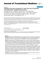
ELISA reactivity of human sera with membrane proteins iso-lated from MN9D neuronal cellsFigure 1
ELISA reactivity of human sera with membrane proteins iso-
lated from MN9D neuronal cells. Black bars represent reac-
tivity of sera from 8 control individuals, and gray bars
represent sera from 19 IPD patients. Bars represent the
mean and standard error of the mean of five individual exper-
iments. *P < 0.05 compared to control sera.
OD (405 nm)
0.0
0.2
0.4
0.6
0.8
1.0
Control
Sera
IPD
Sera
*
Journal of Neuroinflammation 2006, 3:1 />Page 4 of 9
(page number not for citation purposes)
1
streptomycin (Invitrogen Corporation), 3.7 g L
-1
NaHCO
3
(J.T. Baker Chemical Co., Phillipsburg, NJ), and
50 µM 2-mercaptoethanol (Sigma).
Cell culture conditions were set up and analyzed as previ-
ously described (Le et al., 2001), with minor modifica-
tions. In 24-well tissue culture plates (Corning Inc.), 2 ×
10
4
N9 cells were seeded for 24 hr at 37°C under 5% CO
2
.
After 24 hr, medium was removed, and MN9D cells (4 ×
10
4
) in fresh medium were added in the presence of indi-
vidual human sera (1%) samples. The N9:MN9D ratio
was maintained at 1:2, as previously described (Le et al.,
2001). Cells were co-cultured for three days.
Differentiation of MN9D cells was performed as described
[28] by culturing 4.6 × 10
4
cells per well in 4 mL medium
in 6-well tissue culture plates (Corning). Cells were
exposed to 1 mM n-butyrate (Sigma) throughout the
seven day culture and designated wells were harvested
daily, beginning with day 2. Medium was replaced every
48 hr with fresh medium containing 1 mM n-butyrate.
Co-cultures of differentiated MN9D cells were established
by culturing 9.2 × 10
3
MN9D cells in 24-well tissue culture
plates (Corning Inc.) in the presence of 1 mM n-butyrate
(Sigma) and 1% human sera. After 48 hr, the medium was
removed, and fresh medium supplemented with 1 mM n-
butyrate and 1% human sera was added. Twenty-four hr
later, medium was again removed, and cells were washed
twice with 1 mL PBS. Indicated wells received 4.6 × 10
3
N9
microglia and 1% human sera in n-butyrate-free medium.
Cells cultured in the absence of N9 microglia received 1%
human sera in n-butyrate-free medium. Cells were co-cul-
tured for three days.
Quantification of DA expression
At the end of the specified culturing period, plates were
centrifuged at 200 × g for 10 min at 4°C, medium was
removed, and cells were washed with 1 mL PBS. Cells
were exposed to 1 mL 0.2 M HClO
4
and sonicated as
described. This mixture was then centrifuged to remove
proteins from the samples, and DA expression was ana-
lyzed using high performance liquid chromatography
with electrochemical sensors, and quantified using Waters
Millenia software (Waters, Milford, MA), as described
[35]. The protein pellet was resuspended in 50 µL 0.5 M
NaOH, and protein was quantified using the BCA method
with BSA as a standard.
Results
When compared with sera from healthy controls, IgG in
the sera of IPD patients had significantly increased (P <
0.05) binding to ELISA wells coated with MN9D neuronal
membrane proteins (Fig. 1). Western blot analysis also
demonstrated greater IgG reactivity to MN9D cell mem-
brane proteins (Fig. 2); only two sera from healthy indi-
viduals, which had the greatest activity for the control
sera, are shown. The Western analysis of the IPD sera
revealed antibody reactivity to a number of proteins
present in the MN9D neuronal membrane isolates; pro-
teins of 30 to 65 kDa molecular weights were especially
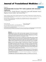
Correlation analysis of clinical score (UPDRS – total) with the sum of the peak areas from the Western analysis (Fig. 2)Figure 3
Correlation analysis of clinical score (UPDRS – total) with
the sum of the peak areas from the Western analysis (Fig. 2).
The r
2
value was assessed by linear regression analysis (Sig-
maPlot 2000) and the significance (p) was calculated by Pear-
son correlation analysis with SigmaStat (Jandel Corp).
UPDRS (total)
020406080100
Total Peak Areas
0
10000
20000
30000
40000
50000
60000
70000
Western blot reactivity of human sera with membrane pro-teins isolated from MN9D neuronal cellsFigure 2
Western blot reactivity of human sera with membrane pro-
teins isolated from MN9D neuronal cells. In this figure, 17
individual IPD sera (lane 1–17) and 2 control sera (lane 18 &
19) were used to probe the PVDF membrane. Numbers des-
ignated to the left of the blot reveal the migration of molecu-
lar weight standards in kDa. Data are representative of two
individual experiments.
Journal of Neuroinflammation 2006, 3:1 />Page 5 of 9
(page number not for citation purposes)
predominant. While this reactivity revealed no consistent
differences between IPD and control sera and no major
common protein band amongst the IPD sera, in general,
there was a noticeable increase in the intensity of bands
with sera from IPD patients with greater unified Parkin-
son's disease rating scale (UPDRS) scores (Table 1). The
UPDRS is a combined score from the physician's evalua-
tion of motor activity including temors, rigidity, posture,
gait, and bradykinesia. For example, the sera from patients
7, 8, 10, 16 and 17 had the most severe IPD (UPDRS-total,
33.5–82.5; UPDRS-motor, 22.5–49), whereas sera from
the least severe (UPDRS-total, 8–16; UPDRS-motor, 6–
10) IPD patients (1, 2, 9, 15) generally had binding as low
as most normal sera.
Pearson correlational analysis does suggest that the anti-
bodies in the IPD sera significantly correlate (r
2
= 0.21; p
= 0.05) with the total UPDRS score (Fig. 3). However,
there was no correlation of the antibody binding to
MN9D antigens (Fig. 2) with regard to UPDRS motor val-
ues or the duration of IPD.
To assess if the observed interactions between IPD sera
and neuronal antigens correlated with any adverse effects
on neuronal cells, in vitro assays were performed. This
analysis revealed that, compared to control sera, IPD sera
had no significant effect on the levels of DA regardless of
whether the quantification was calculated as ng/well or
ng/mg protein (approximately 10 ng/well and 160 ng/mg
protein).
Alternatively, if the N9 microglial cells were co-cultured
with the MN9D neuronal cells and sera (Fig. 4), there was
a noticeable loss of DA. Although there was not a signifi-
Time course of DA expression by MN9D neuronal cells after exposure to n-butyrateFigure 5
Time course of DA expression by MN9D neuronal cells after
exposure to n-butyrate. Black bars represent untreated cells
and gray bars represent cells differentiated in the presence of
1 mM n-butyrate. Results are reported as both (A) ng/well
and (B) ng/mg protein. Results represent the data from three
individual wells per group. *P < 0.05 compared to untreated
cells.
ng DA/well
20
40
60
80
100
Day
123456
ng DA/mg protein
200
400
600
800
1000
After 72 hr exposure to human sera at 37°C, DA levels were assessed in co-cultures of MN9D neuronal cells (4 × 10
4
cells/ml) and N9 microglia (2 × 10
4
cells/ml)Figure 4
After 72 hr exposure to human sera at 37°C, DA levels were
assessed in co-cultures of MN9D neuronal cells (4 × 10
4
cells/ml) and N9 microglia (2 × 10
4
cells/ml). Black bars rep-
resent DA values upon exposure to 8 control sera and gray
bars represent values upon exposure to 19 sera from IPD
patients. Results are reported as both (A) ng/well and (B) ng/
mg protein. Bars represent the mean and standard error of
the mean for four individual experiments. *P < 0.05 com-
pared to control sera.
ng DA/well
2
4
6
8
10
12
14
ng DA/mg protein
50
100
150
200
Control
Sera
IPD
Sera
A
B
*
Journal of Neuroinflammation 2006, 3:1 />Page 6 of 9
(page number not for citation purposes)
cant reduction of DA within these co-cultures on a ng/well
basis (Fig. 4A), the difference between IPD and control
sera became significant (P < 0.05) when corrected for pro-
tein content (Fig. 4B). Additionally, analysis of the viabil-
ity of these cells revealed that the observed reductions in
DA levels within these co-cultures were not due to the via-
bility of these cells (data not shown). Analysis of the
expression of the pro-inflammatory cytokines IL-1β, IL-6,
TNF-α, and IFN-γ revealed no difference between IPD and
control sera with regard to the expression levels of these
cytokines (data not shown). Recent studies have suggested
that LPS-activated microglia cause DA neuronal cell death
via molecules <350 Daltons, which would rule out
cytokines (David Graber, personal communication).
It is known that n-butyrate has the ability to induce differ-
entiation of cells in vitro, including MN9D cells, as evi-
denced by an increased number of outgrowths/
protections [28] and, as shown here, increased DA levels
compared to undifferentiated cells (Fig. 5). DA expression
was increased in MN9D cells exposed to 1 mM n-butyrate,
compared to undifferentiated MN9D cells on days 2–4 on
a ng/well basis (Fig. 5A). However, after Day 4, undiffer-
entiated cells attained the level of DA seen in differenti-
ated cells and, in fact, produced much more DA,
comparatively, through Day 7. Differentiated MN9D cell
expression of DA plateaued on day 4. Upon correction for
protein (Fig 5B), DA values were significantly increased in
differentiated cells compared to undifferentiated cells,
again peaking at Day 3 and eventually dropping until the
level was similar to that seen in undifferentiated cells by
Day 7. This data shows that differentiated cells were more
effective at producing DA, particularly at Day 3, and for
this reason, Day 3 was the day chosen for differentiation
After 72 hr exposure to human sera at 37°C, DA levels were assessed in co-cultures of n-butyrate-differentiated MN9D neuronal cells (4 × 10
4
cells/ml) and N9 microglia (2 × 10
4
cells/ml)Figure 7
After 72 hr exposure to human sera at 37°C, DA levels were
assessed in co-cultures of n-butyrate-differentiated MN9D
neuronal cells (4 × 10
4
cells/ml) and N9 microglia (2 × 10
4
cells/ml). Black bars represent DA values upon exposure to a
pool of 8 control sera; gray bars represent values upon expo-
sure to a pool of 19 sera from IPD patients. Results are
reported as both (A) ng/well and (B) ng/mg protein. Bars rep-
resent the mean and standard error of the mean for three
individual wells. *P < 0.05 compared to control sera.
ng DA/well
1
2
3
4
5
6
Control
ng DA/mg protein
20
40
60
IPD
Sera
*
*
A
B
After 72 hr exposure to human sera at 37°C, DA levels were assessed in cultures of n-butyrate-differentiated MN9D neu-ronal cells (4 × 10
4
cells/ml)Figure 6
After 72 hr exposure to human sera at 37°C, DA levels were
assessed in cultures of n-butyrate-differentiated MN9D neu-
ronal cells (4 × 10
4
cells/ml). Black bars represent DA values
upon exposure to a pool of 8 control sera; gray bars repre-
sent values upon exposure to a pool of 19 sera from IPD
patients. Results are reported as both (A) ng/well and (B) ng/
mg protein. Bars represent the mean and standard error of
the mean for three individual wells. *P < 0.05 compared to
control sera.
ng DA/well
1
2
3
4
5
6
Control
ng DA/mg protein
20
40
60
IPD
Sera
*
*
A
B
Journal of Neuroinflammation 2006, 3:1 />Page 7 of 9
(page number not for citation purposes)
of MN9D neuronal cells prior to removal from n-butyrate
and exposure to N9 microglia.
This system of culturing differentiated cells revealed that,
when cultured alone, n-butyrate-differentiated MN9D
neuronal cells express significantly more DA (P < 0.05) in
the presence of pooled IPD sera, compared to pooled con-
trol sera on both a ng/well and a ng/mg protein basis (Fig.
6). Alternatively, pooled IPD sera decreased DA expres-
sion by differentiated MN9D neuronal cells in the pres-
ence of N9 microglia (Fig. 7). This difference was not
significant on a ng/well basis (Fig. 7A), but was significant
(P < 0.05) on a ng/mg protein basis (Fig. 7B).
Discussion
The results reported here reveal the binding interactions
between IgG antibodies in the sera of IPD patients and
neuronal proteins of a mouse dopaminergic cell line.
These associations were examined using ELISA and West-
ern blot techniques, and significant binding of IgG anti-
bodies to DA neuronal antigens was observed.
Additionally, there were no adverse effects of the IPD sera
in MN9D monocultures, but the IPD sera did significantly
decrease the dopamine content of the MN9D cells when
N9 microglia were present within the cultures. While
there was no evidence that these two activities correlate
with any particular antibody specificity, these results sug-
gest that interactions between IPD sera and neuronal cell
constituents occur, hinting that antibodies may play a role
in IPD. The magnitude of the impact is, as yet, undefined.
Furthermore, it is not clear whether these antibodies are
involved early in the elicitation of IPD symptoms or arise
only after substantial DA neuronal death has occurred.
The reactivity seen between IPD sera and neuronal mem-
brane proteins is striking. The significant differences
observed corroborates previously reported data suggesting
immunoreactivity between sera and CSF from IPD
patients with cellular constituents within the SN of rat
brains [17,19-22,24,27]. In fact, the reactivity with the SN
was reported to be present in as many as 78% of the CSF
samples taken from IPD patients [23]. Surprisingly, aside
from a single report describing reactivity of IPD sera with
a protein modified by DA oxidation [18], there has been
no indication that IPD sera reacts with DA neuronal pro-
tein antigens, as is provided in this report. Furthermore,
analysis of this reactivity by Western blot revealed a
number of proteins that were potentially reactive with
both IPD and control sera making it difficult to pinpoint
specific proteins that were related to a diseased state.
However, the discovery of a number of proteins in the 40–
60 kDa range that reacted to a much greater extent with
IPD sera than with that of controls further limits the pro-
spective candidate proteins that need to be evaluated.
Previous reports have revealed a specific destructive effect
of IPD sera on DA neuronal cells, both in vivo [25,27] and
in vitro [26]. This destructive effect in vivo was specific to
the SN region of the brain and was only seen when IPD
IgG was injected into the SN of rats [25,27]. In vitro utili-
zation of a co-culture system to address the effect of these
interactions revealed that microglia, activated in the pres-
ence of IPD sera have the ability to specifically alter DA
neuron function. Induction of TNF-α was previously
reported to be the microglial factor involved in the loss of
DA [26]; however, we did not observe an increase of TNF-
α or any other proinflammatory cytokine in the co-culture
supernatants with the IPD sera. It is possible that addi-
tional inflammatory microglial products, e.g., nitric oxide
and hydrogen peroxide, are responsible for the loss of DA.
These small oxidative molecules are likely toxicants and
have previously been reported to cause neuronal cell
death [36].
Surprisingly, monocultures of the n-butyrate-treated (dif-
ferentiated) MN9D cells had increased levels of DA when
exposed to IPD sera compared to control sera. A possible
explanation for this increase is that there is a "damage"
signal induced within these neurons upon binding of
antibodies to certain MN9D antigens, which are less
expressed in the non-treated MN9D cells since they were
not affected by the IPD sera in the absence of N9 cells.
This positive signal may induce a hyperactivity of the
"stressed" differentiated neurons, resulting in increased
DA production. Alternatively, antibodies to select surface
proteins on the differentiated MN9D cells may directly
trigger induction of DA production.
Differences between the non-treated and n-butyrate-
treated MN9D cells also were apparent in the presence of
the N9 microglia. Significant reductions in DA levels were
induced by the IPD sera with co-cultures of non-treated or
n-butyrate-treated MN9D cells with N9 cells, when DA
was calculated on a ng/mg protein basis. When DA was
calculated as DA/culture the non-treated MN9D cocul-
tured with N9 microglia and IPD sera did not have signif-
icantly lower levels of DA. This difference is likely due to
the fact that the non-treated MN9D cells are proliferating
to a greater extent and producing less DA per cell, as sug-
gested by the kinetic analyses shown (Figure 5). Thus, the
results with the n-butyrate-treated MN9D cells would
more closely represent the in vivo situation since normal
DA neurons would not be proliferating. The negative
microglial effects on DA neurons corroborate the results
of Le, et al. [26]. It is hypothesized that the antibodies
could cross-link the MN9D cells to Fc receptors on the N9
cells leading to release of neurotoxic factors by the N9
microglia.
Journal of Neuroinflammation 2006, 3:1 />Page 8 of 9
(page number not for citation purposes)
The positive effect of IPD sera on MN9D monocultures
and the negative effect on the co-culture of MN9D and N9
cells with regard to DA production do not address the spe-
cific MN9D antigens involved in these processes. How-
ever, it is clear that the specificity of the antibodies play a
more important role that the amount of antibody, in that
when some individual sera were assayed for binding by
ELISA and for function (DA levels), there was no correla-
tion. This indicates that the specificity (or possibly iso-
type) of certain antibodies in the IPD sera and not their
concentrations are responsible for altering DA produc-
tion. This emphasizes the need to further delineate the
specific DA neuronal antigens being affected. Analyses are
currently underway to isolate and identify the DA neuro-
nal antigens bound by the antibodies, which cause the
observed DA changes. In addition to IPD serum antibod-
ies, it is also possible that the IPD sera may contain addi-
tional factors affecting DA production, e.g.,
tetrahydroisoquinolone, β-carbolines. A number of
potential toxins could be present in some of the IPD sera
[37]. However, this possibility seems unlikely in that the
sera were only inhibitory in the presence of the N9 cells.
While attempting to understand the role of the immune
system in IPD, we have addressed a number of critical
parameters. First, we have shown that there is specific
reactivity of sera from IPD patients with multiple mem-
brane proteins expressed by a mouse DA neuronal cell
line. Second, we have found neuronal antigen bands of
specific reactivity, most notably in the 40–60 kDa range
that may lead to further understanding of what neuronal
proteins are involved in the antibody-induced alteration
of DA production. Finally, through in vitro analyses, we
show that there is a specific effect of IPD sera on co-cul-
tures of MN9D neuronal cells and N9 microglia, suggest-
ing an interaction between the serum factors and the N9
cells.
Competing interests
The author(s) declare that they have no competing inter-
ests.
Authors' contributions
VH carried out most of the in vitro analysis and wrote the
first draft of the manuscript. TM carried out the Western
analyses. SF collected the samples from the IPD patients,
provided the information for Table 1, and reviewed the
manuscript. RS participated in the design of the study,
supervised the HPLC analyses, and reviewed the manu-
script. DL conceived of the study, and participated in its
design and coordination and helped to draft the manu-
script. All authors read and approved the final manu-
script.
Acknowledgements
This work was supported, in part, by an American Parkinson Disease Asso-
ciation Fellowship (VCH, N1402031) and DOD grant and U.S. Army Med-
ical Research and Materiel Command Neurotoxin Exposure Program
Award No: DAMD17-02-1-0173 to RFS.
References
1. Lang A, Lozano A: Parkinson's disease: First of two parts. N Engl
J Med 1998, 339:1044-1053.
2. Lang A, Lozano A: Parkinson's disease: Second of two parts. N
Engl J Med 1998, 339:1130-1143.
3. Beal M: Experimental models of Parkinson's disease. Nat Rev
Neurosci 2001, 2:325-334.
4. Olanow C, Tatton W: Etiology and pathogenesis of Parkinson's
disease. Annu Rev Neurosci 1999, 22:123-144.
5. McInerney-Leo A, Hadley D, Gwinn-Hardy K, Hardy J: Genetic test-
ing in Parkinson's disease. Mov Disord 2005, 20:1-10.
6. McGeer P, Itagaki S, Akiyama H, McGeer E: Rate of cell death in
parkinsonism indicates active neuropathological process.
Ann Neurol 1988, 24:574-576.
7. McGeer P, Itagaki S, Boyes B, McGeer E: Reactive microglia are
positive for HLA-DR in the substantia nigra of Parkinson's
and Alzheimer's disease brains. Neurology 1988, 38:1285-1291.
8. Fiszer U: Does Parkinson's disease have an immunological
basis? The evidence and its therapeutic implications. BioDrugs
2001, 15:351-355.
9. Liberatore G, Jackson-Lewis V, Vukosavic S, Mandir A, Vila M, McAu-
liffe W, Dawson V, Dawson T, Przedborski S: Inducible nitric
oxide synthase stimulates opaminergic neurodegeneration
in the MPTP model of Parkinson disease. Nat Med 1999,
5:1403-1409.
10. Kurkowska-Jastrzebska I, Wronska A, Kohutnicka M, Czlonkowski A,
Czlonkowska A: The inflammatory reaction following 1-
methyl-4-phenyl-1,2,3, 6-tetrahydropyridine intoxication in
mouse. Exp Neurol 1999, 156:50-61.
11. Teismann P, Tieu K, Choi D, Wu D, Naini A, Hunot S, Vila M, Jackson-
Lewis V, Przedborski S: Cyclooxygenase-2 is instrumental in
Parkinson's disease neurodegeneration. Proc Natl Acad Sci USA
2003, 100:5473-5478.
12. Kohutnicka M, Lewandowska E, Kurkowska-Jastrzebska I,
Czlonkowski A, Czlonkowska A: Microglial and astrocytic
involvement in a murine model of Parkinson's disease
induced by 1-methyl-4-phenyl-1,2,3,6-tetrahydropyridine
(MPTP). Immunopharmacology 1998, 39:167-180.
13. Nagatsu T, Mogi M, Ichinose H, Togari A: Changes in cytokines
and neurotrophins in Parkinson's disease. J Neural Transm Suppl
2000:277-290.
14. Czlonkowska A, Kohutnicka M, Kurkowska-Jastrzebska I,
Czlonkowski A: Microglial reaction in MPTP (1-methyl-4-phe-
nyl-1,2,3,6-tetrahydropyridine) induced Parkinson's disease
mice model. Neurodegeneration 1996, 5:137-143.
15. Czlonkowska A, Kurkowska-Jastrzebska I, Czlonkowski A, Peter D,
Stefano G: Immune rocesses in the pathogenesis of Parkin-
son's disease – a potential role for microglia and nitric oxide.
Med Sci Monit 2002, 8:RA165-RA177.
16. Nagatsu T, Mogi M, Ichinose H, Togari A: Cytokines in Parkinson's
disease. J Neural Transm Suppl 2000:143-151.
17. Le W, Rowe D, Jankovic J, Xie W, Appel S: Effects of cerebrospi-
nal fluid from patients with Parkinson disease on dopaminer-
gic cells. Arch Neurol 1999, 56:194-200.
18. Rowe D, Le W, Smith R, Appel S: Antibodies from patients with
Parkinson's disease react with protein modified by
dopamine oxidation. J Neurosci Res 1998, 53:551-558.
19. McRae-Degueurce A, Rosengren L, Haglid K, Booj S, Gottfries C,
Granerus A, Dahlstrom A: Immunocytochemical investigations
on the presence of neuron-specific antibodies in the CSF of
Parkinson's disease cases. Neurochem Res 1988, 13:679-684.
20. McRae D, Gottfries C, Karlsson I, Svennerholm L, Dahlstrom A:
Antibodies in the CSF of a Parkinson patient recognizes neu-
rons in rat mesencephalic regions. Acta Physiol Scand 1986,
126:313-315.
21. Loeffler D, Brickman C, Kapatos G, Peter J, LeWitt P: Anti-Neuro-
nal Antibodies and Other Markers of Immune System Acti-
vation in Parkinson's Disease Cerebrospinal Fluid.
Neurodegeneration 1992, 1:145-153.
Publish with Bio Med Central and every
scientist can read your work free of charge
"BioMed Central will be the most significant development for
disseminating the results of biomedical research in our lifetime."
Sir Paul Nurse, Cancer Research UK
Your research papers will be:
available free of charge to the entire biomedical community
peer reviewed and published immediately upon acceptance
cited in PubMed and archived on PubMed Central
yours — you keep the copyright
Submit your manuscript here:
/>BioMedcentral
Journal of Neuroinflammation 2006, 3:1 />Page 9 of 9
(page number not for citation purposes)
22. Dahlstrom A, Wigander A, Lundmark K, Gottfries C, Carvey P,
McRae A: Investigations on auto-antibodies in Alzheimer's
and Parkinson's diseases, using defined neuronal cultures. J
Neural Transm Suppl 1990, 29:195-206.
23. Carvey P, McRae A, Lint T, Ptak L, Lo E, Goetz C, Klawans H: The
potential use of a dopamine neuron antibody and a striatal-
derived neurotrophic factor as diagnostic markers in Parkin-
son's disease. Neurology 1991, 41:53-58.
24. Pouplard A, Emile J: Autoimmunity in Parkinson's disease. Adv
Neurol 1984, 40:307-313.
25. Chen S, Le W, Xie W, Alexianu M, Engelhardt J, Siklos L, Appel S:
Experimental destruction of substantia nigra initiated by
Parkinson disease immunoglobulins. Arch Neurol 1998,
55:1075-1080.
26. Le W, Rowe D, Xie W, Ortiz I, He Y, Appel S: Microglial activation
and dopaminergic cell injury: an in vitro model relevant to
Parkinson's disease. J Neurosci 2001, 21:8447-8455.
27. He Y, Le W, Appel S: Role of Fcgamma receptors in nigral cell
injury induced by Parkinson disease immunoglobulin injec-
tion into mouse substantia nigra. Exp Neurol 2002, 176:322-327.
28. Choi H, Won L, Kontur P, Hammond D, Fox A, Wainer B, Hoffmann
P, Heller A: Immortalization of embryonic mesencephalic
dopaminergic neurons by somatic cell fusion. Brain Res 1991,
552:67-76.
29. Righi M, Mori L, De Libero G, Sironi M, Biondi A, Mantovani A, Donini
S, Ricciardi-Castagnoli P: Monokine production by microglial
cell clones. Eur J Immunol 1989, 19:1443-1448.
30. Corradin S, Mauel J, Donini S, Quattrocchi E, Ricciardi-Castagnoli P:
Inducible nitric oxide synthase activity of cloned murine
microglial cells. Glia 1993, 7:255-262.
31. Matsuki N: Measurement of cellular 3-(4,5-dimethylthiazol-2-
yl)-2,5-diphenyltetrazolium bromide (MTT) reduction activ-
ity and lactate dehydrogenase release using MTT. Neurosci
Res 2000, 38:325-329.
32. Duncan D, Lawrence D: Residual activation events functional
after irradiation of mouse splenic lymphocytes. Radiation Res
1991, 125:6-13.
33. Narendran A, Hoffman S: Identification of autoantibody reac-
tive integral brain membrane antigens. A two-dimensional
analysis. J Immunol Methods 1988, 114:227-234.
34. Cao L, Lawrence D: Suppression of host resistance to Listeria
moncytogenes by acute cold/restraint stress: lack of direct
IL-6 involvement. J Neuroimmunol 2002, 133:132-143.
35. Seegal R, Brosch K, Bush B: High-performance liquid chroma-
tography of biogenic amines and metabolites in brain, cere-
brospinal fluid, urine and plasma. J Chromatogr 1986,
377:131-144.
36. Wang J, Shum A, Ho Y: Oxidative neurotoxicity in rat cerebral
cortex neurons: synergistic effects of H2O2 and NO on apop-
tosis involving activation of p38 mitogen-activated protein
kinase and caspase-3. J Neurosci Res 2003, 72:508-519.
37. Collins M: Alkaloids, alcohol and Parkinson's disease. Parkinson-
ism Relat Disord 2002, 8:417-422.