báo cáo hóa học:" Stem cells from umbilical cord blood do have myogenic potential, with and without differentiation induction in vitro" pdf
Bạn đang xem bản rút gọn của tài liệu. Xem và tải ngay bản đầy đủ của tài liệu tại đây (1.94 MB, 9 trang )
BioMed Central
Page 1 of 9
(page number not for citation purposes)
Journal of Translational Medicine
Open Access
Research
Stem cells from umbilical cord blood do have myogenic potential,
with and without differentiation induction in vitro
Tatiana Jazedje
1
, Mariane Secco
1
, Natássia M Vieira
1
, Eder Zucconi
1
,
Thomaz R Gollop
2
, Mariz Vainzof
1
and Mayana Zatz*
1
Address:
1
Department of Biology, Human Genome Research Center, São Paulo, Brazil and
2
Fetal Medicine Institute of São Paulo, São Paulo, Brazil
Email: Tatiana Jazedje - ; Mariane Secco - ; Natássia M Vieira - ;
Eder Zucconi - ; Thomaz R Gollop - ; Mariz Vainzof - ; Mayana Zatz* -
* Corresponding author
Abstract
The dystrophin gene, located at Xp21, codifies dystrophin, which is part of a protein complex
responsible for the membrane stability of muscle cells. Its absence on muscle causes Duchenne
Muscular Dystrophy (DMD), a severe disorder, while a defect of muscle dystrophin causes Becker
Muscular Dystrophy (DMB), a milder disease. The replacement of the defective muscle through
stem cells transplantation is a possible future treatment for these patients. Our objective was to
analyze the potential of CD34+ stem cells from umbilical cord blood to differentiate in muscle cells
and express dystrophin, in vitro. Protein expression was analyzed by Immunofluorescence, Western
Blotting (WB) and Reverse Transcriptase – Polymerase Chain Reaction (RT-PCR). CD34+ stem
cells and myoblasts from a DMD affected patient started to fuse with muscle cells immediately after
co-cultures establishment. Differentiation in mature myotubes was observed after 15 days and
dystrophin-positive regions were detected through Immunofluorescence analysis. However, WB
or RT-PCR analysis did not detect the presence of normal dystrophin in co-cultures of CD34+ and
DMD or DMB affected patients' muscle cells. In contrast, some CD34+ stem cells differentiated in
dystrophin producers' muscle cells, what was observed by WB, reinforcing that this progenitor cell
has the potential to originate muscle dystrophin in vitro, and not just in vivo like reported before.
Background
More than 30 different types of muscular dystrophies have
been identified to date, ranging from adult forms with a
mild course to severe childhood forms with a rapid pro-
gression. Among them, the most severe form, X-linked
Duchenne Muscular Dystrophy (DMD), affects 1:3500
living boys. It's caused by a mutation in the dystrophin
gene, leading to the absence of its product, dystrophin. Its
allelic milder form, Becker Muscular Dystrophy (BMD) is
10 times less frequent than DMD [1-3]. It differs from the
first form because patients have some functional dys-
trophin in their muscle, which may be defective in quan-
tity and/or size. Both disorders are characterized by a
progressive degeneration of the skeletal muscle. In DMD,
affected boys are confined to a wheelchair around age 10–
12 and without assisted ventilation death occurs usually
before age 20 of cardiac arrest or respiratory failure. In
BMD, the course is highly variable. Some patients are con-
fined to a wheelchair before age 20 while other may
remain ambulant beyond age 60 depending on how the
gene mutation affects the dystrophin amount and or func-
tion [4-6].
Published: 14 January 2009
Journal of Translational Medicine 2009, 7:6 doi:10.1186/1479-5876-7-6
Received: 21 October 2008
Accepted: 14 January 2009
This article is available from: />© 2009 Jazedje et al; licensee BioMed Central Ltd.
This is an Open Access article distributed under the terms of the Creative Commons Attribution License ( />),
which permits unrestricted use, distribution, and reproduction in any medium, provided the original work is properly cited.
Journal of Translational Medicine 2009, 7:6 />Page 2 of 9
(page number not for citation purposes)
The dystrophin gene, with 2.4 Mb and 79 exons is the larg-
est human gene. Its product, the protein dystrophin has
427 kDa [7-9]. Dystrophin belongs to a complex of pro-
teins (dystrophin-glycoprotein complex) responsible for
the membrane maintenance of muscle cells. A primary
deficiency in any of these proteins induces to a secondary
deficient of the entire complex, causing different types of
muscular dystrophies [10,11].
Many different therapies have been tested in DMD animal
models and patients. A promising approach to the treat-
ment of DMD is to restore dystrophin expression by
repairing the defective muscle through cell therapy. Previ-
ous studies have suggested that hematopoietic stem cells
can contribute to skeletal muscle regeneration. In normal
and mdx mice (murine model of DMD), bone marrow
(BM)-derived cells were shown to participate in skeletal
muscle repair after induced damage [12-14]. However, the
clinical usefulness of hematopoietic cell transplantation
for muscular dystrophies such as DMD [15] depends on
the expansion, homing and myogenic differentiation of
transplanted cells.
In past decades, human umbilical cord blood (HUCB)
has been explored as an alternative source to BM for cell
transplantation and therapy because of its hematopoietic
and nonhematopoietic (mesenchymal) components [16].
In contrast to bone marrow aspiration, HUCB is obtained
by a simple, safe and painless procedure after birth.
Regarding myogenic potential, recent studies have shown
that subpopulations of HUCB cells can differentiate into
muscle cells [17,18]. Additionally, CD34, transmembrane
glycophosphoprotein known to be expressed by human
hematopoietic progenitor cells has recently been associ-
ated with both the quiescent and activated states of myo-
genic progenitor cells. [19]. More recently, the in vivo
myogenic differentiation of human umbilical cord blood
was observed after the injection into the sjl dystrophic
mice, suggesting that human umbilical cord blood has
myogenic precursors [20].
Although the positive results of the in vivo injections, the
interaction of these cells with human dystrophic muscle
cells is still unknown. Here we have investigated, for the
first time, the potential of umbilical cord blood CD34+
stem cells to interact and differentiate into muscle cells
when in direct contact with human DMD/DMB myob-
lasts, and their potential to restore the absent protein. Our
results show CD34+ cells are able to participate in the
myotube formation, resulting in the restoration of dys-
trophin expression. These findings represent a possible
tool for future cell therapy applications in DMD disease
and for other muscular dystrophies.
Materials and methods
Isolation and characterization of human CD34+ cells from
the umbilical cord blood
CD34+ stem cells from human umbilical cord were
obtained from healthy babies, born in Hospital Albert
Einstein, in São Paulo, Brazil. All studies were approved
by the ethical committee and were done after written con-
sent. The cord blood was processed as described in the
SuperMACSII manual (Miltenyi Biotec, Bergisch Glad-
bach, Germany) and the CD34+ stem cells were obtained
by magnetic cell sorting, using the "CD34 progenitor cell
isolation kit" (Miltenyi Biotec, Bergisch Gladbach, Ger-
many).
The purity of CD34+ cells was determined for flow cytom-
etry. Firstly, the immunomagnetically selected cells were
incubated with the conjugated antibody anti-CD34-
PerCP (BD Biosciences), in phosphate-buffered saline
(PBS) at 4°C for 30 minutes, as recommended by the
manufacturer. A total of 10,000 labeled cells were ana-
lyzed using Guava EasyCyte flow cytometer running
Guava ExpressPlus software (Guava Technologies). The
percentage of CD34+ cells present in the sample was
assessed after correction for the percentage of cells reactive
with the isotype control.
Cell cultures
CD34+ cells were cultured and expanded into 25 cm
2
plastic culture flasks (Corning, New York, USA), in 5 mL
with StemSpan SFEM (Serum Free Expansion-Medium)
and with the cytokine cocktail CC100* (Stem Cell Tech-
nologie, British Columbia, Canada), which contains 100
ng/mL rh Flt-3 Ligand, 100 ng/mL rh Stem Cell Factor, 20
ng/mL rh IL-3 and 20 ng/mL rh IL-6. Medium was
replaced once a week, by centrifugation at 1,400 rpm, for
5 minutes. Cells were kept in an incubator at 37°C and
5% CO
2
.
Myoblasts from 3 DMD and 2 DMB affected patients were
obtained from biceps biopsies. They were implanted into
25 cm
2
plastic culture flasks (Corning, New York, USA)
with 5 mL of Dubecco's Modified Medium (DMEM) high
glucose, 20% Fetal Bovine Serum (FBS; Gibco/Invitrogen,
California, USA), 100 U/mL of penicillin and 100 mg/mL
of streptomicyn (Sigma-Aldrich, Missouri, USA) and
amphotericin B (Cultilab, São Paulo, Brazil), and kept in
an incubator at 37°C and 5% CO
2
.
In a ratio 3:1 (3 fold CD34+ stem cells: 1 fold DMD/DMB
muscle cells), co-cultures were performed with 50% of the
medium used for CD34+ stem cells and 50% of the
medium used for myoblasts. They were established into
25 cm
2
plastic culture flasks (Corning, New York, USA)
with 5 mL of medium or into a 10 cm
2
tissue culture
chamber (Nunc, Illinois, USA), with 4 mL of medium.
Journal of Translational Medicine 2009, 7:6 />Page 3 of 9
(page number not for citation purposes)
Co-cultures were kept in an incubator at 37°C and 5%
CO
2
until final analysis.
Dystrophin Immunofluorescence (IF) and Western Blotting
(WB)
Immunolabelling was performed as previous described
[21] and cells were analyzed with an inverted microscope
(Carl Zeiss, Jena, Germany). For WB analysis, myoblasts
of a DMB affected patient, normal muscle cells and co-cul-
tures were trypsinized by standard procedures, washed
with PBS 1× and centrifuged for 7 minutes at 1,400 rpm.
CD34+ cells were washed and centrifuged with PBS 1× for
7 minutes at 1,400 rpm. Cell pellets were transferred to
1,5 mL eppendorfs and processed as previously described
[22]. In both methodologies monoclonal antibodies C
and/or N-terminal anti-human dystrophin were used
(kindly provided by the late Dr. L. V. B. Anderson).
Bisbenzimide H33342 immunofluorescence of living cells
CD34+ stem cells nuclei were dyed with Bisbenzimide
H33342, 5 μg/mL (Sigma-Aldrich, Missouri, USA) for 90
minutes in CO
2
incubator, at 37°C, in the dark. After that,
cells were washed in PBS 1× and cultured protected from
light. Stained stem cells were used in co-cultures of DMD
muscle cells and normal CD34+ stem cells from umbilical
cord blood.
Reverse Transcription – Polymerase Chain Reaction (RT-
PCR)
Total RNA from myoblasts of a DMD affected patient
(with deletion of exons 3–17), normal muscle cells, CD34
positive stem cells and co-cultures were obtained as previ-
ously described [23]. RNA concentration and purity were
determined spectrophotometrically. RT-PCRs reactions
were performed as recommended in the supplier's proto-
col of the kit "SuperScript One-Step RT-PCR with Plati-
num Taq" (Gibco/Invitrogen, California, USA). The
dystrophin primers sequences for the amplification of
exons 8, 12, 13 and 51, are available at Leiden website
. RT-PCRs were performed with Per-
kin-Elmer thermal cycler (PE Applied Biosystems, Califor-
nia, USA) using conditions recommended by the
supplier's protocol. The annealing temperature used was
60°C.
Results
Identification and characterization of CD34+ cells derived
from blood
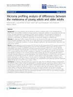
Cells isolated from human umbilical cord blood were
immunomagnetically selected and characterized by flow
cytometry. A representative subpopulation of the cells was
CD34 positive (80.92%), as represented in the graphs
(Figure 1).
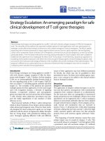
Cells co-cultures
Right after the co-culture establishment, the interaction
between CD34+ and DMD myoblasts was observed (Fig-
ure 2). F, even that blue CD34+ nuclei were found inside
the formed myotubes (Figure 3) the contact between the
cells can ate the fusion, forming multinucleated syncy-
tium. CD34+ stem cells and muscle cells division was also
observed (data not shown).
Dystrophin IF
IF assay was performed after 15 days in culture. Co-cul-
tures of CD34+ stem cells and DMD myoblasts showed
positive dystrophin when compared with the normal
myoblast culture (figure 4). This result suggests that the
fusion of stem cells and muscle cells was sufficient to
induce the stem cells nuclei to express muscle cells pro-
teins, restoring the absent dystrophin expression. More
than 3 different co-cultures of each patient, with different
CD34+ cord blood stem cells donors, were analyzed. The
same result were seen in relation to fusion and IF pattern.
In addition to dystrophin IF analysis, the fusion of CD34+
stem cells and myoblasts from a DMD affected patient
was also followed during the 15 days of culture through
Bisbenzimide H33342 stem cells nuclei staining (figure
5).
Western Blotting (WB) and RT-PCR analysis
We also evaluate the dystrophin expression by WB analy-
sis. We did not detect the presence of normal dystrophin,
by this method, after 15 days of co-cultures with CD34+
stem cells and DMB affected patient muscle cells (data not
shown).
In order to confirm if there was any expression of dys-
trophin from the CD34+ stem cells, we used muscle cells
from a DMD affected patient with deletion of exons 3–17
and total absence of dystrophin. Primers to amplify the
exon 8 (inside the mutation) and exon 51 as a control
were used. However, exon 8 was not amplified in co-cul-
tures, indicating the absence or very low expression of dys-
trophin in co-cultures (data not shown).
Transdifferentiation of CD34+ stem cells into muscle cells
During the expansion of CD34+ stem cells from umbilical
cord blood, we observed the presence, in some cultures, of
a small number of cells that became adherents. These cells
were then kept in culture for 20 days with the same
medium used in co-cultures (50% StemSpan CC100 and
50% DMEM). In this experiment, the used medium was
filtered in a 0,22 μm filter (Millipore, Massachusetts,
EUA) and the pH was adjusted to 7,4 with Hepes and
Sodium Bicarbonate (Sigma-Aldrich, Missouri, USA).
Journal of Translational Medicine 2009, 7:6 />Page 4 of 9
(page number not for citation purposes)
A small number of adherent cells acquired the phenotype
of differentiated muscle cells. At the 20
th
day, a protein
extract of these cells was analyzed by WB and the presence
of normal dystrophin was observed (figures 6 and 7).
Discussion
The possibility to replace a defective tissue by a normal
one through stem cells transplantation has been proposed
as an therapeutic approach for many disorders including
muscular dystrophies. However, many experiments in
vitro and in vivo will have to take place before an effective
treatment for patients affected by muscular dystrophies
will be available. Therefore, the understanding of stem
cell biology is fundamental for their future utilization for
therapeutic purposes.
The experiments showed here, demonstrated that the
hematopoietic stem cells from umbilical cord blood have
the potential to fuse to DMD muscle cells, restoring their
dystrophin expression. However, co-culture experiments
showed dystrophin expression only by IF analysis, sug-
gesting a low expression oh this protein in co-cultured
cells. On the other hand, IF is a much more sensitive
method than WB, which also shows a greater variability.
Previous studies have suggested that hematopoietic stem
cells can contribute to skeletal muscle regeneration.
[16,20,24,25]. The report of a DMD patient who received
a bone marrow (BM) transplantation from his father, at
age 1, due to a severe combined immunodeficiency and
who showed a mild course at age 14 [26] seems very
promising. The presence of BM-derived donor nuclei in
the muscle of this patient, suggested that exogenous
hematopoietic human BM cells had the ability to fuse into
recipient skeletal muscle and to persist for at least 13
years. However, these results have been questioned since
the transplanted patient presents a high level of 44 and 45
exon skiping, leading to the production of an in-frame
transcript, which could be responsible for his milder phe-
notype.
Cell fusion seems to be a rare phenomenon either in vivo
or in vitro (1/100000 cells) and probably occurs in cell
types where polyploidy is common, like hepatocytes, car-
CD34 flow cytometryFigure 1
CD34 flow cytometry. Graphs show forward scatter versus fluorescence intensity. a) Unmarked control before CD34 puri-
fication with MACS columns, where 1.8% were CD34+. b) After CD34 purification with MACS columns, where 80.92% were
CD34+. CD34+ cells are represented by pink points and CD34- cells are represented by blue points.
Journal of Translational Medicine 2009, 7:6 />Page 5 of 9
(page number not for citation purposes)
diac and skeletal muscle or purkinje cells. On the other
hand, transdifferentiation is a process where the nuclei of
the stem cells are reprogrammed, acquiring new expressed
genes and proteins [27]. It was also observed that both
endothelial progenitors in the embryo and differentiated
endothelial cells from the umbilical vein transdifferenti-
ated into beating cardiomyocytes, by fusion, when cocul-
tured with neonatal rat cardiomyocytes or when injected
near to a damaged area of the heart [28]. Transdifferentia-
tion also occurred when murine bone marrow stem cells
fused to murine embryonic stem cells [29]. However, the
real meaning of fusion versus transdifferentiation is still
controversial [30-33].
Adult stem cells transplantation in animal models also
has shown controversial results [13,27,34]. In an attempt
to follow the fate of exogenous stem cells in vivo, specific
markers expression in transplanted stem cells, like GFP
(Green Fluorescent Protein) or β-galactosidase are being
used. However, green autofluorescent artifacts were
observed in IF muscle analysis after stem cells transplanta-
tion in murines [35], calling the attention for the difficulty
in the interpretation of published reports as well as on our
own IF results.
Moreover, in most cases, it was not possible to compare
results because of the differences of conditions in each
experiment, such as the phenotype characterization and
quantity of transplanted stem cells as well as the degener-
ation degree of the recipient musculature. Besides that, the
microenvironmental conditions, presents in vitro or in vivo
experiments are crucial to define and better understand
the observed responses. Until very recently, our group
showed that stem cells from HUCB did not differentiate
into myotubes or express dystrophin when cultured in
muscle-conditioned medium and in the presence of
human muscle cells [25]. Subsequently wehuman Adi-
pose Stem Cells (hASC) can participate in myotube for-
mation when cultured with differentiating human DMD
myoblasts and myotubes even when the co-culture was
maintained in growth media [36]. The present results of
co-culture of CD34+ and DMD myoblasts without the
inductive media show that these cells can interact and
express dystrophin. This data together with our previous
findings [25] suggest that HUCB loose the capacity to fuse
with muscle cells when they are previously committed. In
other words, their pre-differentiation into muscle may
alter or decrease their potential to fuse with muscle cells.
Probably, undifferentiated stem cells can respond to
chemical factors released by the DMD muscle, providing
the signals that contribute to the establishment of a favo-
rable microenvironment to initiate the fusion and myo-
genic differentiation process. Others have also
demonstrated that signals from damaged but not undam-
aged skeletal muscle induce myogenic differentiation of
rat bone-marrow-derived mesenchymal stem cells [37].
Although a comprehensive analysis of the component(s)
responsible for the myogenic effects has not been per-
formed, we do not exclude the possibility that inflamma-
tory and growth factors with myogenic effect, like IL6/LIF,
IGF, HGF, or others [38-40] are present in the medium
and are involved in the reported effects on human stem
cells. Based on our experience, the IGF-1 concentration
was significantly higher in the dystrophic muscle-condi-
tioned medium than normal muscle medium (unpub-
lished data).
Our results on WB analysis confirm the potential of
umbilical cord blood CD34+ stem cells to differentiate in
muscle cells in vitro, although the observed expression of
dystrophin would not be enough for therapeutic poten-
tial. In fact, the skeletal myogenesis is a developmental
Interaction between CD34+ stem cells and DMD myoblastsFigure 2
Interaction between CD34+ stem cells and DMD
myoblasts. a) after 1 hour (630×); b and c) after 24 hours
(200×). arrow indicate syncytium. Microscope Zeiss Axiovert
200.
Journal of Translational Medicine 2009, 7:6 />Page 6 of 9
(page number not for citation purposes)
cascade controlled by a family of myogenic regulatory fac-
tors, that are expressed with a well-defined time course,
during the early stage of myogenic differentiation. Dys-
trophin is one of the last muscle proteins produced at the
time of cell fusion [41]. So, it is possible that once differ-
entiation is triggered, the expression of the genetic reper-
toire of a differentiated tissue in vivo may differ from the
observed in vitro.
Conclusion
Our findings showed that umbilical cord blood CD34+
stem cells have the potential to interact with dystrophic
muscle cells restoring the dystrophin expression of DMD
cells in vitro. Although utilized within the context of
DMD, the results presented here may be valid to other
muscle-related cell therapy applications.
Competing interests
The authors declare that they have no competing interests.
Co-culture after 48 hoursFigure 3
Co-culture after 48 hours. Before the co-culture, stem cell nuclei were previously stained with Bisbenzimide H33342 (blue
fluorescence). a) CD34+ stem cell nuclei with blue fluorescence, been (a) 200× and (a') 630×, respectively. b) Halogen light of
the co-culture, showing the co-existence of both cells: fluctuant CD34+ stem cells and adherent myoblasts, been (b) 200× and
(b') 630×, respectively. c) Pictures from panels a and b superposed, showing blue nuclei inside adherent cells (black arrows),
been (c) 200× and (c') 630×, respectively. Microscope Zeiss Axiovert 200.
Journal of Translational Medicine 2009, 7:6 />Page 7 of 9
(page number not for citation purposes)
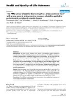
Dystrophin IF in culture cellsFigure 4
Dystrophin IF in culture cells. Anti-human dystrophin (N-terminal) FITC conjugated (green fluorescence) and nuclei dyed
with Bisbenzimide H33342 (blue fluorescence). a) normal muscle cells, 200×; b) muscle cells of patient affected by DMD (dys-
trophin absent), 200×; c) Co-culture of stem cells CD34+ and muscle cells of patient affected by DMD, 200×. Microscope
Zeiss Axiovert 200.
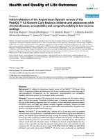
Dystrophin IF after 15 days in cultureFigure 5
Dystrophin IF after 15 days in culture. Antibody anti-dystrophin N-terminal in green fluorescence. a) DMD muscle cells,
after 15 days in culture, with nucleus dyed with Bisbenzimide H33342 (negative control); b) Co-culture after 15 days showing
dystrophin expression and only the CD34+ stem cells' nuclei dyed with Bisbenzimide H33342. Microscope Zeiss Axiovert 200,
400×.
Journal of Translational Medicine 2009, 7:6 />Page 8 of 9
(page number not for citation purposes)
Authors' contributions
TJ and MZ conceived the study and wrote the manuscript.
TJ designed and performed tissue culture, Western Blot-
ting and Immunofluorescence. MS, NMV and EZ helped
with flow cytometric evaluation and with the manuscript
review. MV helped with Western Blotting and Immun-
ofluorescence interpretation. TRG helped providing
umbilical cord blood.
Acknowledgements
The collaboration of the following persons is gratefully acknowledged: Hos-
pital Israelita Albert Einstein, São Paulo, Brazil, especially Dr Andresa
Ribeiro and Dr Eurípides Ferreira. Marta Cánovas and Antonia Cerqueira,
for technical assistance; L.V.B. Anderson, who kindly provided specific anti-
bodies. This work was supported by grants from Fundação de Amparo à
Pesquisa do Estado de São Paulo (FAPESP-CEPID), Conselho Nacional de
Desenvolvimento Científico e Tecnológico (CNPq), PRONEX, and Associ-
ação Brasileira de Distrofia Muscular (ABDIM).
References
1. Emery AEH: Duchenne muscular dystrophy. 2nd edition.
Oxford and Nova York, Oxford University Press; 1993:25-45.
2. Emery AE: The muscular dystrophies. Lancet 2002, 23:687-695.
3. Emery AE: Muscular dystrophy into the new millennium. Neu-
romuscul Disord 2002, 12:343-349.
4. Passos-Bueno MR, Vainzof M, Marie SK, Zatz M: Half the dys-
trophin gene apparently enough for a mild clinical course:
confirmation of its potential use for gene therapy. Hum Mol
Genet 1994, 3:919-922.
5. McNally EM, Passos-Bueno MR, Bönnemann CG, Vainzof M, de Sá
Moreira E, Lidov HG, Othmane KB, Denton PH, Vance JM, Zatz M,
Kunkel LM: Mild and severe muscular dystrophy caused by a
single γ-sarcoglycan mutation. Am J Hum Genet 1996,
59:1040-1047.
6. Bönnemann CG, Passos-Bueno MR, McNally EM, Vainzof M, de Sá
Moreira E, Marie SK, Pavanello RC, Noguchi S, Ozawa E, Zatz M, Kun-
kel LM: Genomic screening for β-sarcoglycan gene mutations:
missense mutations may cause severe limb-girdle muscular
dystrophy type 2E (LGMD 2E). Hum Mol Gen 1996, 5:1953-1961.
7. Koenig M, Hoffman EP, Bertelson CJ, Monaco AP, Feener C, Kunkel
LM: Complete cloning of the Duchenne muscular dystrophy
(DMD) cDNA and preliminary genomic organization of the
DMD gene in normal and affected individuals. Cell 1987,
50:509-517.
8. Zubrzycka-Gaarn EE, Bulman DE, Karpati G, Burghes AH, Belfall B,
Klamut HJ, Talbot J, Hodges RS, Ray PN, Worton RG: The Duch-
enne muscular dystrophy gene product is localized in sarco-
lemma of human skeletal muscle. Nature 1988, 333:466-469.
CD34+ stem cells transdifferentiated in muscle cells in vitroFigure 6
CD34+ stem cells transdifferentiated in muscle cells in vitro. CD34+ stem cells that transdifferentiated in dystrophin
producer muscle cells after 20 days in culture. Microscope Zeiss Axiovert 200.
Western blot for dystrophinFigure 7
Western blot for dystrophin. WB analysis for dystrophin
expression, after transdifferentiation of adherent cells
obtained from prior CD34+ stem cells. Nc) normal control
(human skeletal muscle). SC) CD34+ stem cells. aSC)
adherent stem cells (prior CD34+).
Publish with BioMed Central and every
scientist can read your work free of charge
"BioMed Central will be the most significant development for
disseminating the results of biomedical research in our lifetime."
Sir Paul Nurse, Cancer Research UK
Your research papers will be:
available free of charge to the entire biomedical community
peer reviewed and published immediately upon acceptance
cited in PubMed and archived on PubMed Central
yours — you keep the copyright
Submit your manuscript here:
/>BioMedcentral
Journal of Translational Medicine 2009, 7:6 />Page 9 of 9
(page number not for citation purposes)
9. Arahata K, Ishiura S, Ishiguro T, Tsukahara T, Suhara Y, Eguchi C, Ishi-
hara T, Nonaka I, Ozawa E, Sugita H: Immunostaining of skeletal
and cardiac muscle surface membrane with antibody against
Duchenne muscular dystrophy peptide. Nature 1988,
333:861-863.
10. Ervasti JM, Campbell KP: Membrane organization of the dys-
trophin-glycoprotein complex. Cell 1991, 66:1121-1131.
11. Vainzof M, Passos-Bueno MR, Canovas M, Moreira ES, Pavanello RC,
Marie SK, Anderson LV, Bonnemann CG, McNally EM, Nigro V, Kun-
kel LM, Zatz M: The sarcoglycan complex in the six autosomal
recessive limb-girdle muscular dystrophies. Hum Mol Genet
1996, 5:1963-1969.
12. Ferrari G, Cusella-De Angelis G, Coletta M, Paolucci E, Stornaiuolo
A, Cossu G, Mavilio F: Muscle regeneration by bone marrow-
derived myogenic progenitors. Science 1998,
279(5356):1528-1530.
13. Gussoni E, Soneoka Y, Strickland CD, Buzney EA, Khan MK, Flint AF,
Kunkel LM, Mulligan RC: Dystrophin expression in the mdx
mouse restored by stem cell transplantation. Nature 1999,
401:390-394.
14. Corbel SY, Lee A, Yi L, Duenas J, Brazelton TR, Blau HM, Rossi FM:
Contribution of hematopoietic stem cells to skeletal muscle.
Nat Med 2003, 9:1528-1532.
15. Cossu G, Sampaolesi M: New therapies for muscular dystrophy:
cautious optimism. Trends Mol Med 2004, 10(10):516-520.
Review.
16. Erices A, Conget P, Minguell JJ: Mesenchymal progenitor cells in
human umbilical cord blood. Br J Haematol 2000, 109:235-242.
17. Ishikawa H, Nakao K, Matsumoto K, Ichikawa T, Hamasaki K, Nakata
K, Eguchi K: Antiangiogenic gene therapy for hepatocellular
carcinoma using angiostatin gene. Hepatology 2003,
37(3):696-704.
18. Pesce M, Orlandi A, Iachininoto MG, Straino S, Torella AR, Rizzuti V,
Pompilio G, Bonanno G, Scambia G, Capogrossi MC: Myoendothe-
lial differentiation of human umbilical cord blood-derived
stem cells in ischemic limb tissues. Circ Res 2003, 93(5):51-62.
19. Beauchamp NJ, van Achterberg TA, Engelse MA, Pannekoek H, de
Vries CJ:
Gene expression profiling of resting and activated
vascular smooth muscle cells by serial analysis of gene
expression and clustering analysis. Genomics 2003,
82(3):288-299.
20. Kong KY, Ren J, Kraus M, Finklestein SP, Brown RH Jr: Human
umbilical cord blood cells differentiate into muscle in sjl
muscular dystrophy mice. Stem Cells 2004, 22:981-993.
21. Deval E, Levitsky DO, Marchand E, Cantereau A, Raymond G, Cog-
nard C: Na(+)/Ca(2+) exchange in human myotubes: intracel-
lular calcium rises in response to external sodium depletion
are enhanced in DMD. Neuromuscul Disord 2002, 12:665-673.
22. Sunada Y, Edgar TS, Lotz BP, Rust RS, Campbell KP: Merosin-nega-
tive congenital muscular dystrophy associated with exten-
sive brain abnormalities. Neurology 1995, 45:2084-2089.
23. Chomczynski P, Sacchi N: Single-step method of RNA isolation
by acid guanidinium thiocyanate-phenol-chloroform extrac-
tion. Anal Biochem 1987, 162:156-159.
24. Gang EJ, Jeong JA, Hong SH, Hwang SH, Kim SW, Yang IH, Ahn C,
Han H, Kim H: Skeletal myogenic differentiation of mesenchy-
mal stem cells isolated from human umbilical cord blood.
Stem Cells 2004, 22(4):617-624.
25. Nunes VA, Cavaçana N, Canovas M, Strauss BE, Zatz M: Stem cells
from umbilical cord blood differentiate into myotubes and
express dystrophin in vitro only after exposure to in vivo mus-
cle environment. Biol Cell 2007, 99(4):185-196.
26. Gussoni E, Bennett RR, Muskiewicz KR, Meyerrose T, Nolta JA,
Gilgoff I, Stein J, Chan YM, Lidov HG, Bönnemann CG, Von Moers A,
Morris GE, Den Dunnen JT, Chamberlain JS, Kunkel LM, Weinberg K:
Long-term persistence of donor nuclei in a Duchenne mus-
cular dystrophy patient receiving bone marrow transplanta-
tion. J Clin Invest 2002, 110(6):807-814.
27. Lakshmipathy U, Verfaille C: Stem cell plasticity. Blood Rev 2005,
19:29-38.
28. Condorelli G, Borello U, De Angelis L, Latronico M, Sirabella D,
Coletta M, Galli R, Balconi G, Follenzi A, Frati G, Cusella De Angelis
MG, Gioglio L, Amuchastegui S, Adorini L, Naldini L, Vescovi A,
Dejana E, Cossu G: Cardiomyocyto induce endothelial cells to
trans-differentiate into cardiac muscle: implications for
myocardium regeneration. PNAS 2001, 98:10733-10738.
29. Terada N, Hamazaki T, Oka M, Hoki M, Mastalerz DM, Nakano Y,
Meyer EM, Morel L, Petersen BE, Scott EW: Bone marrow cells
adopt the phenotype of other cells by spontaneous cell
fusion. Nature 2002, 416:542-545.
30. Ying QL, Nichols J, Evans EP, Smith AG: Changing potency by
spontaneous fusion. Nature 2002, 416:545-548.
31. Wurmser AE, Gage FH: Stem cells: cell fusion causes confusion.
Nature 2002, 416:485-487.
32. Wang X, Willenbring H, Akkari Y, Torimaru Y, Foster M, Al-Dhalimy
M, Lagasse E, Finegold M, Olson S, Grompe M: Cell fusion is the
principal source of bone-marrow-derived hepatocytes.
Nature 2003, 422:897-901.
33. Sohn RL, Gussoni E: Stem cell therapy for muscular dystrophy. Expert
Opin Biol Ther 2004, 4:1-9.
34. Ferrari G, Stornaiuolo A, Mavilio F: Failure to correct murine
muscular dystrophy. Nature 2001, 411:1014-1015.
35. Jackson KA, Snyder DS, Goodell MA: Skeletal muscle fiber-spe-
cific green autofluorescence: potential for stem cell engraft-
ment artifacts. Stem Cells 2004, 22(2):180-187.
36. Vieira NM, Brandalise V, Zucconi E, Jazedje T, Secco M, Nunes VA,
Strauss BE, Vainzof M, Zatz M: Human multipotent adipose-
derived stem cells restore dystrophin expression of Duch-
enne skeletal-muscle cells in vitro. Biol Cell 2008,
100(4):231-241.
37. María LS, Rojas CV, Minguell JJ: Signals from damaged but not
undamaged skeletal muscle induce myogenic differentiation
of rat bone-marrow-derived mesenchymal stem cells. Exp
Cell Res 2008, 300:418-426.
38. Chen G, Quinn LS: Partial characterization of skeletal myob-
last mitogens in mouse crushed muscle extract. J Cell Physiol
1992, 153:563-574.
39. Chen G, Birnbaum RS, Yablonka Reuveni Z, Quinn LS: Separation
of mouse crushed muscle extract into distinct mitogenic
activities by heparin affinity chromatography.
J Cell Physiol
1994, 160:563-572.
40. Tatsumi R, Anderson JE, Nevoret CJ, Halevy O, Allen RE: HGF/SF is
present in normal adult skeletal muscle and is capable of
activating satellite cells. Dev Biol 1998, 194:114-128.
41. Edmondson DG, Olson EN: Helix-loop-helix proteins as regula-
tors of muscle-specific transcription. J Biol Chem 1993,
268(2):755-758. Review.