báo cáo hóa học: " Interleukin-1 mediates Alzheimer and Lewy body pathologies" potx
Bạn đang xem bản rút gọn của tài liệu. Xem và tải ngay bản đầy đủ của tài liệu tại đây (547.71 KB, 9 trang )
BioMed Central
Page 1 of 9
(page number not for citation purposes)
Journal of Neuroinflammation
Open Access
Research
Interleukin-1 mediates Alzheimer and Lewy body pathologies
W Sue T Griffin*
1,3,4,5
, Ling Liu
1
, Yuekui Li
1
, Robert E Mrak
2,3
and
Steven W Barger
1,3,4
Address:
1
Department of Geriatrics, University of Arkansas for Medical Sciences, Little Rock, Arkansas 72205, USA,
2
Department of Pathology,
University of Arkansas for Medical Sciences, Little Rock, Arkansas 72205, USA,
3
Department of Neurobiology & Developmental Sciences,
University of Arkansas for Medical Sciences, Little Rock, Arkansas 72205, USA,
4
Geriatric Research, Education and Clinical Center, Department of
Veterans' Affairs Medical Center, Little Rock, Arkansas 72205, USA and
5
Mental Illness Research Education Center, Department of Veterans' Affairs
Medical Center, Little Rock, Arkansas 72205, USA
Email: W Sue T Griffin* - ; Ling Liu - ; Yuekui Li - ;
Robert E Mrak - ; Steven W Barger -
* Corresponding author
Abstract
Background: Clinical and neuropathological overlap between Alzheimer's (AD) and Parkinson's
disease (PD) is now well recognized. Such cases of concurrent AD and Lewy body disease (AD/
LBD) show neuropathological changes that include Lewy bodies (α-synuclein aggregates), neuritic
amyloid plaques, and neurofibrillary tangles (hyperphosphorylated tau aggregates). The co-
occurrence of these clinical and neuropathological changes suggests shared pathogenic mechanisms
in these diseases, previously assumed to be distinct. Glial activation, with overexpression of
interleukin-1 (IL-1) and other proinflammatory cytokines, has been increasingly implicated in the
pathogenesis of both AD and PD.
Methods: Rat primary cultures of microglia and cortical neurons were cultured either separately
or as mixed cultures. Microglia or cocultures were treated with a secreted fragment (sAPPα) of
the β-amyloid precursor protein (βAPP). Neurons were treated with IL-1β or conditioned medium
from sAPPα-activated microglia, with or without IL-1 receptor antagonist. Slow-release pellets
containing either IL-1β or bovine serum albumin (control) were implanted in cortex of rats, and
mRNA for various neuropathological markers was analyzed by RT-PCR. Many of the same markers
were assessed in tissue sections from human cases of AD/LBD.
Results: Activation of microglia with sAPPα resulted in a dose-dependent increase in secreted IL-
1β. Cortical neurons treated with IL-1β showed a dose-dependent increase in sAPPα release, an
effect that was enhanced in the presence of microglia. IL-1β also elevated the levels of α-synuclein,
activated MAPK-p38, and phosphorylated tau; a concomitant decrease in levels of synaptophysin
occurred. Delivery of IL-1β by slow-release pellets elevated mRNAs encoding α-synuclein, βAPP,
tau, and MAPK-p38 compared to controls. Finally, human cases of AD/LBD showed colocalization
of IL-1-expressing microglia with neurons that simultaneously overexpressed βAPP and contained
both Lewy bodies and neurofibrillary tangles.
Conclusion: Our findings suggest that IL-1 drives production of substrates necessary for
formation of the major neuropathological changes characteristic of AD/LBD.
Published: 16 March 2006
Journal of Neuroinflammation2006, 3:5 doi:10.1186/1742-2094-3-5
Received: 30 December 2005
Accepted: 16 March 2006
This article is available from: />© 2006Griffin et al; licensee BioMed Central Ltd.
This is an Open Access article distributed under the terms of the Creative Commons Attribution License ( />),
which permits unrestricted use, distribution, and reproduction in any medium, provided the original work is properly cited.
Journal of Neuroinflammation 2006, 3:5 />Page 2 of 9
(page number not for citation purposes)
Background
Parkinson's disease (PD), once thought of as a purely
motor disorder, is linked to Alzheimer's disease (AD) in
several ways. Many PD patients develop memory deficits,
delirium, and other hallmarks of dementia late in the
course of their disease [1,2]. Dementia with Lewy bodies
is now a well-recognized entity featuring clinical cognitive
impairment combined with the neuropathological find-
ing of Lewy bodies in non-motor regions of the cerebral
cortex. Such cortical Lewy bodies are often found in asso-
ciation with the amyloid plaques and neurofibrillary tan-
gles pathognomonic for AD, and Lewy bodies correlate
with clinical dementia in cases of mixed pathology [2].
Lewy bodies are composed of fibrillar aggregates of α-
synuclein, and Trojanowski and colleagues [3] have
shown that full length α-synuclein is present in Lewy bod-
ies in cases of familial AD arising from βAPP mutation.
From this, they propose that "the mechanisms of Lewy
bodies formation are identical regardless of the biological
trigger."
Our present study addresses potential roles for neuroin-
flammation and interleukin-1 (IL-1) in Lewy body forma-
tion. Pro-inflammatory cytokines such as IL-1, as well as
other indicators of microglial activation, have been sug-
gested as drivers of neuropathological changes in several
neurodegenerative conditions. AD has been more exten-
sively explored in this regard [5], but PD has also been
associated with microglial activation [4]. In addition,
other conditions that are associated with precocious
development of AD-type neuropathological changes also
show microglial activation and overexpression of IL-1 at
early stages; these include Down's syndrome [6], AIDS-
associated dementia [7,8], epilepsy [9,10], and traumatic
brain injury [11].
It is increasingly clear that glial neuroinflammatory events
are associated with neurodegenerative consequences. For
instance, neuroinflammatory factors deleteriously impact
normal regulation of neurotransmitters such as acetylcho-
line [12-14] and excitotoxins like glutamate [15] and D-
serine [17], both of which could contribute to general
declines in synapses [18] and specific declines in hippoc-
ampal NMDA-R1 receptors [16]. Moreover, each of these
effects on neuronal function results in increases in the
production of neuronal β-amyloid precursor protein
(βAPP) and its secreted fragments (e.g., sAPPα) which, in
turn, activate microglia and induce IL-1 synthesis and
release [12]. These observations suggest that a chronic
inflammatory process, driven mainly by activated micro-
glia overexpressing IL-1, is primary in the development of
many neurodegenerative conditions.
In this study, we used several experimental approaches to
assess the effects of microglia-neuron interactions on neu-
ronal production of βAPP, sAPP, α-synuclein, and phos-
phorylated tau. In addition, we demonstrated
colocalization of activated microglia overexpressing IL-1
with neurons that overexpressing βAPP and simultane-
ously manifesting neurofibrillary tangles and Lewy bod-
ies. Based on our results, we propose that neuronal-glial
interactions give rise to co-stimulation of expression and
release of sAPPα from neurons and excessive expression of
IL-1 in activated microglia and that these interactions are
key links in an array of self-propagating, molecular and
cellular interactions that are neurodegenerative in nature,
triggering the overlapping clinicopathological spectrums
of AD, LBD, and PD.
Methods
Patients
Autopsy material was obtained from five patients with
diagnoses of combined AD/LBD. In addition, one patient
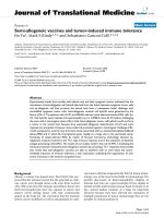
Neuron-microglia interactions induce elevated expression of secreted APP (sAPP)Figure 1
Neuron-microglia interactions induce elevated
expression of secreted APP (sAPP). Relative levels of
sAPP were determined in medium from primary cortical neu-
ron cultures treated with IL-1β (0.1–10 ng/mL) and in
medium from IL-1β-treated neuron/microglia mixed cultures.
Proteins in the medium were concentrated by Centricon,
and sAPP was detected by western immunoblot analysis. A)
Representative sAPP immunoblot; B) Densitometric quantifi-
cation of immunoblot results. Values represent the mean ±
SEM of three experiments (error bars are smaller than the
datapoint symbols). At each IL-1β dose, the sAPP levels in
medium of microglial neuronal-mixed cultures were greater
than in medium from neurons cultured alone (p < 0.05).
Journal of Neuroinflammation 2006, 3:5 />Page 3 of 9
(page number not for citation purposes)
had a clinical diagnosis of PD. There were 2 males and 3
females, with an age range of 68–85 y. The patients whose
tissue was examined postmortem, with appropriate con-
sent, were participants in the University of Arkansas for
Medical Sciences National Institute on Aging-sponsored
Alzheimer's Disease Center under University Institutional
Review Board guidelines.
Reagents
Secreted APP was purified from a recombinant expression
system as described previously [15]. Mouse IL-1ra was
from R&D System (Minneapolis MN). Medium, serum,
and B27 supplement for cell cultures were from Invitro-
gen (Grand Island NY).
Cell cultures
Primary neuronal cultures were derived from the cerebral
cortex of fetal Spraque-Dawley rats (embryonic day 18),
as described previously [19]. Experiments using primary
neuronal cells were performed after 8–12 days in culture.
Primary cultures of rat microglia were generated from the
cortical tissue of neonatal (0–3 days) Sprague-Dawley
rats, as described previously [15]. When mixed cultures
were used, primary microglia were added directly to cul-
tures of primary neurons and exposed for 24 h to 0.0 to 10
ng/mL IL-1β. To test the potential role of factors derived
from activated microglia in driving neuronal expression of
substrates of AD- and PD-associated neuropathologies,
cultures of primary neurons were incubated for 24 h with
medium from unactivated primary microglia, or with
medium derived from sAPP-activated (30 nM) microglia.
To determine if such changes in expression were mediated
by IL-1, some of the neuron cultures were treated with IL-
1 receptor antagonist (IL-1ra, 50 ng/mL) before incuba-
tion with conditioned medium from activated microglia.
Pellet implantation
Pellets impregnated with IL-1β (100 ng of recombinant
mouse IL-1β) or control pellets (containing 100 ng of ace-
tone-extracted bovine serum albumin, BSA) were
implanted 2.8 mm caudal to bregma;4.5 mm right of the
midline, and 2.5 mm deep to the pial surface [12].
Twenty-one male Sprague-Dawley rats weighing 264 ± 6 g
were randomly divided into three groups. Eight rats
received implants of IL-1-containing pellets, seven rats
received pellets with BSA impregnation, and six rats
served as un-operated controls. Twenty-one days after
implantation, cortex from the hemisphere contralateral to
the implant was collected for RNA isolation.
RT-PCR
Total RNA was extracted from rat brain tissue with Tri-Rea-
gent (Molecular Research, Cincinnati, OH). RT-PCR was
performed as previously described [12]. The level of α-
synuclein PCR product in each sample was normalized to
that of GAPDH in the same sample.
Western immunoblot assay
Western immunoblot analyses were performed as
described previously [18]. Briefly, equal amounts of total
protein (determined by bicinchonic acid assay) were
resolved on 8–15% SDS/polyacrylamide gels and trans-
ferred to nitrocellulose blots. Blots were probed overnight
at 4°C with either a rabbit anti-α-synuclein polyclonal
antibody, diluted 1:1000 (Chemicon, Temecula CA); or a
rabbit anti-βAPP polyclonal antibody, diluted 0.5 µg/mL
(Stressgen, San Diego, CA; Vector Lab, Burlingame, CA);
or monoclonal phosphorylated-Tau antibody, diluted
1:1000 (AT8, Pierce, Rockford, IL); or MAPK-p38, diluted
1:1000 (BioLabs, Inc., Beverly, MA); or monoclonal syn-
aptophysin antibody, diluted 1:2000 (Roche, Indianapo-
lis, IN); or IL-1β, diluted 0.1 µg/mL (Chemicon,
Temecula, CA) After washing, blots were reacted with
HRP-conjugated goat anti-rabbit antibodies, for polyclo-
nal antibodies or anti-mouse for monoclonal antibodies.
Detection of antibody-antigen binding was performed
with Western-Light™ Chemiluminescent Detection Sys-
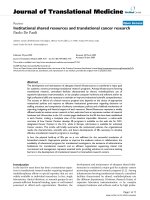
Secreted APP (sAPP) induces dose-dependent increases in the levels of IL-1β released from primary microglial culturesFigure 2
Secreted APP (sAPP) induces dose-dependent
increases in the levels of IL-1β released from primary
microglial cultures. Primary microglia were treated with
the indicated doses of recombinant sAPPα for 24 h. Proteins
in the medium were concentrated by Centricon filter and IL-
1β was analyzed by western immunoblots. A) Representative
IL-1β immunoblot; and B) Densitometric quantification of
immunoblot data from at least three experiments.
Journal of Neuroinflammation 2006, 3:5 />Page 4 of 9
(page number not for citation purposes)
tem (Applied Biosystems, Foster City, CA). For Western
immunoblot analysis of culture medium to detect either
sAPP or IL-1β, media samples were concentrated via filtra-
tion through Centricon YM-10 centrifugal concentrators
(Millipore, Bedford, MA) before analysis as above.
Triple label immunohistochemistry
Analysis was performed on 6-micron thick tissue sections
from formalin-fixed, paraffin-embedded blocks of para-
hippocampal gyri from brains of patients with AD/LBD.
Tissue sections were deparaffinized by passing through a
series of xylenes, and rehydrated through a graded series
of alcohols, to deionized water. For the triple-labeled
immunoreactions in the present study, the four antibod-
ies were combined as follows: AT8 and α-synuclein with
either IL-1α or βAPP. All incubations were followed by
washing in PBS and, unless stated otherwise, were per-
formed at room temperatures. For immunoreactions that
included IL1α, AT8, and α-synuclein, sections were pre-
treated with an antigen retrieval enzyme digestion system
(Digest-All 2, Zymed, South San Francisco, CA), immuno-
reacted at overnight with polyclonal IL-1α antibody
(Peprotech, Rocky Hill, NJ), diluted 1:10 in 2.5% normal
horse serum in PBS, immunoreacted with an ImmPRESS
Reagent anti-rabbit Ig-peroxidase-micropolymerized
reporter enzyme staining system (Vector), followed by
detection with DAB (Zymed) and a 30-min treatment
with Double Stain Enhancer (Zymed). Then the sections
were pretreated for antigen epitope retrieval by microwav-
ing in boiling 1 mM EDTA buffer (pH 8) for 10 min. Next,
the sections were immunoreacted with AT8 antibody
overnight, followed by a 30-min incubation with bioti-
nylated goat-anti mouse antibody, diluted 1:200 in 2%
normal goat serum in PBS, and then a 30-min treatment
with avidin D-conjugated β-galactosidase (Vector),
diluted 1:200 in 2% normal goat serum in PBS, and
finally, detection using X-Gal Substrate Set (KPL). This
antigen detection was followed by α-synuclein antigen
epitope retrieval using two pretreatments: first, a 10-min
microwave pretreatment in boiling 1 mM EDTA buffer
(pH 8), and second, a 2-min incubation in concentrated
formic acid, followed by immunoreaction with a mono-
clonal anti-α-synuclein antibody (Vector), diluted 1:30,
for 2.5 h. This was followed by incubation with polymer-
alkaline-phosphatase-conjugated secondary antibody
(DAKO, Envision Kit), and labeled with permanent red
(DAKO). When the three antigens to be detected were
AT8, βAPP, and α-synuclein, monoclonal anti-βAPP anti-
body (Pierce) was immunoreacted in the place of polyclo-
nal anti-IL-1α antibody in the protocol above as follows:
sections were immunoreacted 30 min with a monoclonal
AT8 antibody (Pierce), diluted 1:1000 in 2.5% normal
horse serum in PBS, and immunoreacted with an
ImmPRESS Reagent anti-mouse Ig-peroxidase-micropoly-
merized reporter enzyme staining system (Vector), fol-
lowed by detection with DAB (Zymed), and then by a 30-
min treatment with Double Stain Enhancer (Zymed). This
was followed by α-synuclein antibody immunoreaction
and detection by the β-galactosidase-avidin D/X-Gal sys-
tem. Finally, anti-βAPP was applied and detected with the
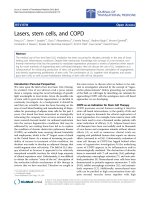
IL-1 mediates microglia-induced elevation of neuronal levels of substrates of the neuropathological changes characteristic of AD and PD and Lewy body pathologiesFigure 3
IL-1 mediates microglia-induced elevation of neuro-
nal levels of substrates of the neuropathological
changes characteristic of AD and PD and Lewy body
pathologies. Primary neurons were cultured with the con-
ditioned medium from either untreated primary microglia
(unCM), or with medium from microglia that had been acti-
vated with 30 nM sAPPα (CM). Parallel neuron cultures
were pretreated for 1 h with IL-1ra (50 ng/mL), followed by
treatment with medium from sAPP-activated microglia (IL-
1ra + CM). A) Representative western immunoblot using
antibodies recognizing βAPP, α-synuclein, phosphorylated
tau, synaptophysin, or p38-MAP kinase (MAPK-p38); B) Den-
sitometric quantification of immunoblot results from dupli-
cate experiments. * = p < 0.05, ** = p < 0.01.
Journal of Neuroinflammation 2006, 3:5 />Page 5 of 9
(page number not for citation purposes)
alkaline-phosphatase-conjugated secondary antibody and
permanent red.
Statistical analyses
The unpaired, two-tailed t-test was used to determine sig-
nificance using the StatView 4.1 statistical analysis pro-
gram.
Results
Mutual induction of IL-1
β
and sAPP in neuronal-microglial
cultures
To test specific degenerative feedback relationships
between neurons and microglia, primary neuronal and
microglial culture models were employed. IL-1β treat-
ment of purified primary neurons, cultured alone, pro-
duced dose-dependent increases in sAPP release over an
IL-1 concentration range of 0.1 and 1.0 ng/mL. These IL-
1β effects were enhanced when the neurons were cultured
together with microglia (Fig. 1). For instance, in such neu-
ronal-microglial cultures, neuronal sAPP induction
required as little as one third the amount of IL-1β required
for a similar elevation of sAPP in the absence of microglia.
One possible explanation for the enhanced overexpres-
sion of sAPP in mixed cultures, relative to that seen in neu-
rons cultured alone, would be a positive feedback
mechanism involving (i) IL-1β-induced sAPP release from
neurons, (ii) sAPP-induced activation of microglia with
induction of further IL-1β, and (iii) further induction of
neuronal sAPP release by the increased levels of IL-1β. In
support of this possibility, 24-h exposures of purified
microglial cultures to recombinant sAPPα resulted in
dose-dependent elevations of IL-1β expression (Fig. 2).
The effects of conditioned media from sAPP-activated
microglia on neuronal expression of
α
-synuclein,
β
APP,
phospho-MAPK-p38, and phospho-tau are mediated by
IL-1
To test the consequences of neuron-microglia interactions
as they may apply to AD- and PD-related aspects of neu-
rodegeneration, we assessed the role of IL-1 in the effects
of conditioned medium from sAPP-activated microglia on
neuronal cultures. Such exposure to sAPP resulted in an
increase in the levels of α-synuclein that was mediated by
IL-1, as demonstrated by suppression of α-synuclein
expression by pretreatment of the neuron cultures with
the natural IL-1 receptor antagonist IL-1ra (Fig. 3). α-
Synuclein is a necessary component of the Lewy bodies
that characteristize neuropathological features of both PD
and dementia with Lewy bodies, and that often co-exist
with the characteristic neuropathological features of AD.
In addition to increased levels of α-synuclein, we found
altered expression of other markers of neuropathological
changes that have been found in AD, PD and AD/LBD. We
found increases in the expression of i) βAPP – the precur-
sor of Aβ and sAPP; ii) activated (phosphorylated) MAPK-
p38, a tau kinase; and iii) phosphorylated tau, of the pri-
mary component of paired helical filaments and neurofi-
brillary tangles. Conversely, and coincident with these
increases, we found iv) decreased levels of synaptophysin,
a marker of synaptic integrity (Fig. 3). To test the role of
IL-1 in these events, parallel cultures of neurons were pre-
treated for 1 h with IL-1ra (50 ng/mL) before application
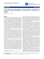
IL-1 induction of α-synuclein is dose- and time-dependentFigure 4
IL-1 induction of α-synuclein is dose- and time-
dependent. Representative western α-synuclein (α-synuc)
immunoblots of proteins from neurons treated with IL-1β
for 24 h at the doses indicated (A) or with IL-1β at 30 ng/mL
for the times indicated (C). B) and D) Densitometric quanti-
fication of immunoblot results from duplicate experiments.
Journal of Neuroinflammation 2006, 3:5 />Page 6 of 9
(page number not for citation purposes)
of the conditioned medium from sAPP-activated micro-
glia. This pretreatment suppressed the increases in α-synu-
clein, βAPP, phosphorylated-tau, and activated MAPK-
p38.
To more carefully characterize the effect of IL-1β on α-
synuclein expression, dependencies on dose and time
were assessed in cultures of neurons. As little as 3.0 ng/mL
IL-1β was an adequate dose to effect a substantial eleva-
tion of α-synuclein levels in neurons (Fig. 4A, B). Such
elevations were apparent within 6 h of treatment (Fig. 4C,
D).
IL-1
β
induces
α
-synuclein in vivo
We next sought to extend our in vitro findings regarding
IL-1-induced elevation of α-synuclein and of other pro-
teins associated with neurodegenerative events in AD to
an in vivo model. Pellets that slowly release IL-1β over a
period of 21 days were implanted into the cerebral cortex
of Sprague-Dawley rats; "sham" rats received pellets con-
taining BSA [12]. IL-1β administration resulted in signifi-
cant elevations in α-synuclein mRNA levels relative to
either the levels observed in BSA "sham" animals (p <
0.02) or unoperated controls (p = 0.0003) (Fig. 5). Other
markers of neurodegeneration elevated by IL-1β in vitro
also showed elevated expression in cortices of rats bearing
IL-1β pellets (Fig. 5).
To determine whether the IL-1-mediated events observed
in vitro and in brains of experimental animals are paral-
leled and mutually associated in AD/LBD, we examined
histological sections from autopsy material. Figure 6 illus-
trates triple-label immunohistochemistry confirming
colocalization of neurofibrillary tangles (anti-AT8) with
Lewy body (anti-α-synuclein) staining (Fig. 6A), and the
tell-tale accompaniment of microglial cell activation with
over-expression of IL-1 (Fig. 6B).
Discussion
We examined the effects of microglial activation on sub-
strates of neuropathological elements of AD, PD, and AD/
LBD using in vitro cell culture experiments and an in vivo
pellet-implantation model. Activated microglia were
found to elevate neuronal synthesis of βAPP and release of
sAPP and increase levels of α-synuclein and phosphor-
ylated-tau; microglial release of IL-1 appeared to be essen-
tial for each of these neuronal events. Moreover, a positive
feedback mechanism was documented through which IL-
1-induced sAPPα release stimulates further microglial IL-
1β production. Chronic intracerebral delivery of IL-1β
produced gene-expression changes consistent with the
events observed in culture. Finally, we found that acti-
vated microglia overexpressing IL-1 colocalize with both
AD- and PD-associated markers of neuropathology in
human brain. These results, together with previous obser-
vations [6,20-22] suggest that microglia-derived IL-1 and
neuron-derived sAPP are involved in a vicious circle of
glia-neuronal interactions, which over time can precipi-
tate neurodegenerative consequence common to both AD
and PD.
The elevation of IL-1β levels by sAPPα and sAPPβ is a
component of the microglial response to these proteins.
In addition to this effect on IL-1β, nanomolar concentra-
tions of both sAPPα and sAPPβ can activate microglia to
express inducible nitric oxide synthase (iNOS), to release
glutamate and D-serine, and to exhibit neurotoxicity; but
sAPPβ lacks the balancing neuroprotective action of
Chronic exposure to excess IL-1β elevates expression of α-synuclein and other markers of AD and PD pathologies in vivoFigure 5
Chronic exposure to excess IL-1β elevates expression
of α-synuclein and other markers of AD and PD
pathologies in vivo. Slow-release pellets containing IL-1β
(IL-1 pellet) or BSA (Sham) were implanted into the cere-
brum of adult Sprague-Dawley rats, and unoperated age-
matched rats served as controls (Control). After 21 days in
all cases, cerebral tissue was homogenized and RNA was iso-
lated. RT-PCR was performed using primers specific for α-
synuclein (α-synuc), βAPP, tau, or MAPK-p38. GAPDH was
also analyzed as a quantitative control. A) RT-PCR results;
B) Quantification of results by densitometric analyses. * = p
< 0.05, ** = p < 0.01.
Journal of Neuroinflammation 2006, 3:5 />Page 7 of 9
(page number not for citation purposes)
sAPPα [15,17,18,20]. As sAPPα and sAPPβ differ only at
the carboxyterminus, their similar effects on microglial
activation imply that a domain in the aminoterminus is
responsible for such proinflammatory activity, confirmed
by mutagenesis [15,20]. A receptor-binding event is fur-
ther suggested by the fact that this same region of sAPP is
required for binding by apolipoprotein E, an event that
blocks microglial activation by sAPPs, possibly by stearic
interference with sAPP binding events [20]. A receptor
that mediates these effects has not been identified, but it
may be related to that which mediates an activation of
ERKs in PC12 cells [23]. This is especially compelling con-
sidering that sAPPα activates ERKs in microglia, where
MAP kinases are necessary for microglial activation by
sAPPα [24]. Interestingly, elevation of iNOS levels by
sAPP appears to be mechanistically distinct from the ele-
vation of iNOS by Aβ or LPS [25].
We have previously shown that neuronal stress increases
neuronal synthesis of βAPP and secretion of sAPP, which
in turn activates microglia and increases IL-1 synthesis
[20] and release [12]. IL-1 is sufficient to elevate expression
and processing of βAPP, which could favor production of
either amyloid β-peptide or sAPPα [26]. The mechanisms
involved in the elevation of βAPP processing appear to be
complex. IL-1 can apparently elevate the expression of
βAPP by transcriptional [27] and post-transcriptional
events [28]. Once expressed, βAPP can be dramatically
altered in its processing by IL-1 [29,30]. The cytokine has
been shown to elevate processing by both γ-secretase [31]
and α-secretase [32]. The latter, relevant to the stimula-
tion of sAPP levels by IL-1 reported here, appears to
require MAP kinases of the MKK1 and JNK classes, at least
in neuroglioma cells [32].
In addition to its sufficiency for βAPP expression and
processing, we show here that IL-1 is also necessary for the
pathogenic signaling that activated microglia exert on
neurons leading to diverse pathological changes, includ-
ing elevation of α-synuclein levels. IL-1 also induces tau
phosphorylation and depression of synaptophysin levels
[18], actions that are consistent with AD pathology. Thus,
an inflammatory cytokine can lead to the convergence of
neuropathological events that are traditionally considered
AD-associated (e.g., NFTs) with some that are not (e.g.,
Lewy bodies). The particular constellation of neuropatho-
logical markers occurring in association with one another
in a given individual or a given neuron may depend upon
coincidences of genetics, neurochemical profile, or vari-
ous other autonomous variables rather than any differen-
tial the exposure to IL-1.
Our results suggest that the contributions of IL-1 to AD
pathology include influences of this cytokine on synaptic
function [33]; namely, (i) a decrease in synaptophysin,
perhaps indicative of inflammation-related synapse loss,
and (ii) an increase in α-synuclein, perhaps in an attempt
to repair this very event [34]. The effect of IL-1β on α-
synuclein in cultures of primary neurons is generally con-
sistent with prior reports that the cytokine induces α-
synuclein protein in macrophages [35] and a glioma cell
line but not in primary astrocytes [36]. Negative data were
also acquired in a teratocarcinoma cell line, albeit one
that is similar to neurons when differentiated [37]. These
findings do not refute the data we obtained with post-
mitotic, primary neurons. α-Synuclein is normally found
as an abundant synaptic vesicle protein of unclear func-
tion. And while it can be found in glia under both normal
[38] and pathological conditions [39], its accumulation is
much more prominent as Lewy bodies in neurons in PD
and AD/LBD [40,41]. Mice transgenically modified to
overproduce human forms of both Aβ and α-synuclein
have higher rates of α-synuclein aggregation [42], a phe-
nomenon that now appears to have been confirmed in
humans with Lewy body disease [43].
Conclusion
Our findings regarding the involvement of IL-1 in micro-
glia activation-induced neuronal overexpression of βAPP
and α-synuclein provide a mechanistic link between neu-
ronal stress, microglial activation, IL-1 overexpression,
and sAPP-driven events, leading to the recognized neu-
ropathological changes that encompass both AD and PD.
These interrelated events appear to be triggered in condi-
Colocalization of activated microglia, overexpressing IL-1α, and markers of neuropathological changes in AD-LBDFigure 6
Colocalization of activated microglia, overexpressing
IL-1α, and markers of neuropathological changes in
AD-LBD. A) A neuron with a Lewy body (blue), neurofibril-
lary tangles (brown), and βAPP (red) colocalization. B) Local-
ization of an activated microglia, overexpressing IL-1α
(brown), with a neuron-like structure, containing a Lewy
body (red) and neurofibrillary tangles (blue). Scale bar repre-
sents 15 µ (A) or 5 µ (B).
Journal of Neuroinflammation 2006, 3:5 />Page 8 of 9
(page number not for citation purposes)
tions that increase risk for precocious development of AD,
as well [22,44]. Based on the quantitative studies reported
here, particularly the observations in rats implanted with
IL-1-containing pellets, one could predict that any neural
condition involving IL-1 overexpression would exhibit
concomitant pernicious alterations in the substrates asso-
ciated with the hallmark neuropathologies in either AD or
PD, alone or together. IL-1-mediated downstream conse-
quences may favor manifestations or precocious develop-
ment of AD, PD, or AD/LBD, a distinction likely
dependent on the originating insult or the specific neuro-
nal cell type first affected. For instance, in Down's syn-
drome and familial AD, genetic forces are the most likely
culprits; in head injury and epilepsy, direct neuronal
trauma; in AIDS, viral infection of microglia; and in PD
and sporadic AD, unknown insults directed in a cell-spe-
cific manner to the affected neurons. As we show here,
excessive levels of IL-1 can alter the expression of the neu-
ronal substrates of AD, PD, and AD/LBD, thus predispos-
ing the brain for establishment of a cycle of neuronal
compromise, consequential activation of glia, and further
IL-1 overexpression. Therefore, because of the potential of
IL-1 to drive alterations in neuronal expression of disease
substrates, we propose overexpression of CNS IL-1 as a
drug discovery target for therapeutic intervention in IL-1-
mediated self-propagating cycles.
Competing interests
The author(s) declare that they have no competing inter-
ests.
Authors' contributions
This study is based on an original idea of WSTG, who
directed the work with SWB. SWB, LL, and WSTG wrote
the manuscript. REM provided expertise on Lewy body
disease, provided neuropathological evaluation of tissues,
and participated in preparation of the final manuscript.
SWB produced the recombinant sAPP. LL, in collabora-
tion with YL, performed the experiments, constructed the
figures, and made important conceptual contributions.
Acknowledgements
The authors especially thank Richard Jones, Sue Woodward, and Rajshek-
har Kore for technical support and Pam Free for secretarial support. This
work was supported in part by NIH grants AG12411, AG19606, HD37989,
AG17498; by a grant from the Alzheimer's Association; and by endow-
ments from The Pat and Willard Walker Family Foundation and the Donald
W. Reynolds Foundation. The authors particularly thank the generous
donations from Alzheimer's Disease Center participants and their families.
References
1. Galvin JE, Lee VM, Trojanowski JQ: Synucleinopathies: clinical
and pathological implications. Arch Neurol 2001, 58:186-190.
2. Hurtig HI, Trojanowski JQ, Galvin J, Ewbank D, Schmidt ML, Lee VM,
Clark CM, Glosser G, Stern MB, Gollomp SM, Arnold SE: α-Synu-
clein cortical Lewy bodies correlate with dementia in Parkin-
son's disease. Neurology 2000, 54:1916-1921.
3. Lippa CF, Schmidt ML, Lee VM, Trojanowski JQ: α-Synuclein in
familial Alzheimer disease: epitope mapping parallels
dementia with Lewy bodies and Parkinson disease. Arch Neu-
rol 2001, 58:1817-1820.
4. Mirza B, Hadberg H, Thomsen P, Moos T: The absence of reactive
astrocytosis is indicative of a unique inflammatory process in
Parkinson's disease. Neuroscience 2000, 95:425-432.
5. Mrak RE, Griffin WS: Glia and their cytokines in progression of
neurodegeneration. Neurobiol Aging 2005, 26:349-354.
6. Griffin WS, Stanley LC, Ling C, White L, MacLeod V, Perrot LJ, White
CLd, Araoz C: Brain interleukin 1 and S-100 immunoreactivity
are elevated in Down syndrome and Alzheimer disease. Proc
Natl Acad Sci USA 1989, 86:7611-7615.
7. Stanley LC, Mrak RE, Woody RC, Perrot LJ, Zhang S, Marshak DR,
Nelson SJ, Griffin WS: Glial cytokines as neuropathogenic fac-
tors in HIV infection: pathogenic similarities to Alzheimer's
disease. J Neuropathol Exp Neurol 1994, 53:231-238.
8. Esiri MM, Biddolph SC, Morris CS: Prevalence of Alzheimer
plaques in AIDS. J Neurol Neurosurg Psychiatry 1998, 65:29-33.
9. Sheng JG, Boop FA, Mrak RE, Griffin WS: Increased neuronal β-
amyloid precursor protein expression in human temporal
lobe epilepsy: association with interleukin-1 α immunoreac-
tivity. J Neurochem 1994, 63:1872-1879.
10. Mackenzie IR, Miller LA: Senile plaques in temporal lobe epi-
lepsy. Acta Neuropathol (Berl) 1994, 87:504-510.
11. Griffin WS, Sheng JG, Gentleman SM, Graham DI, Mrak RE, Roberts
GW: Microglial interleukin-1 α expression in human head
injury: correlations with neuronal and neuritic β-amyloid
precursor protein expression. Neurosci Lett 1994, 176:133-136.
12. Li Y, Liu L, Kang J, Sheng JG, Barger SW, Mrak RE, Griffin WS: Neu-
ronal-glial interactions mediated by interleukin-1 enhance
neuronal acetylcholinesterase activity and mRNA expres-
sion. J Neurosci 2000, 20:149-155.
13. Toiber D, Soreq H: Cellular stress reactions as putative cholin-
ergic links in Alzheimer's disease. Neurochem Res 2005,
30:909-919.
14. Wenk GL, McGann K, Hauss-Wegrzyniak B, Rosi S: The toxicity of
tumor necrosis factor-α upon cholinergic neurons within the
nucleus basalis and the role of norepinephrine in the regula-
tion of inflammation: implications for Alzheimer's disease.
Neuroscience 2003, 121:719-729.
15. Barger SW, Basile AS: Activation of microglia by secreted amy-
loid precursor protein evokes release of glutamate by cys-
tine exchange and attenuates synaptic function. J Neurochem
2001, 76:846-854.
16. Rosi S, Ramirez-Amaya V, Hauss-Wegrzyniak B, Wenk GL: Chronic
brain inflammation leads to a decline in hippocampal
NMDA-R1 receptors. J Neuroinflammation 2004, 1:12.
17. Wu SZ, Bodles AM, Porter MM, Griffin WS, Basile AS, Barger SW:
Induction of serine racemase expression and D-serine
release from microglia by amyloid β-peptide. J Neuroinflamma-
tion 2004, 1:2.
18. Li Y, Liu L, Barger SW, Griffin WS: Interleukin-1 mediates path-
ological effects of microglia on tau phosphorylation and on
synaptophysin synthesis in cortical neurons through a p38-
MAPK pathway. J Neurosci 2003, 23:1605-1611.
19. Li Y, Wang J, Sheng JG, Liu L, Barger SW, Jones RA, Van Eldik LJ, Mrak
RE, Griffin WS: S100 β increases levels of β-amyloid precursor
protein and its encoding mRNA in rat neuronal cultures. J
Neurochem 1998, 71:1421-1428.
20. Barger SW, Harmon AD: Microglial activation by Alzheimer
amyloid precursor protein and modulation by apolipopro-
tein E. Nature 1997, 388:878-881.
21. Sheng JG, Jones RA, Zhou XQ, McGinness JM, Van Eldik LJ, Mrak RE,
Griffin WS: Interleukin-1 promotion of MAPK-p38 overex-
pression in experimental animals and in Alzheimer's disease:
potential significance for tau protein phosphorylation. Neuro-
chem Int 2001, 39:341-348.
22. Griffin WS, Mrak RE: Interleukin-1 in the genesis and progres-
sion of and risk for development of neuronal degeneration in
Alzheimer's disease. J Leukoc Biol 2002, 72:233-238.
23. Greenberg SM, Koo EH, Selkoe DJ, Qiu WQ, Kosik KS: Secreted β-
amyloid precursor protein stimulates mitogen-activated
protein kinase and enhances tau phosphorylation. Proc Natl
Acad Sci USA 1994, 91:7104-7108.
Publish with BioMed Central and every
scientist can read your work free of charge
"BioMed Central will be the most significant development for
disseminating the results of biomedical research in our lifetime."
Sir Paul Nurse, Cancer Research UK
Your research papers will be:
available free of charge to the entire biomedical community
peer reviewed and published immediately upon acceptance
cited in PubMed and archived on PubMed Central
yours — you keep the copyright
Submit your manuscript here:
/>BioMedcentral
Journal of Neuroinflammation 2006, 3:5 />Page 9 of 9
(page number not for citation purposes)
24. Bodles AM, Barger SW: Secreted β-amyloid precursor protein
activates microglia via JNK and p38-MAPK. Neurobiol Aging
2005, 26:9-16.
25. Barger SW, Chavis JA, Drew PD: Dehydroepiandrosterone
inhibits microglial nitric oxide production in a stimulus-spe-
cific manner. J Neurosci Res 2000, 62:503-509.
26. Mrak RE, Griffin WS: Interleukin-1 and the immunogenetics of
Alzheimer disease. J Neuropathol Exp Neurol 2000, 59:471-476.
27. Goldgaber D, Harris HW, Hla T, Maciag T, Donnelly RJ, Jacobsen JS,
Vitek MP, Gajdusek DC: Interleukin 1 regulates synthesis of
amyloid β-protein precursor mRNA in human endothelial
cells. Proc Natl Acad Sci USA 1989, 86:7606-7610.
28. Rogers JT, Randall JD, Cahill CM, Eder PS, Huang X, Gunshin H, Leiter
L, McPhee J, Sarang SS, Utsuki T, Greig NH, Lahiri DK, Tanzi RE, Bush
AI, Giordano T, Gullans SR: An iron-responsive element type II
in the 5'-untranslated region of the Alzheimer's amyloid pre-
cursor protein transcript. J Biol Chem 2002, 277:45518-45528.
29. Blasko I, Veerhuis R, Stampfer-Kountchev M, Saurwein-Teissl M, Eike-
lenboom P, Grubeck-Loebenstein B: Costimulatory effects of
interferon-γ and interleukin-1β or tumor necrosis factor α on
the synthesis of Aβ 1–40 and Aβ 1–42 by human astrocytes.
Neurobiol Dis 2000, 7:682-689.
30. Buxbaum JD, Oishi M, Chen HI, Pinkas-Kramarski R, Jaffe EA, Gandy
SE, Greengard P: Cholinergic agonists and interleukin 1 regu-
late processing and secretion of the Alzheimer β/A4 amyloid
protein precursor. Proc Natl Acad Sci USA 1992, 89:10075-10078.
31. Liao YF, Wang BJ, Cheng HT, Kuo LH, Wolfe MS: Tumor necrosis
factor-α, interleukin-1β, and interferon-γ stimulate γ-secre-
tase-mediated cleavage of amyloid precursor protein
through a JNK-dependent MAPK pathway. J Biol Chem 2004,
279:49523-49532.
32. Ma G, Chen S, Wang X, Ba M, Yang H, Lu G: Short-term inter-
leukin-1(β) increases the release of secreted APP(α) via
MEK1/2-dependent and JNK-dependent α-secretase cleav-
age in neuroglioma U251 cells. J Neurosci Res 2005, 80:683-692.
33. Vereker E, O'Donnell E, Lynch MA: The inhibitory effect of inter-
leukin-1β on long-term potentiation is coupled with
increased activity of stress-activated protein kinases. J Neuro-
sci 2000, 20:6811-6819.
34. Chandra S, Gallardo G, Fernandez-Chacon R, Schluter OM, Sudhof
TC: α-Synuclein cooperates with CSPα in preventing neuro-
degeneration. Cell 2005, 123:383-396.
35. Tanji K, Mori F, Imaizumi T, Yoshida H, Matsumiya T, Tamo W, Yoshi-
moto M, Odagiri H, Sasaki M, Takahashi H, Satoh K, Wakabayashi K:
Upregulation of α-synuclein by lipopolysaccharide and inter-
leukin-1 in human macrophages. Pathol Int 2002, 52:572-577.
36. Tanji K, Imaizumi T, Yoshida H, Mori F, Yoshimoto M, Satoh K,
Wakabayashi K: Expression of α-synuclein in a human glioma
cell line and its up-regulation by interleukin-1β. Neuroreport
2001, 12:1909-1912.
37. Satoh JI, Kuroda Y: α-Synuclein expression is up-regulated in
NTera2 cells during neuronal differentiation but unaffected
by exposure to cytokines and neurotrophic factors. Parkinson-
ism Relat Disord 2001, 8:7-17.
38. Mori F, Tanji K, Yoshimoto M, Takahashi H, Wakabayashi K: Dem-
onstration of α-synuclein immunoreactivity in neuronal and
glial cytoplasm in normal human brain tissue using protein-
ase K and formic acid pretreatment. Exp Neurol 2002,
176:98-104.
39. Wakabayashi K, Hayashi S, Yoshimoto M, Kudo H, Takahashi H:
NACP/α-synuclein-positive filamentous inclusions in astro-
cytes and oligodendrocytes of Parkinson's disease brains.
Acta Neuropathol (Berl) 2000, 99:14-20.
40. Baba M, Nakajo S, Tu PH, Tomita T, Nakaya K, Lee VM, Trojanowski
JQ, Iwatsubo T: Aggregation of α-synuclein in Lewy bodies of
sporadic Parkinson's disease and dementia with Lewy bod-
ies. Am J Pathol 1998, 152:879-884.
41. Campbell BC, Li QX, Culvenor JG, Jakala P, Cappai R, Beyreuther K,
Masters CL, McLean CA: Accumulation of insoluble α-synuclein
in dementia with Lewy bodies. Neurobiol Dis 2000, 7:192-200.
42. Masliah E, Rockenstein E, Veinbergs I, Sagara Y, Mallory M, Hashimoto
M, Mucke L: β-amyloid peptides enhance α-synuclein accumu-
lation and neuronal deficits in a transgenic mouse model
linking Alzheimer's disease and Parkinson's disease. Proc Natl
Acad Sci USA 2001, 98:12245-12250.
43. Pletnikova O, West N, Lee MK, Rudow GL, Skolasky RL, Dawson TM,
Marsh L, Troncoso JC: Aβ deposition is associated with
enhanced cortical α-synuclein lesions in Lewy body diseases.
Neurobiol Aging 2005, 26:1183-1192.
44. Rothwell N: Interleukin-1 and neuronal injury: mechanisms,
modification, and therapeutic potential. Brain Behav Immun
2003, 17:152-157.