báo cáo hóa học: " Boosting with intranasal dendrimeric Aβ1–15 but not Aβ1–15 peptide leads to an effective immune response following a single injection of Aβ1–40/42 in APP-tg mice" ppt
Bạn đang xem bản rút gọn của tài liệu. Xem và tải ngay bản đầy đủ của tài liệu tại đây (629.65 KB, 10 trang )
BioMed Central
Page 1 of 10
(page number not for citation purposes)
Journal of Neuroinflammation
Open Access
Research
Boosting with intranasal dendrimeric Aβ1–15 but not Aβ1–15
peptide leads to an effective immune response following a single
injection of Aβ1–40/42 in APP-tg mice
Timothy J Seabrook, Liying Jiang, Katelyn Thomas and Cynthia A Lemere*
Address: Center for Neurologic Diseases, Brigham and Women's Hospital, Harvard Medical School, Boston, MA 02115, USA
Email: Timothy J Seabrook - ; Liying Jiang - ;
Katelyn Thomas - ; Cynthia A Lemere* -
* Corresponding author
Abstract
Background: Immunotherapy for Alzheimer's disease (AD) is emerging as a potential treatment.
However, a clinical trial (AN1792) was halted after adverse effects occurred in a small subset of
subjects, which may have been caused by a T cell-mediated immunological response. In general,
aging limits the humoral immune response, therefore, immunogens and vaccination regimes are
required that induce a strong antibody response with less potential for an adverse immune
response.
Method: In the current study, we immunized both wildtype and J20 APP-tg mice with a priming
injection of Aβ1–40/42, followed by multiple intranasal boosts with the novel immunogen dAβ1–
15 (16 copies of Aβ1–15 on a lysine tree), Aβ1–15 peptide or Aβ1–40/42 full length peptide.
Results: J20 APP-tg mice primed with Aβ1–40/42 subcutaneously and subsequently boosted
intranasally with Aβ1–15 peptide did not generate a cellular or humoral immune response. In
contrast, J20 APP-tg mice boosted intranasally with dAβ1–15 or full length Aβ1–40/42 produced
high levels of anti-Aβ antibodies. Splenocyte proliferation was minimal in mice immunized with
dAβ1–15. Wildtype littermates of the J20 APP-tg mice produced higher amounts of anti-Aβ
antibodies compared to APP-tg mice but also had low T cell proliferation. The anti-Aβ antibodies
were mainly composed of IgG2b and directed to an epitope within the Aβ1–7 region, regardless of
the immunogen. Examination of the brain showed a significant reduction in Aβ plaque burden in
the J20 APP-tg mice producing antibodies compared to controls. Biochemically, Aβ40 or Aβ42
were also reduced in brain homogenates and elevated in plasma but the changes did not reach
significance.
Conclusion: Our results demonstrate that priming with full length Aβ40/42 followed by boosting
with dAβ1–15 but not Aβ1–15 peptide led to a robust humoral immune response with a minimal
T cell response in J20 APP-tg mice. In addition, Aβ plaque burden was reduced in mice producing
anti-Aβ antibodies. Interestingly, wildtype mice produced higher levels of anti-Aβ antibodies,
indicating that immune tolerance may be present in J20 APP-tg mice. Together, these data suggest
that dAβ1–15 but not Aβ1–15 peptide may be useful as a boosting immunogen in an AD vaccination
regime.
Published: 05 June 2006
Journal of Neuroinflammation 2006, 3:14 doi:10.1186/1742-2094-3-14
Received: 14 April 2006
Accepted: 05 June 2006
This article is available from: />© 2006 Seabrook et al; licensee BioMed Central Ltd.
This is an Open Access article distributed under the terms of the Creative Commons Attribution License ( />),
which permits unrestricted use, distribution, and reproduction in any medium, provided the original work is properly cited.
Journal of Neuroinflammation 2006, 3:14 />Page 2 of 10
(page number not for citation purposes)
Background
Alzheimer's disease (AD) is a devastating disease charac-
terized by a progressive deterioration in cognitive abili-
ties, eventually leading to severe dementia. Pathologically
there is localized deposition of cerebral β-amyloid (Aβ)
protein, neuritic plaques, glial activation, neurofibrillary
tangle formation, and neuronal loss [1]. The cause of AD
is still an area of debate however, there is accumulating
epidemiologic, pathologic and genetic evidence that Aβ
has a pivotal role in the pathogenesis of AD, suggesting
that therapies to inhibit its production, enhance its degra-
dation or improve its clearance from the brain would be
therapeutic [2]. One such avenue of investigation is Aβ
immunotherapy. Schenk et al. demonstrated that immu-
nizing APP transgenic mice (APP-tg) with Aβ peptide
resulted in a lowering of cerebral Aβ deposition [3]. Sev-
eral subsequent studies demonstrated the importance of
antibody mediated clearance of Aβ and its role in improv-
ing cognition [4-7]. Recently it has been demonstrated
that non-B cell mechanisms may also have a role in clear-
ing cerebral Aβ [8]. A multi-center Aβ vaccine human clin-
ical trial (AN1792) was initiated but was suspended when
approximately 6% of the subjects experienced symptoms
of meningoencephalitis [9-11]. To date, three autopsy
case reports from AN1792 participants demonstrated a
reduction in Aβ plaque number compared to controls [12-
14]. However, a T cell infiltrate was present in the lep-
tomeninges, perivascular spaces and parenchyma of the
brain in two of the cases, suggesting a T cell mediated
immune response to the vaccination. In the other report,
there was little evidence of overt inflammation at the time
of death [14] however, this does not rule out that inflam-
mation had been present but resolved by the time of
autopsy. Therefore, Aβ based immunotherapy has poten-
tial but more research is required to determine why a sub-
set of patients experienced adverse outcomes.
We and others have demonstrated that the B cell epitope
in humans [15], monkeys [16] and mice [17-19] is located
in the Aβ1–15 region, whereas the T cell epitope has been
mapped to within Aβ15–42 [20,21]. Based on these non-
overlapping epitopes, fragments of Aβ spanning the B cell
epitope but not the T cell epitopes may avoid a deleterious
cellular immune response. Prior reports have suggest that
this may be true as shorter Aβ fragments conjugated to T
cell helper epitopes [22] or mutated Aβ [23,24], have lead
to a humoral immune response. Recent reports using mul-
tiple antigen peptides (MAP) have demonstrated that Aβ
fragments on a branching lysine tree results in a humoral
immune response [18,25]. We have previously demon-
strated that immunization using Aβ1–15 peptide plus the
adjuvant LT(R192G) as both the priming and boosting
immunogen does not induce a humoral immune
response. However, when Aβ1–15 was given intranasally
as a boosting immunogen following a priming injection
of Aβ1–40/42, specific Aβ antibodies were detected in
wildtype B6D2F1 mice [26]. To date there has been little
data regarding the T cell response to various Aβ immuno-
gens, which is important for the design of a safer AD vac-
cine.
The purpose of these experiments was to determine if a
single subcutaneous injection of full length Aβ plus the
adjuvant LT(R192G) (priming dose) followed by multiple
intranasal boosts with either dendrimeric Aβ1–15 (dAβ1–
15, 16 copies of Aβ1–15 on a branching lysine tree), Aβ1–
15 peptide (a single copy of Aβ1–15) or Aβ1–40/42
would result in a humoral immune response. The main
advantage of the prime/boost strategy is the increased
immune response due to the priming with the full-length
peptide and then focusing the immune response to a spe-
cific region by boosting with a smaller peptide. The use of
the whole peptide increases the chances that the immune
system will recognize the peptide and initiate the immune
response. The ability of these vaccination strategies to
avoid a T cell response was measured using splenocyte
proliferation assays and cytokine specific ELISAs. The effi-
cacy of the different immunogens plus the adjuvant
LT(R192G) to reduce cerebral Aβ and the attending
pathology was examined.
Methods
Animals
Heterozygous J20 APP-tg mice (APP
sw
and
V717F
) (C57BL/
6 X DBA2 background) [27] and non-transgenic litterma-
tes were from our breeding colony and vaccination was
begun at 4.0 ± 0.1 months of age. Mice were genotyped
using PCR. All animal use was approved by the Harvard
Standing Committee for Animal Use and in compliance
with all state and federal regulations
Immunization
All peptides used in these studies, Aβ1–40, Aβ1–42, Aβ1–
15 and dAβ1–15, were synthesized by the Biopolymer
Laboratory, Center for Neurologic Diseases (Boston, MA).
Aβ1–15 peptide is a single copy of the first 15 amino acids
of Aβ, whereas dAβ1–15 consists of 16 copies of the same
peptide on a lysine backbone. The peptides were diluted
in distilled water at 4 mg/ml. For the full length Aβ1–40/
42 immunogen, a mixture of Aβ1–40 (3 mg/ml) and
Aβ1–42 (1 mg/ml) in distilled water was incubated over-
night at 37°C. Synthetic Aβ1–42 assembles into a variety
of structures in aqueous buffers, including low n-oligom-
ers, ADDLs, protofibrils and fibrils [28]. The solutions of
synthetic Aβ used in this study probably contained a mix-
ture of these assemblies, but biophysical analysis was not
performed to determine the presence or relative abun-
dance of these species. Circular dichroism analysis of the
dAβ1–15 peptide demonstrated a random structure, with-
out α or β sheet structures (data not shown). The immu-
Journal of Neuroinflammation 2006, 3:14 />Page 3 of 10
(page number not for citation purposes)
nogens were aliquotted and frozen at -20°C. The
adjuvant, 5 µg mutant heat labile E.coli enterotoxin,
LT(R192G) (kind gift of J. Clements, Tulane University
School of Medicine, New Orleans, LA) [29], was mixed
with immunogen just prior to immunization.
Mice were given a priming dose of vaccine by a single s.c.
injection composed of 75 µg Aβ40 and 25 µg Aβ42 mixed
with 10 µg LT(R192G). Vehicle control mice received
LT(R192G) alone. Boosting by intranasal vaccination was
performed on a weekly basis as previously reported [30].
Briefly, 100 µg of immunogen plus 5 µg LT(R192G) was
mixed and applied to the naris in 2 separate 15 µl doses,
allowing capillary action to draw the liquid into the nasal
cavity. Control mice received LT(R192G) alone. All vacci-
nations were administered weekly for 6 months.
Plasma and tissue collection
Blood was collected from the tail bi-weekly and the
plasma frozen at -20°C. One week following the final
immunization, mice were sacrificed by CO
2
inhalation.
Blood was collected by cardiac puncture followed by per-
fusion with 10 ml Tris buffered saline (TBS). The spleen
was aseptically removed and placed in RPMI on ice for cell
culture studies. The brain was removed and divided sagit-
tally into two hemispheres. One hemi-brain, as well as
pieces of liver, kidney and spleen was placed in 10% buff-
ered neutral buffered formalin for 2 hours, processed, and
embedded in paraffin. The other hemi-brain was snap fro-
zen and stored at -80°C for biochemical analysis of Aβ.
Anti-A
β
antibody ELISA
Anti-Aβ antibodies in plasma were measured by ELISA as
previously described [30]. ELISAs for antibody isotypes
and epitope mapping were performed as previously
reported [31]. Briefly, quantitative Ig isotype-specific ELI-
SAs for IgG1, IgG2a, IgG2b, IgA and IgM anti-Aβ antibod-
ies were performed by adding a standard curve of the
appropriate Ig isotype (Southern Biotechnology Associ-
ates, Birmingham, AL) to the standard immunoassay and
using biotinylated isotype specific secondary antibodies
(Zymed, San Francisco, CA). Peptide competition assays
to determine antibody B cell epitopes were performed as
previously described [32]. The following overlapping Aβ
fragments (CND Biopolymer Laboratory) were used for
antibody epitope mapping; Aβ1–15, Aβ1–7, Aβ3–9, Aβ6–
20, Aβ11–25, Aβ26–42, and Aβ1–40. Diluted plasma
samples were co-incubated with peptide fragments over-
night and applied to Aβ1–40-coated ELISA plates.
Immunohistochemistry and image analysis
Immunohistochemistry was performed on 12 µm paraffin
sections using the ELITE ABC method (Vector Laborato-
ries, Burlingame, CA) as previously described. [33]. The
following antibodies and dilutions were used to examine
T cells (CD5, 1:50; BD PharMingen, San Jose, CA), B cells
(CD45RC, 1:500; BD PharMingen), activated microglia/
macrophage, (CD45, 1:5000; Serotec, Raleigh, NC) or
astrocytes (GFAP, 1:1000; Dakocytomation, Carpinteria,
CA). Rabbit polyclonal Aβ antibodies DW14 1:1000 and
R1282 1:1000 (gifts of D. Walsh and D. Selkoe, respec-
tively, Center for Neurologic Diseases, Boston, MA) were
used to visualize Aβ deposition. Positive controls (sec-
tions of spleen and brain from mice with experimental
autoimmune encephalitis and aged APP-tg mice) and neg-
ative controls (normal immunoglobulin) were included.
To screen plasma for antibody binding to AD plaques,
paraffin-embedded human AD brain tissue was used as
previously reported [34].
The Perl's Prussian blue method of hemosiderin staining
was performed to examine brains for ferric iron (found in
hemaglobin) as a measure of hemorrhage. Briefly, de-
waxed sections were submerged in a solution of 2% potas-
sium ferrocyanide and 2% HCl for 20 minutes, followed
by rinsing and eosin counterstaining.
Images were captured for quantification from 4–6 sec-
tions of hippocampus using a 4× objective for R1282
staining (Aβ) or a 10× objective for CD45 (microglia) and
GFAP (astrocytes). Acquisition of images was performed
in a single session using a SPOT camera (Sterling Heights,
MI). Computer-assisted image analysis was performed
using IP Lab Spectrum 3.1 Image Analyzer software (Fair-
fax, VA), with the hippocampus as the region of interest
(ROI).
Splenocyte cultures
All cell culture reagents were from Invitrogen (Los Ange-
les, CA). Splenocytes were isolated and harvested using
standard methods as previously reported [32]. Aβ pep-
tides were added to cultures in triplicate at a final concen-
tration of 0, 0.5, 5 or 50 µg/ml. At 48 and 72 hours,
supernatants were collected and analyzed by ELISA for
cytokines. To measure proliferation, 1 µCi of [
3
H]-thymi-
dine was added to cells at 72 h. Eighteen hours later cells
were harvested and thymidine incorporation determined
using a liquid scintillation counter. A stimulation index
was calculated using the following formula: CPM of well
with antigen/CPM with no antigen.
Cytokine ELISA
Cytokine levels were measured in splenocyte supernatants
using matching antibody pairs composed of a capture and
detection antibody for IL-4 and IFN-γ (BD PharMingen).
A
β
ELISA
Both soluble and insoluble brain Aβ levels were deter-
mined. For soluble Aβ levels, frozen hemibrains were
homogenized in 4 volumes of TBS with a protease inhib-
Journal of Neuroinflammation 2006, 3:14 />Page 4 of 10
(page number not for citation purposes)
itor cocktail (Sigma, Saint Louis, MO)). The samples were
centrifuged at 60,000 RPM for 30 minutes at 4°C. The
supernatant was collected and stored at -20°C. TBS insol-
uble Aβ protein was extracted as previously described [35]
using 10 volumes of ice cold guanidine buffer (5 M guani-
dine-HCl/50 mM Tris, pH 8.0). ELISAs specific for human
Aβ
40
, Aβ
42
andtotal Aβ were performed (using antibodies
kindly supplied byELAN Pharmaceuticals) as previously
described [36].
Statistical analysis
Kruskal-Wallis nonparametric one-way ANOVA analysis
was used to determine statistical significance between
groups using Prism version 4.0 (GraphPad Software, San
Diego, CA).
Results
Immunization with dA
β
1–15 or A
β
1–40/42 but not A
β
1–15
peptide results in anti-A
β
antibody production in APP-tg
and wildtype mice
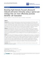
Using a specific ELISA the levels of plasma anti-Aβ anti-
bodies were measured throughout the period of immuni-
zation. Anti-Aβ antibodies were detected after only 3
treatments in mice receiving either dAβ1–15 or full length
Aβ1–40/42 after a priming dose of full length Aβ1–40/42
(Figure 1). No mice receiving either LT(R192G) adjuvant
alone or Aβ1–15 peptide produced antibodies, even
though the Aβ1–15 peptide group received a priming
dose of Aβ1–40/42. Interestingly, the immunized
wildtype mice produced greater amounts of anti-Aβ anti-
bodies compared to their APP-tg littermates. All wildtype
mice produced antibodies in response to immunization,
whereas anti-Aβ antibodies were not detected in a small
percentage of APP-tg mice (Table 1). Plasma from mice
producing anti-Aβ antibodies as measured by ELISA
bound Aβ plaques in cerebral tissue obtained from AD
subjects (data not shown).
Isotypes were examined using specific secondary antibod-
ies in ELISAs. There was a mixture of IgG1, IgG2b, IgG2a
and IgA anti-Aβ antibodies in all mice that generated anti-
bodies in response to vaccination (Table 1). However,
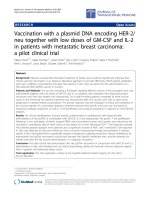
IgG2b and IgG1 were the predominant isotypes. Epitope
mapping demonstrated that the antibodies bound a
region located in the 1–7 region of Aβ, regardless of the
immunogen used (Figure 2).
Limited anti-A
β
cellular immune response found in A
β
vaccinated J20 APP-tg and wildtype mice
Splenocytes were isolated from all J20 APP-tg mice and re-
stimulated in vitro with either Aβ1–40, Aβ1–42 or a mix-
ture of Aβ1–40/42. All peptides were incubated for 24
hours at 37°C to allow fibrils to form. Considering a S.I.
above 2 to be proliferation above background, only J20
APP-tg mice immunized with Aβ1–40/42 showed spleno-
Table 1: Percentage of responders and Aβ antibody isotypes
Strain Immunogen Prime (Y/N) Responder^ IgG1* IgG2a* IgG2b* IgM* IgA*
WT dAβ1–15 Y 4/4 265 ± 89 71 ± 47 257 ± 130 68 ± 22 116 ± 35
WT dAβ1–15 N 4/4 238 ± 105 77 ± 49 742 ± 178 86 ± 21 122 ± 30
APP-tg dAβ1–15 Y 5/7 148 ± 48 75 ± 59 214 ± 99 106 ± 15 46 ± 11
APP-tg Aβ40/42 Y 5/6 45 ± 27 50 ± 48 234 ± 103 76 ± 21 66 ± 17
^ number of mice with detectable plasma anti-Aβ antibodies/total number mice immunized
* µg/ml ± SEM; Y = yes, N = no.
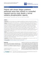
Anti-Aβ antibodies are produced by priming with Aβ1–40/42, followed by boosting with Aβ40/42 or dAβ1–15Figure 1
Anti-Aβ antibodies are produced by priming with
Aβ1–40/42, followed by boosting with Aβ40/42 or
dAβ1–15. J20 APP-tg mice primed with a single subcutane-
ous dose of Aβ1–40/42 with LT(R192G) and subsequently
boosted intranasally with Aβ1–40/42 (n = 6) or dAβ1–15 (n
= 7) produce anti-Aβ antibodies. However, J20 APP-tg mice
receiving adjuvant alone (n = 6) or boosted with Aβ1–15
peptide (n = 8) did not produce any detectable antibodies.
Wildtype littermates immunized with a priming dose of Aβ1–
40/42 and boosted with dAβ1–15 (n = 4) generated signifi-
cantly higher levels of anti-Aβ antibodies compared to any of
the immunized J20 APP-tg mice regardless of treatment
group (*p < 0.05, Kruskal-Wallis one-way ANOVA).
Wildtype mice immunized by dAβ1–15 alone (n = 4) pro-
duced high levels of anti-Aβ antibodies that were significantly
greater than those produced by APP-tg mice primed with
Aβ1–40/42 and boosted with Aβ1–15 and vehicle controls
(**p < 0.05, Kruskal-Wallis one-way ANOVA). Mean ± SEM.
Journal of Neuroinflammation 2006, 3:14 />Page 5 of 10
(page number not for citation purposes)
cyte proliferation after re-stimulation with full length Aβ
peptide, indicating a T cell immune response (Figure 3).
Mice immunized with dAβ1–15, Aβ1–15 peptide or
LT(R192G) alone did not show a significant splenocyte
proliferation (S.I. < 2.0) Cultured splenocytes were re-
stimulated with either Aβ1–40, Aβ1–42 or Aβ1–40/42 to
determine if the species of Aβ had an effect on splenocyte
proliferation. There was no significant difference in the
proliferation when the different peptides were compared.
Using specific and sensitive ELISAs, IFN-γ and IL-4 were
not detected in the splenocyte culture supernatants when
stimulated with any of the tested peptides.
The cellular immune response in dAβ1–15 treated
wildtype littermates was also examined. No splenocyte
proliferation was detected in the group receiving a prim-
ing injection of Aβ40/42, followed by intranasal boosting
with dAβ1–15 or those mice receiving i.n. dAβ1–15 alone
(Figure 4). In addition, cervical lymph nodes were cul-
tured to examine whether these draining lymph nodes
contained Aβ reactive lymphocytes. Similar to the spleno-
cyte cultures, no proliferation was observed (Figure 4).
IFN-γ and IL-4 were not detected in the supernatants from
either the splenocyte or lymph node cultures.
Immunization of J20 APP-tg mice reduces cerebral A
β
plaques and attending pathology
Computer-assisted quantification of immunohistochem-
istry was used to examine the hippocampus of 10 month-
old J20 APP-tg mice following 6 months of immuniza-
tion. Mice that did not produce anti-Aβ antibodies (i.e.,
"non-responders") were excluded from this analysis, as
the purpose of these experiments was to determine if suc-
cessful treatment would decrease cerebral Aβ and its
attending pathology. Immunohistochemistry using poly-
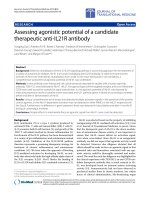
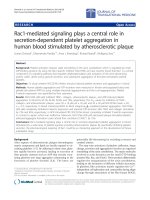
Minimal T cell reactivity to Aβ peptides in dAβ1–15 immu-nized J20 APP-tg miceFigure 3
Minimal T cell reactivity to Aβ peptides in dAβ1–15
immunized J20 APP-tg mice. Splenocytes were isolated,
pooled into treatment groups and stimulated with Aβ1–40
(A), Aβ1–42 (B) or a combination of Aβ1–40/42 (C) in tripli-
cate. Following 72 hours of incubation, 3[H] thymidine was
added and the radioactivity measured after 18 hours. Mice
primed by s.c. injection of Aβ1–40/42 and boosted intrana-
sally with Aβ1–40/42 proliferated more than mice in the
other treatment groups. J20 APP-tg mice boosted with
LT(R192G), Aβ1–15 peptide or dAβ1–15 did not proliferate
to any of the peptides (S.I. <2).
Anti-Aβ antibodies recognize an epitope within the Aβ1–7 regionFigure 2
Anti-Aβ antibodies recognize an epitope within the
Aβ1–7 region. Absorption of diluted plasma from J20 APP-
tg mice producing anti-Aβ antibodies with Aβ1–7, Aβ1–15 or
Aβ1–40 peptide reduced binding to plate-bound Aβ, thereby
demonstrating that the antibodies recognized an epitope
within the Aβ1–7 region.
Journal of Neuroinflammation 2006, 3:14 />Page 6 of 10
(page number not for citation purposes)
clonal anti-Aβ antibodies demonstrated a significant
decrease in the Aβ plaque burden in those mice receiving
a subcutaneous prime of Aβ1–40/42, followed by i.n.
boosting with Aβ1–40/42 or dAβ1–15 (Figures 5). This
decrease was not seen in the groups of J20 APP-tg mice
receiving Aβ1–15 peptide as a boosting immunogen.
To examine the glial response to immunization, GFAP
and CD45, markers for reactive astrocytes and activated
microglia respectively, were examined. At 10 months of
age, limited numbers of compacted plaques and very few
activated microglia were observed in hippocampus of
non-immunized J20 APP-tg mice. For example, CD45
immunoreactivity occupied only 0.56 ± 0.19% of the hip-
pocampus area. There were no significant differences
between any of the treatment groups for either microglial
labeling with anti-CD45 or astrocyte labeling with anti-
GFAP (data not shown), most likely due to the low num-
bers of compacted plaques at this age.
To determine if autoimmune encephalitis was induced by
the immunization protocols used in these experiments,
the brain was examined for the presence of lymphocytes.
No T or B cells were found in any regions of the brain.
Additionally, no instances of micro-hemorrhages were
found as examined by hemosiderin staining.
To confirm the decrease in cerebral Aβ detected immuno-
histochemically, biochemical analysis was performed on
J20 APP-tg mice using specific ELISAs for Aβx-40 and Aβx-
42. There was a non-significant reduction of Aβ40 and
Aβ42 TBS and guanidine soluble levels (commonly
referred to as soluble and insoluble fractions) in both the
Aβ1–40/42 and dAβ1–15 boosted J20 APP-tg groups,
compared to the LT(R192G) and Aβ1–15 peptide groups
(Table 2). Plasma levels of total Aβ were elevated in the
J20 APP-tg mice boosted with Aβ1–40/42 compared to
the other treatment groups.
Discussion
These experiments were performed to examine if a prim-
ing and boosting vaccination protocol using different
immunogens would lead to an effective humoral immune
response and a clearing of cerebral Aβ in an APP-tg mouse
model. This strategy has been successful in the induction
of a cellular immune response [37], however we were
interested if this same immunization regime could be uti-
lized to induce a humoral immune response. We report
that an initial injection of full length Aβ1–40/42 (prim-
ing), followed by subsequent weekly intranasal immuni-
zation with either Aβ1–40/42 or dAβ1–15 (boosting) led
to a humoral immune response. However, no anti-Aβ
antibodies could be detected in the plasma of mice
primed with Aβ1–40/42 and boosted with Aβ1–15 pep-
No T cell reactivity in dAβ1–15 immunized wildtype miceFigure 4
No T cell reactivity in dAβ1–15 immunized wildtype
mice. Cells from the spleen and cervical lymph nodes were
isolated from B6D2F1 mice that received either a prime
injection with Aβ1–40/42 and subsequent boosting intrana-
sally with dAβ1–15 or received intranasal dAβ1–15 alone.
The cells were pooled with respect to treatment group and
tissue and stimulated in vitro with a mixture of Aβ1–40/42.
There was no significant proliferation of cells from either tis-
sue, regardless of the treatment (S.I. <2), thus indicating no
population of anti-AβT cells in the draining lymph nodes of
the nasal mucosa.
Table 2: Cerebral and plasma Aβ levels in J20 APP-tg mice
TBS soluble* Guanidine soluble* Plasma^
Treatment group† Aβx-40 Aβx-42 Aβx-40 Aβx-42 Aβ1-total
LT(R192G) 8.1 ± 1.8 12.3 ± 1.1 485.8 ± 123.0 2017.5 ± 659.1 370 ± 29
Aβ40/42 6.0 ± 1.8 10.1 ± 3.0 178.1 ± 49.8 757.6 ± 186.7 522 ± 81
Aβ1–15 9.4 ± 1.6 13.3 ± 1.9 331.7 ± 71.2 2024 ± 829.9 316 ± 46
dAβ1–15 5.8 ± 1.9 7.8 ± 2.5 224.1 ± 58.9 1345 ± 551.5 344 ± 86
* ng/ml
^ pg/ml
† all mice except the LT(R192G) control mice received a priming dose of Aβ40/42 before the boosting doses of the immunogens listed in this table
Journal of Neuroinflammation 2006, 3:14 />Page 7 of 10
(page number not for citation purposes)
tide. This is in contrast to our previous report using
wildtype B6D2F1 mice, which are a similar genetic back-
ground as the J20 APP-tg mice used in the present study
[26]. In our previous report, we demonstrated that i.n.
boosting with Aβ1–15 peptide following an intraperito-
neal priming injection of Aβ1–40/42 resulted in a long
lasting humoral immune response. The difference
between studies may be due to the intrinsic immune tol-
erance seen in the J20 APP-tg mice. These mice, unlike
their wildtype littermates and B6D2F1 mice, are exposed
to human Aβ throughout their lives due to their transgene
expression. Therefore, this protein is considered a self-
antigen and requires the breaking of immune tolerance to
generate anti-Aβ antibodies. In addition, this is likely the
phenomena responsible for the lower anti-Aβ antibodies
seen in the J20 APP-tg mice compared to their wildtype lit-
termates in the present experiment. Regardless of the
mechanism it appears that a combination of Aβ1–40/42
priming and i.n. boosting with Aβ1–15 peptide does not
result in a humoral immune response in J20 APP-tg mice.
Mechanisms to overcome immune tolerance include the
use of adjuvants and presenting the peptide in a novel
manner. The use of the adjuvant LT(R192G) was not
enough to overcome tolerance as seen with the lack of a
immune response to Aβ1–15 peptide. Therefore, we con-
structed a novel immunogen, dAβ1–15 and tested it in
both wildtype and J20 APP-tg mice. Dendrimeric immu-
nogens have the unique ability to present multiple copies
of the peptide and are larger molecules compared to a sin-
gle copy of the peptide [38-40]. These two properties
allow the immunogen to be longer lived, thus increasing
its potential to be phagocytosed, presented by antigen pre-
senting cells and allowing an immune response to be gen-
erated. This is the likely the reason full length fibrillar Aβ
can induce a humoral immune response in humans
[41,42], monkeys [16] and mice [3]. Dendrimeric Aβ1–15
when utilized as a boosting immunogen induced anti-Aβ
antibodies in a similar amount as boosting with full
length Aβ1–40/42. Indeed, in wildtype littermates, a
priming injection of Aβ1–40/42 was not required to stim-
ulate a humoral immune response. In all immunized
groups, regardless if the mice received Aβ1–40/42 or
dAβ1–15, the antibodies were directed to an epitope
found in the Aβ1–7 region, similar to other studies from
our laboratory [16,32,36,43] and others [18,20,22,42].
Therefore, it appears that dAβ1–15, unlike Aβ1–15 pep-
tide, is an effective boosting immunogen in J20 APP-tg
mice. The higher number of immunogens and its repeti-
tive structure may allow dAβ1–15 to overcome tolerance
in humans. For an AD vaccine to be successful, it should
induce a humoral immune response in the majority of
subjects, which was not the case in the AN1792 trial, as
many subjects did not produce anti-Aβ antibodies [44].
The use of novel immunogens, including dAβ1–15, may
help overcome this problem.
The construction of dAβ1–15 includes the B cell epitope
but avoids the reported T cell epitope [18,21]. As a T cell
mediated immune response is hypothesized to be the
basis of the meningoencephalitis reported in the AN1792
trial [10], this may be an important consideration in the
Prime/boost immunization of J20 APP-tg mice with Aβ1–40/42 or dAβ1–15 leads to a reduction in hippocampal Aβ plaque burdenFigure 5
Prime/boost immunization of J20 APP-tg mice with
Aβ1–40/42 or dAβ1–15 leads to a reduction in hippoc-
ampal Aβ plaque burden. Following a 6-month immuniza-
tion regime, the cerebral tissue was harvested,
immunohistochemistry performed and computer assisted
quantification completed. J20 APP-tg mice primed with an
injection of Aβ1–40/42 and boosted with Aβ1–40/42 or
dAβ1–15 had significantly less hippocampal area covered by
Aβ immunoreactivity compared to LT(R192G) treated con-
trols and mice boosted with Aβ1–15 peptide (A). Each group
is composed of the mean of 2–6 sections from 5–7 different
mice in each treatment group. Statistical significance was
determined using Kruskal-Wallis nonparametric one-way
ANOVA. (B) Immunostaining with the anti-Aβ rabbit poly-
clonal antibody, R1282, demonstrates a reduction in cerebral
Aβ plaques. Vehicle control mice and mice boosted with
Aβ1–15 peptide had mostly diffuse Aβ deposits with a small
number of compacted plaques in the hippocampus at 10
months of age. However, in mice boosted with dAβ1–15 or
Aβ1–40/42 diffuse Aβ was absent and somewhat fewer com-
pacted Aβ plaques remained in the hippocampus, following 6
months of treatment. Scale bar = 100 µm.
Journal of Neuroinflammation 2006, 3:14 />Page 8 of 10
(page number not for citation purposes)
design of a safe, effective vaccine. A strong humoral
immune response was found in both wildtype and J20
APP-tg mice, following i.n. boosting with either Aβ1–40/
42 or dAβ1–15. Splenocyte cultures demonstrated that
there was no proliferation in response to stimulation with
Aβ1–40, Aβ1–42 or Aβ1–40/42 in dAβ1–15 immunized
APP-tg or wildtype mice. To determine if a small popula-
tion of T cells was present, we cultured the cervical lymph
nodes, known to be the draining lymph nodes of the nasal
mucosa. Therefore, i.n. immunization may enrich Aβ-
reactive T cells in these lymph nodes compared to the
spleen. However, there was no proliferation or cytokine
production detected in these cultures, similar to that seen
in the spleen. Interestingly, in cervical lymph nodes in
mice not primed with Aβ1–40/42 but receiving only i.n.
dAβ1–15, the stimulation index was below 1. One expla-
nation for this is the induction of regulatory T cells follow-
ing i.n. dAβ1–15, which may suppress Aβ reactive T cells,
as shown in studies with other peptides [45,46]. Further
research is required but this may be an important mecha-
nism to reduce the induction of effector T cells. In con-
trast, J20 APP-tg mice receiving Aβ1–40/42 as both the
priming and boosting immunogen, demonstrated a mod-
erate proliferation of splenocytes in response to Aβ1–40,
Aβ1–42 or their combination. Together these data dem-
onstrate that dAβ1–15 boosting, subsequent to a priming
dose of Aβ1–40/42, results in a humoral but not a T cell
response in J20 APP-tg and wildtype mice.
The effects of i.n. boosting with dAβ1–15, Aβ1–15 and
Aβ1–40/42 on the cerebral levels of Aβ was investigated in
J20 APP-tg mice. Plaque burden was significantly reduced
in the mice receiving dAβ1–15 and Aβ1–40/42 as com-
pared to LT(R192G) and Aβ1–15 peptide immunized
mice. This is not surprising as the former treatments
resulted in the production of anti-Aβ antibodies, whereas
no anti-Aβ antibodies could be detected in the latter
groups. A reduction was also noted in the biochemical lev-
els of both TBS and guanidine soluble fractions of Aβ in
those groups producing Aβ specific antibodies, though it
did not reach significance. The lack of a significant
decrease was likely due to the variability in the Aβ protein
levels in J20 APP-tg mice found at 10 months of age, as
noted by the large standard error values seen in Table 2,
and a dilution effect observed when the entire hemibrain
is homogenized. The reduction in hippocampal Aβ
plaque burden is in agreement with other reports demon-
strating that immunization of APP-tg mice reduced cere-
bral Aβ plaque burden [3,24,36]. Reactive microglia and
activated astrocytes were not significantly different
between treatment groups and vehicle controls. This is
likely attributable to the low numbers of compacted
plaques and activated microglia detected in non-immu-
nized J20 APP-tg mice at 10 months of age. Interestingly,
after immunization, only compacted plaques were
detected in hippocampus; diffuse Aβ deposits were not
observed. The number of activated microglia was not
increased in the Aβ immunized mice, indicating a lack of
cerebral inflammatory reaction and correlating well with
the lack of cellular immune response to Aβ in the spleno-
cyte cultures. No instances of micro-hemorrhage, T or B
cells were observed in the CNS of any of the wildtype or
APP-tg mice regardless of the immunization regime, pro-
viding further evidence for the lack of a deleterious
immune response in brain. These data provide further
data towards the safety and efficacy of dAβ1–15 immuni-
zation as a potential Aβ vaccine, although it is acknowl-
edged that the adverse events observed in the AN1792
trial in humans have not yet been replicated in APP-tg
mice after active Aβ immunization. Therefore, improved
mouse models mimicking the human adverse events are
needed to better assess the safety of our new immunogens
and immunization regimens.
Conclusion
These studies are the first to explore the concept of a prime
and boost Aβ immunization strategy using different Aβ
immunogens in APP-tg mice. Following a single subcuta-
neous priming dose of full length Aβ1–40/42, intranasal
boosting with a peptide composed of a single copy of
Aβ1–15 peptide was not effective in inducing a humoral
immune response in J20 APP-tg mice,. In contrast, boost-
ing with dAβ1–15 resulted in a robust humoral immune
response, with a minimal T cell response in either the
spleen or draining lymph nodes. Immunization with full
length Aβ1–40/42 resulted in a lowering of cerebral Aβ
and a humoral immune response, but was accompanied
by a modest T cell response. Taken together these data sug-
gest that a prime/boost immunization regime with Aβ1–
40/42 and dAβ1–15 may be an effective alternative com-
pared to full length Aβ immunization in a potential AD
vaccine.
Competing interests
Cynthia Lemere has a research contract from ELAN/Wyeth
Pharmaceuticals to study Aβ immunotherapy in non-
human primates. The other authors have no competing
interests.
Authors' contributions
TJS performed animal treatments, collected plasma, per-
formed necropsies, immunohistochemistry and ELISAs,
and guided or performed image analysis, analyzed data
and drafted the manuscript. LJ performed tissue prepara-
tion, immunohistochemistry, image analysisand ELISAs.
KT carried out ELISAs and immunohistochemistry. CAL
developed the design of the study, analyzed data, guided
study execution, edited the manuscript, and submitted it
online for publication. All authors read and approved the
final manuscript.
Journal of Neuroinflammation 2006, 3:14 />Page 9 of 10
(page number not for citation purposes)
Acknowledgements
Supported by NIH grant AG20159 (CAL). We thank Drs. J. Clements, D.
Walsh and D. Selkoe and Elan Pharmaceuticals for providing reagents. We
gratefully acknowledge J. Sears for expert technical assistance.
References
1. Selkoe DJ: Alzheimer's disease: genes, proteins, and therapy.
Physiol Rev 2001, 81:741-766.
2. Golde TE: Alzheimer disease therapy: can the amyloid cas-
cade be halted? J Clin Invest 2003, 111:11-18.
3. Schenk D, Barbour R, Dunn W, Gordon G, Grajeda H, Guido T, Hu
K, Huang J, Johnson-Wood K, Khan K, Kholodenko D, Lee M, Liao Z,
Lieberburg I, Motter R, Mutter L, Soriano F, Shopp G, Vasquez N,
Vendevert C, Walker S, Wogulis M, Yednock T, Games D, Seubert P:
Immunization with amyloid-ß attenuates Alzheimer-dis-
ease-like pathology in the PDAPP mouse. Nature 1999,
400:173-177.
4. Bard F, Cannon C, Barbour R, Burke RL, Games D, Grajeda H, Guido
T, Hu K, Huang J, Johnson-Wood K, Khan K, Kholodenko D, Lee M,
Lieberburg I, Motter R, Nguyen M, Soriano F, Vasquez N, Weiss K,
Welch B, Seubert P, Schenk D, Yednock T: Peripherally adminis-
tered antibodies against amyloid beta-peptide enter the cen-
tral nervous system and reduce pathology in a mouse model
of Alzheimer disease. Nat Med 2000, 6:916-919.
5. Dodart JC, Bales KR, Gannon KS, Greene SJ, DeMattos RB, Mathis C,
DeLong CA, Wu S, Wu X, Holtzman DM, Paul SM: Immunization
reverses memory deficits without reducing brain Aß burden
in Alzheimer's disease model. Nature Neurosci 2002, 5:452-457.
6. Janus C, Pearson J, McLaurin J, Mathews PM, Jiang Y, Schmidt SD,
Chishti MA, Horne P, Heslin D, French J, Mount HT, Nixon RA, Mer-
cken M, Bergeron C, Fraser PE, St George-Hyslop P, Westaway D: A
beta peptide immunization reduces behavioural impairment
and plaques in a model of Alzheimer's disease. Nature 2000,
408:979-982.
7. Morgan D, Diamond DM, Gottschall PE, Ugen KE, Dickey C, Hardy J,
Duff K, Jantzen P, DiCarlo G, Wilcock D, Connor K, Hatcher J, Hope
C, Gordon M, Arendash GW: A beta peptide vaccination pre-
vents memory loss in an animal model of Alzheimer's dis-
ease. Nature 2000, 408:982-985.
8. Frenkel D, Maron R, Burt DS, Weiner HL: Nasal vaccination with
a proteosome-based adjuvant and glatiramer acetate clears
beta-amyloid in a mouse model of Alzheimer disease. J Clin
Invest 2005, 115:2423-2433.
9. Schenk D: Amyloid-ß Immunotherapy for Alzheimer's dis-
ease: the end of the beginning. Nature 2002, 3:824-828.
10. Orgogozo JM, Gilman S, Dartigues JF, Laurent B, Puel M, Kirby LC,
Jouanny P, Dubois B, Eisner L, Flitman S, Michel BF, Boada M, Frank
A, Hock C: Subacute meningoencephalitis in a subset of
patients with AD after Abeta42 immunization. Neurology
2003, 61:46-54.
11. Gilman S, Koller M, Black RS, Jenkins L, Griffith SG, Fox NC, Eisner L,
Kirby L, Boada Rovira M, Forette F, Orgogozo JM: Clinical effects
of A{beta} immunization (AN1792) in patients with AD in an
interrupted trial. Neurology 2005.
12. Nicoll JA, Wilkinson D, Holmes C, Steart P, Markham H, Weller RO:
Neuropathology of human Alzheimer disease after immuni-
zation with amyloid-beta peptide: a case report. Nat Med
2003, 9:448-452.
13. Ferrer I, Boada Rovira M, Sanchez Guerra ML, Rey MJ, Costa-Jussa F:
Neuropathology and pathogenesis of encephalitis following
amyloid-beta immunization in Alzheimer's disease. Brain
Pathol 2004, 14:11-20.
14. Masliah E, Hansen L, Adame A, Crews L, Bard F, Lee C, Seubert P,
Games D, Kirby L, Schenk D: Abeta vaccination effects on
plaque pathology in the absence of encephalitis in Alzheimer
disease. Neurology 2005, 64:129-131.
15. Geylis V, Kourilov V, Meiner Z, Nennesmo I, Bogdanovic N, Steinitz
M: Human monoclonal antibodies against amyloid-beta from
healthy adults. Neurobiol Aging 2005, 26:597-606.
16. Lemere CA, Beierschmitt A, Iglesias M, Spooner ET, Bloom JK, Leve-
rone JF, Zheng JB, Seabrook TJ, Louard D, Li D, Selkoe DJ, Palmour
RM, Ervin FR: Alzheimer's disease abeta vaccine reduces cen-
tral nervous system abeta levels in a non-human primate,
the Caribbean vervet. Am J Pathol 2004, 165:283-297.
17. Lemere CA, Maron R, Spooner ET, Grenfell TJ, Mori C, Desai R, Han-
cock WW, Weiner HL, Selkoe DJ: Nasal Aß treatment induces
anti-Aß antibody production and decreases cerebral amyloid
burden in PD-APP mice. Ann N Y Acad Sci 2000, 920:328-331.
18. Agadjanyan MG, Ghochikyan A, Petrushina I, Vasilevko V, Movsesyan
N, Mkrtichyan M, Saing T, Cribbs DH: Prototype Alzheimer's dis-
ease vaccine using the immunodominant B cell epitope from
beta-amyloid and promiscuous T cell epitope pan HLA DR-
binding peptide. J Immunol 2005, 174:1580-1586.
19. Seabrook TJ, Bloom JK, Iglesias M, Spooner ET, Walsh DM, Lemere
CA: Species-specific immune response to immunization with
human versus rodent A beta peptide. Neurobiol Aging 2004,
25:1141-1151.
20. Cribbs DH, Ghochikyan A, Vasilevko V, Tran M, Petrushina I,
Sadzikava N, Babikyan D, Kesslak P, Kieber-Emmons T, Cotman CW,
Agadjanyan MG: Adjuvant-dependent modulation of Th1 and
Th2 responses to immunization with beta-amyloid. Int Immu-
nol 2003, 15:505-514.
21. Monsonego A, Zota V, Karni A, Krieger JI, Bar-Or A, Bitan G, Budson
AE, Sperling R, Selkoe DJ, Weiner HL: Increased T cell reactivity
to amyloid beta protein in older humans and patients with
Alzheimer disease. J Clin Invest 2003, 112:415-422.
22. Bard F, Barbour R, Cannon C, Carretto R, Fox M, Games D, Guido
T, Hoenow K, Hu K, Johnson-Wood K, Khan K, Kholodenko D, Lee
C, Lee M, Motter R, Nguyen M, Reed A, Schenk D, Tang P, Vasquez
N, Seubert P, Yednock T: Epitope and isotype specificities of
antibodies to beta-amyloid for protection against Alzhe-
imer's disease-like neuropathology. Proc Natl Acad Sci 2003,
100:2023-2028.
23. Sigurdsson EM, Knudsen E, Asuni A, Fitzer-Attas C, Sage D, Quarter-
main D, Goni F, Frangione B, Wisniewski T: An attenuated
immune response is sufficient to enhance cognition in an
Alzheimer's disease mouse model immunized with amyloid-
beta derivatives. J Neurosci 2004, 24:6277-6282.
24. Sigurdsson EM, Scholtzova H, Mehta PD, Frangione B, Wisniewski T:
Immunization with a non-toxic/non-fibrillar amyloid-ß
homologous peptide reduces Alzheimer's disease-associated
pathology in transgenic mice. Am J Pathol 2001, 159:439-447.
25. Zhou J, Fonseca MI, Kayed R, Hernandez I, Webster SD, Yazan O,
Cribbs DH, Glabe CG, Tenner AJ: Novel Abeta peptide immuno-
gens modulate plaque pathology and inflammation in a
murine model of Alzheimer's disease. J Neuroinflammation 2005,
2:28.
26. Leverone JF, Spooner ET, Lehman HK, Clements JD, Lemere CA:
Abeta1-15 is less immunogenic than Abeta1-40/42 for intra-
nasal immunization of wild-type mice but may be effective
for "boosting". Vaccine 2003, 21:2197-2206.
27. Mucke L, Masliah E, Yu GQ, Mallory M, Rockenstein EM, Tatsuno G,
Hu K, Kholodenko D, Johnson-Wood K, McConlogue L: High-level
neuronal expression of Aß1-42 in wild-type human amyloid
protein precursor transgenic mice: synaptotoxicity without
plaque formation. J Neurosci 2000, 20:4050-4058.
28. Walsh DM, Lomakin A, Benedek GB, Maggio JE, Condron MM,
Teplow DB: Amyloid b-protein fibrillogenesis: Detection of a
protofibrillar intermediate. J Biol Chem 1997, 272:22364-22374.
29. Dickinson BL, Clements JD: Dissociation of Escherichia coli
heat-labile enterotoxin adjuvanticity from ADP-ribosyl-
transferase activity. Infect Immun 1995, 63:1617-1623.
30. Spooner ET, Desai RV, Mori C, Leverone JF, Lemere CA: The gen-
eration and characterization of potentially therapeutic
Abeta antibodies in mice: differences according to strain and
immunization protocol. Vaccine 2002, 21:290-297.
31. Lemere CA, Spooner ET, Leverone JF, Mori C, Clements JD: Intra-
nasal immunotherapy for the treatment of Alzheimer's dis-
ease: Escherichia coli LT and LT(R192G) as mucosal
adjuvants. Neurobiol Aging 2002, 23:991-1000.
32. Seabrook TJ, Iglesias M, Bloom JK, Spooner ET, Lemere CA: Differ-
ences in the immune response to long term Abeta vaccina-
tion in C57BL/6 and B6D2F1 mice. Vaccine 2004, 22:4075-4083.
33. Stoltzner SE, Grenfell TJ, Mori C, Wisniewski KE, Wisniewski TM,
Selkoe DJ, Lemere CA: Temporal accrual of complement pro-
teins in amyloid plaques in Down's syndrome with Alzhe-
imer's disease. Am J Pathol 2000, 156:489-499.
34. Lemere CA, Blustzjan JK, Yamaguchi H, Wisniewski T, Saido TC,
Selkoe DJ: Sequence of deposition of heterogeneous amyloid
b-peptides and Apo E in Down syndrome: implications for
Publish with Bio Med Central and every
scientist can read your work free of charge
"BioMed Central will be the most significant development for
disseminating the results of biomedical research in our lifetime."
Sir Paul Nurse, Cancer Research UK
Your research papers will be:
available free of charge to the entire biomedical community
peer reviewed and published immediately upon acceptance
cited in PubMed and archived on PubMed Central
yours — you keep the copyright
Submit your manuscript here:
/>BioMedcentral
Journal of Neuroinflammation 2006, 3:14 />Page 10 of 10
(page number not for citation purposes)
initial events in amyloid plaque formation. Neurobiol Disease
1996, 3:16-32.
35. Johnson-Wood K, Lee M, Motter R, Hu K, Gordon G, Barbour R,
Khan K, Gordon M, Tan H, Games D, Lieberburg I, Schenk D, Seubert
P, McConlogue L: Amyloid precursor protein processing and A
beta42 deposition in a transgenic mouse model of Alzheimer
disease. Proc Natl Acad Sci U S A 1997, 94:1550-1555.
36. Weiner HL, Lemere CA, Maron R, Spooner ET, Grenfell TJ, Mori C,
Issazadeh S, Hancock WW, Selkoe DJ: Nasal administration of
amyloid-beta peptide decreases cerebral amyloid burden in
a mouse model of Alzheimer's disease. Ann Neurol 2000,
48:567-579.
37. Woodland DL: Jump-starting the immune system: prime-
boosting comes of age. Trends Immunol 2004, 25:98-104.
38. Tam JP: Synthetic peptide vaccine design: synthesis and prop-
erties of a high-density multiple antigenic peptide system.
Proc Natl Acad Sci U S A 1988, 85:5409-5413.
39. Munesinghe DY, Clavijo P, Calle MC, Nussenzweig RS, Nardin E:
Immunogenicity of multiple antigen peptides (MAP) con-
taining T and B cell epitopes of the repeat region of the P.
falciparum circumsporozoite protein. Eur J Immunol 1991,
21:3015-3020.
40. Ciesielski MJ, Kazim AL, Barth RF, Fenstermaker RA: Cellular anti-
tumor immune response to a branched lysine multiple anti-
genic peptide containing epitopes of a common tumor-
specific antigen in a rat glioma model. Cancer Immunol Immu-
nother 2005, 54:107-119.
41. Hock C, Konietzko U, Papassotiropoulos A, Wollmer A, Streffer J,
von Rotz RC, Davey G, Moritz E, Nitsch RM: Generation of anti-
bodies for beta-amyloid by vaccination of patients with
Alzheimer disease. Nat Med 2002, 8:1270-1275.
42. Lee M, Bard F, Johnson-Wood K, Lee C, Hu K, Griffith SG, Black RS,
Schenk D, Seubert P: Abeta42 immunization in Alzheimer's
disease generates Abeta N-terminal antibodies. Ann Neurol
2005, 58:430-435.
43. Maier M, Seabrook TJ, Lemere CA: Modulation of the humoral
and cellular immune response in Abeta immunotherapy by
the adjuvants monophosphoryl lipid A (MPL), cholera toxin
B subunit (CTB) and E. coli enterotoxin LT(R192G). Vaccine
2005, 23:5149-5159.
44. Bayer AJ, Bullock R, Jones RW, Wilkinson D, Paterson KR, Jenkins L,
Millais SB, Donoghue S: Evaluation of the safety and immuno-
genicity of synthetic Abeta42 (AN1792) in patients with AD.
Neurology 2005, 64:94-101.
45. Unger WW, Jansen W, Wolvers DA, van Halteren AG, Kraal G, Sam-
som JN: Nasal tolerance induces antigen-specific CD4+CD25-
regulatory T cells that can transfer their regulatory capacity
to naive CD4+ T cells. Int Immunol 2003, 15:731-739.
46. Unger WW, Hauet-Broere F, Jansen W, van Berkel LA, Kraal G, Sam-
som JN: Early events in peripheral regulatory T cell induction
via the nasal mucosa. J Immunol 2003, 171:4592-4603.