báo cáo hóa học: " Apolipoprotein E isoform-dependent dendritic recovery of hippocampal neurons following activation of innate immunity" pot
Bạn đang xem bản rút gọn của tài liệu. Xem và tải ngay bản đầy đủ của tài liệu tại đây (403.68 KB, 9 trang )
BioMed Central
Page 1 of 9
(page number not for citation purposes)
Journal of Neuroinflammation
Open Access
Research
Apolipoprotein E isoform-dependent dendritic recovery of
hippocampal neurons following activation of innate immunity
Izumi Maezawa
1
, Snjezana Zaja-Milatovic
1
, Dejan Milatovic
1
,
Christina Stephen
1
, Izabela Sokal
1
, Nobuyo Maeda
1
, Thomas J Montine
1
and
Kathleen S Montine*
1,2
Address:
1
Department of Pathology, University of Washington, Seattle, WA, USA and
2
Department of Pathology, University of North Carolina,
Chapel Hill, NC, USA
Email: Izumi Maezawa - ; Snjezana Zaja-Milatovic - ;
Dejan Milatovic - ; Christina Stephen - ; Izabela Sokal - ;
Nobuyo Maeda - ; Thomas J Montine - ;
Kathleen S Montine* -
* Corresponding author
Abstract
Background: Innate immune activation, including a role for cluster of differentiation 14/toll-like receptor 4 co-
receptors (CD14/TLR-4) co-receptors, has been implicated in paracrine damage to neurons in several
neurodegenerative diseases that also display stratification of risk or clinical outcome with the common alleles of
the apolipoprotein E gene (APOE): APOE2, APOE3, and APOE4. Previously, we have shown that specific stimulation
of CD14/TLR-4 with lipopolysaccharide (LPS) leads to greatest innate immune response by primary microglial
cultures from targeted replacement (TR) APOE4 mice and greatest p38MAPK-dependent paracrine damage to
neurons in mixed primary cultures and hippocampal slice cultures derived from TR APOE4 mice. In contrast, TR
APOE2 astrocytes had the highest NF-kappaB activity and no neurotoxicity. Here we tested the hypothesis that
direct activation of CD14/TLR-4 in vivo would yield different amounts of paracrine damage to hippocampal sector
CA1 pyramidal neurons in TR APOE mice.
Methods: We measured in vivo changes in dendrite length in hippocampal CA1 neurons using Golgi staining and
determined hippocampal apoE levels by Western blot. Neurite outgrowth of cultured primary neurons in
response to astrocyte conditioned medium was assessed by measuring neuron length and branch number.
Results: Our results showed that TR APOE4 mice had slightly but significantly shorter dendrites at 6 weeks of
age. Following exposure to intracerebroventricular LPS, there was comparable loss of dendrite length at 24 hr
among the three TR APOE mice. Recovery of dendrite length over the next 48 hr was greater in TR APOE2 than
TR APOE3 mice, while TR APOE4 mice had failure of dendrite regeneration. Cell culture experiments indicated
that the enhanced neurotrophic effect of TR APOE2 was LDL related protein-dependent.
Conclusion: The data indicate that the environment within TR APOE2 mouse hippocampus was most supportive
of dendrite regeneration while that within TR APOE4 hippocampus failed to support dendrite regeneration in this
model of reversible paracrine damage to neurons from innate immune activation, and suggest an explanation for
the stratification of clinical outcome with APOE seen in several degenerative diseases or brain that are associated
with activated innate immune response.
Published: 25 August 2006
Journal of Neuroinflammation 2006, 3:21 doi:10.1186/1742-2094-3-21
Received: 30 June 2006
Accepted: 25 August 2006
This article is available from: />© 2006 Maezawa et al; licensee BioMed Central Ltd.
This is an Open Access article distributed under the terms of the Creative Commons Attribution License ( />),
which permits unrestricted use, distribution, and reproduction in any medium, provided the original work is properly cited.
Journal of Neuroinflammation 2006, 3:21 />Page 2 of 9
(page number not for citation purposes)
Background
Innate immune activation has been associated with sev-
eral neurodegenerative diseases including Alzheimer's dis-
ease (AD), Parkinson's disease (PD), amyotrophic lateral
sclerosis (ALS), traumatic brain injury, and HIV-encepha-
litis [1]. Confounding a clear interpretation of the contri-
bution of innate immune activation to
neurodegeneration in human diseases and their corre-
sponding animal models is the simultaneous occurrence
of multiple pathogenic processes [2]. For this reason, we
and others have used a model of selective innate immune
activation, intracerebroventricular (ICV) injection of
lipopolysaccharide (LPS) [3-10]. LPS specifically activates
cluster of differentiation (CD) 14/toll-like receptor (TRL)
4 co-receptors, expressed on glia and especially microglia
in brain, with subsequent increase in gene transcription
mediated by a required adaptor protein, myeloid differen-
tiation factor 88 (MyD88), and then a bifurcated signaling
pathway that is dependent on nuclear factor-kappaB (NF-
κB) and p38 mitogen-activated protein kinase
(p38MAPK) activities [11,12]. Importantly, a role for
CD14/TLR-4 co-receptors is not limited to endotoxemia,
as they are important in innate immune response to sev-
eral endogenous ligands [13], including amyloid beta
(Aβ) fibril-stimulated microglial-mediated neurotoxicity
[14], as well as peptides and neoantigens expressed by
apoptotic cells [15].
We have previously determined for wild-type (wt) C57Bl/
6 mice that 24 hrs after LPS treatment there is significant,
reversible, free radical damage to neuronal membranes
and dendritic degeneration in the absence of febrile
response, discernable tissue damage, adaptive immune
cell infiltrate, or detectable neuron loss [3]. We investi-
gated changes in both proximal and distal components of
the dendritic tree using Golgi staining, which allows direct
examination of the dendritic compartment of neurons
that is transparent to standard histological techniques,
and Sholl analysis, which uses concentric circles centered
on the neuron cell body to give a measure of dendritic
complexity [16]. We found that hippocampal CA1 pyram-
idal neurons undergo delayed, reversible spino-dendritic
degeneration without neuron death that reaches its nadir
at approximately 24 hr and recovers to near basal levels by
72 hr [3]. Furthermore, we have shown that reversible
spino-dendritic degeneration in this LPS model is depend-
ent on expression of CD14, TLR4 (but not TLR2), MyD88,
the p50 subunit of NF-κB, inducible nitric oxide synthase
(iNOS), and the prostaglandin E
2
receptor EP2 [3,17,18].
Humans, unlike other mammals, have 3 common alleles
of the apolipoprotein (apo) E gene (APOE), the ε2
(APOE2), ε3 (APOE3), and ε4 (APOE4) alleles [19], and
inheritance of APOE4 is associated with increased risk,
earlier onset, or poorer clinical outcome for a number of
neurologic diseases that broadly overlap with those asso-
ciated with innate immune activation cited above: AD,
PD, ALS, traumatic brain injury, and HIV-encephalitis
[20-28]; at least for AD, inheritance of APOE2 also is asso-
ciated with apparent neuroprotection [29]. While apoE
isoform-specific interactions with Aβ peptides likely con-
tribute to the stratification of AD by APOE, the mecha-
nisms by which apoE isoforms may influence the clinical
outcomes of these several other neurologic diseases is not
at all clear. Indeed, results from several groups that inves-
tigated aspects of innate immunity have suggested greater
response from apoE4- than apoE3-expressing cells or ani-
mals [30-33].
ApoE has an established immune modulatory function in
the peripheral adaptive immune response to some bacte-
ria and viruses [34]. It also modulates inflammation in
cell culture and in vivo models of brain injury, where an
apoE mimetic therapeutic peptide has been shown to
reduce CNS inflammation [35-38]. We have recently pub-
lished that microglia from targeted replacement (TR) mice
that express "humanized" apoE show isoform-dependent
innate immune activation from CD14/TLR4 activation
and paracrine damage to neurons in mixed primary cul-
tures and hippocampal slices that is least with TR APOE2,
intermediate with TR APOE3, and greatest with TR
APOE4; these apoE isoform-dependent differences in
paracrine damage to neurons are p38MAPK-dependent
[39]. Primary astrocyte cultures from these same TR APOE
mice show LPS-mediated innate immune activation that
is greatest with TR APOE2, less with TR APOE3, and least
with TR APOE4 that correlates with NF-κB activity; acti-
vated TR APOE2 astrocytes produce the least (none) para-
crine damage to neurons among the TR APOE mice [40].
From these cell and organotypic culture data, we hypoth-
esize that apoE isoforms modulate the neurotoxic vs. neu-
rotrophic environment following innate immune
activation through their differing actions in microglia and
astrocytes. This study sought to test that hypothesis in vivo
by determining whether there are apoE isoform-specific
differences in dendritic degeneration and recovery follow-
ing activation of innate immune response by LPS.
Methods
Materials
LPS and RAP (receptor-associated protein) were pur-
chased from Calbiochem (La Jolla, CA). The FD Rapid
GolgiStain Kit Stain was purchased from FD Neurotech-
nologies (Ellicott City, MD). Cell culture media and sup-
plements were purchased from GIBCO (Grand Island,
NY). Precast 4–15% SDS-polyacrylamide gels were pur-
chased from BioRad (Hercules, CA). Polyclonal anti-
human apoE was purchased from Dako (Carpinteria, CA)
and polyclonal anti-MAP2 was purchased from Chemicon
International (Temecula, CA). Alexa Fluor
®
594 goat anti-
Journal of Neuroinflammation 2006, 3:21 />Page 3 of 9
(page number not for citation purposes)
rabbit secondary antibody was purchased from Invitrogen
(Carlsbad, CA). BCA Protein Assay was purchased from
Pierce (Rockford, IL).
Animals
All experiments were performed exactly as approved by
the University of Washington IACUC. All mice were
female between 5 and 7 weeks of age, were housed at 21
± 1°C, humidity 50 ± 10%, light/dark cycle of 12 hr/12 hr,
and had free access to pelleted food (Rodent Laboratory
Chow, Purina Mills, Inc., St. Louis, MO) and water.
Homozygous APOE2, APOE3 and APOE4 targeted
replacement (TR) mice 'humanized' at apoE were devel-
oped by Dr. Maeda and colleagues and are backcrossed
greater than six generations to a C67Bl/6 genetic back-
ground [41,42]. Briefly, all three lines contain mouse apoE
5' regulatory sequences continuous with mouse exon 1
(noncoding) followed by human APOE exons (and
introns) 2–4. ICV LPS or vehicle (phosphate-buffered
saline) was delivered exactly as previously described [5].
Briefly, following anesthesia, a 5 μl ICV injection was
delivered over 1 minute into the left lateral ventricle. LPS
was dissolved in PBS (pH 7.4) at a final concentration of
1 mg/ml for a total ICV dose of 5 μg. Animals were euth-
anized and the ipsilateral cerebral hemisphere was
removed and either cut in two and immediately immersed
in Golgi impregnation solution, or the ipsilateral hippoc-
ampus dissected out, flash-frozen in liquid N
2
, and stored
at -80°C for western blotting. Sixteen mice with each TR
APOE were used: eight exposed to LPS and eight PBS con-
trols. These were then stratified to sacrifice at 24 or 72 hr
(n = 4 in each group) and these equally divided between
morphometric and biochemical assays. We have previ-
ously shown that ICV saline injection results in no change
in LPS-sensitive endpoints such as F
2
-isprostane, F
4
-neu-
roprostane and citrulline levels [5].
Morphometric measurement
Golgi staining of 50 micron thick sections from paraffin-
embedded blocks was as previously described by us [3]
following the manufacturer's specifications (FD Rapid
GolgiStain Kit). Eight mice from each genotype were proc-
essed for Golgi staining: four exposed to LPS and four
exposed to PBS. These were divided equally between the
two time points to give two mice per genotype, exposure,
and time point. Six Golgi-impregnated pyramidal neu-
rons with no breaks in staining along the dendrites from
CA1 sector of hippocampus were randomly selected from
each mouse and traced according to the methods previ-
ously described by us using Neurolucida (MicroBright-
Field, Williston, VT) at ×1000 [3]. SZM and CS were
blinded to APOE and treatment (LPS or saline) of each
section. Dendrite length was calculated by Neuroexplorer
(MicroBrightField) utilizing the Sholl method of concen-
tric circles [16] and is presented as total dendrite length
(μm) per Sholl compartment.
Western blotting of hippocampal apoE
Hippocampal homogenates were prepared by the addi-
tion of 200 μl of lysis buffer (60 mM Tris pH6.8, 2% SDS,
10% glycerol) to each frozen ipsilateral hippocampus (~5
mg) followed by sonication, boiling, and centrifugation
to remove insoluble debris. The resulting homogenate
was assayed for protein concentration and 10 μg of each
sample separated by gel electrophoresis, transferred and
western blotted as previously described by us [43]. Signal
was developed by enhanced chemiluminescence (Amer-
sham, Piscataway, NJ) and band intensity analyzed by
scanning densitometry using ImageJ software to deter-
mine background-corrected absorbance units.
Astrocyte conditioned medium (CM)
Preparation of primary cultures of 1-day-old mouse cere-
bral cortical astrocytes from TR APOE mice was as
described previously [43]. Briefly, 1 week-old astrocytes (1
× 10
6
cells/T75 flask) were washed with PBS and cultured
with serum-free Neurobasal medium containing 125 mM
glutamine and B27 supplement (1 ml/50 ml) for 72 hrs to
generate astrocyte CM.
Neurite outgrowth
Primary cortical neurons were derived from the cerebral
cortex of newborn mice following the protocol of Xiang et
al. [44] as previously done by us [39]. 10
5
cells were plated
onto poly-D-lysine coated coverslips and used after 10
days in vitro (DIV). 10 DIV neurons were incubated with
50% astrocyte CM (see above) and 50% Neurobasal
medium or 100% Neurobasal medium (control) for 48
hrs. At this time, cells were rinsed, fixed, and prepared for
immunocytochemistry as previously described [43]. Cells
were incubated overnight with MAP2 antibody (1:500),
washed, and incubated with Alexa Fluor
®
594 goat anti-
rabbit secondary antibody (1:1000) for 2 hr. The cells
were washed and mounted in Vectashield mounting
media (Vector Lab, Burlington, CA) containing 4'-6-dia-
midino-2-phenylindole (DAPI) for nuclear label. Images
were examined with a confocal microscope using Laser-
Sharp software (BioRad). Five or more MAP2-stained neu-
rons were randomly selected and dendrite length and
branch number measured with Neurolucida. SZM and CS
were blinded to APOE of the astroctyes used to generate
CM.
Results
Six week-old TR APOE mice were ICV injected with LPS or
vehicle (phosphate-buffered saline) and sacrificed after 24
or 72 hrs. Ipsilateral cerebral hemispheres were removed
and the hippocampal CA1 region Golgi stained for mor-
phometric analysis as previously described by us [3]. Fig-
Journal of Neuroinflammation 2006, 3:21 />Page 4 of 9
(page number not for citation purposes)
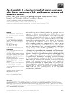
ure 1 shows representative Neurolucida images for two
pyramidal neurons (with or without LPS) used to calcu-
late total dendrite length for each Sholl compartment. The
Table summarizes our results for the three genotypes (n =
8 to 12 neurons examined for each condition and geno-
type) at 24 hrs. Our results for saline treatment showed
that there was a significant difference in basal dendrite
length in the distal (101–150 μm) Sholl compartment of
CA1 pyramidal neurons among the TR APOE mice in the
absence of a specifically applied stress with reduced length
in TR APOE4 mice. Because of this variation, data for ICV
LPS-induced changes after 24 hrs were analyzed as the
percentage of basal values for that TR APOE line (Table).
Similar to our previous findings with wt mice [3,17,18],
dendrite length was significantly reduced in all TR APOE
mice in all three compartments 24 hrs after LPS treatment.
We found no difference in the extent of reduction among
genotypes, with two-way ANOVA showing no significant
difference in decrease in dendrite length among TR APOE
in any of the Sholl compartments at 24 hr post-ICV LPS.
We have previously found in wt mice that following ICV
LPS injection, several indices of neuronal damage, includ-
ing morphometric measures, have returned to near basal
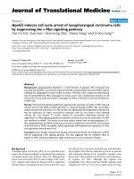
levels by 72 hr [3]. We compared recovery at 72 hrs among
the three genotypes and found a striking difference in the
recovery of TR APOE4 in all 3 Sholl compartments (Figure
2). Similar to our previous wt results, dendrite length of
neurons in both TR APOE2 and TR APOE3 mice recovered
extensively in all 3 compartments, with TR APOE2 recov-
ery better than TR APOE3 (P < 0.05 in the intermediate
and distal Sholl compartments). In contrast, dendrite
length of pyramidal neurons in TR APOE4 mice did not
show any recovery in any of the compartments (P <
0.001).
Dr. Maeda and colleagues have previously demonstrated
that despite brain regional differences in apoE expression,
there are no isoform-specific differences in levels of apoE
in brain tissue from these TR APOE mice, even with ele-
vated apoE2 plasma levels [45]. We have also previously
found no difference in apoE concentration in CM from
cultured astrocytes or microglia from the three genotypes
with or without LPS treatment [39,40,43]. To confirm
similar basal levels of apoE and determine the effects (if
any) of LPS, we performed Western blot analysis of hip-
pocampal homogenates from parallel ICV-injected mice
and compared apoE levels among the three TR APOE mice
24 and 72 hrs after saline or LPS ICV injection. There was
no significant difference in cerebral apoE levels among the
three TR APOE mice, and no effect of LPS on apoE levels
24 or 72 hrs after LPS activation. We also compared the
effects of conditioned medium (CM) from TR APOE2 and
APOE4 astrocytes on neurite length and branch number
in primary neuronal cultures, since these measurements
are comparable to our morphometric in vivo data. We
incubated primary cultures of wt cerebral neurons with
CM from primary cultures of TR APOE2 or TR APOE4
Representative neurons 24 hrs after treatment with saline or LPSFigure 1
Representative neurons 24 hrs after treatment with
saline or LPS. Neurolucida tracings of two CA1 hippocam-
pal pyramidal neurons stained by Golgi method; blue is soma
and first order dendrites, red is second order dendrites,
green is third order dendrites, purple is fourth order den-
drites. Sholl compartments are indicated by circles that rep-
resent 0–50 μm (P: proximal), 51–100 μm (I: intermediate),
and 101–150 μm (D: distal) from the center of the soma. For
the two neurons shown here, saline had total dendrite
lengths of 614.1 (P), 617.3 (I), and 64 (D) microns as calcu-
lated by Neuroexplorer. LPS had dendrite lengths of 382.3
(D), 321.4 (I), and 35.9 (D) microns. Data for all neurons
examined are shown in the Table.
TR APOE3 saline 24 hr
TR APOE3 LPS 24 hr
P
I
I
D
P
D
Journal of Neuroinflammation 2006, 3:21 />Page 5 of 9
(page number not for citation purposes)
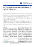
astrocytes and found that TR APOE2 astrocyte CM sup-
ported significant neurite growth while TR APOE4 astro-
cyte CM had no apparent effect on neurite length or
branch number (Figure 3). Since we have previously ruled
out differences in apoE protein levels in astrocyte CM
among the TR APOEs [40,43], we turned to apoE receptor
binding as a mechanistic basis for this difference. We
tested the effect of an apoE receptor antagonist, receptor-
associated protein (RAP), on neurite stimulation from TR
APOE2 astrocyte CM by preincubating neuron cultures
with a concentration (200 nM) of RAP that specifically
inhibits the LDL receptor-related protein (LRP) [46-48].
RAP treatment reduced the effect of TR APOE2 astrocyte
CM so that it no longer had a significant effect on either
neurite length or branch number (Figure 3).
Discussion
Several investigators have reported direct neurotrophic
and neurotoxic actions of recombinant or purified apoE
isoforms using a variety of culture systems (reviewed in
[49]). In addition to these direct effects on neurons, we
have recently published that glia from TR APOE mice
show apoE isoform-dependent innate immune activation
following stimulation of CD14/TLR4 co-receptors with
LPS [39,40]. Specifically, our experiments have shown
greater innate immune response by TR APOE4 microglia
and greater p38MAPK-dependent paracrine damage to
neurons in mixed primary cultures and hippocampal slice
cultures from TR APOE4 mice. In contrast, TR APOE2
astrocytes had the highest NF-κB activity and least (none)
neurotoxicity following innate immune activation. Thus,
in addition to the direct effects of apoE on neurons, our
cell culture data show that apoE isoform-dependent
actions in glia may also alter paracrine effects of astrocytes
and microglia on neurons and thereby contribute to neu-
rotoxic vs. neurotrophic outcomes associated with inherit-
ance of different APOE alleles. Our in vivo results
presented here are consistent with this model and further
demonstrate that neurite repair following paracrine dam-
age from innate immune activation is modulated by TR
APOE and that the neurotrophic actions of TR APOE2 CM
may be largely RAP-dependent.
Several laboratories have investigated apoE isoform-spe-
cific neurotrophic actions in a variety of cell culture mod-
els. The initial studies were performed using fetal rabbit
dorsal root ganglion cultures and showed that, in the pres-
ence of beta-migrating very low density lipoproteins,
apoE3 increased neurite outgrowth whereas apoE4
decreased neurite outgrowth [50]. Using immortalized
and transfected neuronal cell line, subsequent investiga-
tions confirmed the neurotrophic effects of apoE3 relative
to apoE4 and showed that the neurotrophic effect of
apoE3 was dependent on interaction with the heparin sul-
fate proteoglycan-low density lipoprotein receptor-related
protein (LRP) pathway [51-54] when incorporated into
several different lipid particles and even without the addi-
tion of exogenous lipid [55,56]. The next series of experi-
ments used primary cultures derived from transgenic mice
that express apoE3 or apo4 under the glial fibrillary acidic
protein promoter. Again, these showed greater relative
neurotrophism of apoE3 expression compared to apoE4
that was largely LRP-dependent [57,58]. Using these same
transgenic mice aged 1 to 2 years, a final in vivo study dem-
onstrated greater spine density in transgenic mice express-
ing human apoE3 than human apoE4 [59]. It is important
to note that while all of these studies support the proposal
that apoE4 expression could underlie relative regenerative
failure of injured central nervous system neurons, none
have investigated the relative neurotrophic actions of
apoE2.
Dendrite length recovery 72 hours after LPSFigure 2
Dendrite length recovery 72 hours after LPS. Data are
total dendrite length in each Sholl compartment of hippoc-
ampal CA1 pyramidal neurons from TR APOE mice 72 hr
post ICV LPS expressed as percent change from 24 hr post-
ICV LPS for each genotype. n = 12 neurons for each group (6
neurons per mouse for each genotype, time point, and expo-
sure). Two-way ANOVA for recovery of dendrite length had
P < 0.0001 for TR APOE but P > 0.05 for Sholl compartment.
In the proximal Sholl compartment, one-way ANOVA had P
< 0.0001 for % change in dendrite length among TR APOE;
Bonferroni-corrected repeated pair analysis had *P < 0.001
for TR APOE2 or TR APOE3 vs. TR APOE4 but ^P > 0.05
for TR APOE2 vs. TR APOE3. In both the intermediate and
distal Sholl compartments, one-way ANOVA had P < 0.0001
for % change in dendrite length among TR APOE; Bonfer-
roni-corrected repeated pair analysis had *P < 0.001 for TR
APOE2 or TR APOE3 vs. TR APOE4 and ^P < 0.05 for TR
APOE2 vs. TR APOE3.
Journal of Neuroinflammation 2006, 3:21 />Page 6 of 9
(page number not for citation purposes)
Neurite outgrowth following incubation with astrocyte conditioned medium (CM)Figure 3
Neurite outgrowth following incubation with astrocyte conditioned medium (CM). CM was generated from TR
APOE primary astrocytic cultures and collected after 72 hours. Primary wt neurons were treated with astrocyte CM (50% of
total volume) or 100% Neurobasal medium (control). For RAP treatment, neurons were preincubated with 200 nM RAP for 2
hrs prior to the addition of CM. After 48 hours, neurons were immunostained with anti-MAP2 antibody (1:500) and examined
with a confocal microscope using LaserSharp software (BioRad). Five or more MAP2-stained neurons were randomly selected
and dendrite length (A) and branch number (B) determined by Neurolucida. Data are expressed as mean length or branch
number ± SEM (n = 5). One-way ANOVA for neurite length (A) had P < 0.0001 and Bonferroni-corrected repeated pair anal-
ysis had *P < 0.001 for TR APOE2 vs. control but P > 0.05 for TR APOE4 or TR APOE2+RAP vs. control. For branch number
(B), one-way ANOVA had P < 0.05 and Bonferroni-corrected repeated pair analysis had
#
P < 0.01 for TR APOE2 vs. control, P
> 0.05 for TR APOE4 or TR APOE2+RAP vs. control, and ^P < 0.05 for TR APOE2 vs. TR APOE2+RAP.
Journal of Neuroinflammation 2006, 3:21 />Page 7 of 9
(page number not for citation purposes)
Our in vivo results with TR mice are consonant with these
cell culture studies and agree with the single other in vivo
study that used transgenic mice by indicating a relatively
more neurotrophic environment in the presence of apoE3
vs. apoE4. We add to this knowledge by showing that
expression of apoE2 also supports a more neurotrophic
environment than apoE4. Indeed, our experiments with
ICV LPS provide in vivo support for the proposal made
from cell culture data that apoE4 may play a role in neur-
ite regenerative failure [58]. Our observations on apoE2
go further because we observed enhanced neurite regener-
ation in TR APOE2 vs. TR APOE3 mice; this is a novel
observation that resonates with genetic association stud-
ies that indicate that inheritance of APOE2 reduces the
risk of AD. Like the enhanced neurotrophic action of
apoE3, our cell culture data suggest that the greater neuro-
trophic actions of apoE2 may derive, at least in part, from
astrocyte-secreted apoE2 and may be dependent on
apoE2-LRP interaction. However, conditioned medium
from primary astrocytes is an artificial system with many
active factors and so these results must be interpreted with
caution.
In summary, our results from TR APOE mice that express
each of the common apoE isoforms indicate a relatively
least neurotrophic environment in TR APOE4 mice com-
pared to TR APOE3 or TR APOE2 under basal conditions.
Moreover, following reversible paracrine damage to neu-
rons from direct activation of CD14/TLR4 receptor there
was failure of neurite regeneration in TR APOE4 mice and
greater regeneration in TR APOE2 mice compared to TR
APOE3 mice. The observations offer an explanation for
the stratification of clinical outcome with APOE seen in
several degenerative diseases or brain that are associated
with activated innate immune response.
Conclusion
The data indicate that the environment within TR APOE2
mouse hippocampus was most supportive of dendrite
regeneration while that within TR APOE4 hippocampus
failed to support dendrite regeneration in this model of
reversible paracrine damage to neurons from innate
immune activation, and suggest an explanation for the
stratification of clinical outcome with APOE seen in sev-
eral degenerative diseases or brain that are associated with
activated innate immune response.
Abbreviations
AD: Alzheimer's disease; ALS: amyotrophic lateral sclero-
sis; apo: apolipoprotein; Aβ: amyloid beta; CD: cluster of
differentiation; CM: conditioned medium; DAPI: 4'-6-dia-
midino-2-phenylindole ; DIV: days in vitro; ICV: intracer-
ebroventricular; iNOS: inducible nitric oxide synthase;
LPS: lipopolysaccharide; LRP: LDL receptor-related pro-
tein; MAP2: microtubule-associated protein 2; MyD88:
myeloid differentiation primary response protein; NF-κB:
nuclear factor kappaB; p38MAPK: p38 mitogen-associ-
ated protein kinase; PD: Parkinson's disease; RAP: recep-
tor-associated protein; TLR: toll-like receptor; TR: targeted
replacement; wt: wild type.
Competing interests
The author(s) declare that they have no competing inter-
ests.
Authors' contributions
IM, SZM, DM, CS and IS performed the experiments
described. NM developed the mouse line that was used in
all experiments. TJM conceived the study and its design
and helped to draft the manuscript. KSM analyzed the
data, prepared the figures, and drafted the manuscript.
Table 1: Dendrite length 24 hrs after ICV injection of saline or LPS.
apoE2 apoE3 apoE4
saline LPS Saline LPS saline LPS
Sholl r (
μ
m) microns % of saline microns % of saline microns % of saline
0–50 523 ± 38 52.9 ± 5.1 497 ± 48 44.7 ± 3.3 542 ± 36 44.6 ± 4.7
51–100 497 ± 36 40.1 ± 4.9 456 ± 47 36.8 ± 10.0 513 ± 39 36.9 ± 7.2
101–150 98 ± 13 42.2 ± 10.4 80 ± 10 30.0 ± 10.1 58 ± 8^ 33.1 ± 9.8
Total dendrite length for each Sholl compartment for mouse pyramidal neurons within hippocampal sector CA1 was determined by Golgi staining
followed by analysis with Neurolucida. Data are from the three lines of TR APOE mice with four mice from each genotype examined at this time
point. For PBS-treated mice (n = 6 neurons per mouse in each group for a total of 12 neurons per genotype and exposure), two-way ANOVA had
P < 0.0001 for Sholl compartment but P > 0.05 for TR APOE; however, one-way ANOVA in the distal Sholl compartment of the three genotypes
had P < 0.05 and Bonferroni-corrected repeated pair comparisons had ^P < 0.05 for TR APOE2 vs. TR APOE4 but no other paired comparisons.
For ICV LPS-exposed mice, two-way ANOVA was not significant for TR APOE or Sholl compartment at 24 hr post ICV LPS (n = 6 neurons per
mouse in each group for a total of 12 neurons per genotype and exposure), nor was one-way ANOVA across any of the Sholl compartments.
These values encompass our previous results with ICV LPS in C57Bl/6 mice [3, 17, 18]
Journal of Neuroinflammation 2006, 3:21 />Page 8 of 9
(page number not for citation purposes)
Acknowledgements
This work was supported by the Alvord Endowed Chair in Neuropathology
as well as grants from the NIH including AG027526 and AG24011.
References
1. Polazzi E, Contestabile A: Reciprocal interactions between
microglia and neurons: from survival to neuropathology. Rev
Neurosci 2002, 13:221 -2242.
2. Wyss-Coray T, Mucke L: Inflammation in neurodegenerative
disease a double-edged sword. Neuron 2002, 35:419-432.
3. Milatovic D, Zaja-Milatovic S, Montine KS, Horner PJ, Montine TJ:
Pharmacologic suppression of neuronal oxidative damage
and dendritic degeneration following direct activation of
glial innate immunity in mouse cerebrum. J Neurochem 2003,
87:1518-1526.
4. Stern EL, Quan N, Proescholdt MG, Herkenham M: Spatiotempo-
ral induction patterns of cytokine and related immune signal
molecule mRNAs in response to intrastriatal injection of
lipopolysaccharide. J Neuroimmunol 2000, 106:114-129.
5. Montine TJ, Milatovic D, Gupta RC, Valyi-Nagy T, Morrow JD, Breyer
RM: Neuronal oxidative damage from activated innate
immunity is EP2 receptor-dependent. J Neurochem 2002,
83:463-470.
6. Nadeau S, Rivest S: Glucocorticoids play a fundamental role in
protecting the brain during innate immune response. J Neu-
rosci 2003, 23:5536-5544.
7. Nadeau S, Rivest S: Endotoxemia prevents the cerebral inflam-
matory wave induced by intraparenchymal lipopolysaccha-
ride injection: role of glucocorticoids and CD14. J Immunol
2002, 169:3370-3381.
8. Hauss-Wegrzyniak B, Lynch MA, Vraniak PD, Wenk GL: Chronic
brain inflammation results in cell loss in the entorhinal cor-
tex and impaired LTP in perforant path-granule cell syn-
apses. Exp Neurol 2002, 176:336-341.
9. Lehnardt S, Massillon L, Follett P, Jensen FE, Ratan R, Rosenberg PA,
Volpe JJ, Vartanian T: Activation of innate immunity in the CNS
triggers neurodegeneration through a Toll-like receptor 4-
dependent pathway. Proc Natl Acad Sci U S A 2003, 100:8514-8519.
10. Wenk GL, McGann-Gramling K, Hauss-Wegrzyniak B, Ronchetti D,
Maucci R, Rosi S, Gasparini L, Ongini E: Attenuation of chronic
neuroinflammation by a nitric oxide-releasing derivative of
the antioxidant ferulic acid. J Neurochem 2004, 89:484-493.
11. Imler JL, Hoffmann JA: Toll receptors in innate immunity. Trends
Cell Biol 2001, 11:304-311.
12. Akira S: Toll-like receptor signaling. J Biol Chem 2003,
278:38105-38108.
13. Johnson GB, Brunn GJ, Platt JL: Activation of mammalian Toll-
like receptors by endogenous agonists. Crit Rev Immunol 2003,
23:15-44.
14. Fassbender K, Walter S, Kuhl S, Landmann R, Ishii K, Bertsch T, Stal-
der AK, Muehlhauser F, Liu Y, Ulmer AJ, Rivest S, Lentschat A, Gul-
bins E, Jucker M, Staufenbiel M, Brechtel K, Walter J, Multhaup G,
Penke B, Adachi Y, Hartmann T, Beyreuther K: The LPS receptor
(CD14) links innate immunity with Alzheimer's disease.
FASEB J 2004, 18:203-205.
15. Moffatt OD, Devitt A, Bell ED, Simmons DL, Gregory CD: Macro-
phage recognition of ICAM-3 on apoptotic leukocytes. J
Immunol 1999, 162:6800-6810.
16. Sholl DA: Dendritic organization in the neurons of the visual
and motor cortices of the cat. J Anat 1953, 87:387-406.
17. Milatovic D, Zaja-Milatovic S, Montine KS, Shie FS, Montine TJ: Neu-
ronal oxidative damage and dendritic degeneration follow-
ing activation of CD14-dependent innate immune response
in vivo. J Neuroinflammation 2004, 1:20.
18. Milatovic D, Zaja-Milatovic S, Montine KS, Nivison M, Montine TJ:
CD14-dependent innate immunity-mediated neuronal dam-
age in vivo is suppressed by NSAIDS and ablation of a pros-
taglandin E2 receptor, EP2. Current Medicinal Chemistry 2005,
5:151-156.
19. Mahley RW: Apolipoprotein E: cholesterol transport protein
with expanding role in cell biology. Science 1988, 240:622-630.
20. Strittmatter WJ, Roses AD: Apolipoprotein E and Alzheimer's
disease. Proc Natl Acad Sci 1995, 92:4725-4727.
21. Alberts MJ, Graffagnino C, McClenny C, DeLong D, Strittmatter WJ,
Saunders AM, Roses AD: APOE genotype and survival from
intracerebral hemorrhage. Lancet 1995, 346:575.
22. Newman MF, Croughwell ND, Blumenthal JA, Lowry E, White WD,
Spillane W, Davis RD, Glower DD, Smith LR, Mahanna EP: Predic-
tors of cognitive decline after cardiac operation. Ann Thorac
Surg 1995, 59:1326-1330.
23. Li YJ, Hauser MA, Scott WK, Martin ER, Booze MW, Qin XJ, Walter
JW, Nance MA, Hubble JP, Koller WC, Pahwa R, Stern MB, Hiner BC,
Jankovic J, Goetz CG, Small GW, Mastaglia F, Haines JL, Pericak-
Vance MA, Vance JM: Apolipoprotein E controls the risk and
age at onset of Parkinson disease. Neurology 2004,
62:2005-2009.
24. Li YJ, Pericak-Vance MA, Haines JL, Siddique N, McKenna-Yasek D,
Hung WY, Sapp P, Allen CI, Chen W, Hosler B, Saunders AM, Delle-
fave LM, Brown RHJ, Siddique T: Apolipoprotein E is associated
with age at onset of amyotrophic lateral sclerosis. Neurogenet-
ics 2004, 5:209-213.
25. Enzinger C, Ropele S, Smith S, Strasser-Fuchs S, Poltrum B, Schmidt
H, Matthews PM, Fazekas F: Accelerated evolution of brain atro-
phy and "black holes" in MS patients with APOE-epsilon 4.
Ann Neurol 2004, 55:563-569.
26. Nathoo N, Chetty R, van Dellen JR, Barnett GH: Genetic vulnera-
bility following traumatic brain injury: the role of apolipopro-
tein E. Mol Pathol 2003, 56:132-136.
27. Jordan BD, Relkin NR, Ravdin LD, Jacobs AR, Bennett A, Gandy S:
Apolipoprotein E epsilon4 associated with chronic traumatic
brain injury in boxing. JAMA 1997, 278:136-140.
28. Corder EH, Robertson K, Lannfelt L, Bogdanovic N, Eggertsen G,
Wilkins J, Hall C: HIV-infected subjects with the E4 allele for
APOE have excess dementia and peripheral neuropathy. Nat
Med 1998, 4:1182 -11184.
29. Corder EH, Saunders AM, Risch NJ, Strittmatter WJ, Schmechel DE,
Gaskell PJ, Rimmler JB, Locke PA, Conneally PM, Schmader KE, al. :
Protective effect of apolipoprotein E type 2 allele for late
onset Alzheimer disease. Nat Genet 1994, 7:180-184.
30. Ophir G, Amariglio N, Jacob-Hirsch J, Elkon R, Rechavi G, Michaelson
DM: Apolipoprotein E4 enhances brain inflammation by
modulation of the NF-kappaB signaling cascade. Neurobiol Dis
2005, 20:709-718.
31. Ophir G, Meilin S, Efrati M, Chapman J, Karussis D, Roses A, Michael-
son DM: Human apoE3 but not apoE4 rescues impaired
astrocyte activation in apoE null mice. Neurobiol Dis 2003,
12:56-64.
32. Colton CA, Brown CM, Cook D, Needham LK, Xu Q, Czapiga M,
Saunders AM, Schmechel DE, Rasheed K, Vitek MP: APOE and the
regulation of microglial nitric oxide production: a link
between genetic risk and oxidative stress. Neurobiol Aging 2002,
23:777-785.
33. Brown CM, Wright E, Colton CA, Sullivan PM, Laskowitz DT, Vitek
MP: Apolipoprotein E isoform mediated regulation of nitric
oxide release. Free Radic Biol Med 2002, 32:1071-1075.
34. Mahley RW, Rall SCJ: Apolipoprotein E: far more than a lipid
transport protein. Annu Rev Genomics Hum Genet 2000, 1:507-537.
35. Lynch JR, Tang W, Wang H, Vitek MP, Bennett ER, Sullivan PM,
Warner DS, Laskowitz DT: APOE genotype and an ApoE-
mimetic peptide modify the systemic and central nervous
system inflammatory response. J Biol Chem 2003,
278:48529-48533.
36. Li FQ, Sempowski GD, McKenna SE, Laskowitz DT, Colton CA, Vitek
MP: Apolipoprotein E-derived peptides ameliorate clinical
disability and inflammatory infiltrates into the spinal cord in
a murine model of multiple sclerosis. J Pharmacol Exp Ther 2006.
37. McAdoo JD, Warner DS, Goldberg RN, Vitek MP, Pearlstein R,
Laskowitz DT: Intrathecal administration of a novel apoE-
derived therapeutic peptide improves outcome following
perinatal hypoxic-ischemic injury. Neurosci Lett 2005,
381:305-308.
38. Aono M, Bennett ER, Kim KS, Lynch JR, Myers J, Pearlstein RD,
Warner DS, Laskowitz DT: Protective effect of apolipoprotein
E-mimetic peptides on N-methyl-D-aspartate excitotoxicity
in primary rat neuronal-glial cell cultures. Neuroscience 2003,
116:437-445.
39. Maezawa I, Nivison M, Montine KS, Maeda N, Montine TJ: Neuro-
toxicity from innate immune response is greatest with tar-
Publish with Bio Med Central and every
scientist can read your work free of charge
"BioMed Central will be the most significant development for
disseminating the results of biomedical research in our lifetime."
Sir Paul Nurse, Cancer Research UK
Your research papers will be:
available free of charge to the entire biomedical community
peer reviewed and published immediately upon acceptance
cited in PubMed and archived on PubMed Central
yours — you keep the copyright
Submit your manuscript here:
/>BioMedcentral
Journal of Neuroinflammation 2006, 3:21 />Page 9 of 9
(page number not for citation purposes)
geted replacement of E4 allele of apolipoprotein E gene and
is mediated by microglial p38MAPK. Faseb J 2006, 20:797-799.
40. Maezawa I, Maeda N, Montine TJ, Montine KS: Apolipoprotein E-
specific innate immune response in astrocytes from targeted
replacement mice. J Neuroinflammation 2006, 3:10.
41. Sullivan PM, Mezdour H, Aratani Y, Knouff C, Najib J, Reddick RL,
Quarfordt SH, Maeda N: Targeted replacement of the mouse
apolipoprotein E gene with the common human APOE3
allele enhances diet-induced hypercholesterolemia and
atherosclerosis. J Biol Chem 1997, 272:17972-17980.
42. Sullivan PM, Mezdour H, Quarfordt SH, Maeda N: Type III hyperli-
poproteinemia and spontaneous atherosclerosis in mice
resulting from gene replacement of mouse Apoe with
human Apoe*2. J Clin Invest 1998, 102:130-135.
43. Maezawa I, Jin LW, Woltjer RL, Maeda N, Martin GM, Montine TJ,
Montine KS: Apolipoprotein E isoforms and apolipoprotein AI
protect from amyloid precursor protein carboxy terminal
fragment-associated cytotoxicity. J Neuorchem 2004,
91:1312-1321.
44. Xiang J, Chao DT, Korsmeyer SJ: BAX-induced cell death may
not require interleukin 1 beta-converting enzyme-like pro-
teases. Proc Natl Acad Sci U S A 1996, 93:14559-14563.
45. Sullivan PM, Mace BE, Maeda N, Schmechel DE: Marked regional
differences of brain human apolipoprotein E expression in
targeted replacement mice. Neuroscience 2004, 124:725-733.
46. LaDu MJ, Shah JA, Reardon CA, Getz GS, Bu G, Hu J, Guo L, Van Eldik
LJ: Apolipoprotein E and apolipoprotein E receptors modu-
late A beta-induced glial neuroinflammatory responses. Neu-
rochem Int 2001, 39:427-434.
47. Herz J, Qiu SQ, Oesterle A, DeSilva HV, Shafi S, Havel RJ: Initial
hepatic removal of chylomicron remnants is unaffected but
endocytosis is delayed in mice lacking the low density lipo-
protein receptor. Proc Natl Acad Sci U S A 1995, 92:4611-4615.
48. Howard GC, Roberts BC, Epstein DL, Pizzo SV: Characterization
of alpha 2-macroglobulin binding to human trabecular mesh-
work cells: presence of the alpha 2-macroglobulin signaling
receptor.
Arch Biochem Biophys 1996, 333:19-26.
49. Mahley RW, Weisgraber KH, Huang Y: Apolipoprotein E4: a caus-
ative factor and therapeutic target in neuropathology,
including Alzheimer's disease. Proc Natl Acad Sci U S A 2006,
103:5644-5651.
50. Nathan BP, Bellosta S, Sanan DA, Weisgraber KH, Mahley RW, Pitas
RE: Differential effects of apolipoproteins E3 and E4 on neu-
ronal growth in vitro. Science 1994, 264:850-852.
51. Nathan BP, Chang KC, Bellosta S, Brisch E, Ge N, Mahley RW, Pitas
RE: The inhibitory effect of apolipoprotein E4 on neurite out-
growth is associated with microtubule depolymerization. J
Biol Chem 1995, 270:19791-19799.
52. Holtzman DM, Pitas RE, Kilbridge J, Nathan B, Mahley RW, Bu G,
Schwartz AL: Low density lipoprotein receptor-related pro-
tein mediates apolipoprotein E-dependent neurite out-
growth in a central nervous system-derived neuronal cell
line. Proc Natl Acad Sci U S A 1995, 92:9480-9484.
53. Bellosta S, Nathan BP, Orth M, Dong LM, Mahley RW, Pitas RE: Sta-
ble expression and secretion of apolipoproteins E3 and E4 in
mouse neuroblastoma cells produces differential effects on
neurite outgrowth. J Biol Chem 1995, 270:27063-27071.
54. Narita M, Bu G, Holtzman DM, Schwartz AL: The low-density lipo-
protein receptor-related protein, a multifunctional apolipo-
protein E receptor, modulates hippocampal neurite
development. J Neurochem 1997, 68:587-595.
55. Fagan AM, Bu G, Sun Y, Daugherty A, Holtzman DM: Apolipopro-
tein E-containing high density lipoprotein promotes neurite
outgrowth and is a ligand for the low density lipoprotein
receptor-related protein. J Biol Chem 1996, 271:30121-30125.
56. DeMattos RB, Curtiss LK, Williams DL: A minimally lipidated
form of cell-derived apolipoprotein E exhibits isoform-spe-
cific stimulation of neurite outgrowth in the absence of exog-
enous lipids or lipoproteins. J Biol Chem
1998, 273:4206-4212.
57. Sun YL, Wu S, Bu GJ, Onifade MK, Patel SN, Ladu MJ, Fagan AM,
Holtzman DM: Glial fibrillary acidic protein-apolipoprotein E
(apoE) transgenic mice- astrocyte-specific expression and
differing biological effects of astrocyte-secreted apoE3 and
apoE4 lipoproteins. J Neurosci 1998, 18:3261-3272.
58. Nathan BP, Jiang Y, Wong GK, Shen F, Brewer GJ, Struble RG: Apol-
ipoprotein E4 inhibits, and apolipoprotein E3 promotes neu-
rite outgrowth in cultured adult mouse cortical neurons
through the low-density lipoprotein receptor-related pro-
tein. Brain Res 2002, 928:96-105.
59. Ji Y, Gong Y, Gan W, Beach T, Holtzman DM, Wisniewski T: Apoli-
poprotein E isoform-specific regulation of dendritic spine
morphology in apolipoprotein E transgenic mice and Alzhe-
imer's disease patients. Neuroscience 2003, 122:305-315.