báo cáo hóa học: " Secreted phospholipase A2 activity in experimental autoimmune encephalomyelitis and multiple sclerosis" potx
Bạn đang xem bản rút gọn của tài liệu. Xem và tải ngay bản đầy đủ của tài liệu tại đây (1.73 MB, 8 trang )
BioMed Central
Page 1 of 8
(page number not for citation purposes)
Journal of Neuroinflammation
Open Access
Research
Secreted phospholipase A2 activity in experimental autoimmune
encephalomyelitis and multiple sclerosis
Timothy J Cunningham*
1
, Lihua Yao
1
, Michelle Oetinger
1
, Laura Cort
2
,
Elizabeth P Blankenhorn
2
and Jeffrey I Greenstein
3
Address:
1
Departments of Neurobiology and Anatomy, Drexel University College of Medicine, 2900 Queen Lane, Philadelphia, PA 19129, USA,
2
Department of Microbiology and Immunology, Drexel University College of Medicine, 2900 Queen Lane, Philadelphia, PA 19129, USA and
3
The
Multiple Sclerosis Institute, 1740 South Street Philadelphia, PA 19146, USA
Email: Timothy J Cunningham* - ; Lihua Yao - ; Michelle Oetinger - ;
Laura Cort - ; Elizabeth P Blankenhorn - ; Jeffrey I Greenstein -
* Corresponding author
Abstract
Background: There is increased interest in the contribution of the innate immune system to
multiple sclerosis (MS), including the activity of acute inflammatory mediators. The purpose of this
study was to test the involvement of systemic secreted phospholipase A2 (sPLA2) enzymes in
experimental autoimmune encephalomyelitis (EAE), an MS model, and to determine if enzyme
activity is elevated in MS patients.
Methods: A non-invasive urinary assay was developed in order to monitor enzymatically active
sPLA2 levels in Dark Agouti rats after induction of EAE. Some Rats were treated with nonapeptide
CHEC-9, an uncompetitive sPLA2 enzyme inhibitor, during the initial rise in urinary enzyme levels.
Body weight and clinical EAE score were measured for 18 days post immunization (PI), after which
the rats were sacrificed for H&E and myelin staining, and for ED-1 immunocytochemistry, the latter
to quantify macrophages and activated microglia. The urinary sPLA2 assay was also applied to un-
timed samples collected from a cross section of 44 MS patients and 14 healthy controls.
Results: Mean levels of enzymatically active sPLA2 in the urine increased following immunization
and peaked between days 8–10 PI which was just prior to the onset of EAE symptoms. At this time,
a transient attenuation of activity was detected in the urine of CHEC-9 treated rats consistent with
the activity-dependent properties of the inhibitor. The peptide also reduced or abolished EAE
symptoms compared to vehicle-injected controls. Histopathological changes in the spinal cords of
the EAE rats correlated generally with clinical score including a significant reduction in ED-1+ cells
after peptide treatment. Multiple Sclerosis patients also showed elevations in sPLA2 enzyme
activity. Mean levels of sPLA2 were increased 6-fold in the urine of patients with active disease and
4-fold for patients in remission, regardless of immunomodulating therapy.
Conclusion: The results suggest that sPLA2 enzymes, traditionally thought to be part the acute
phase inflammatory response, are therapeutic targets for MS.
Published: 11 September 2006
Journal of Neuroinflammation 2006, 3:26 doi:10.1186/1742-2094-3-26
Received: 10 June 2006
Accepted: 11 September 2006
This article is available from: />© 2006 Cunningham et al; licensee BioMed Central Ltd.
This is an Open Access article distributed under the terms of the Creative Commons Attribution License ( />),
which permits unrestricted use, distribution, and reproduction in any medium, provided the original work is properly cited.
Journal of Neuroinflammation 2006, 3:26 />Page 2 of 8
(page number not for citation purposes)
Background
The pathophysiology of multiple sclerosis (MS) involves
both antigen specific mechanisms and the innate immune
system, including elements of the acute inflammatory
response [1-4]. Since axonal loss is likely to begin at dis-
ease onset, the inflammation that accompanies this
degeneration may be a persistent contributing factor in
MS, as it all disorders in which there is destruction of nerv-
ous tissue. Increased hydrolysis of membrane phospholi-
pids by phospholipase A2 is a well-known early response
to tissue damage in all organ systems including the nerv-
ous system [5-7]. These enzymes are responsible for the
production of arachidonic acid and lysophospholipids
and therefore also control levels of inflammatory media-
tors and cytotoxic metabolites including prostaglandins,
leukotrienes, and reactive oxygen species. PLA2 enzymes
and products also influence several aspects of the cellular
and cytokine involvement in the inflammatory response.
Recent studies of rodent experimental autoimmune
encephalomyelitis (EAE) models of MS suggest PLA2
enzymes are involved in the onset and genesis of this dis-
ease [8,9]. Pinto, et al.[8], found that systemic infusion of
anchored lipid conjugates, targeting extracellular or
secreted (s)PLA2s, attenuated aspects of the autoimmune
response and EAE clinical disease. We have identified a
nonapeptide sPLA2 inhibitor called CHEC-9 that has
properties that may be particularly advantageous for
applications in vivo [32]. CHEC-9 is an internal fragment
of the endogenous protein DSEP/Dermcidin/PIF [10-16].
Both the parent polypeptide and isolated peptide mimet-
ics, including CHEC-9, have been used previously to
increase cell survival and inhibit inflammation in a variety
of experimental paradigms.
In the present study, we used a non-invasive urinary assay
to monitor changes in systemic levels of active sPLA2
enzymes following a standard EAE immunization proto-
col. We also examined the effects of CHEC-9 on the devel-
opment of EAE symptoms, including correlated
morphological changes in the spinal cord. Finally, this
same urinary assay was applied to randomly collected
urine samples from MS patients to determine if systemic
levels of enzyme are also elevated in this disease.
Methods
EAE production and analysis
The animal experiments were conducted under the aus-
pices of an IACUC protocol from Drexel University. All
personnel involved were experimentally blinded for all
procedures. Mild to moderate EAE was induced in 20
female Dark Agouti rats (130–150 g) by bilateral foot pad
immunizations of 100 μg guinea pig myelin basic protein
in saline emulsified with Complete Freund's Adjuvant
[17]. The results presented are from 2 separate sets of
experiments with 10 in each cohort, equally divided
between peptide and vehicle-treated rats. The rats were
weighed and scored for EAE symptoms daily using a 1–4
rating scale for clinical disease (1-tail drop, 2 hind limb
paresis, 3 hind limb paralysis, 4 moribund), and 2–3
independent investigators were responsible for the scor-
ing. We scored 2.5 for complete paralysis of one hind limb
which was the most severe disease encountered in this
experimental system. The experiments lasted 18 days fol-
lowing immunization, after which the rats were perfused
with 4% paraformaldehyde in 0.1 M phosphate buffer
and their spinal cords removed for histology.
Urine collection and CHEC-9 treatment
Urine was collected in metabolic cages from all rats start-
ing 1–2 days before immunization and then every other
day for 18 days. The urine was collected between 9:00–
11:00 AM, immediately sterile filtered, and frozen at -40°
prior to use. CHEC-9 treatment started 5 days after immu-
nization during the rise in urinary sPLA2 activity (see
Results). Treatment consisted of a subcutaneous injection
of 60μg CHEC-9 in clear DMEM vehicle on the first day,
followed by daily 30μg doses for 9 days. CHEC-9,
CHEASAAQC, was made by Celtek (Nashville, TN), puri-
fied and cross-linked as described previously[10].
sPLA2 enzyme activity
Twenty-five μl samples of urine were reacted with 600μM
1,2-bis (heptanoylthio) glycerophosphocholine, a sub-
strate for all PLA2s with the exception of cPLA2 and PAF-
AH (Caymen Chemical, Ann Arbor MI). Reaction buffer
consisted of 50 mM tris, 0.1 M NaCl (pH = 7.4) contain-
ing 1 mM DTNB (Ellman's reagent) and 2 mM CaCl
2
.
Reaction rates were determined with a Deltasoft (Prince-
ton NJ) supported ELX 808 reader (Biotek, Burlington
VT). The urinary sPLA2 assay was developed because in a
previous study we found that monitoring active enzyme
in blood plasma may be subject to considerable variabil-
ity due to the stress of restraint and collection procedures,
as well as handling of samples for ex vivo monitoring [32].
Enzyme activity in urine was found to be much more sta-
ble, specifically in those rats tested prior to immunization
or in healthy human controls. As in previous studies in
which the active enzyme concentration was of interest, we
confirmed that product formation in the presence of an
excess of substrate was linear during a 40 minute measure-
ment period, and therefore that the measured rate of the
reaction is proportional to urinary concentration of active
sPLA2. These rates were expressed relative to total protein
in the samples and normalized either to average baseline
values for the rats (obtained in the two days prior to
immunization), or in the patient studies, to average value
obtained from healthy controls.
Journal of Neuroinflammation 2006, 3:26 />Page 3 of 8
(page number not for citation purposes)
Statistical tests
Data comparisons were by a two-tailed Mann Whitney
test, nonparametric (Spearmann) linear correlation, or
Friedman's repeated measures ANOVA.
Histology and immunohistochemistry
Serial 20μm transverse (n = 10) or parasagittal sections (n
= 10) were cut through spinal cord blocks that extended
from the conus medullaris to the lumbar enlargement.
Alternate transverse sections were stained with hemotoxy-
lin & eosin and cyanin R the latter for myelin and myelin
debris (e.g. see ref. [18]). The parasaggital sections
allowed for greater areal coverage of this part of the spinal
cord and therefore these were used for quantification of
macrophages, ameboid microglia, and activated microglia
using the ED-1 monoclonal antibody (Serotec, UK). The
cells were counted by experimentally blinded investiga-
tors in 5-vehicle treated rats and 5 peptide-treated rats
using three equivalent parasaggital sections from each rat.
The counts were expressed per area of the sections and
then normalized to the average value obtained in the vehi-
cle-treated rats.
Patients
Forty-four patients with a diagnosis of relapsing/remitting
Multiple Sclerosis provided urine samples for this study
(Table 1). Healthy controls were recruited by advertise-
ment. The Institutional Review Boards of Drexel Univer-
sity and Graduate Hospital of Philadelphia approved the
study and informed consent was obtained from all sub-
jects. Twelve of the patients presented with active disease
at the time of sample collection, which was defined as a
change of one or more points on the functional neuro-
logic status score in the absence of fever or infection [19].
Therapies noted in Table 1 were interferon beta-1a
(Avonex
®
, Biogen, 30μg/week I.M.) or glatiramer acetate
(Copaxone
®
, Teva Pharmaceutical Industries 20 mg/day,
S.C.). Subjects were carefully screened and those with
peripheral infections, inflammatory disorders, or those
using anti-inflammatory drugs within the 24 hours prior
to sample collection, were excluded from the study. Urine
was collected in a sterile 50 ml tube at random times dur-
ing the day (9:00 AM-4:00 PM), immediately sterile fil-
tered, frozen and analyzed as above.
Results
Body weight
There was an initial loss of body weight in all rats post-
immunization (PI). From PI day 5 (the start of treatment)
onward, peptide treated animals gained weight at a signif-
icantly higher rate than vehicle treated rats. Weight gain in
the peptide-treated animals was 0.517 g/day compared to
0.392 g/day in vehicle-treated rats. The difference between
groups was significant (p = 0.011, Friedman's repeated
measures ANOVA).
sPLA2 activity and clinical score
Mean urinary sPLA2 activity increased steadily for the first
8 days following immunization in both treatment groups
(Fig. 1, Top). In vehicle treated rats, the mean activity lev-
eled off and declined at this point. The effects of CHEC-9
treatment were not detected until days 10 and 12 PI, when
the peptide significantly attenuated sPLA2 activity. It was
during this period that the behavioral deficits appeared in
the rats (Fig. 1, Bottom). However, only 3/10 peptide-
treated rats showed symptoms of disease and one of these
had a late onset tail paresis (at day 17 post-immuniza-
tion). This compared to 8/10 in the vehicle treated group
showing generally more severe symptoms that appeared
between days 10 and 13 PI. As might be expected from
these differences, the statistical difference between the two
groups was very significant (p = 0.0002, Friedman's
repeated measures ANOVA).
Histology and immunohistochemistry
Hematoxylin & eosin together with cyanin R staining in
alternate sections of the spinal cord revealed pathology
consistent with the clinical score. The most conspicuous
changes appeared in rats showing the most severe deficits
(clinical score 2.0 or greater). In the caudal-most regions
of the cord of these rats, the region around the conus med-
ullaris was severely shrunken and contained large areas of
degenerating myelin. In more rostral sections, large EAE
lesions were found often localized to the white matter
(Fig. 2). Adjacent sections stained for cyanin R revealed
that these areas were also characterized by dense myelin
debris (Fig. 2). Symptom-free rats had limited and more
scattered small cell infusion and myelin debris. To quan-
tify the immune response, we determined the number of
macrophages and microglia by counts of ED-1+ cells
within the spinal cord. The counts were made in repre-
Table 1: Patient population and control subjects used for the sPLA2 measurements.
Group n (F/M) Age yrs ± sd MS Onset yrs ± sd MS Duration yrs ± sd βIF GA None
Active 12 (8/4) 38.2 ± 9.5 29.5 ± 8.9 7.3 ± 4.5 9 1 2
Stable 32 (25/7) 43.6 ± 8.1 33.4 ± 8.6 10.6 ± 9.3 15 3 14
Control 14 (9/5) 37.5 ± 9.0 - - - - -
The means of subject age, age of MS onset, duration of the disease, and ongoing therapies are listed. F/M = female/male; βIF = beta-interferon; GA
= glatiramer acetate; sd = standard deviation.
Journal of Neuroinflammation 2006, 3:26 />Page 4 of 8
(page number not for citation purposes)
sentative vehicle-treated rats (n = 5, mean clinical score,
1.6) and peptide-treated rats (n = 5, mean clinical score,
0.2). Both groups of rats showed immunoreactive cells on
the spinal cord surface with a variable numbers of cells
occupying sub-adjacent white matter and parenchyma.
The ED-1+ cells included large round macrophages,
smaller round ameboid microglia, and "activated" micro-
glia, which were large (presumably hypertrophied) cells
with processes. The latter cell type dominated the tissue
especially in the vehicle treated rats (Fig. 3). In fact,
CHEC-9 treated rats had over 60% fewer ED-1+ cells per
unit area of the sections (p < 1 × 10
-3
, Mann Whitney test).
In addition, there was an obvious difference in the extent
to which the cells had infiltrated the spinal cord in the
control-treated rats. However, the calculated difference in
density may be inflated because in two of the vehicle
treated rats used for this part of the study, there was
shrinkage of the caudal spinal cord (EAE scores 2.0, 2.5).
Nevertheless, these results showed clearly that clinical EAE
score reflected the histopathological changes in the spinal
cord, as has been described in numerous other studies
with the EAE model [20]. The results also suggested that
the peptide's therapeutic effects included inhibition of
some aspects of the cellular immune response, as has been
shown previously after traumatic CNS lesions [10].
MS patients
The mean level of active sPLA2 enzymes was significantly
elevated in the urine of patients with both active and sta-
ble MS (Fig. 4). Enzyme concentrations were higher on
average in relapsing patients compared to non-relapsing
patients although the difference did not reach statistical
significance (p = 0.107, Mann Whitney test). Interestingly,
the increased concentration of enzyme found in stable
patients receiving no treatment was nearly identical to
that from patients treated with beta interferon (mean 393
± 130% of controls n = 15, versus 409 ± 98% s.e.m., n =
14, p = 0.585, Mann Whitney test), although both these
patient populations were somewhat heterogeneous as
shown by the large variances.
In order to rule out differences in renal function between
the groups, we compared total urinary protein with sPLA2
enzyme levels. Enzyme and protein levels were not corre-
lated in any group (p values for protein-sPLA2 correlation,
Active = 0.572; Stable = 0.350; Control = 0.885, Spear-
mann's nonparametric linear correlation). Therefore, it
was unlikely that the patients simply leaked more sPLA2
because of a systematic and unrecognized renal/urinary
tract pathology associated with MS. Notably, untimed
'spot' samples as used in this study are routinely applied
and accepted as an indicator of excess protein in the urine
(proteinuria) [21], which is expected to correlate with ele-
vated PLA2 enzyme levels under conditions of compro-
mised renal function [22,23].
Discussion
The present results suggest that increased levels of sPLA2
enzymes, long associated with inflammation outside the
nervous system[24], also characterize Multiple Sclerosis
and Experimental Autoimmune Encephalomyelitis.
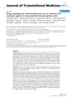
Secreted phospholipase A2 (sPLA2) activity and clinical dis-ease in EAE rats treated with sPLA2 inhibitor CHEC-9Figure 1
Secreted phospholipase A2 (sPLA2) activity and clin-
ical disease in EAE rats treated with sPLA2 inhibitor
CHEC-9. Top: Urinary sPLA2 enzymatic activity, normal-
ized to average pre-immunization values, increased steadily
to day 8 in both CHEC-9 and vehicle-treated rats. A signifi-
cant reduction in activity was observed on days 10 and 12
post-immunization in the peptide treated group either by
comparing values of peptide and vehicle directly (p = 0.049,
0.026 respectively, Mann Whitney), or by peak to trough
comparison between days 8 and 12, where reduction in
sPLA2 levels with peptide treatment was significant (p = 10
-
3
). Bottom: Mean clinical scores from day 10 onwards were
also significantly lower in the peptide treated rats (see text).
Journal of Neuroinflammation 2006, 3:26 />Page 5 of 8
(page number not for citation purposes)
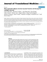
Low power photomicrograph of hematoxylin & eosin (A, B) and cyanin R (C, D) stained sections through the lumbar spinal cordFigure 2
Low power photomicrograph of hematoxylin & eosin (A, B) and cyanin R (C, D) stained sections through the lumbar spinal
cord. Left panels (A, C): Spinal cord of a peptide-treated rat that was symptom-free showing limited small cell infusion and
myelin degeneration. Right panels (B, D): Spinal cord of a vehicle treated rat that had an EAE score of 2.0. The tissue was char-
acterized by EAE lesions consisting of small darkly staining cells in the ventral and lateral white matter (arrows), and myelin
degeneration as shown in an adjacent section (D).
Journal of Neuroinflammation 2006, 3:26 />Page 6 of 8
(page number not for citation purposes)
Although the EAE model has been questioned recently in
terms of its validity for identifying potential MS therapies
[25,26], the model is clearly useful to help define the role
of inflammatory enzymes, specifically the PLA2s, in
autoimmune attack of the nervous system. Using a simple
urinary assay for active enzyme concentration, we found
that rats immunized to produce EAE had evidence of
increased systemic sPLA2 following immunization. The
same assay applied to samples collected from MS patients
also showed increased levels of active enzyme even with
random or "spot" sampling. Based in part on the parallel
results in the rodent model, we conclude that sPLA2
inflammatory activity is ongoing in the majority of MS
patients, active or stable, regardless of treatment. The
highest levels of enzyme were found in patients with
active disease, i.e., during relapse. Interestingly, asympto-
matic EAE rats had elevated enzyme activity but became
symptomatic only after a peak in activity was detected.
Finally, and most important, EAE symptoms were attenu-
ated by sPLA2 inhibitor CHEC-9. This finding, along with
similar results by others [8,9], support the idea that PLA2
enzymes play a direct role in the pathogenesis of MS and
related autoimmune disorders.
It important to recognize that increased PLA2 activity is
found after a variety of inflammatory stimuli, from local-
ized tissue damage to systemic infections [32]. In addi-
tion, a variety of antigenic stimuli are expected to raise
systemic sPLA2 levels, which suggests the nature of the
antigen is also critical in determining the resulting pathol-
ogy. On the other hand, the contribution of some types of
peripheral or systemic infections to induction and exacer-
bation of autoimmune disorders, including those affect-
ing the nervous system, is well known [27,28]. This non-
specific regulation of specific autoimmune disorders, or
cross talk between innate and acquired immune
responses, could depend in part on acute phase mediators
like sPLA2. A key element in this interaction may be lyso-
phosphatidyl choline, an important but often overlooked
product of sPLA2 activity. LysoPC has been implicated in
several aspects of acquired immunity including lym-
phocyte and monocyte chemotaxis, as well as dendritic
cell differentiation [29-31].
The appearance of symptoms at or shortly after the peak
of sPLA2 activity is interesting, especially if this peak
reflects maximal levels of inflammation and oxidative
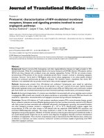
Macrophages and microglia were reduced by CHEC-9 treatment of EAEFigure 3
Macrophages and microglia were reduced by CHEC-9 treatment of EAE. Graph (left panel): The density of ED-1+ cells in the
spinal cord was reduced over 60% following peptide treatment (p < 1 × 10
-3
). Although the immunostained cells tended to
accumulate at or near the surface of both groups, fewer cells, occupying a smaller area within the cord were found CHEC-9
treated rats (B) compared to vehicle treated rats (C). The majority of intraspinal ED-1 immunoreactive cells were activated
microglia (arrows).
Journal of Neuroinflammation 2006, 3:26 />Page 7 of 8
(page number not for citation purposes)
stress. For example, it is under these conditions that sPLA2
may exert the most profound effects on the activity of
cytosolic PLA2 (cPLA2 [32]). The specific involvement of
cPLA2 in EAE has also been demonstrated [9]. It is also
under such conditions that the sPLA2 inhibition by the
peptide is expected to be maximal because of its proper-
ties as an uncompetitive inhibitor, including activity-
dependent inhibition [32]. These properties may also
explain our ability to detect a significant attenuation of
enzyme activity in urine of peptide treated rats only at this
time but not before or after, even with peptide treatment.
However, measurements every 48 hours are not likely to
be sensitive enough to reveal the actual pharmakinetics of
CHEC-9, which is currently under study. Therefore the
peptide's full effects on enzyme activity during the course
of these experiments are unknown. In addition, small
intermittent reductions of activity, that may go undetected
in urine, could be sufficient to slow or even prevent the
onset or appearance of more severe EAE symptoms.
The above discussion of the therapeutic effects of CHEC-9
assumes that the peptide's effects are due to enzyme inhi-
bition, i.e., decreases in sPLA2-mediated inflammation
and in the inflammatory products of PLA2-driven metab-
olism, directly and via cross talk with cPLA2. This explana-
tion is especially attractive since CHEC-9 is the third of
three different PLA2 inhibitors that has been used to treat
the disease. However, there are other properties of these
enzymes, including their direct influence on neuron via-
bility. For example, sPLA2 enzymes potentiate excitoxic-
ity, a mechanism suggested to be involved in most
instances of neurodegeneration [33, 34]. Also, as noted
above, there are likely to be interactions of the PLA2
enzymes with elements of antigen-specific immunity. Fur-
thermore, sPLA2s influence levels of proinflammatory
cytokines in a variety of cell types (either directly or via
cPLA2), especially during periods of oxidative stress [24,
32, 35, 36, 37]. In some instances this influence does not
depend on enzymatic activity. Thus, sPLA2 enzymes may
be an important therapeutic target for a variety neurode-
generative disorders associated with exaggerated autoim-
mune or inflammatory reactions, and especially for
diseases such as MS, where elements of both processes
contribute to pathology.
Conclusion
Further study of sPLA2-regulated processes in EAE models
may provide new insights into therapies for autoimmune
disorders affecting the nervous system. Amelioration of
EAE by uncompetitive sPLA2 inhibitor CHEC-9 suggests a
direct role for these enzymes in such disorders. Further-
more, monitoring sPLA2 activity in MS patients, for exam-
ple in relation to their susceptibility to relapse, could help
define periods of vulnerability in these patients as well as
appropriate regimens for application of therapies involv-
ing sPLA2 inhibition.
Competing interests
TJC, LY, and Drexel University have applied for patent
protection of CHEC-9, a peptide inhibitor used in these
experiments.
Authors' contributions
TJC drafted the manuscript, organized the assay data for
computer analysis and participated in the histological
analysis. LY carried out the enzyme assays, immunohisto-
chemistry, and assisted in the EAE studies. MO assisted in
the histological analysis, EAE studies, and in the patient
studies. EB criticized the manuscript and together with LC
designed and implemented the EAE studies. JG screened
subjects for the human studies, conducted neurological
exams and collected samples from patients and controls.
Level of sPLA2 enzyme activity in MS patients with active or stable disease compared to controls (see Table 1)Figure 4
Level of sPLA2 enzyme activity in MS patients with
active or stable disease compared to controls (see
Table 1). All measurements were made using 600 μM sub-
strate and normalized to the average control value. There
was a significant 4 and 6-fold increase in PLA2 activity com-
pared to controls in the stable and active MS patients respec-
tively, (p = 0.049*; 0.0019**, for comparison with controls,
Mann Whitney test). Treated and untreated stable patients
were grouped since their average levels of active enzyme
were almost identical (see text). Patients with active disease
were mostly undergoing treatment with beta-interferon (9/
12, Table 1).
Publish with Bio Med Central and every
scientist can read your work free of charge
"BioMed Central will be the most significant development for
disseminating the results of biomedical research in our lifetime."
Sir Paul Nurse, Cancer Research UK
Your research papers will be:
available free of charge to the entire biomedical community
peer reviewed and published immediately upon acceptance
cited in PubMed and archived on PubMed Central
yours — you keep the copyright
Submit your manuscript here:
/>BioMedcentral
Journal of Neuroinflammation 2006, 3:26 />Page 8 of 8
(page number not for citation purposes)
He also assisted in data interpretation, and in preparing
the manuscript. All authors approved of the final version
of the manuscript.
Acknowledgements
This work was supported by grant PP1055 from the National Multiple Scle-
rosis Society.
References
1. Prat A, Antel J: Pathogenesis of multiple sclerosis. Curr Opin
Neurol 2005, 18(3):225-230.
2. Martino G, Adorini L, Rieckmann P, Hillert J, Kallmann B, Comi G,
Filippi M: Inflammation in multiple sclerosis: the good, the
bad, and the complex. Lancet Neurol 2002, 1(8):499-509.
3. Hendriks JJ, Teunissen CE, de Vries HE, Dijkstra CD: Macrophages
and neurodegeneration. Brain Res Brain Res Rev 2005,
48(2):185-195.
4. Trapp BD: Pathogenesis of multiple sclerosis: the eyes only
see what the mind is prepared to comprehend. Ann Neurol
2004, 55(4):455-457.
5. Cummings BS, McHowat J, Schnellmann RG: Phospholipase A(2)s
in cell injury and death. J Pharmacol Exp Ther 2000,
294(3):793-799.
6. Bazan NG, Colangelo V, Lukiw WJ: Prostaglandins and other
lipid mediators in Alzheimer's disease. Prostaglandins Other Lipid
Mediat 2002, 68-69:197-210.
7. Farooqui AA, Horrocks LA: Phospholipase A2-generated lipid
mediators in the brain: the good, the bad, and the ugly. Neu-
roscientist 2006, 12(3):245-260.
8. Pinto F, Brenner T, Dan P, Krimsky M, Yedgar S: Extracellular
phospholipase A2 inhibitors suppress central nervous sys-
tem inflammation. Glia 2003, 44(3):275-282.
9. Kalyvas A, David S: Cytosolic phospholipase A2 plays a key role
in the pathogenesis of multiple sclerosis-like disease. Neuron
2004, 41(3):323-335.
10. Milicevic I, Pekovic S, Subasic S, Mostarica-Stojkovic M, Stosic-Grujicic
S, Medic-Mijacevic L, Pejanovic V, Rakic L, Stojiljkovic M: Ribavirin
reduces clinical signs and pathological changes of experi-
mental autoimmune encephalomyelitis in Dark Agouti rats.
J Neurosci Res 2003, 72(2):268-278.
11. Cunningham TJ, Souayah N, Jameson B, Mitchell J, Yao L: Systemic
treatment of cerebral cortex lesions in rats with a new
secreted phospholipase A2 inhibitor. J Neurotrauma 2004,
21(11):1683-1691.
12. Dailey AT, Avellino AM, Benthem L, Silver J, Kliot M:
Complement
depletion reduces macrophage infiltration and activation
during Wallerian degeneration and axonal regeneration. J
Neurosci 1998, 18(17):6713-6722.
13. Kurtzke JF: Rating neurologic impairment in multiple sclero-
sis: an expanded disability status scale (EDSS). Neurology 1983,
33(11):1444-1452.
14. Ercolini AM, Miller SD: Mechanisms of immunopathology in
murine models of central nervous system demyelinating dis-
ease. J Immunol 2006, 176(6):3293-3298.
15. Eknoyan G: On testing for proteinuria: time for a methodical
approach. Cleve Clin J Med 2003, 70(6):493, 496-7, 501.
16. Bijian K, Cybulsky AV: Stress proteins in glomerular epithelial
cell injury. Contrib Nephrol 2005, 148:8-20.
17. Sharma M, McCarthy ET, Sharma R, Fish BL, Savin VJ, Cohen EP,
Moulder JE: Arachidonic acid metabolites mediate the radia-
tion-induced increase in glomerular albumin permeability.
Exp Biol Med (Maywood) 2006, 231(1):99-106.
18. Triggiani M, Granata F, Giannattasio G, Marone G: Secretory phos-
pholipases A2 in inflammatory and allergic diseases: not just
enzymes. J Allergy Clin Immunol 2005, 116(5):1000-1006.
19. Sriram S, Steiner I: Experimental allergic encephalomyelitis: A
misleading model of multiple sclerosis. Ann Neurol 2005,
58(6):939-945.
20. Skundric DS: Experimental models of relapsing-remitting
multiple sclerosis: current concepts and perspective. Curr
Neurovasc Res 2005, 2(4):349-362.
21. Kamradt T, Goggel R, Erb KJ: Induction, exacerbation and inhi-
bition of allergic and autoimmune diseases by infection.
Trends Immunol 2005, 26(5):260-267.
22. Nogai A, Siffrin V, Bonhagen K, Pfueller CF, Hohnstein T, Volkmer-
Engert R, Bruck W, Stadelmann C, Kamradt T: Lipopolysaccharide
injection induces relapses of experimental autoimmune
encephalomyelitis in nontransgenic mice via bystander acti-
vation of autoreactive CD4+ cells. J Immunol 2005,
175(2):959-966.
23. Kabarowski JH, Xu Y, Witte ON: Lysophosphatidylcholine as a
ligand for immunoregulation. Biochem Pharmacol 2002,
64(2):161-167.
24. Coutant F, Perrin-Cocon L, Agaugue S, Delair T, Andre P, Lotteau V:
Mature dendritic cell generation promoted by lysophos-
phatidylcholine. J Immunol 2002, 169(4):1688-1695.
25. Murphy AA, Santanam N, Morales AJ, Parthasarathy S: Lysophos-
phatidyl choline, a chemotactic factor for monocytes/T-lym-
phocytes is elevated in endometriosis. J Clin Endocrinol Metab
1998, 83(6):2110-2113.
26. Han WK, Sapirstein A, Hung CC, Alessandrini A, Bonventre JV:
Cross-talk between cytosolic phospholipase A2 alpha
(cPLA2 alpha) and secretory phospholipase A2 (sPLA2) in
hydrogen peroxide-induced arachidonic acid release in
murine mesangial cells: sPLA2 regulates cPLA2 alpha activ-
ity that is responsible for arachidonic acid release. J Biol Chem
2003, 278(26):24153-24163.
27. DeCoster MA, Lambeau G, Lazdunski M, Bazan NG: Secreted
phospholipase A2 potentiates glutamate-induced calcium
increase and cell death in primary neuronal cultures. J Neuro-
sci Res 2002, 67(5):634-645.
28. Wei S, Ong WY, Thwin MM, Fong CW, Farooqui AA, Gopalakrishna-
kone P, Hong W: Group IIA secretory phospholipase A2 stim-
ulates exocytosis and neurotransmitter release in
pheochromocytoma-12 cells and cultured rat hippocampal
neurons. Neuroscience 2003, 121(4):891-898.
29. Thommesen L, Sjursen W, Gasvik K, Hanssen W, Brekke OL, Skatte-
bol L, Holmeide AK, Espevik T, Johansen B, Laegreid A: Selective
inhibitors of cytosolic or secretory phospholipase A2 block
TNF-induced activation of transcription factor nuclear fac-
tor-kappa B and expression of ICAM-1. J Immunol 1998,
161(7):3421-3430.
30. Triggiani M, Granata F, Balestrieri B, Petraroli A, Scalia G, Del Vecchio
L, Marone G: Secretory phospholipases A2 activate selective
functions in human eosinophils. J Immunol 2003,
170(6):3279-3288.
31. Granata F, Petraroli A, Boilard E, Bezzine S, Bollinger J, Del Vecchio
L, Gelb MH, Lambeau G, Marone G, Triggiani M: Activation of
cytokine production by secreted phospholipase A2 in human
lung macrophages expressing the M-type receptor. J Immunol
2005, 174(1):464-474.
32. Cunningham TJ, Maciejewski J, Yao L: Inhibition of secreted phos-
pholipase A2 by neuron survival and anti-inflammatory pep-
tide CHEC-9. J Neuroinflammation 2006, 3(1):25.