báo cáo hóa học: " All-trans retinoic acid induces COX-2 and prostaglandin E2 synthesis in SH-SY5Y human neuroblastoma cells: involvement of retinoic acid receptors and extracellular-regulated kinase 1/2" potx
Bạn đang xem bản rút gọn của tài liệu. Xem và tải ngay bản đầy đủ của tài liệu tại đây (582.89 KB, 9 trang )
BioMed Central
Page 1 of 9
(page number not for citation purposes)
Journal of Neuroinflammation
Open Access
Research
All-trans retinoic acid induces COX-2 and prostaglandin E
2
synthesis
in SH-SY5Y human neuroblastoma cells: involvement of retinoic
acid receptors and extracellular-regulated kinase 1/2
Matilde Alique, Juan F Herrero and Francisco Javier Lucio-Cazana*
Address: Facultad de Medicina, Departamento de Fisiología, Campus Universitario, Universidad de Alcalá, 28871 Madrid, Spain
Email: Matilde Alique - ; Juan F Herrero - ; Francisco Javier Lucio-Cazana* -
* Corresponding author
Abstract
Background: Our recent results show that all-trans retinoic acid (ATRA), an active metabolite of
vitamin A, induces COX-dependent hyperalgesia and allodynia in rats. This effect was mediated by
retinoic acid receptors (RARs) and was associated with increased COX-2 expression in the spinal
cord. Since ATRA also up-regulated COX-2 expression in SH-SY5Y human neuroblastoma cells,
the current study was undertaken to analyze in these cells the mechanism through which ATRA
increases COX activity.
Methods: Cultured SH-SY5Y neuroblastoma cells were treated with ATRA. COX expression and
kinase activity were analyzed by western blot. Transcriptional mechanisms were analyzed by RT-
PCR and promoter assays. Pharmacological inhibitors of kinase activity and pan-antagonists of RAR
or RXR were used to assess the relevance of these signaling pathways. Production of prostaglandin
E
2
(PGE
2
) was quantified by enzyme immunoabsorbent assay. Statistical significance between
individual groups was tested using the non-parametric unpaired Mann-Whitney U test.
Results: ATRA induced a significant increase of COX-2 expression in a dose- and time-dependent
manner in SH-SY5Y human neuroblastoma cells, while COX-1 expression remained unchanged.
Morphological features of differentiation were not observed in ATRA-treated cells. Up-regulation
of COX-2 protein expression was followed by increased production of PGE
2
. ATRA also up-
regulated COX-2 mRNA expression and increased the activity of a human COX-2 promoter
construct. We next explored the participation of RARs and mitogen-activated peptide kinases
(MAPK). Pre-incubation of SH-SY5Y human neuroblastoma cells with either RAR-pan-antagonist
LE540 or MAP kinase kinase 1 (MEK-1) inhibitor PD98059 resulted in the abolition of ATRA-
induced COX-2 promoter activity, COX-2 protein expression and PGE
2
production whereas the
retinoid X receptor pan-antagonist HX531, the p38 MAPK inhibitor SB203580 or the c-Jun kinase
inhibitor SP600125 did not have any effect. The increase in RAR-β expression and extracellular-
regulated kinase 1/2(ERK1/2) phosphorylation in ATRA-incubated cells suggested that RARs and
ERK1/2 were in fact activated by ATRA in SH-SY5Y human neuroblastoma cells.
Conclusion: These results highlight the importance of RAR-dependent and kinase-dependent
mechanisms for ATRA-induced COX-2 expression and activity.
Published: 04 January 2007
Journal of Neuroinflammation 2007, 4:1 doi:10.1186/1742-2094-4-1
Received: 28 November 2006
Accepted: 04 January 2007
This article is available from: />© 2007 Alique et al; licensee BioMed Central Ltd.
This is an Open Access article distributed under the terms of the Creative Commons Attribution License ( />),
which permits unrestricted use, distribution, and reproduction in any medium, provided the original work is properly cited.
Journal of Neuroinflammation 2007, 4:1 />Page 2 of 9
(page number not for citation purposes)
Background
The initiation and maintenance of central sensitization
involve numerous neuromediators. The expression of
cyclooxygenase-2 (COX-2), for example, is enhanced rap-
idly in the spinal cord during sensitization, along with the
production of prostaglandins like prostaglandin E
2
(PGE
2
) [1]. Interleukin-1β (IL-1β) is also up-regulated fol-
lowing inflammation and induces up-regulation of COX-
2 in the spinal cord [1]. The mechanisms underlying the
up-regulation of COX-2 are not known. Retinoids might
be one of these unidentified systems [2].
Biologically active retinoids, a family of vitamin A metab-
olites or analogues, such as all-trans retinoic acid (ATRA)
[3], play an essential activity in the embryological devel-
opment of several tissues and organs [4], including the
brain and the spinal cord [3,5]. Retinoids are also present
in the brain and spinal cord of adult rats and mice [6,7]
and are involved in functions such as spatial learning and
memory [8,9]. ATRA is the carboxylic acid form of vitamin
A and is considered its major metabolite.
Physiological retinoids are characterized by their capacity
to bind and activate retinoid nuclear receptors, including
retinoic acid receptors (RARs) and/or retinoid X receptors
(RXRs), each having three isotypes, α, β and γ. RARs and
RXRs have been identified in numerous tissues including
spinal cord [10]. The actions of ATRA are generally medi-
ated by binding to RARs, which act as ligand-regulated
transcription factors by binding as hetetodimers with the
RXRs to ATRA response elements (RAREs) located in reg-
ulatory regions of target genes [11]. Other signalling path-
ways may also mediate the effects of retinoids and, in the
context of the present work, it is particularly relevant the
fact that ATRA enhances extracellular-regulated kinase 1/2
(ERK1/2) phosphorylation [12-15], since we have
recently found ATRA in human mesangial cells that ERK1/
2 plays a key role in the up-regulation of COX-2 by ATRA
[16].
In a previous work carried out in our laboratory [2] we
observed that rats with inflammation treated with ATRA
p.o. showed a more intense development of allodynia and
hyperalgesia than control rats. Also, the recovery to base-
line was slower in animals treated with ATRA. We also
observed that ATRA up-regulated COX-2 expression in
SH-SY5Y human neuroblastoma cells, a clonal derivative
of the human neuroblastoma SK-N-SH cell line that
expresses RARs and RXRs [17,18], and in whole spinal
cord of animals treated with ATRA. Further studies [19]
indicated that oral treatment with ATRA in normal rats
induces a sensitization-like effect on spinal cord neuronal
responses similar to that observed in animals with inflam-
mation, and might explain the enhancement of allodynia
and hyperalgesia observed in previously published behav-
ioral experiments. The mechanism of action involved an
over-expression of COX-2, but not COX-1, in the lumbar
spinal cord [19]. When ATRA was administered intrathe-
cally, the sensitization-like effect was inhibited by a RAR-
pan-antagonist and associated with a modulation of
COX-2 and IL-1 activities [20].
The current study was undertaken to analyze in SH-SY5Y
human neuroblastoma cells the mechanism through
which ATRA increases COX activity. Preliminary results
have been published in abstract form [21].
Materials and methods
Drugs and other reagents
The RARs pan-antagonist ATRA (all trans-retinoic acid)
was purchased from Sigma (St. Louis, MO). The selective
RAR pan-antagonist (LE540) and RXR pan-antagonist
(HX531) were kindly provided by Dr. Kagechika (School
of Biomedical Science, Tokyo Medical and Dental Univer-
sity, Tokyo, Japan). The mitogen-activated protein kinase
(MAPK) inhibitors: PD98059 (MAPK kinase (MKK1)
inhibitor), SB203580 (p38 MAPK inhibitor) and
SP600125 (JNK MAPK inhibitor) were purchased from
Calbiochem (La Jolla, CA). Interleukin-1β was purchased
from Roche (Indianapolis, IN). All reagents were prepared
in DMSO so that the final concentration was < 0.1%,
except ATRA, which was dissolved in ethanol, and inter-
leukin-1β, which was dissolved in sterile water. The
human COX-2 luciferase reporter construct phPES2 con-
taining the promoter fragment -327 to +59 [22] was a gift
from Dr. Hiroyasu Inoue (Nara Women's University,
Nara, Japan). Primary antibodies against COX-1, COX-2,
RAR-β and total ERK2 were purchased from Santa Cruz
Biotechnology (Santa Cruz, CA). Antibody against phos-
phorylated form of ERK1/2 was purchased from Cell Sig-
naling Technology (Danvers, MA) and an anti-α-actin
antibody was from Sigma Chemical Co (St. Louis, MO).
All antibodies were used at 1:1000 dilution.
Cell culture
The SH-SY5Y human neuroblastoma cell line (N-type
cells, derived from the parental cell line SK-N-SH; Biedler
et al. 1973) was obtained from American Type Culture
Collection (Cat #: CRL-2266; ATCC, Manasas, VA). The
culture medium was DMEM (Invitrogen, CA) supple-
mented with 10% fetal bovine serum (FBS), 20 mM L-
glutamine and antibiotics (penicillin 100 U/ml and strep-
tomycin 100 μg/ml). Confluent cultures were used and
they were made quiescent when appropriate by a 24 h
incubation with medium supplemented with 0.5% FBS.
ATRA treatment did not induce morphological features of
differentiation.
Journal of Neuroinflammation 2007, 4:1 />Page 3 of 9
(page number not for citation purposes)
Transient transfection and luciferase assay
Cells, 3.5 × 10
5
per well, were plated in 6-well plates 24 h
before transfection. The cells in every well were then incu-
bated 8 h at 37°C with 2 ml Opti-MEM (Invitrogen, CA)
cointaining complexes of 5 μg lipofectAMINE (Invitrogen,
CA), 1 μg human COX-2 reporter and 0.1 μg renilla luci-
ferase reporter as an internal control. Transfected cells
were next incubated with complete growth medium for 16
h and then treated with either ATRA (10 μM, 24 h) or with
IL-1β (10 ng/ml, 24 h), and in other experiments pre-
incubated with for 1 h with either 2.5 μM LE540 or 50 μM
PD98059 and then with ATRA (10 μM, 24 h). Finally, fire-
fly luciferase activity of the COX-2 reporter was measured
with a Lumat LB9506 luminometer (Berthold Technolo-
gies, Herts, UK) and normalized against the renilla luci-
ferase activity by using the dual-luciferase reporter assay
system (Promega, Madison, WI). The experiments were
performed in triplicate and repeated four times (for statis-
tical purposes n = 4).
Western blot analysis
Cells were homogenized in a solution containing 150 mM
NaCl, 10 mM Tris-HCl (pH 7.4), 5 mM EDTA, 1% deoxy-
cholic acid, 0.1% SDS, 1% Triton X-100 and protease
inhibitors 1 mM phenyl-methyl-sulfonyl-fluoride, 10 μ/
ml aprotinin, 2 μg/ml leupeptin and the phosphatase
inhibitir 0.2 mM NaVO
4
. Cell proteins (30–40 μg) were
run in 8–10% SDS-polyacrilamide gels, transferred onto a
nitrocellulose membrane (Trans-Blot Transfer Medium,
Bio-Rad, CA) and incubated overnight at 4°C with anti-
bodies recognizing specifically COX-1, COX-2, RAR-β, P-
ERK1/2 as previously described [2]. This incubation was
followed by a second incubation with peroxidase-conju-
gated secondary antibody and immunoreactive products
were detected by chemiluminiscence using the ECL West-
ern Blotting Detection Reagents (Amersham Biosciences,
UK) following the protocol provided by the manufac-
turer. As a loading control, blots probed with anti-COX-1,
anti-COX-2 and anti-RAR-β were subsequently re-probed
with anti-α-actin, whereas blots probed with anti-P-ERK1/
2 were re-probed with anti-total ERK2. Each experiment
was performed at least three times.
RT-PCR analysis of COX-2 expression
Total RNA was extracted using the TriPure isolation rea-
gent (Roche Diagnostics, GmbH, Mannheim, Germany)
according to the manufacturer's instructions and spectro-
photometrically quantified. The RT-PCR reaction was per-
formed with the cMaster RTplusPCR system (Eppendorf
AG, Hamburg, Germany) with specific primers for human
COX-2 purchased to Ambion (Austin, TX) [(F) 5'-CAT-
TCTTTGCCCAGCACTTCAC-3'; (R) GACCAGGCACCA-
GACCAAAGAC; Accession number: D28235
]. Modified
18S primers (QuantumRNA 18S Internal Standards;
Ambion) were used for 18S coamplification, as constitu-
tive controls.
The reaction mixture was incubated for 60 min at 42°C
and 2 min at 94°C, followed by 35 cycles of 30 sec at
94°C, 30 sec at 59°C and 30 sec at 72°C, with a final
extension of 5 min at 72°C. Preliminary experiments
established that these conditions provided a linear cDNA
amplification. PCR products were separated on 2% agar-
ose gels, and bands were visualized by ethidium bromide
staining. Each experiment was performed three times.
Determination of PGE
2
formation
The cultured medium of SH-SY5Y human neuroblastoma
cells growth in 6-plates and treated as described in the leg-
ends of Figures 1e and 3b was collected and diluted 2
times. PGE
2
concentrations in the medium were deter-
mined in triplicate using a commercially available
enzyme immunoabsorbent assay (EIA) kit (Cayman
Chemical Company, Ann Arbor, MI) following the man-
ufacturer's protocol. The assay was performed in a total
volume of 150 μl, with the following components being
added in 50 μl volumes: standards or biological samples,
enzymatic tracer and specific antiserum. After overnight
incubation at 4°C, the plates were washed, and 200 μl Ell-
man's reagent was added into each well. After 1–3 h, the
absorbance at 414 nm of each well was measured. A
standard curve, with values ranging from 50 to 0.39 pg/
ml, was used to evaluate the concentrations. The reliable
limit of quantification for PGE
2
was 15 pg/ml, and the
coefficient of variation was less than 14% within the cali-
bration range (15–1000 pg/ml). Results were calculated
by using the nonlinear regression of a four-parameter
logistic model. Each experiment was performed four times
(for statistical purposes n = 4).
Data analysis and statistical procedures
All values are presented as mean ± standard error of the
mean (s.e.m). All experiments were repeated a minimum
of three times. Statistical significance between individual
groups was tested using the non-parametric unpaired
Mann-Whitney U test. A P value of < 0.05 was considered
significant.
Results
ATRA up-regulates the expression of COX-2 protein and
COX-2 mRNA as well as the production of PGE
2
in SH-
SY5Y human neuroblastoma cells
The effect of ATRA on the levels of COX-1 and COX-2 pro-
teins was examined in typical dose-response and time-
course experiments. Serum-deprived cultured SH-SY5Y
human neuroblastoma cells constitutively expressed both
COX-1 and COX-2 in the absence of stimulation (Figure
1a, b). COX-1 expression was not modified by incubation
with the retinoid (Figure 1a) whereas ATRA treatment up-
Journal of Neuroinflammation 2007, 4:1 />Page 4 of 9
(page number not for citation purposes)
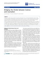
ATRA up-regulates COX-2 expression and increases PGE
2
in SH-SY5Y human neuroblastoma cellsFigure 1
ATRA up-regulates COX-2 expression and increases PGE
2
in SH-SY5Y human neuroblastoma cells. (a,b) Expression of
COX-1 and COX-2 proteins was analyzed by western blot in SH-SY5Y human neuroblastoma cells incubated for 24 h with the
indicated concentrations of ATRA or incubated with 10 μM ATRA for the indicated times. (c) For comparison, expression of
COX-2 protein was also analyzed in cells incubated with the inflammatory cytokine interleukin-1 β (IL-1β,10 ng/ml). (d)
Expression of COX-2 mRNA, analyzed by semiquantitative RT-PCR in cells incubated for 24 h with 10 μM ATRA or 10 ng/ml
IL-1β. (e) PGE
2
production in SH-SY5Y human neuroblastoma cells incubated for 24 h with 10 μM ATRA or 10 ng/ml IL-1β.
PGE
2
in the medium was determined in triplicate in four separate experiments (for statistical purposes n = 4; * P < 0.01 vs con-
trol). (a,b,c,d) Each photograph represents at least three repeated experiments. Equal protein or mRNA loading were con-
firmed by probing with an anti-α-actin antibody or by co-amplification of 18 S RNA, respectively Normalized density ratio of
COX-2 over either α-actin or 18 S RNA is indicated for each band.
c
α
-actin
COX-2
α
-actin
COX-2
IL-1
β
0 15 30 60 90 min
IL-1
β
0 8 24 h
a
b
ATRA 0 0.01 0.1 1 5 10
μ
M
α
-actin
COX-1
ATRA 0 15´ 30´ 60´ 90´ 24 h
α
-actin
COX-1
ATRA 0 0.01 0.1 1 5 10
μ
M
α
-actin
COX-2
ATRA 0 15´ 30´ 60´ 90´ 24 h
α
-actin
COX-2
e
0
20
40
60
80
100
120
140
PGE
2
(pg/ml/mg
protein)
CONTROL
ATRA
IL-1
β
*
*
d
18S
COX-2
C ATRA IL-1
β
Ratio 1 1.32 1.16 1.36 1.38 1.29
Ratio 1 0.93 0.97 1.10 1 0.95
Ratio 1 0.96 1.62 2.13 2.85 3.46
Ratio 1 1.37 1.88 2.96 3.31 3.60
Ratio 1 1.08 2.24 2.83 3.11 Ratio 1 3.79 4.36
Ratio 1 2.72 3.10
Journal of Neuroinflammation 2007, 4:1 />Page 5 of 9
(page number not for citation purposes)
regulated COX-2 expression in dose- and time-depend-
ently manner. As shown in Figure 1b, the COX-2 protein
levels increased early after the treatment with 10 μM
ATRA, modestly after 30 minutes and markedly after 24
hours. Treatment with the pro-inflammatory mediator IL-
1β (10 ng/ml) rendered similar results (Figure 1c). Equal
protein loading was confirmed by re-probing with an
anti-α-actin antibody.
We next examined the effect of ATRA on the expression of
COX-2 mRNA. Serum-deprived human SH-SY5Y human
neuroblastoma cells were treated with or without ATRA
for 24 hours, and semiquantitative RT-PCR was per-
formed. Basal COX-2 mRNA expression in SH-SY5Y
human neuroblastoma cells was up-regulated by treat-
ment with ATRA (Figure 1d) to a similar extent than that
found in cells treated with IL-1β (10 ng/ml).
We finally confirmed that the up-regulation of COX-2
protein expression was followed by increased production
of PGE
2
. Basal release of PGE
2
over 24 h was substantially
increased after incubation with 10 μM ATRA (Figure 1e).
The increase in PGE
2
production was similar to that found
when SH-SY5Y human neuroblastoma cells were incu-
bated with IL-1β (10 ng/ml).
ATRA increases the activity of the human COX-2 promoter
and its effect is inhibited by RAR-pan-antagonist LE540 or
MEK-1 inhibitor PD98059
To examine whether ATRA can induce transcription from
the COX-2 promoter, we used phPES2 (-327/+59; Figure
2a), a plasmid that expresses firefly luciferase (LUC)
under the control of the human COX-2 gene promoter (-
327/+59). Transient transfection assay showed that ATRA
increased the activity of the human COX-2 gene promoter
than that observed with IL-1β treatment (Figure 2b).
The actions of ATRA are generally mediated by binding to
RARs and we have recently found that ATRA-induces spi-
nal cord sensitization through a mechanism involving
RAR-mediated COX-2 up-regulation [16]. Nevertheless,
ATRA may also have RAR-independent effects being
ERK1/2-dependent mechanisms particularly important
for ATRA-induced COX-2 up-regulation in renal mesang-
ial cells [23]. Activation of RAR-dependent mechanisms
by ATRA was indirectly assessed in SH-SY5Y human neu-
roblastoma cells by western blot analysis of ATRA-
induced RAR-β expression, since this receptor contains a
RARE in its promoter [23] (Figure 2c, left). In turn, ERK1/
2 phosphorylation by incubation with ATRA was con-
firmed by western blot analysis (Figure 2c, right). We
therefore designed experiments involving pharmacologic
inhibitors to analyze the specific contribution of RAR-
and/or ERK1/2-dependent mechanisms to ATRA-induced
COX-2 promoter activity. Preincubation for 1 h with
either the RAR pan-antagonist LE540 or the selective
inhibitor of MEK-1 PD98059 abolished ATRA-induced
COX-2 promoter activity (Figure 2d) whereas the RXR
pan-antagonist HX531, the p38 MAPK inhibitor
SB203580 or the c-Jun kinase inhibitor SP600125 did not
have any effect (data not shown).
ATRA-induced COX-2 protein expression and PGE
2
production are inhibited by RAR pan-antagonist LE540 or
MEK-1 inhibitor PD98059
To confirm the relevance of the findings described above,
we studied the effect of LE540 and PD98059 on ATRA-
induced COX-2 protein expression and PGE
2
production.
Cells were treated for 1 hour with the inhibitors and then
they were stimulated by ATRA for 24 h. The results
showed that ATRA-induced COX-2 protein expression
and PGE
2
production were both inhibited by either RAR
pan-antagonist LE540 or MEK-1 inhibitor PD98059 (Fig-
ure 3a left, 3a right, 3b).
Discussion
The aim of the present work was to examine the mecha-
nisms involved in ATRA-induced COX-2 up-regulation in
human neuroblastoma SH-SY5Y. Our results indicate that
ATRA up-regulates COX-2 but not COX-1 expression and
that transcriptional mechanisms and ERK1/2 are specifi-
cally involved in COX-2 up-regulation. These results are
identical to those previously obtained in human [20] and
rat [24] renal mesangial cells in culture. Since ATRA also
up-regulates COX-2 expression in rat spinal cord [17], in
rat kidney [2] and in canine kidney tubular cells MDCK
(unpublished observations), our results suggest that ATRA
is a main regulator of COX-2 expression in nervous and
renal tissues.
Increased activity of the COX-2 promoter resulting in
COX-2 mRNA up-regulation (Figures 1d and 2b) is likely
involved in COX-2 up-regulation by ATRA, although post-
transcriptional mechanisms may also be involved. The
transcriptional effects of ATRA are most commonly medi-
ated by binding to nuclear receptors RARs, which nor-
mally act as ligand-inducible transcription factors by
binding, as heterodimers with the RXRs, to DNA response
elements known as retinoic acid response elements [25].
Since there are no retinoic acid-response elements identi-
fied in the human COX-2 promoter [26], ATRA-activated
RAR-RXR heterodimers are not expected to be involved in
a direct activation of the COX-2 promoter. In this context,
the abolition by RAR pan-antagonist LE540 of ATRA-
induced COX-2 promoter activation (Figure 2d) is more
likely due to the inhibition of a RAR-dependent effect on
an unidentified target. This unknown target of ATRA
would, in turn, be responsible for the activation of a sig-
nalling pathway leading to the activation of the COX-2
promoter. Previous studies have found contradictory
Journal of Neuroinflammation 2007, 4:1 />Page 6 of 9
(page number not for citation purposes)
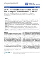
ATRA increases the activity of the human COX-2 promoter and its effect is inhibited by RAR-pan-antagonist LE540 or MEK-1 inhibitor PD98059Figure 2
ATRA increases the activity of the human COX-2 promoter and its effect is inhibited by RAR-pan-antagonist LE540 or
MEK-1 inhibitor PD98059. (a) Schematic of the COX-2 human promoter construct phPES2 containing the promoter frag-
ment -327 to +59 (b) ATRA (10 μM, 24 h) increases the activity of the human COX-2 gene promoter transfected in SH-SY5Y
human neuroblastoma cells. For comparison, the effect of IL-1β (10 ng/ml, 24 h) is also shown. COX-2 promoter activity was
determined in triplicate in four separate experiments (for statistical purposes n = 4; *P < 0.01 vs other groups; **P < 0.01 vs
control). (c) ATRA (10 μM, 24 h) increases the expression of RAR-β (left) and induces ERK1/2 phosphorylation (right). Nor-
malized density ratio of either RAR-β or ERK1/2 over α-actin is indicated for each band. Each photograph represents at least
three repeated experiments. (d) Inhibition of ATRA-induced COX-2 promoter activity by the RAR-pan-antagonist LE540 and
the MEK-1 inhibitor PD98059. Transiently transfected cells were pre-incubated for 1 h with either 2.5 μM LE540 or 50 μM
PD98059 and then with ATRA (10 μM, 24 h). COX-2 promoter activity was measured in triplicate in four separate experi-
ments (for statistical purposes n = 4) (*P < 0.01 vs other groups)
c
a
0
0,5
1
1,5
2
2,5
3
3,5
pCOX-2-LUC activity
(RLU fold increase)
CONTROL
ATRA
IL-1
β
**
*
b
ATRA 0 24 h
total ERK
P-ERK1/2
d
0
0,5
1
1,5
2
2,5
3
3,5
pCOX-2-LUC activity
(RLU fold increase)
CONTROL
LE540
ATRA
LE540+ATRA
PD98059
PD98059+ATRA
*
ATRA 0 24 h
α
-actin
RAR-
β
Ratio 1 6.16 Ratio 1 2.73
Journal of Neuroinflammation 2007, 4:1 />Page 7 of 9
(page number not for citation purposes)
results on the effect of ATRA on COX-2 expression: For
instance, ATRA repressed COX-2 promoter activity and
COX-2 mRNA expression in several murine lung tumour-
derived cell lines, yet it increased promoter activity and
COX-2 mRNA expression significantly in another lung
tumour-derived cell line [27]. In other studies ATRA did
not induce COX-2 in phorbol 12-myristate 13-acetate-dif-
ferentiated U937 cells [28] whereas it suppressed COX-2
transcription in human mammary epithelial cells [29].
There are also observations showing that, in certain cell
lines, RAR and RXR inhibit COX-2 promoter activity
[30,31]. Finally, our own studies in renal mesangial cells
indicate that RARs are involved in ATRA-induced COX-2
expression in cells cultured from rats [24] but not in cells
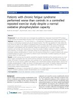
ATRA-induced COX-2 protein expression and PGE
2
production are inhibited by RAR pan-antagonist LE540 or MEK-1 inhibi-tor PD98059Figure 3
ATRA-induced COX-2 protein expression and PGE
2
production are inhibited by RAR pan-antagonist LE540 or MEK-1
inhibitor PD98059. (a) Western blot analysis of the expression of COX-2 protein in SH-SY5Y human neuroblastoma cells
pre-incubated for 1 h with either RAR pan-antagonist LE540 2.5 μM (left) or 50 μM MEK-1 inhibitor PD98059 (right) and then
incubated with ATRA (10 μM, 24 h). Equal protein loading was confirmed by probing with an anti-α-actin antibody. Normalized
density ratio of COX-2 over α-actin is indicated for each band. Each experiment represents at least three repeated experi-
ments. (b) PGE
2
production in SH-SY5Y human neuroblastoma cells incubated for 24 hours with ATRA 10 μM in cells and pre-
treated for 1 hour with either RAR pan-antagonist LE540 2.5 μM or 50 μM MEK-1 inhibitor PD98059. PGE
2
in the medium was
determined in triplicate in four separate experiments (for statistical purposes n = 4) (*P < 0.01 vs other groups)
a
b
α
-actin
ATRA - + - +
COX-2
LE540
ATRA - + - +
α
-actin
COX-2
PD98059
0
20
40
60
80
100
120
140
PGE
2
(pg/ml/mg protein)
*
CONTROL
LE540
ATRA
LE540+ATRA
PD98059
PD98059+ATRA
Ratio 1 2.27 0.94 0.98 Ratio 1 3.03 0.88 0.92
Journal of Neuroinflammation 2007, 4:1 />Page 8 of 9
(page number not for citation purposes)
cultured from human donors [20]. These data indicate
that the effect of ATRA on COX-2 expression is likely to be
cell-specific.
We and others have reported that ATRA enhances ERK1/2
phosphorylation [14,32-34] and that ERK1/2 plays a key
role in the up-regulation of COX-2 by ATRA in human
and rat mesangial cells [20,24]. Here we observed that
ATRA also induces ERK1/2 phosphorylation in SH-SY5Y
human neuroblastoma cells (Figure 2c, right) and we con-
firmed the key role of ERK1/2 phosphorylation for COX-
2 up-regulation by ATRA since treatment with PD098059,
the selective inhibitor of mitogen-activated protein kinase
kinase 1 (MEK-1), was sufficient to abrogate COX-2 pro-
moter activation, to increase COX-2 protein expression
and to increase PGE
2
production (Figure 2d and Figure 3a
right and 3b). The mechanism by which ATRA can cause
ERK1/2 activation is still unknown. For neuronal cells, it
has been suggested that a subpopulation of classical RAR
receptors, localized at or near the cell membrane, could be
responsible for ATRA-induced CREB (cyclic AMP-
response-element-binding protein) activation through
ERK1/2 phosphorylation [14]. We have proposed that
such mechanism could also be responsible for ATRA-
induced COX-2 up-regulation through ERK1/2 phospho-
rylation in rat mesangial cells [24]. In summary, the
induction of ERK1/2 phosphorylation by ATRA is a RAR-
independent key event though which the retinoid
increases COX-2 expression and PGE
2
production in SH-
SY5Y human neuroblastoma cells.
Intrathecal administration of ATRA to normal rats
enhances nociceptives responses as well as responses to
innocuous stimulation through a COX-2 dependent
mechanism [20]. We describe here, in SH-SY5Y human
neuroblastoma cells, that ATRA induces COX-2 expres-
sion resulting in increased PGE
2
production through a
mechanism involving ERK1/2. A confirmation of these
data in primary cultures of neuronal cells is needed to sup-
port a role in spinal cord sensitization of ATRA-induced
PGE
2
production.
Conclusion
In conclusion, our results indicate that ATRA induces
activity of the COX-2 promoter and synthesis of COX-2
mRNA and COX-2 protein, resulting in increased PGE
2
production in SH-SY5Y human neuroblastoma cells; and
that RARs and ERK1/2 were required for these ATRA
effects. This highlights the importance of RAR-dependent
and kinase-dependent mechanisms for ATRA-induced
COX-2 expression and activity.
Abbreviations
Cyclooxygenase (COX); All-trans retinoic acid (ATRA);
Prostaglandins (PGs); Prostaglandin E
2
(PGE
2
); Extracel-
lular signal-regulated kinase 1/2 (ERK1/2); Interleukin-1β
(IL-1β); Mitogen-activated protein kinase (MAPK);
Enzyme immunoabsorbent assay (EIA); Retinoic acid
receptor (RAR); Retinoid X receptor (RXR); MAP kinase
kinase 1(MEK-1); Jun N-terminal kinase, (JNK).
Competing interests
The author(s) declare that they have no competing inter-
ests.
Authors' contributions
MA carried out all the experiments. JFH participated in the
design of the study and performed the statistical analysis.
FJLC conceived of the study, and participated in its design
and coordination and helped to draft the manuscript. All
authors have read and approved the final manuscript.
Acknowledgements
This work was supported by the Spanish Ministry of Education (SAF2005-
06242-C03-01 and 03). Alique is a fellow of the Spanish Ministry of Educa-
tion and Science. We thank Dr. H. Kagechika (Tokyo Medical and Dental
University, Tokyo, Japan) for LE540 and HX531 and Dr. H. Inoue (Nara
Women's University, Nara, Japan) for the human COX-2 luciferase
reported construct phPES2.
References
1. Samad TA, Moore KA, Sapirstein A, Billet S, Alchorne A, Poole S,
Bonventre JV, Woolf CJ: Interleukin-1-beta-mediated induction
of Cox-2 in the CNS contributes to inflammatory pain hyper-
sensitivity. Nature 2001, 410:471-475.
2. Romero-Sandoval EA, Alique M, Moreno-Manzano V, Molina C, Lucio
FJ, Herrero JF: The oral administration of retinoic acid
enhances nociceptive withdrawal reflexes in rats with softh-
tissue inflammation. Inflamm Res 2004, 53:297-303.
3. Duester G, Mic FA, Molotkov A: Cytosolic retinoid dehydroge-
nases govern ubiquitous metabolism of retinol to retinalde-
hyde followed by tissue specific metabolism to retinoic acid.
Chemico Biol Interact 2003, 143:201-210.
4. Sporn MB, Roberts AB: Cervical dysplasia regression induced
by all-trans-retinoic acid. J Natl Cancer Inst 1994, 86:476-7.
5. Solomin L, Johansson CB, Zetterstrom RH, Bissonnette RP, Heyman
RA, Olson L, Lendahl U, Frisen J, Perlmann T: Retinoid-X receptor
signalling in the developing spinal cord. Nature 1998,
395:398-402.
6. Zetterstrom RH, Simon A, Giacobini MM, Eriksson U, Olson L:
Localization of cellular retinoid-binding proteins suggests
specific roles dor retinoids in the adult central nervous sys-
tem. Neuroscience 1994, 62:899-918.
7. Werner EA, Deluca HF: Retinoic acid is detected at relatively
high levels in the CNS of adult rats. Am J Physiol Endocrinol Metab
2002, 282:672-678.
8. Misner DL, Jacobs S, Shimizu Y, De Urquiza AM, Solomin L, Perlmann
T, DeLuca LM, Stevens CF, Evans RM: Vitamin A deprivation
results in reversible loss of hippocampal long-term synaptic
plasticity. Proc Natl Acad Sci USA 2001, 98:11714-11719.
9. Cocco S, Diaz G, Stancampiano R, Diana A, Carta M, Curreli R, Sarais
L, Fadda D: Vitamin a deficiency produces spatial learning and
memoory impairment in rats. Neuroscience 2002, 115:475-482.
10. Krezel W, Kastner P, Chambon P: Differential expression of
retinoid receptors in the adult mouse central nervous sys-
tem. Neuroscience
1999, 89:1291-300.
11. Thacher SM, Vasudevan J, Chandraratna RAS: Therapeutic applica-
tions for ligands of retinoid receptors. Therapeutic applica-
tions for ligands of retinoid receptors. Curr Pharm Design 2000,
6:25-58.
12. Hong HY, Varvayanis S, Yen A: Retinoic acid causes MEK-
dependent RAF phosphorylation through RARalpha plus
RXR activation in HL-60 cells. Differentiation 2001, 68:55-66.
Publish with BioMed Central and every
scientist can read your work free of charge
"BioMed Central will be the most significant development for
disseminating the results of biomedical research in our lifetime."
Sir Paul Nurse, Cancer Research UK
Your research papers will be:
available free of charge to the entire biomedical community
peer reviewed and published immediately upon acceptance
cited in PubMed and archived on PubMed Central
yours — you keep the copyright
Submit your manuscript here:
/>BioMedcentral
Journal of Neuroinflammation 2007, 4:1 />Page 9 of 9
(page number not for citation purposes)
13. Xu Q, Konta T, Furusu A, Nakayama K, Lucio-Cazana FJ, Fine L, Kita-
mura M: Transcriptional induction of mitogen-activated pro-
tein kinase phosphatase 1 by retinoids. Selective roles of
nuclear receptors and contribution to the antiapoptotic T
effect. J Biol Chem 2002, 277:41693-41700.
14. Canon E, Cosgaya JM, Scsucova S, Aranda A: Rapid effects of retin-
oic acid on CREB and ERK phosphorylation in neuronal cells.
Mol Biol Cell 2004, 15:5583-5592.
15. Aggarwal S, Kim SW, Cheon K, Tabassam F, Joon JH, Koo JS: Non-
classical action of retinoic acid on the activation of the camp
response element-binding protein in normal human bron-
chial epithelial cells. Mol Biol Cell 2006, 17:566-575.
16. Alique M, Lucio JF, Herrero JF: Vitamin A active metabolite, all-
trans retinoic acid, induces spinal cord sensitization. II.
Effects after intrathecal administration. Br J pharmacol 2006,
149:65-72.
17. Lovat PE, Pearson AD, Malcom A, Redfern CP: Retinoic acid recep-
tor expression during the in vitro differentiation of human
neuroblastoma. Retinoic acid receptor expression during
the in vitro differentiation of human neuroblastoma. Neurosci
Lett 1993, 162:109-113.
18. Rana B, Veal GJ, Pearson AD, Redfern CP: Retinoid X receptors
and retinoid response in neuroblastoma cells. J Cell Biochem
2002, 86:67-78.
19. Romero-Sandoval EA, Molina C, Alique M, Moreno-Manzano V, Lucio
JF, Herrero JF: Vitamin A active metabolite, all-trans retinoic
acid, induces spinal cord sensitization. I. Effects after oral
administration. Br J Pharmacol 2006, 149:56-64.
20. Alique M, Moreno-Manzano V, Xu Q, Kitamura M, Lucio FJ: Kinase-
dependent, retinoic acid receptor-independent up-regula-
tion of cyclooxygenase-2 by all-trans retinoic acid in human
mesangial cells. Br J Pharmacol 2006, 149:215-25.
21. Alique M, Romero-Sandoval EA, Herrero JF, Lucio J: All trans-retin-
oic acid (ATRA) up-regulates cyclooxygenase-2 (COX-2)
and prostaglandin E2 (PGE2) production in SH-SY5Y cells.
Role of retinoic acid receptors (RARs) and ERK1/2 pathways.
36th Annual Meeting, Society for Neuroscience 14–18 October 2006,
Atlanta .
22. Inoue H, Yokoyama C, Hara S, Tone Y, Tanabe T:
Transcriptional
regulation of human prostaglandin-endoperoxide synthase-2
gene by lypopolysaccharide and phorbol ester in vascular
endotelial cell. J Biol Chem 1995, 270:24965-24971.
23. Chambon P: A decade of molecular biology of retinoic acid
receptors. FASEB J 1996, 10:940-954.
24. Alique A, Lucio-Cazaña FJ, Moreno V, Xu Q, Konta T, Nakayama K,
Furusu A, Sepúlveda JC, Kitamura M: Up-regulation of cyclooxy-
genases by retinoic acid in rat mesangial cells. Pharmacology
2007, 79:57-64.
25. Thacher SM, Vasudevan J, Chandraratna RAS: Therapeutic applica-
tions for ligands of retinoid receptors. Curr Pharm Design 2000,
6:25-58.
26. Tanabe T, Tohnai N: Cyclooxygenase isozymes and their gene
structures and expresión. Prostaglandins Other Lipid Mediat 2002,
68–69:95-114.
27. Wardlaw SA, Zhang N, Belinsky SA: Transcriptional regulation of
basal cyclooxygenase-2 expression in murine lung tumor-
derived cell lines by CCAAT/enhancer-binding protein and
activating transcription factor/cAMP response element-
binding protein. Mol Pharmacol 2002, 62:326-33.
28. Tsukamoto H, Hishinuma T, Tayama R, Narahara K, Suzuki N, Tomi-
oka Y, Goto J: The induction of prostaglandin E synthase and
upregulation of cyclooxygenase-2 by 9-cis retinoic acid. Pros-
taglandins Other Lipid Mediat 2004, 74:61-74.
29. Subbaramaiah K, Cole PA, Dannenberg AJ: Retinoids and carnasol
suppress cyclooxygenase-2 transcription by CREB-binding
protein/p300-dependent and -independent mechanisms.
Cancer Res 2002, 62:2522-2530.
30. Kong G, Kim HT, Wu K, DeNardo D, Hilsenbeck SG, Xu XC, Lamph
WW, Bissonnette R, Dannenberg AJ, Brown PH: The retinoid X
receptor-selective retinoid, LGD down-regulates cyclooxy-
genase-2 expression in human breast cells through transcrip-
tion factor crosstalk: implications for molecular-based
chemoprevention. Cancer Res 2005, 65:3462-9.
31. Song S, Lippman SM, Zou Y, Ye X, Ajani JA, Xu XC: Induction of
cyclooxygenase-2 by benzo[a]pyrene diol epoxide though
inhibition of retinoic acid receptor-beta 2 expression. Onco-
gene 2005, 24:8268-8276.
32. Hong HY, Varvayanis S, Yen A: Retinoic acid causes MEK-
dependent RAF phosphorylation through RARalpha plus
RXR activation in HL-60 cells.
Differentiation 2001, 68:55-66.
33. Xu Q, Konta T, Furusu A, Nakayama K, Lucio-Cazana FJ, Fine L, Kita-
mura M: Transcriptional induction of mitogen-activated pro-
tein kinase phosphatase 1 by retinoids. Selective roles of
nuclear receptors and contribution to the antiapoptotic
effect. J Biol Chem 2002, 277:41693-41700.
34. Agarwal C, Chandraratna RA, Johnson AT, Rorke EA, Eckert RL:
AGN193109 is a highly effective antagonist of retinoid action
in human ectocervical epithelial cells. J Biol Chem 1996,
271:12209-12212.