báo cáo hóa học: " Formation of multinucleated giant cells and microglial degeneration in rats expressing a mutant Cu/Zn superoxide dismutase gene" ppt
Bạn đang xem bản rút gọn của tài liệu. Xem và tải ngay bản đầy đủ của tài liệu tại đây (3.25 MB, 12 trang )
BioMed Central
Page 1 of 12
(page number not for citation purposes)
Journal of Neuroinflammation
Open Access
Research
Formation of multinucleated giant cells and microglial
degeneration in rats expressing a mutant Cu/Zn superoxide
dismutase gene
Sarah E Fendrick, Qing-Shan Xue and Wolfgang J Streit*
Address: Department of Neuroscience, University of Florida College of Medicine and McKnight Brain Institute, 100 Newell Drive, Gainesville FL
32611, USA
Email: Sarah E Fendrick - ; Qing-Shan Xue - ; Wolfgang J Streit* -
* Corresponding author
Abstract
Background: Microglial neuroinflammation is thought to play a role in the pathogenesis of
amyotrophic lateral sclerosis (ALS). The purpose of this study was to provide a histopathological
evaluation of the microglial neuroinflammatory response in a rodent model of ALS, the SOD1
G93A
transgenic rat.
Methods: Multiple levels of the CNS from spinal cord to cerebral cortex were studied in
SOD1
G93A
transgenic rats during three stages of natural disease progression, including
presymptomatic, early symptomatic (onset), and late symptomatic (end stage), using immuno- and
lectin histochemical markers for microglia, such as OX-42, OX-6, and Griffonia simplicifolia isolectin
B4.
Results: Our studies revealed abnormal aggregates of microglia forming in the spinal cord as early
as the presymptomatic stage. During the symptomatic stages there was prominent formation of
multinucleated giant cells through fusion of microglial cells in the spinal cord, brainstem, and red
nucleus of the midbrain. Other brain regions, including substantia nigra, cranial nerve nuclei,
hippocampus and cortex showed normal appearing microglia. In animals during end stage disease
at 4–5 months of age virtually all microglia in the spinal cord gray matter showed extensive
fragmentation of their cytoplasm (cytorrhexis), indicative of widespread microglial degeneration.
Few microglia exhibiting nuclear fragmentation (karyorrhexis) indicative of apoptosis were
identified at any stage.
Conclusion: The current findings demonstrate the occurrence of severe abnormalities in
microglia, such as cell fusions and cytorrhexis, which may be the result of expression of mutant
SOD1 in these cells. The microglial changes observed are different from those that accompany
normal microglial activation, and they demonstrate that aberrant activation and degeneration of
microglia is part of the pathogenesis of motor neuron disease.
Published: 28 February 2007
Journal of Neuroinflammation 2007, 4:9 doi:10.1186/1742-2094-4-9
Received: 12 January 2007
Accepted: 28 February 2007
This article is available from: />© 2007 Fendrick et al; licensee BioMed Central Ltd.
This is an Open Access article distributed under the terms of the Creative Commons Attribution License ( />),
which permits unrestricted use, distribution, and reproduction in any medium, provided the original work is properly cited.
Journal of Neuroinflammation 2007, 4:9 />Page 2 of 12
(page number not for citation purposes)
Background
Amyotrophic lateral sclerosis (ALS) is an adult onset neu-
rodegenerative disease characterized by selective loss of
upper and lower motor neurons. Loss of motor neurons
results in muscle paralysis and ultimately death due to res-
piratory failure. 5–10% of ALS cases are familial inherited
in an autosomal dominant pattern, and of familial ALS
cases 20% have been linked to mutations located in the
Cu/Zn superoxide dismutase 1 (SOD1) gene [1-4]. The
discovery that SOD1 gene mutations are linked to motor
neuron disease has facilitated development of transgenic
rodent models to mimic human disease [1,2,5], and these
have provided important leads towards understanding the
molecular pathology of ALS. Since SOD1 is critically
involved in eliminating superoxide, an undesirable
byproduct of oxidative phosphorylation and a potential
source of oxidative damage, the fact that transgenic ani-
mals with SOD1 mutations show unchanged or even ele-
vated SOD1 activity has led to the conclusion that it is not
a lack of enzymatic activity that contributes to disease
development but rather some acquired toxic property of
the enzyme [6,7]. Thus the question arises, what are the
cellular targets of this toxicity? Several studies have shown
that expression of mutant SOD1 limited to motor neu-
rons is insufficient to cause motor neuron degeneration
[8,9], and work by Cleveland and co-workers has gener-
ated findings, which show that toxicity to motor neurons
requires damage from mutant SOD1 acting within non-
neuronal cells [10] and, more specifically, that microglial
cells are important for late stage disease development
[11]. These findings point towards a critical involvement
of microglia in motor neuron disease development, yet
the nature of microglial-neuronal interactions that lead to
motor neuron degeneration remains unknown. One pos-
sibility, which has also been studied extensively in the
context of other neurodegenerative diseases, notably
Alzheimer's disease, is the notion of chronic and detri-
mental microglial neuroinflammation [12]. According to
this theory, activated microglia are seen as the main cellu-
lar source of inflammatory mediators in the CNS and as
such are thought to be potentially neurotoxic [13,14].
Chronic neuroinflammation is thought to be involved
also in the pathogenesis of ALS based on a variety of in
vivo and in vitro studies concerned with studying micro-
glial activation using both human and animal tissues [15-
20].
In order to learn more about the role of microglia in the
pathogenesis of motor neuron disease, we set out to inves-
tigate microglial activation in the G93A SOD1 mutant rat
during natural disease progression. The results reported
here are unexpected in that they reveal a highly abnormal
microglial reaction that does not meet the criteria of an
anticipated, characteristic neuroinflammatory response.
Methods
Animals
Animal use protocols were approved by the University of
Florida Institutional Use and Care of Animals Committee
(IUCAC). All transgenic animals used in this study were
male Sprague Dawley NTac:SD-TgN(SOD1G93A)L26H
rats obtained from Taconic Farms where animals were
screened extensively for infections prior to shipping.
Upon arrival animals were housed under SPF conditions.
Age-matched, wild type Sprague Dawley rats were pur-
chased from Harlan. The time course of disease progres-
sion varied among individual animals, but in general
once symptoms developed disease progression was quite
rapid causing death of most animals by 5 months of age.
To examine microglial morphology, microglial markers
were used at three stages of the disease: 1) presympto-
matic stage, where animals had no apparent muscle weak-
ness. Animals studied in this group were aged 74–84 days;
2) early symptomatic stage (onset), where animals first
showed evidence of hind limb weakness. Animals studied
in this group were aged 113–117 days; 3) late sympto-
matic (end stage), where animals were no longer able to
right themselves after 30s. Animals studied in this group
were aged 135–156 days. For each of the three disease
stages, 4 transgenic and 4 age-matched wild type control
animals were used.
Tissue processing and immunohistochemistry
Animals were deeply anesthetized with pentobarbital and
perfused transcardially with phosphate buffer saline
(PBS) followed by a fixative solution containing 4% para-
formaldehyde in PBS. The spinal cord and brain were dis-
sected out and fixed overnight in 4% paraformaldehyde at
4°C, transferred to 30% sucrose and then frozen. Lumbar
spinal cord, cortical, and brainstem sections were cut in
the coronal plane at 20 μm on a cryostat, mounted on
slides and air dried. Sections were pretreated in PBS with
0.5% Triton X-100 for 15 min, blocked in 10% normal
goat serum for 30 min and incubated overnight at room
temperature in the primary antibody diluted in buffer.
The primary antibodies included MRC OX-42 (Serotec,
Cambridge, UK) and MRC OX-6 (Serotec, Cambridge,
UK) at 1:500. The slides were rinsed in PBS and incubated
in secondary antibody (1:500) for 1 h. Following incuba-
tion, slides were rinsed and Horseradish Peroxidase Avi-
din D was applied (1:500; Vector, Burlingame, CA) and
incubated for 30 min. Slides were washed and immunore-
activity was visualized with 3,3'-diaminobenzidine
(DAB)-H
2
O
2
substrate. After a brief rinse, slides were
dehydrated in increasing concentrations of ethanols,
cleared in xylene, and coverslipped using Permount
mounting medium (Fisher Scientific).
Journal of Neuroinflammation 2007, 4:9 />Page 3 of 12
(page number not for citation purposes)
OX-42 immunoreactivity in the ventral spinal cord was
quantified using Image Pro Plus software (version 4.5.1,
Media Cybernetics, Carlsbad, CA). The area occupied by
stained cells was highlighted and measured for each sec-
tion of spinal cord (6 sections per animal) then expressed
as a percentage of total area of ventral spinal cord. Using
GraphPad Prism software (San Diego, CA) a t-test was per-
formed to determine statistical significance between trans-
genic SOD 1 and control animals at each time point. A
one-way ANOVA was performed to compare differences
among the transgenic animals followed by a Tukey multi-
ple comparison test.
Paraffin processing and lectin histochemistry
Animals were deeply anesthetized and transcardially per-
fused with phosphate buffer saline (PBS) followed by a
fixative solution containing 4% paraformaldehyde. The
spinal cord and brain were dissected out and fixed 2 h in
4% paraformaldehyde. The tissue was dehydrated
through ascending alcohols, cleared in xylenes and
embedded in paraffin. Serial 7 μm coronal sections were
collected and mounted on slides. Sections were deparaffi-
nized through xylenes, graded alcohols and rinsed in PBS.
Next, the slides were trypsin treated (0.1% trypsin, 0.1%
CaCl
2
) for 12 min at 37°C. Following a 10 min wash the
slides were incubated overnight at 4°C in lectin GSA I-B
4
-
HRP (Sigma Chemical Co.) diluted 1:10 in PBS contain-
ing cations (0.1 mM of CaCl
2
, MgCl
2
and MnCl
2
) and
0.1% Triton X-100. After overnight incubation slides were
briefly rinsed in PBS and visualized with 3,3'-diabi-
mobenzidine (DAB)- H
2
O
2
substrate. Sections were coun-
terstained with cresyl violet, dehydrated through
ascending alcohols, cleared in xylenes and coverslipped
with Permount.
Results
Development of microgliosis during natural disease
progression in the spinal cord
The CR3 complement receptor recognized by OX-42 anti-
body is expressed constitutively by all resting and acti-
vated microglial cells [21]. OX-42 immunoreactivity
observed in presymptomatic SOD1 transgenic rats was
similar to that seen in wild type control, i.e. there was uni-
form staining of all resting microglia (Figs. 1A,D). Occa-
sionally, in these presymptomatic animals cell fusions
involving several microglia were observed (Fig. 1A, inset).
The onset of symptoms was associated with a dramatic
increase in OX-42 staining in the ventral horn due to
much greater microglial cell numbers (Fig. 1B). Many of
these seemingly activated microglia were clustered and/or
fused into multi-cellular aggregates. In end stage animals,
overall immunoreactivity with OX-42 was decreased com-
pared to that seen in animals with disease onset (Fig. 1C).
This unexpected diminution in microglial staining was
due to widespread degenerative cytoplasmic fragmenta-
tion affecting most, if not all microglia within the ventral
horn (see below). The qualitatively evident increases and
decreases in immunoreactivity were confirmed through
quantitative morphometric measurements (Fig. 1E).
With onset of symptoms, there was apparent activation of
microglia as judged by the dramatic increase in OX-42
immunoreactivity in the spinal gray matter. Examining
sections at low power clearly revealed pronounced spots
of enhanced OX-42 staining in the ventral horns (Fig. 2A),
and these were judged initially to be due to the formation
of microglial phagocytic clusters around dying motor neu-
rons, as this would be a normal response to motor neuron
death. However, when spots of intense OX-42 immunore-
activity were examined at higher power (Fig. 2B,D) they
appeared unusual in that individual microglial phago-
cytes were not discernable. Subsequent counterstaining of
these sections with cresyl violet allowed us to conclude
that the OX-42 reactive structures were, in fact, not phago-
cytic clusters but represented multinucleated giant cells
(Figs. 2C,E). These giant cells were found in all SOD1
G93A
transgenic rats studied. They formed apparently as a result
of multiple microglial cells fusing together into sizable
syncytia (40–50 μm) that often showed a circular arrange-
ment of microglial nuclei about their periphery (Fig. 2E).
This kind of nuclear arrangement is classically associated
with multinucleated giant cells of the Langhans type. The
cytoplasmic interior of Langhans giant cells appeared
granular and fragmented, suggesting ongoing deteriora-
tion. A few of the giant cells revealed the presence of apop-
totic bodies, evident as nuclear fragments (Fig. 2F), but
overall apoptotic bodies either inside or outside of giant
cells were sparse. Microglia dispersed in between giant
cells revealed relatively normal process-bearing morphol-
ogy and lacked the conspicuous hypertrophy that is char-
acteristic of activated microglia. (Fig. 2E). However, some
sections showed ongoing microglial cytorrhexis, i.e. frag-
mentation of the cytoplasm. Cytorrhexis became conspic-
uous in animals that were in the terminal stages of the
disease process (Fig. 3) and was evident as a loss of dis-
cernable microglial cell structure and presence of abun-
dant OX-42 immunoreactive fragments of microglial
cytoplasm dispersed throughout the spinal gray matter
(Figs. 3A,B,D,E). Occasional giant cells could still be
observed during end stage disease, however, most of these
showed signs of deterioration evident by increased irregu-
larity of their shape and nuclear arrangement, as well as by
increased granularity and fragmentation (Figs. 3A,B). In
some sections, neurons remained stained with cresyl vio-
let suggesting residual preservation of neuronal integrity.
However, pathological features were evident in motor
neurons, including most notably intense hyperchromia
with formation of a nuclear cap consisting of condensed
chromatin material (Fig. 3C). This neuronal appearance
Journal of Neuroinflammation 2007, 4:9 />Page 4 of 12
(page number not for citation purposes)
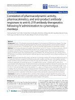
Microglial staining with OX-42 immunohistochemistry in the spinal cord during three different stages of motor neuron disease progressionFigure 1
Microglial staining with OX-42 immunohistochemistry in the spinal cord during three different stages of motor neuron disease
progression. A, presymptomatic stage; inset shows early microglial fusion in spinal cord. B, disease onset; C, end stage; D, wild
type control. Note the dramatic increase in microglial staining with OX-42 during onset (B) and its subsequent decline during
end stage (C). Scale bar: 200 μm. E, morphometric quantification of microglial immunostaining with OX-42 during disease
development; * p < 0.05 and ** p < 0.001 with respect to age-matched controls; # p < 0.05 with respect to onset group.
Journal of Neuroinflammation 2007, 4:9 />Page 5 of 12
(page number not for citation purposes)
stood in stark contrast to that of normal motor neurons as
seen in wild type animals (Fig. 3F).
Histopathology in the brain stem
Sections from the brainstem at the level of cranial nerve
VII during disease onset and end stage were marked by
changes indicative of severe neuropathology (Fig. 4). They
included prominent, widespread vacuolization of the
extracellular space and hyperchromia of neuronal proc-
esses. Often neurites appeared physically separated (as if
torn) from neuronal cell bodies leaving one or more dis-
tinct stumps on the perikaryon (Fig. 4D,E). The changes
affecting microglia were striking in that multinucleated
giant cells were present throughout any given section.
These consisted of fused microglial cells that gave rise to a
variety of bizarrely shaped cellular fusions which, in some
cases, extended for more than one hundred micrometers
in length (Figs. 4B,C,F). Microglial fusions varied in size,
sometimes involving only a few cells, and other times
twenty or more. Although not obviously associated with
vascular channels, some microglial giant cells due to their
elongated shape seemed to have formed along blood ves-
sels (Fig. 4C). Presence of giant cells was observed in all
animals regardless of whether they were at an early or late
symptomatic stage of motor neuron disease. They were
scattered seemingly at random throughout the brainstem
and not limited to any particular nucleus or tract, and
often displayed the classic morphological features of
Langhans type giant cells (Fig. 4G).
Within vacuolated spaces rounded, shrunken microglia
exhibiting nuclear fragmentation or shrinkage (pyknosis)
were identified using lectin histochemical staining (Figs.
4H).
Microglia in midbrain and cerebral cortex
Microglial fusions similar to those seen in the spinal cord
and brainstem level were found also in the red nucleus of
the midbrain (Figs. 5A–D). The specificity with which
these microglial fusions were restricted to the red nucleus
area was remarkable, as they were visible even at the low-
est magnification (Fig. 5A). Microglia outside of the red
nucleus displayed normal, ramified morphology. Rubros-
pinal neurons appeared normal in size and morphology,
as well as in number, and there was no evidence to suggest
that any of these neurons were undergoing degeneration.
Rubrospinal neurons were not encircled by activated
microglia. It is noteworthy also that motor neurons in the
oculomotor nucleus, which appears with the red nucleus
in the same sections, revealed no evidence of degenerative
changes, and microglia here were normal and non-acti-
vated in appearance. Similarly, microglia in the substantia
nigra appeared completely normal (Fig. 5F). Somewhat
surprisingly, we also found no evidence at all for micro-
glial activation or abnormalities in the motor cortex of
animals, regardless of disease stage, with any of the micro-
glial markers employed (Figs. 5G,H).
Discussion
The purpose of the current study was to perform an inves-
tigation of microgliosis in a recently developed rat model
of ALS involving expression of a mutated human SOD1
transgene (G93A) [5]. Although these animals, similar to
their murine counterparts, reportedly mimic many of the
histopathological features of human ALS, including glial
activation [5,19], until now a detailed analysis of reactive
microgliosis has not been performed. Our current results
show that the microgliosis that occurs in SOD1
G93A
rats is
atypical and marked by some highly unusual features in
microglial cells that are indicative of cellular dysfunction.
The key microglial aberrations found consist of fusion
into giant cells and cytorrhexis (Fig. 6). These features are
not observed normally during microglial activation and
they lead us to conclude that this particular animal model
of ALS is characterized by microglial degeneration rather
than by microglial neuroinflammation. It is therefore con-
ceivable that neurodegeneration occurs as a consequence
of glial cell deterioration.
Prior work in ALS rodent models involving SOD1 muta-
tions has generated clues about an involvement of glial
cells. Damage to astrocytes has been described to occur
concomitant with degeneration of motor neurons
prompting the hypothesis that astrocytic damage pro-
motes motor neuron degeneration [22]. However, subse-
quent experiments showed that restricted expression of
mutant SOD1 genes in astrocytes is not sufficient to cause
motor neuron degeneration [23]. Notwithstanding these
findings, more recently it was determined using chimeric
animals consisting of mixtures of normal cells and cell
expressing human mutant SOD1 that nonneuronal cells
containing mutant SOD1 are indeed required to cause
damage to motor neurons, whereas wildtype nonneuro-
nal cells promote motor neuron survival [10,24]. In addi-
tion, recent work has shown that mutant SOD1 acting
within microglial cells specifically is a primary determi-
nant of late stage disease progression [11]. These observa-
tions gain added significance when considered together
with the current findings showing widespread microglial
degeneration in the spinal cord gray matter of end stage
animals, because it now seems clear that mutant SOD1 is
particularly toxic to microglia and that SOD1-mediated
microglial degeneration is linked to a terminal neurode-
generative disease state. Thus, loss of microglial cells
could be very detrimental to neuronal survival [25].
Future research may be directed towards elucidating the
molecular mechanisms that underlie SOD1's selective
microglial toxicity, and towards ways of inhibiting it as a
strategy for new ALS treatments.
Journal of Neuroinflammation 2007, 4:9 />Page 6 of 12
(page number not for citation purposes)
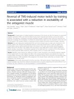
OX-42 immunohistochemistry during symptomatic phase of diseaseFigure 2
OX-42 immunohistochemistry during symptomatic phase of disease. A, low power view reveals intensified immunoreactivity in
spinal cord ventral horns; multiple large, rounded spots are visible. B, higher power view of large immunoreactive spots is sug-
gestive of phagocytic clusters. C, same field as in B; counterstaining with cresyl violet facilitates identification of large immuno-
reactive spots as multinucleated giant cells. D, E, the same microscopic field prior to and after cresyl violet counterstaining
reveals a well-formed multinucleated giant cell of the Langhans type. F, enlargement of framed area in C shows apoptotic
microglial nucleus (arrow) within a giant cell. Scale bars: 500 μm (A), 40 μm (B, C), 20 μm (D-F).
Journal of Neuroinflammation 2007, 4:9 />Page 7 of 12
(page number not for citation purposes)
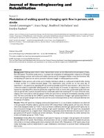
OX-42 immunohistochemistry during end stage disease demonstrates extensive microglial cytoplasmic fragmentation (A-E)Figure 3
OX-42 immunohistochemistry during end stage disease demonstrates extensive microglial cytoplasmic fragmentation (A-E).
A, D, two different views of spinal ventral gray matter demonstrate loss of microglial cell integrity and widespread punctate
staining indicative of cytorrhexis. Note that many neurons remain stained with cresyl violet. B, enlargement of framed area in
A shows detail of microglial cytorrhexis, including a disintegrating giant cell on the right. E, enlargement of framed area in D
shows detail of microglial cytorrhexis. C, motor neuron in SOD1
G93A
rat reveals intense hyperchromasia with cresyl violet and
nuclear cap. F, normal motor neuron and microglia from wild type spinal cord. Scale bars: 40 μm (A, D); 20 μm (B, C, E, F).
Journal of Neuroinflammation 2007, 4:9 />Page 8 of 12
(page number not for citation purposes)
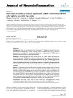
Lectin staining of microglia in the brainstem (level of cranial nerve VII) in wildtype animals (A) and in late symptomatic/end stage animals (B-H)Figure 4
Lectin staining of microglia in the brainstem (level of cranial nerve VII) in wildtype animals (A) and in late symptomatic/end
stage animals (B-H). Cresyl violet counterstain. A, microglia show normal ramified morphology. B, a large lectin-positive
aggregate of fused microglia is evident in severely vacuolated brainstem tissue. Note enlarged perineuronal spaces to the right.
C, string-like microglial fusions extend over long distances. D, breakage of neuronal process, probably a dendrite, from cell
body within markedly vacuolated space (arrows). E, two multinucleated microglial giant cells are seen below a neuron with
broken off process (arrow). F, large multinucleated giant cell displaying vacuolization is present amidst numerous microglial
cytoplasmic fragments. G, multinucleated giant cell of the Langhans type displaying characteristic peripheral arrangement of
nuclei. H, rounded lectin-positive microglial cell (arrow) within vacuolated space displays nuclear fragmentation indicative of
apoptosis. Scale bars: 20 μm (A-H).
Journal of Neuroinflammation 2007, 4:9 />Page 9 of 12
(page number not for citation purposes)
Visualization of microglia in midbrain with GSA-I-B
4
lectin (A-F) and in motor cortex with OX-42 (G) and OX-6 (H) during symptomatic diseaseFigure 5
Visualization of microglia in midbrain with GSA-I-B
4
lectin (A-F) and in motor cortex with OX-42 (G) and OX-6 (H) during
symptomatic disease. A, low power view of midbrain reveals enhanced lectin staining in the red nucleus. B, higher magnifica-
tion shows that enhanced lectin reactivity is confined strictly to red nucleus region (arrows indicate perimeter of red nucleus).
C, microglial fusions are interspersed with rubrospinal neurons that appear undamaged. D, lectin-positive microglial fusion
(giant cell) within red nucleus. E, oculomotor nucleus reveals normal-appearing motor neurons and lack of microgliosis. F, sub-
stantia nigra (pars compacta) shows presence of normal, ramified microglial cells. G, motor cortex shows normal, ramified
microglia. H, single, ramified microglial cell positive with OX-6 (arrow) near lateral ventricle. Scale bars: 400 μm (A); 200 μm
(E); 100 μm (B,H); 50 μm (C,F,G); 20 μm (D).
Journal of Neuroinflammation 2007, 4:9 />Page 10 of 12
(page number not for citation purposes)
We use the term "cytorrhexis" to describe the kind of
microglial degeneration we observed in SOD1
G93A
rats
because it involves disintegration of the cell's cytoplasm
rather than of its nucleus. Cytorrhexis has been used pre-
viously only to describe neuronal necrosis resulting from
excitotoxicity [26], but extending its use to describe micro-
glial cytoplasmic deterioration is appropriate since this
form of cell death does not involve the nuclear disintegra-
tion (karyorrhexis) that is characteristic of apoptosis. Cyt-
orrhexis therefore describes accidental, rather than
programmed, microglial cell death. Our inability to detect
large numbers of apoptotic microglia in the tissues stud-
ied indirectly supports the idea that cytorrhexis is the "pre-
ferred" mode of microglial cell death during the toxic
disease state thought to be generated by mutant SOD1
expression. Finding widespread microglial degeneration
in this particular animal model of neurodegenerative dis-
ease strongly supports the broader concept that microglial
abnormalities characterize other neurodegenerative con-
ditions as well [27-29].
Perhaps the earliest sign of an aberrant microglial
response in SOD1 mutant rats is reflected in our observa-
tion of occasional microglial fusions in presymptomatic
animals. We suspect that with disease onset these progress
to produce the conspicuous multinucleated giant cells
composed of many microglia fused into large syncytia.
The occurrence of microglial giant cells throughout the
lumbar spinal gray matter, as well as the brainstem, and
especially their selective localization in the red nucleus,
raises the intriguing possibility that their formation is
related to the fact that these regions all give rise to fibers
that project onto ventral motor neurons. It is conceivable
therefore that a signal is transmitted retrogradely from
ventral horn cells to these supraspinal regions to trigger
formation of microglial fusions, consistent with the
notion of disease spread from an initially affected region
[11]. However, at the same time the notable absence of
microglial abnormalities and/or activation in the motor
cortex reported here would argue against this idea. Addi-
tional studies providing more detailed mapping of the
location of giant cells could be helpful in this regard.
Fusion of microglia into giant cells represents an anoma-
lous type of cellular behavior, since microglia are nor-
mally "territorial" and exhibit strong contact inhibition.
Microglial giant cells have never been described to occur
in situ in rat brain, but they can form spontaneously in
vitro using cultured microglia from a variety of species [30-
33]. Multinucleated giant cells are a pathological hallmark
in human brain during infectious diseases, most notably
in HIV/AIDS encephalopathy [34,35], and since microglia
are the main cellular target of HIV-1 in the brain it is
thought that presence of virus within microglia causes the
Schematic depicting the approximate time course of motor neuron disease development and the accompanying microglial changes in SOD1
G93A
ratsFigure 6
Schematic depicting the approximate time course of motor neuron disease development and the accompanying microglial
changes in SOD1
G93A
rats. Note that disease onset and subsequent development of end stage disease is variable among individ-
ual animals.
Journal of Neuroinflammation 2007, 4:9 />Page 11 of 12
(page number not for citation purposes)
cells to fuse with each other. However, the exact mecha-
nisms that produce microglial fusions in the SOD1
G93A
rat
are unknown and require additional studies. But regard-
less of the mechanism(s) involved, it seems clear from the
current observations that fusion of microglia into giant
cells is an abnormal cellular response that can progress
further to produce frank microglial degeneration evident
as cytorrhexis. It is unknown currently whether microglial
cytorrhexis occurs in HIV encephalopathy, but the fact
that neurodegenerative changes accompany advanced,
untreated HIV encephalopathy (clinically evident as
AIDS-dementia complex) raises the intriguing possibility
that microglial degeneration may be part of the pathogen-
esis of cognitive dysfunction associated with HIV
encephalitis. In addition, descriptions of an ALS-like syn-
drome in HIV-infected subjects [36-38] leads us to
hypothesize that dysfunctional or degenerating microglia
may be a common denominator in these two seemingly
unrelated conditions.
The thought that microglial neuroinflammation pro-
motes motor neuron degeneration can be traced back to
the initial descriptions of activated microglia in human
ALS tissues [16]. Since then, the idea of detrimental neu-
roinflammation has received additional support from a
variety of studies documenting microglial activation and
increased production of proinflammatory substances in
human ALS tissues, serum and CSF as well as in SOD1
transgenic mice [15,20,39-44]. With regard to the SOD1
transgenic rat, the only report thus far concerning neu-
roinflammation has described elevated gene expression of
proinflammatory mediators in the spinal cord, as well as
downregulation of the neuroprotective factor VEGF [45].
Conclusion
Our current findings provide a perspective on the patho-
genesis of neurodegenerative disease that is different from
the neuroinflammation theory which claims that chronic
neuroinflammation leads to motor neuron degeneration
[14]. We propose that, rather than being overly activated,
the brain's immune cells fail in performing normal, neu-
roprotective functions and that degeneration of microglia
contributes to neurodegeneration. This may significantly
change future approaches towards treatment by pharma-
cological and other means.
Competing interests
The author(s) declare that they have no competing inter-
ests.
Authors' contributions
SF carried out the histopathological studies and drafted
the manuscript. QX participated in analysis of histopatho-
logical findings and design of figures and schematics. WS
conceived and directed the study, and completed the final
version of the manuscript.
Acknowledgements
Supported by NIH grant NS49185.
References
1. Rosen DR, Siddique T, Patterson D, Figlewicz DA, Sapp P, Hentati A,
Donaldson D, Goto J, O'Regan JP, Deng HX, et al.: Mutations in Cu/
Zn superoxide dismutase gene are associated with familial
amyotrophic lateral sclerosis. Nature 1993, 362(6415):59-62.
2. Deng HX, Hentati A, Tainer JA, Iqbal Z, Cayabyab A, Hung WY, Get-
zoff ED, Hu P, Herzfeldt B, Roos RP, et al.: Amyotrophic lateral
sclerosis and structural defects in Cu,Zn superoxide dis-
mutase. Science 1993, 261(5124):1047-1051.
3. Siddique T, Deng HX: Genetics of amyotrophic lateral sclerosis.
Hum Mol Genet 1996, 5 Spec No:1465-1470.
4. Mulder DW, Kurland LT, Offord KP, Beard CM: Familial adult
motor neuron disease: amyotrophic lateral sclerosis. Neurol-
ogy 1986, 36(4):511-517.
5. Nagai M, Aoki M, Miyoshi I, Kato M, Pasinelli P, Kasai N, Brown RH
Jr., Itoyama Y: Rats expressing human cytosolic copper-zinc
superoxide dismutase transgenes with amyotrophic lateral
sclerosis: associated mutations develop motor neuron dis-
ease. J Neurosci 2001, 21(23):9246-9254.
6. Cleveland DW, Rothstein JD: From Charcot to Lou Gehrig:
deciphering selective motor neuron death in ALS. Nat Rev
Neurosci 2001, 2(11):806-819.
7. Gurney ME, Pu H, Chiu AY, Dal Canto MC, Polchow CY, Alexander
DD, Caliendo J, Hentati A, Kwon YW, Deng HX, et al.: Motor neu-
ron degeneration in mice that express a human Cu,Zn
superoxide dismutase mutation. Science 1994,
264(5166):1772-1775.
8. Lino MM, Schneider C, Caroni P: Accumulation of SOD1
mutants in postnatal motoneurons does not cause motone-
uron pathology or motoneuron disease. J Neurosci 2002,
22(12):4825-4832.
9. Pramatarova A, Laganiere J, Roussel J, Brisebois K, Rouleau GA: Neu-
ron-specific expression of mutant superoxide dismutase 1 in
transgenic mice does not lead to motor impairment. J Neuro-
sci 2001, 21(10):3369-3374.
10. Clement AM, Nguyen MD, Roberts EA, Garcia ML, Boillee S, Rule M,
McMahon AP, Doucette W, Siwek D, Ferrante RJ, Brown RH Jr., Jul-
ien JP, Goldstein LS, Cleveland DW:
Wild-type nonneuronal cells
extend survival of SOD1 mutant motor neurons in ALS
mice. Science 2003, 302(5642):113-117.
11. Boillee S, Yamanaka K, Lobsiger CS, Copeland NG, Jenkins NA, Kas-
siotis G, Kollias G, Cleveland DW: Onset and Progression in
Inherited ALS Determined by Motor Neurons and Microglia.
Science 2006, 312(5778):1389-1392.
12. Streit WJ, Mrak RE, Griffin WS: Microglia and neuroinflamma-
tion: a pathological perspective. J Neuroinflammation 2004,
1(1):14.
13. McGeer PL, McGeer EG: Inflammation, autotoxicity and Alzhe-
imer disease. Neurobiol Aging 2001, 22(6):799-809.
14. McGeer PL, McGeer EG: Inflammatory processes in amyo-
trophic lateral sclerosis. Muscle Nerve 2002, 26(4):459-470.
15. Alexianu ME, Kozovska M, Appel SH: Immune reactivity in a
mouse model of familial ALS correlates with disease pro-
gression. Neurology 2001, 57(7):1282-1289.
16. Kawamata T, Akiyama H, Yamada T, McGeer PL: Immunologic
reactions in amyotrophic lateral sclerosis brain and spinal
cord tissue. Am J Pathol 1992, 140(3):691-707.
17. Sargsyan SA, Monk PN, Shaw PJ: Microglia as potential contribu-
tors to motor neuron injury in amyotrophic lateral sclerosis.
Glia 2005, 51(4):241-253.
18. Weydt P, Yuen EC, Ransom BR, Moller T: Increased cytotoxic
potential of microglia from ALS-transgenic mice. Glia 2004,
48(2):179-182.
19. Hall ED, Oostveen JA, Gurney ME: Relationship of microglial and
astrocytic activation to disease onset and progression in a
transgenic model of familial ALS. Glia 1998, 23(3):249-256.
20. Henkel JS, Engelhardt JI, Siklos L, Simpson EP, Kim SH, Pan T, Good-
man JC, Siddique T, Beers DR, Appel SH: Presence of dendritic
Publish with Bio Med Central and every
scientist can read your work free of charge
"BioMed Central will be the most significant development for
disseminating the results of biomedical research in our lifetime."
Sir Paul Nurse, Cancer Research UK
Your research papers will be:
available free of charge to the entire biomedical community
peer reviewed and published immediately upon acceptance
cited in PubMed and archived on PubMed Central
yours — you keep the copyright
Submit your manuscript here:
/>BioMedcentral
Journal of Neuroinflammation 2007, 4:9 />Page 12 of 12
(page number not for citation purposes)
cells, MCP-1, and activated microglia/macrophages in amyo-
trophic lateral sclerosis spinal cord tissue. Ann Neurol 2004,
55(2):221-235.
21. Graeber MB, Streit WJ, Kreutzberg GW: Axotomy of the rat
facial nerve leads to increased CR3 complement receptor
expression by activated microglial cells. J Neurosci Res 1988,
21(1):18-24.
22. Bruijn LI, Becher MW, Lee MK, Anderson KL, Jenkins NA, Copeland
NG, Sisodia SS, Rothstein JD, Borchelt DR, Price DL, Cleveland DW:
ALS-linked SOD1 mutant G85R mediates damage to astro-
cytes and promotes rapidly progressive disease with SOD1-
containing inclusions. Neuron 1997, 18(2):327-338.
23. Gong YH, Parsadanian AS, Andreeva A, Snider WD, Elliott JL:
Restricted expression of G86R Cu/Zn superoxide dismutase
in astrocytes results in astrocytosis but does not cause
motoneuron degeneration. J Neurosci 2000, 20(2):660-665.
24. Beers DR, Henkel JS, Xiao Q, Zhao W, Wang J, Yen AA, Siklos L,
McKercher SR, Appel SH: Wild-type microglia extend survival
in PU.1 knockout mice with familial amyotrophic lateral
sclerosis. Proc Natl Acad Sci U S A 2006, 103(43):16021-16026.
25. Streit WJ: Microglia as neuroprotective, immunocompetent
cells of the CNS. Glia 2002, 40(2):133-139.
26. Ingvar M, Morgan PF, Auer RN: The nature and timing of excito-
toxic neuronal necrosis in the cerebral cortex, hippocampus
and thalamus due to flurothyl-induced status epilepticus.
Acta Neuropathol (Berl) 1988, 75(4):362-369.
27. Streit WJ: Microglia and Alzheimer's disease pathogenesis. J
Neurosci Res 2004, 77(1):1-8.
28. v Eitzen U, Egensperger R, Kosel S, Grasbon-Frodl EM, Imai Y, Bise K,
Kohsaka S, Mehraein P, Graeber MB: Microglia and the develop-
ment of spongiform change in Creutzfeldt-Jakob disease. J
Neuropathol Exp Neurol 1998, 57(3):246-256.
29. Wierzba-Bobrowicz T, Lewandowska E, Kosno-Kruszewska E,
Lechowicz W, Pasennik E, Schmidt-Sidor B: Degeneration of
microglial cells in frontal and temporal lobes of chronic
schizophrenics. Folia Neuropathol 2004, 42(3):157-165.
30. Lee TT, Martin FC, Merrill JE: Lymphokine induction of rat
microglia multinucleated giant cell formation. Glia 1993,
8(1):51-61.
31. Tambuyzer BR, Nouwen EJ: Inhibition of microglia multinucle-
ated giant cell formation and induction of differentiation by
GM-CSF using a porcine in vitro model. Cytokine 2005,
31(4):270-279.
32. Suzumura A, Tamaru T, Yoshikawa M, Takayanagi T: Multinucle-
ated giant cell formation by microglia: induction by inter-
leukin (IL)-4 and IL-13. Brain Res 1999, 849(1-2):239-243.
33. Hassan NF, Prakash K, Chehimi J, McCawley LJ, Douglas SD: Isola-
tion and characterization of newborn rabbit brain-derived
microglia. Clin Immunol Immunopathol 1991, 59(3):426-435.
34. Dickson DW: Multinucleated giant cells in acquired immuno-
deficiency syndrome encephalopathy. Origin from endog-
enous microglia? Arch Pathol Lab Med 1986, 110(10):967-968.
35. Michaels J, Price RW, Rosenblum MK: Microglia in the giant cell
encephalitis of acquired immune deficiency syndrome: pro-
liferation, infection and fusion. Acta Neuropathol (Berl) 1988,
76(4):373-379.
36. Moulignier A, Moulonguet A, Pialoux G, Rozenbaum W: Reversible
ALS-like disorder in HIV infection. Neurology 2001,
57(6):995-1001.
37. Sinha S, Mathews T, Arunodaya GR, Siddappa NB, Ranga U, Desai A,
Ravi V, Taly AB: HIV-1 clade-C-associated "ALS"-like disorder:
first report from India. J Neurol Sci 2004, 224(1-2):97-100.
38. Verma A, Berger JR: ALS syndrome in patients with HIV-1
infection. J Neurol Sci 2006, 240(1-2):59-64.
39. Elliott JL: Cytokine upregulation in a murine model of familial
amyotrophic lateral sclerosis. Brain Res Mol Brain Res 2001, 95(1-
2):172-178.
40. Hensley K, Floyd RA, Gordon B, Mou S, Pye QN, Stewart C, West M,
Williamson K: Temporal patterns of cytokine and apoptosis-
related gene expression in spinal cords of the G93A-SOD1
mouse model of amyotrophic lateral sclerosis. J Neurochem
2002, 82(2):365-374.
41. Wilms H, Sievers J, Dengler R, Bufler J, Deuschl G, Lucius R: Intrath-
ecal synthesis of monocyte chemoattractant protein-1
(MCP-1) in amyotrophic lateral sclerosis: further evidence
for microglial activation in neurodegeneration. J Neuroimmu-
nol 2003, 144(1-2):139-142.
42. Poloni M, Facchetti D, Mai R, Micheli A, Agnoletti L, Francolini G,
Mora G, Camana C, Mazzini L, Bachetti T: Circulating levels of
tumour necrosis factor-alpha and its soluble receptors are
increased in the blood of patients with amyotrophic lateral
sclerosis. Neurosci Lett 2000, 287(3):211-214.
43. Yoshihara T, Ishigaki S, Yamamoto M, Liang Y, Niwa J, Takeuchi H,
Doyu M, Sobue G: Differential expression of inflammation- and
apoptosis-related genes in spinal cords of a mutant SOD1
transgenic mouse model of familial amyotrophic lateral scle-
rosis. J Neurochem 2002, 80(1):158-167.
44. Hensley K, Fedynyshyn J, Ferrell S, Floyd RA, Gordon B, Grammas P,
Hamdheydari L, Mhatre M, Mou S, Pye QN, Stewart C, West M, West
S, Williamson KS: Message and protein-level elevation of
tumor necrosis factor alpha (TNF alpha) and TNF alpha-
modulating cytokines in spinal cords of the G93A-SOD1
mouse model for amyotrophic lateral sclerosis. Neurobiol Dis
2003, 14(1):74-80.
45. Xie Y, Weydt P, Howland DS, Kliot M, Moller T: Inflammatory
mediators and growth factors in the spinal cord of G93A
SOD1 rats. Neuroreport 2004, 15(16):2513-2516.