báo cáo hóa học: " Cerebral Amyloid Angiopathy-related Inflammation Presenting with Steroid-responsive Higher Brain Dysfunction: Case Report and Review of the Literature" doc
Bạn đang xem bản rút gọn của tài liệu. Xem và tải ngay bản đầy đủ của tài liệu tại đây (2.55 MB, 10 trang )
CAS E REP O R T Open Access
Cerebral Amyloid Angiopathy-related
Inflammation Presenting with Steroid-responsive
Higher Brain Dysfunction: Case Report and
Review of the Literature
Hideya Sakaguchi
*
, Akihiko Ueda, Takayuki Kosaka, Satoshi Yamashita, En Kimura, Taro Yamashita, Yasushi Maeda,
Teruyuki Hirano and Makoto Uchino
Abstract
A 56-year-old man noticed discomfort in his left lower limb, followed by convulsion and numbness in the same
area. Magnetic resonance imaging (MRI) showed white matter lesions in the right parietal lobe accompanied by
leptomeningeal or leptomeningeal and cortical post-contrast enhancement along the parietal sulci. The patient
also exhibited higher brain dysfunction corresponding with the lesions on MRI. Histological pathology disclosed
b-amyloid in the blood vessels and perivascular inflammation, which highlights the diagnosis of cerebral amyloid
angiopathy (CAA)-related inflammation. Pulse steroid therapy was so effective that clinical and radiological findings
immediately improved.
CAA-related inflammation is a rare disease, defined by the deposition of amyloid proteins within the
leptomeningeal and cortical arteries associated with vasculitis or perivasculitis. Here we report a patient with CAA-
related inflammation who showed higher brain dysfunction that improved with steroid therapy. In cases with
atypical radiological lesions like our case, cerebral biopsy with histolog ical confirmation remains necessary for an
accurate diagnosis.
Keywords: cerebral amyloid angiopathy, CAA-related inflammation, higher brain dysfunction
Background
Cerebral amyloid angiopathy (CAA) is a common pathol-
ogy in the elderly characterized by the deposition of amy-
loid proteins within the leptomeningeal and cortical
arteries [1]. Recently, coexisting inflammations in CAA
patients, such as vasculitis or peri vasculitis, which clini-
cally resemble central nervous system vasculitis, have
been recognized as CAA-related inflammation [2,3]. The
inflammation typically responds well to steroid therapy
[4], and recent studies have pointed out its similarities
with meningoencephalitis induced by immunization to
Ab in Alzheimer disease patients [4-6]. Herein we report
apatientwithCAA-related inflammation who showed
convulsion in the left lower extremity and higher brain
dysfunction; both were dramatically improved by steroid
therapy.
Case presentation
A 56-year-old man first noticed discomfort in his left
lower limb in January 2010. After 7 days, convulsion in
the left lower li mb suddenly occurred, and he was trans-
ported to the emergency hospital. Magnetic resonance
imaging (MRI) showed increased white matter intensities
in the right parietal lobe on T2-weighted and fluid attenu-
ated inversion-recovery (FLAIR) images. T1-weighted
gadolinium (Gd)-enhanced images revealed enhanced lep-
tomeningeal lesions along the parietal sulci (Figure 1A-B).
No microhemorrhages were observed with Gradient-
recalled echo (GRE)-T2* imaging (1.5T). He was referred
to our institution.
On admission, neurological exam showed mild hyper-
esthesia in the left lower limb and mild hypalgesia in the
* Correspondence:
Department of Neurology, Faculty of Life Sciences, Kumamoto University 1-
1-1 Honjo, Kumamoto 860-0811, Japan
Sakaguchi et al. Journal of Neuroinflammation 2011, 8:116
/>JOURNAL OF
NEUROINFLAMMATION
© 2011 Sakaguchi et al; licensee BioMed Cen tral Ltd. This is an Open Acc ess art icle distributed under the terms of the Creative
Commons Attribution License (http://cr eativecommons.org/licenses/by/2.0), which permits unrestricted use, distribution, and
reproduction in any medium, provided the original work is properly cited.
left crus. No other abnormal findings were present. Bio-
chemical screening tests were generally normal except
for serum C-reactive protein (0.77 mg/dL), soluble inter-
leukin-2 receptor antibody (462 U/mL), erythrocyte sedi-
mentation rate (26/1 h, 72/2 h), and c arcinoembryonic
antigen (4.5 ng/mL). In the cerebrospinal fluid, protein
levels were elevated (72 mg/dl) and the cell count was
mildly elevated (12/μL).
Because a follow-up MRI revealed progression of the
white matter lesions and parenchymal enhanced lesions
without microhemorrhages (GRE-T2* imaging; 3T) (Fig-
ure 1C-G), a brain biopsy was performed in March
2010. Histological patholo gy showed nonspecific menin-
goencephalitis involving perivasculitis of the leptome-
ninges and cortical gray matter (Figure 2A-D).
Starting in April 2010, the patien t complained of diffi-
culty with his handwriting. Neuropsychological tests of
higher brain functions re vealed mild constructional apraxia,
line imbalance for words and numbers, difficulty drawing a
figure following oral instructions, and problems with visual
reproduction. No apathy or dementia w as observed.
After the episode, further histological analysis with
Congo-red staining disclosed amyloid laden blood vessels.
Immunohistochemical staining for b-amyloid led to the
diagnosis of CAA-related inflammation (Figure 2F-G).
Steroid pulse therapy (methylprednisolone 1 g/day for 3
days) was performed. The abnormal Gd-enhanced findings
immediate ly improved wi th gradually decre asin g FLAIR
findings, and the higher brain dysfunctions also gradually
resolved (Figure 3).
DC
AB
G
E
F
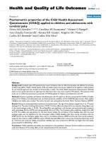
Figure 1 Axial MRI from the referring hospita l and on ad mission to our ho spital. MRI findings of FLAIR (A) and T1-weighted image with
Gd enhancement (B) from the referring hospital (1.5T). Increased white matter lesions are visible in the right parietal lobe on FLAIR images (A),
and a T1-weighted Gd enhanced image revealed abnormal enhanced parenchymal lesions along the parietal sulci (B). On admission, these
lesions worsened in both FLAIR (C) and T1-weighted enhanced images (D). High signal intensity in the apparent diffusion coefficient (ADC) map
(E) and low signal intensity in the diffusion-weighted image (F) suggested its edematous nature. No microhemorrhages were observed with
Gradient recalled echo-T2* imaging (3T) (G).
Sakaguchi et al. Journal of Neuroinflammation 2011, 8:116
/>Page 2 of 10
100ȝm
100ȝm
100ȝm
A
E
D
C
B
G
F
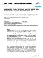
Figure 2 Histological and immune-histological exami nation of brain biopsy. Microscopic examination showed nonspecific
meningoencephalitis involving perivasculitis of leptomeninges (arrows) and cortical gray matter (A). The cellular infiltrate was mainly composed
of CD-3-positive T-lymphocytes (B) and CD-68-positive macrophages (C) with minimal CD-20-positive B-lymphocytes (D). PAS staining showed no
deposits (E). Congo-red staining revealed amyloid positive blood vessels (F); the amyloid was disclosed to be amyloid-b by
immunohistochemical staining (G).
Sakaguchi et al. Journal of Neuroinflammation 2011, 8:116
/>Page 3 of 10
2010
March
2010
April
2010
Ma
y
2010
June
2010
Jul
y
2010
August
Oral steroid
70ঌ
ঌঌ
ঌ/day
Steroid pulse therapy
FLAIR
T1-Gd(+)
Three
dimentional
house
Sunflower
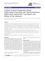
Figure 3 Clinical course of treatment with steroid. Abnormal T1 Gd-enhanced findings immediately improved in the fifth course of steroid
pulse therapy, accompanied by a gradual decrease of FLAIR findings and a gradual improvement in higher brain function. As the MRI lesions
improved (05/28), the descriptions of the 3D-house and sunflower were made more vivid (05/25). Because T1 Gd-enhanced lesions almost
disappeared after the fifth course of the steroid (05/28), we stopped the steroid therapy, and the lesion relapsed (06/04). However, after the
initiation of oral steroid therapy, no relapse was observed either clinically or radiologically (08/17).
Sakaguchi et al. Journal of Neuroinflammation 2011, 8:116
/>Page 4 of 10
After the fifth course of stero id pulse tr eatment, the
T1-enhanced lesions had almost disappeared, and we
stopped the treatment. However, 2 weeks later, the
lesions had relapsed on a follow-up MRI, although no
clinical signs were observed. We performed pulse ster-
oid therapy again, followed by oral methylprednisolone
therapy (70 mg/day). After the oral steroid therapy was
initiated, no relapses were observed either clinically or
radiologically. Two months later, the oral steroid was
tapered at a rate of 5 mg/week, and he was discharged
on a regimen of methylprednisolone 30 mg/day.
Discussion
CAA is defi ned by the deposition of amyloid proteins
within leptomeningeal and cortical arteries, arterioles, and
capillaries [1]. Recently, a subset of patients who presented
with seizures, subacute cognitive decline, or headaches
with hyperintensities on T2-weighted or FLAIR MRI
images with microh emorrhages were described as having
CAA-related inflammation [2,3]. Neuropathologic exami-
nation has generally revealed angiitis of CAA-affected ves-
sels and peripheral infla mmation, presenting as vasculitis
or perivasculitis [7]. Both pathologic forms can co-exist,
and it has been suggested that the prognosis is bette r for
the perivascular type [8]. This inflammation appears to
represent an autoimmune response to vascular b-amyloid
deposits. The mechanism by which this immune response
occurs is not well understood, although one possible factor
is the increased frequency of apolipoprotein E ε4/ε4 geno-
type [9].
The clinical spectrum of CAA-related inflammation is
mainly composed of rapidly progressive dementia and
seizure. Although the initial presentation of our case was
seizure and numbness, the subsequent higher brain dys-
function is uncommon. To clarify how of ten higher brain
dysfunction has been observed, w e reviewed previous
cases including our case (Table 1) [1,3,4,7-37]. In 64
cases, 10 presented with higher brain dysfunction without
encephalopathy or dementia (15.3%). The most frequent
symptom was aphasia (6 cases: 9.3%), followed by hemi-
neglect (2 cases: 3.1%). One other case was reported of
various higher bra in dysfunction without mental change
or dementia, like our case [23]. In these ten cases with
higher brain dysfunction, MRI lesions and the presence
of leptomeningeal enhancement were inconsistent, and
thus the presentation of higher brain dysfunction was
considered to be derived from the observed lesion rather
than specific to CAA-related inflammation.
The MRI presentation for CAA-related inflammation
was previously described as characterized by large conflu-
ent areas of predominantly white matter hyperintense
signal on T2-weighted or FLAIR images [34]. These
lesions are typically asymmetric and involve one or more
cortical lesions without evident preferential laterality. T2-
weighted gradient-echo sequence images usually showed
multiple scattered cortical or subcortical microhemor-
rhages [34]. However, these microhemorrhages were not
observed in our case, resulting in a delayed diagnosis. In
our review, 13 cases were examined by MRI with an echo
gradient sequence, and microhemorrhages were not seen
in 2 cases including our case (13.3%). A possible explana-
tion is that the inflammatio n caused by the immunoreac-
tivity to amyloid might precede the vascular change of
cerebellar amyloid angiopathy in some cases, such that
microhemorrhages were not observed in radiological
exams. This suggests that the gradient-echo sequence
image might not be adequate for diagnosis of CAA-
related inflammation in all cases. Brain biopsy should be
considered if CAA-related inflammation is highly sus-
pected from clinical presentation, even if microhemor-
rhages were not radiologically observed.
Approximately three quarters of all patients described
had a good clinical response to corticosteroid therapy.
Additionally, patients presenting with CAA and menin-
geal enhancement seem to have less progressive disease
[29]. In our review, the leptomeningeal enhancement sta-
tus of 42 patients was mentioned, and the clinical courses
of 39 patients were described. Among 19 patients with
leptomeningeal enhancement, only one patient died
(5.3%) and the remaini ng 18 patients survived. However,
among the other 20 patients without enhancement, 7
patients died (35%), suggesting that leptomeningea l
enhancement might be a good prognostic factor.
The distinctive pattern of asymmetric MRI lesions in
CAA-related inflammation appears to be distinguishable
from both non-inflammatory CAA and other causes.
This observation raises the possibility that typical MRI
findings should prove sufficient to diagnose CAA-related
inflammation without necessitating brain biopsy [4].
However, in our case, preoperative imaging did not
show the typical microhemorrhages associa ted with
CAA, and the diagnosis could not have been established
before biopsy. Therefore, we suggest that cerebral biopsy
with histological confirmation remains necessary for an
accurate diagnosis.
Conclusion
We described a patient with CAA-related inflammation
whose higher brain functions were dramatically
improved by steroid therapy. Because the improvement
of cognitive function paralleled resolution of the lesions
seen on MRI, this report demonstrates clinically and
radiologically progressive improvement of CAA-related
inflammation. Our case also suggests the import ance of
brain biopsy for diagnosis in a case with atypical radi-
ological findings, because correct diagnosis and treat-
ment are crucial for successful recovery and good
prognosis.
Sakaguchi et al. Journal of Neuroinflammation 2011, 8:116
/>Page 5 of 10
Table 1 Review of reported cases of CAA-related inflammation
Reference n Age Sex Clinical presentation MRI lesion Micro bleeds in T2*-
weighted images
MRI
enhanced
lesion
Pathology treatment Outcome
Greenberg et
al. 1993 [10]
1 72 F dementia headache left frontal NA (-) vasculitis NA NA
Ortiz et al.
1996 [11]
1 68 F headache right temporal/parietal NA (-) vasculitis steroid NA
Fountain et
al. 1996 [12]
266 M
fluent aphasia right
hemianopia
bilateral temporal/parietal NA (-) vasculitis
perivasculitis
steroid
cyclophosphamide
alive relapse
(+)
69 F headache confusion focal
neurology seizure
bilateral confluent multifocal NA NA vasculitis steroid
cyclophosphamide
died relapse
(+)
Anders et al.
1997 [13]
2 70 M mental status change right frontal NA NA vasculitis NA NA
69 M headache lethargy behavior
change
bilateral white matter NA (+) vasculitis NA NA
Fountain et
al. 1999 [14]
1 71 M headache confusion gait
difficulty left hand apraxia
right temporal/parietal NA NA vasculitis cyclophosphamide alive relapse
(+)
Scully et al.
2000 [15]
1 63 M behavior change ataxia bilateral white matter NA (+) perivasculitis cyclophosphamide alive
Oide et al.
2002 [16]
1 69 M dizziness dementia seizure bilateral symmetrical periventricular NA NA vasculitis (-) NA
Schwab et al.
2003 [8]
2 74 M seizure dementia headache bilateral multifocal NA (+) perivasculitis steroid alive relapse
(+)
70 F dementia headache right temporal NA (+) perivasculitis steroid alive relapse
(+)
Tamargo et
al. 2003 [17]
1 80 F dementia left-side
hemineglect word finding
difficulty
bilateral left frontal right parietal NA (+) vasculitis steroid alive
Oh et al.
2004 [1]
2 80 F Headache
aphasia bilateral right parietal/occipital left
frontal
NA (-) perivasculitis steroid alive
77 M
aphasia left temporal NA (-) vasculitis steroid alive
Safriel et al.
2004 [18]
1 49 M seizure right occipital/temporal NA (-) vasculitis steroid alive
Hashizume et
al. 2004 [19]
1 65 M headache left hemianopsia
left-side hemineglect
right temporal/occipital NA (+) vasculitis steroid
cyclophosphamide
died
Harkness et
al. 2004 [20]
1 72 F dementia bilateral frontal NA (-) vasculitis no specific therapy alive
Jacobs et al.
2004 [21]
1 81 F confusion Balint’s syndrome
agraphia right-left confusion
finger anomia left-side
neglect
bilateral parietal/occipital NA (+) vasculitis steroid alive
Sakaguchi et al. Journal of Neuroinflammation 2011, 8:116
/>Page 6 of 10
Table 1 Review of reported cases of CAA-related inflammation (Continued)
Scolding et
al. 2005 [3
6 69.3* M 3
F3
encephalopathy 6 focal
neurology 2 seizure 1
headache 2
NA mutifocal 1 frontal 1 diffuse
white matter 1 right occipital
1 left frontal 1 bilateral
confluent 1
NA (+) 1 (-) 5 vasculitis steroid 3 steroid
cyclophosphamide 2
tumor resection
steroid 1
alive 4
(relapse NA)
died 2
Mikolaenko
et al. 2006
[22]
1 50 M seizure right frontal NA (+) vasculitis surgery alive
Wong et al.
2006 [23]
179 F
higher brain dysfunction
fatigue
right frontal/temporal/parietal NA NA vasculitis steroid alive relapse
(+)
Kinnecom et
al. 2007 [4]
1 62.3* M 9
F3
encephalopathy 9 headache
5 seizure 7
aphasia 1
presyncope 1
NA NA NA (the presence of
microbleeds are
mentioned but the
proportion is not
mentioned)
NA perivasculitis steroid 9 steroid
cyclophosphamide 3
alive 11
(relapse (+) 3)
died 1
Greenberg et
al. 2007 [24]
1 63 M headache behavioral change
cognitive change
bilateral multiple NA (+) vasculitis cyclophosphamide alive relapse
(+)
Marotti et al.
2007 [25]
1 57 F headache seizure bilateral frontal/temporal/insular right
thalamus
(+) (+) vasculitis seizure control died
McHugh et
al. 2007 [26]
1 80 F confusion incontinent urine
global aphasia seizure right
hemianopia right
hemiparesis
bilateral frontal (+) (-) vasculitis
perivasculitis
steroid alive relapse
(+)
Takada et al.
2007 [27]
1 69 F headache cognitive decline bilateral right frontal/parietal bilateral
parietal/occipital
(+) (-) vasculitis steroid died
Machida et
al. 2008 [28]
1 69 F cognitive decline bilateral multifocal (-) (+) perivasculitis steroid alive relapse
(+)
Salvarani et
al. 2008 [29]
8 63* M6
F2
encephalopathy 6 focal
neurology 2 headache 3
only aphasia with alexia 1
bilateral
8
multifocal NA (+) 5 (-) 3 vasculitis steroid 3 steroid
cyclophosphamide 5
improved 6
died 1
worsened 1
Amick et al.
2008 [30]
1 79 F transient right sided
weakness
left occipital/parietal NA (-) vasculitis (-) died
Alcalay et al.
2009 [31]
1 92 F mental status change bilateral multifocal (+) (+) (-) steroid alive
Daniëls et al.
2009 [32]
1 80 F mental status change right
sided hemiparesis dysphasia
seizure
bilateral left hemisphere right
parietal/occipital
(+) (-) (-) steroid alive relapse
(+)
Greenberg et
al. 2010 [9]
1 87 F seizure cognitive impairment bilateral multifocal (+) NA perivasculitis steroid died
Kloppenborg
et al. 2010 [ 7]
1 74 M increased sleepiness loss of
initiative seizure
bilateral frontal (+) (+) perivasculitis steroid alive
Morishige et
al. 2010 [33]
1 78 F motor aphasia dementia left frontal NA (+) vasculitis steroid alive
Savoiardo et
al. 2010 [34]
1 76 M fatigue confusion bilateral temporal/occcipital/frontal (+) (-) (-) steroid alive
Sakaguchi et al. Journal of Neuroinflammation 2011, 8:116
/>Page 7 of 10
Table 1 Review of reported cases of CAA-related inflammation (Continued)
Cano et al.
2010 [35]
176 M transient motor aphasia
transient headache
bilateral temporal (+) NA (-) (-) alive
DiFrancesco
et al. 2011
[36]
1 68 M memory loss mood disorder bilateral multifocal (+) (-) NA steroid alive
Chung et al.
2011 [37]
3 83 F seizure bilateral multifocal NA NA vasculitis steroid died
forties F headache mild hemiparesis
sensory loss
right parietal/occipital (+) NA vasculitis steroid alive
haemorrhage
(+)
72 M seizure
left-side neglect left
hemianopia
bilateral multifocal NA NA vasculitis
perivasculitis
steroid
cyclophosphamide
died
our case 1 56 M Seizure sensory disturbance
higher brain dysfunction
right parietal (-) (+) perivasculitis steroid alive relapse
(+)
From the literature, we extracted the cases of CAA-related inflammation in which an MRI was evaluated. If autopsy or biopsy was examined, the cases without inflammation were excluded. All cases satisfy the
diagnostic criteria of definite or probable CAA-related inflammation proposed by Chung et al. [37]. In 64 cases, 10 presented with higher brain dysfunction without encephalopathy or dementia (15.3%). The most
frequent symptom was aphasia (6 cases: 9.3%), followed by hemineglect (2 cases: 3.1%). One case besides the current presented with various higher brain dysfunction without mental change or dementia [23]. In
these 10 cases with higher brain dysfunction, MRI lesions and the presence of leptomeningeal enhancement were inconsistent. Thirteen cases were examined with MRI with an echo gradient sequence, and
microhemorrhages were not seen in 2 cases, including our case (13.3%).
The leptomeningeal enhancement status of 42 patients was mentioned, and the clinical courses of 39 patients were described. Only one patient among 19 patients with leptomeningeal enhancement died (5.3%);
however, 7 of 20 patients without enhancement died (35%), suggesting that leptomeningeal enhancement might be a factor in good prognosis. *: calculated mean
Sakaguchi et al. Journal of Neuroinflammation 2011, 8:116
/>Page 8 of 10
Consent
Written informed consent was obtained from the patient
for publication of this case report and any accompany-
ing images. A copy of the written consent is available
for review by the Editor-in-Chief of this journal.
List of abbreviations
Aβ: amyloid β; ADC: apparent diffusion coefficient; CAA: cerebral amyloid
angiopathy; FLAIR: fluid attenuated inversion-recovery; Gd: gadolinium; MRI:
magnetic resonance imaging; GRE: gradient-recalled echo.
Acknowledgements
The authors are very grateful to Professor Hitoshi Takahashi of the Brain
Research Institute at the University of Niigata for his expert suggestions
regarding pathology.
Authors’ contributions
HS designed this article and direction for investigations and drafted the
manuscript. AU, TK, SY, EK, TY, YM, TH, and MU contributed to
interpretations of clinical, radiological and pathological details. All authors
read and approved the final manuscript.
Authors’ information
All authors are members of the Department of Neurology, Faculty of Life
Sciences, Kumamoto University, and TK was also a graduate student of the
Brain Research Institute, University of Niigata until March 2011.
Competing interests
The authors declare that they have no competing interests.
Received: 20 May 2011 Accepted: 14 September 2011
Published: 14 September 2011
References
1. Oh U, Gupta R, Krakauer JW, Khandji AG, Chin SS, Elkind MS: Reversible
leukoencephalopathy associated with cerebral amyloid angiopathy.
Neurology 2004, 62(3):494-497.
2. Eng JA, Frosch MP, Choi K, Rebeck GW, Greenberg SM: Clinical
manifestations of cerebral amyloid angiopathy-related inflammation.
Ann Neurol 2004, 55(2):250-256.
3. Scolding NJ, Joseph F, Kirby PA, Mazanti I, Gray F, Mikol J, Ellison D,
Hilton DA, Williams TL, MacKenzie JM, et al: Abeta-related angiitis: primary
angiitis of the central nervous system associated with cerebral amyloid
angiopathy. Brain 2005, 128(Pt 3):500-515.
4. Kinnecom C, Lev MH, Wendell L, Smith EE, Rosand J, Frosch MP,
Greenberg SM: Course of cerebral amyloid angiopathy-related
inflammation. Neurology 2007, 68(17):1411-1416.
5. Nicoll JA, Wilkinson D, Holmes C, Steart P, Markham H, Weller RO:
Neuropathology of human Alzheimer disease after immunization with
amyloid-beta peptide: a case report. Nat Med 2003, 9(4):448-452.
6. Orgogozo JM, Gilman S, Dartigues JF, Laurent B, Puel M, Kirby LC,
Jouanny P, Dubois B, Eisner L, Flitman S, et al: Subacute
meningoencephalitis in a subset of patients with AD after Abeta42
immunization. Neurology 2003, 61(1):46-54.
7. Kloppenborg RP, Richard E, Sprengers ME, Troost D, Eikelenboom P,
Nederkoorn PJ: Steroid responsive encephalopathy in cerebral amyloid
angiopathy: a case report and review of evidence for
immunosuppressive treatment. J Neuroinflammation 2010, 7:18.
8. Schwab P, Lidov HG, Schwartz RB, Anderson RJ: Cerebral amyloid
angiopathy associated with primary angiitis of the central nervous
system: report of 2 cases and review of the literature. Arthritis Rheum
2003, 49(3):421-427.
9. Greenberg SM, Rapalino O, Frosch MP: Case records of the Massachusetts
General Hospital. Case 22-2010. An 87-year-old woman with dementia
and a seizure. N Engl J Med 2010, 363(4):373-381.
10. Greenberg SM, Vonsattel JP, Stakes JW, Gruber M, Finklestein SP: The
clinical spectrum of cerebral amyloid angiopathy: presentations without
lobar hemorrhage. Neurology 1993, 43(10):2073-2079.
11. Ortiz O, Reed L: Cerebral amyloid angiopathy presenting as a
nonhemorrhagic, infiltrating mass. Neuroradiology 1996, 38(5) :449-452.
12. Fountain NB, Eberhard DA: Primary angiitis of the central nervous system
associated with cerebral amyloid angiopathy: report of two cases and
review of the literature. Neurology 1996, 46(1):190-197.
13. Anders KH, Wang ZZ, Kornfeld M, Gray F, Soontornniyomkij V, Reed LA,
Hart MN, Menchine M, Secor DL, Vinters HV: Giant cell arteritis in
association with cerebral amyloid angiopathy: immunohistochemical
and molecular studies. Hum Pathol 1997, 28(11):1237-1246.
14. Fountain NB, Lopes MB:
Control of primary angiitis of the CNS associated
with
cerebral amyloid angiopathy by cyclophosphamide alone.
Neurology 1999, 52(3):660-662.
15. Case records of the Massachusetts General Hospital. Weekly
clinicopathological exercises. Case 10-2000. A 63-year-old man with
changes in behavior and ataxia. N Engl J Med 2000, 342(13):957-965.
16. Oide T, Tokuda T, Takei Y, Takahashi H, Ito K, Ikeda S: Serial CT and MRI
findings in a patient with isolated angiitis of the central nervous
system associated with cerebral amyloid angiopathy. Amyloid 2002,
9(4):256-262.
17. Tamargo RJ, Connolly ES, McKhann GM, Khandji A, Chang Y, Libien J,
Adams D: Clinicopathological review: primary angiitis of the central
nervous system in association with cerebral amyloid angiopathy.
Neurosurgery 2003, 53(1):136-143, discussion 143.
18. Safriel Y, Sze G, Westmark K, Baehring J: MR spectroscopy in the diagnosis
of cerebral amyloid angiopathy presenting as a brain tumor. AJNR Am J
Neuroradiol 2004, 25(10):1705-1708.
19. Hashizume Y, Yoshida M, Suzuki E, Hirayama M: A 65-year-old man with
headaches and left homonymous hemianopsia. Neuropathology 2004,
24(4):350-353.
20. Harkness KA, Coles A, Pohl U, Xuereb JH, Baron JC, Lennox GG: Rapidly
reversible dementia in cerebral amyloid inflammatory vasculopathy. Eur
J Neurol 2004, 11(1):59-62.
21. Jacobs DA, Liu GT, Nelson PT, Galetta SL: Primary central nervous system
angiitis, amyloid angiopathy, and Alzheimer’s pathology presenting with
Balint’s syndrome. Surv Ophthalmol 2004, 49(4) :454-459.
22. Mikolaenko I, Conner MG, Jinnah HA: A 50-year-old man with acute-onset
generalized seizure. Cerebral amyloid angiopathy and associated giant
cell reaction. Arch Pathol Lab Med 2006, 130(1) :e5-7.
23. Wong SH, Robbins PD, Knuckey NW, Kermode AG: Cerebral amyloid
angiopathy presenting with vasculitic pathology. J Clin Neurosci 2006,
13(2):291-294.
24. Greenberg SM, Parisi JE, Keegan BM: A 63-year-old man with headaches
and behavioral deterioration. Neurology 2007, 68(10):782-787.
25. Marotti JD, Savitz SI, Kim WK, Williams K, Caplan LR, Joseph JT: Cerebral
amyloid angiitis processing to generalized angiitis and
leucoencephalitis. Neuropathol Appl Neurobiol 2007, 33(4):475-479.
26. McHugh JC, Ryan AM, Lynch T, Dempsey E, Stack J, Farrell MA, Kelly PJ:
Steroid-responsive recurrent encephalopathy in a patient with cerebral
amyloid angiopathy. Cerebrovasc Dis 2007, 23(1):66-69.
27. Takeda A, Tatsumi S, Yamashita M, Yamamoto T: [Granulomatous angiitis
of the CNS associated with cerebral amyloid angiopathy
–an
autopsied
case with widespread involvement]. Brain Nerve 2007, 59(5):537-543.
28. Machida K, Tojo K, Naito KS, Gono T, Nakata Y, Ikeda S: Cortical petechial
hemorrhage, subarachnoid hemorrhage and corticosteroid-responsive
leukoencephalopathy in a patient with cerebral amyloid angiopathy.
Amyloid 2008, 15(1):60-64.
29. Salvarani C, Brown RD, Calamia KT, Christianson TJ, Huston J, Meschia JF,
Giannini C, Miller DV, Hunder GG: Primary central nervous system
vasculitis: comparison of patients with and without cerebral amyloid
angiopathy. Rheumatology (Oxford) 2008, 47(11):1671-1677.
30. Amick A, Joseph J, Silvestri N, Selim M: Amyloid-beta-related angiitis: a
rare cause of recurrent transient neurological symptoms. Nat Clin Pract
Neurol 2008, 4(5):279-283.
31. Alcalay RN, Smith EE: MRI showing white matter lesions and multiple
lobar microbleeds in a patient with reversible encephalopathy. J
Neuroimaging 2009, 19(1):89-91.
32. Daniels R, Geurts JJ, Bot JC, Schonewille WJ, van Oosten BW: Steroid-
responsive edema in CAA-related inflammation. J Neurol 2009,
256(2):285-286.
33. Morishige M, Abe T, Kamida T, Hikawa T, Fujiki M, Kobayashi H, Okazaki T,
Kimura N, Kumamoto T, Yamada A, et al: Cerebral vasculitis associated
Sakaguchi et al. Journal of Neuroinflammation 2011, 8:116
/>Page 9 of 10
with amyloid angiopathy: case report. Neurol Med Chir (Tokyo) 2010,
50(4):336-338.
34. Savoiardo M, Erbetta A, Storchi G, Girotti F: Case 159: cerebral amyloid
angiopathy-related inflammation. Radiology 2010, 256(1):323-327.
35. Cano LM, Martinez-Yelamos S, Majos C, Alberti MA, Boluda S, Velasco R,
Rubio F: Reversible acute leukoencephalopathy as a form of
presentation in cerebral amyloid angiopathy. J Neurol Sci 2010, 288(1-
2):190-193.
36. DiFrancesco JC, Brioschi M, Brighina L, Ruffmann C, Saracchi E,
Costantino G, Galimberti G, Conti E, Curto NA, Marzorati L, et al: Anti-Abeta
autoantibodies in the CSF of a patient with CAA-related inflammation: a
case report. Neurology 2011, 76(9):842-844.
37. Chung KK, Anderson NE, Hutchinson D, Synek B, Barber PA: Cerebral
amyloid angiopathy related inflammation: three case reports and a
review. J Neurol Neurosurg Psychiatry 2011, 82(1):20-26.
doi:10.1186/1742-2094-8-116
Cite this article as: Sakaguchi et al.: Cerebral Amyloid Angiopathy-
related Inflammation Presenting with Steroid-responsive Higher Brain
Dysfunction: Case Report and Review of the Literature. Journal of
Neuroinflammation 2011 8:116.
Submit your next manuscript to BioMed Central
and take full advantage of:
• Convenient online submission
• Thorough peer review
• No space constraints or color figure charges
• Immediate publication on acceptance
• Inclusion in PubMed, CAS, Scopus and Google Scholar
• Research which is freely available for redistribution
Submit your manuscript at
www.biomedcentral.com/submit
Sakaguchi et al. Journal of Neuroinflammation 2011, 8:116
/>Page 10 of 10