báo cáo hóa học: " Treatment with gelsolin reduces brain inflammation and apoptotic signaling in mice following thermal injury" potx
Bạn đang xem bản rút gọn của tài liệu. Xem và tải ngay bản đầy đủ của tài liệu tại đây (2.01 MB, 18 trang )
RESEARCH Open Access
Treatment with gelsolin reduces brain
inflammation and apoptotic signaling in mice
following thermal injury
Qing-Hong Zhang
1
, Qi Chen
1
, Jia-Rui Kang
2
, Chen Liu
3
, Ning Dong
1
, Xiao-Mei Zhu
1
, Zhi-Yong Sheng
1
and
Yong-Ming Yao
1,4*
Abstract
Background: Burn survivors develop long-term cognitive impairment with increased inflammation and apoptosis
in the brain. Gelsolin, an actin-binding protein with capping and severing activities, plays a crucial role in the septic
response. We investigated if gelsolin infusion could attenuate neural damage in burned mice.
Methods: Mice with 15% total body surface area burns were injected intravenously with bovine serum albumin as
placebo (2 mg/kg), or with low (2 mg/kg) or high doses (20 mg/kg) of gelsolin. Samples were harvested at 8, 24,
48 and 72 hours postburn. The immune function of splenic T cells was analyzed. Cerebral pathology was examined
by hematoxylin/eosin staining, while activated glial cells and infiltrating leukocytes were detected by
immunohistochemistry. Cerebral cytokine mRNAs were further assessed by quantitative real-time PCR, while
apoptosis was evaluated by caspase-3. Neural damage was determined using enzyme-linked immunosorbent assay
of neuron-specific enolase (NSE) and soluble protein-100 (S-100). Finally, cerebral phospho-ERK expression was
measured by western blot.
Results: Gelsolin significantly improved the outcomes of mice following major burns in a dose-dependent manner.
The survival rate was improved by high dose gelsolin treatment compared with the placebo group (56.67% vs.
30%). Although there was no significant improvement in outcome in mice receiving low dose gelsolin (30%),
survival time was prolonged against the placebo control (43.1 ± 4.5 h vs. 35.5 ± 5.0 h; P < 0.05). Burn-induced T
cell suppression was greatly alleviated by high dose gelsolin treatment. Concurrently, cerebral abnormalities were
greatly ameliorated as shown by reduced NSE and S-100 content of brain, decreased cytokine mRNA expressions,
suppressed microglial activation, and enhanced infiltration of CD11b+ and CD45+ cells into the brain. Furthermore,
the elevated caspase-3 activity seen following burn injury was remarkably reduced by high dose gelsolin treatment
along with down-regulation of phospho-ERK expression.
Conclusion: Exogenous gelsolin infusion improves survival of mice following major burn injury by partially
attenuating inflammation and apoptosis in brain, and by enhancing peripheral T lymphocyte function as well.
These data suggest a novel and effective strategy to combat excessive neuroinflammation and to preserve
cognition in the setting of major burns.
Keywords: Burns, Gelsolin, Septic encephalopathy, Neuroinflammation, Caspase-3, Apoptosis
* Correspondence:
1
Department of Microbiology and Immunology, Burns Institute, First Hospital
Affiliated to the Chinese PLA General Hospital, Beijing 100048, PR China
Full list of author information is available at the end of the article
Zhang et al. Journal of Neuroinflammation 2011, 8:118
/>JOURNAL OF
NEUROINFLAMMATION
© 2011 Zhang et al; licensee BioMed Central Ltd. This is a n Open Access article distributed under the terms of the Creative Commons
Attribution License (http://creativecommons .org/licenses/by/2.0), which permits unrestricted use, distribution, and reproduction in
any medium, provided the original work is properly cited.
Background
Brain is one of the remote organs subjected to injurious
effects of severe burns [1]. Survivors suffering from
extensive burn injury present long-term cognitive
impairment, including depression, anxiety, post-trau-
matic stress disorder [2,3], and alterat ion in painful sen-
sation as well as sensory sensitivity in later life [4]. In
animal studies, magnetic resonance imaging has iden ti-
fied marked changes in the brain up to 3 days postburn
(pb), most notably swelling and lesions [5], changes in
cerebral blood flow [6], dysregulation of g lucose meta-
bolism [7], and disruption of the blood-brain barrier
(BBB) [8,9].
Neuroinflammation is a frequent consequence of sep-
sis and septic shock [10]. Approximately 93% of burn
patients show clinical signs of a systemic inflammato ry
response syndrome before succumbing to their injuries
[11], and this syndrome can deteriorate and develop
into severe sepsis [12]. After burn injury, there is a dra-
matic increase in proinflammatory cytokines in brain as
early as 3 hours (h) [13,14] and a compromised BBB
leading to a large infiltration of macrophages [9]. Benefi-
cial as well as deleterious effects have been ascribed to
immune cells that infiltrate the nervous system after
neural injury [15-19]. Despite the correlation between
cerebral complications in severe burn victims and mor-
tality, burn-induced neuroinflammation continues to be
an underestimated entity in critically ill burn patients
[10].
Gelsolin was first described as a ~90 kDa cytoplasm
actin-binding protein with capping and severing activ-
ities [20]. Further studies have confirmed a secrete d gel-
solin isoform in blood plasma [21]. Recent reports have
documented that it also participates in the regul ation of
the systemic immune response. Extracellular gelsolin is
involved in host immune recognition of bacterial wall
molecules during cell division or attack by immune
components, while cytoplasmic gelsolin is necessary for
macrophage motility in culture, and its absence is likely
to impair recruitment of macrophages to a site of crush
injury of sciatic nerve [22]. In fact, overexpression of
gelsolin could alter actin dynamics in Jurkat T cells, cor-
relating with inhibition of activation-dependent signaling
pathways [23]. Moreover, cytoplasmic gelsolin depletion
is observed in diverse states of inflammation that are
associated with tissue injury and actin release, includ ing
hemorrhagic shock [24], early sepsis, trauma, and rheu-
matoid arthritis [25]. In addition, its deficiency has been
found to correlate with septic mortality [26] and prog-
nosis [27], suggesting that gelsolin might play a crucial
protective role in the course of sepsis.
Accordingly, gelsolin replacement might be considered
as a potential therapy for the lethal condition of sepsis
[28]. It could solubilize circulating actin aggregates and
shift expressed cytokines toward an anti-inflammatory
profile [28], resulting in a s ignificant reduction of mor-
tality in endotoxemic mic e. Since gelsolin has been
shown to significantly blunt neutrophil recruitment to
lungs [29] and to markedly attenuate vascular perme-
ability in burn injury in rats [30], we hypothesized that,
in severe burn injury of mice, a single dose of gelsolin
might attenuate neuroinflam mation, which might ulti-
mately protect the brain from injurious effects following
the acute insult.
Methods
Animal model of burn injury
Male Balb/c mice (20-25 g, 8-9 weeks old, obtained
from the Laboratory Animal Institute, Beijing, China)
were anesthetized, and the dorsal and lateral surfaces of
the mice were shaved. Mice were secured in a protective
template on their backs wit h an opening corresponding
to 15% of the total body surface area (TBSA), and the
exposed skin was immersed in 95°C water for 8 seconds
(s). This procedure has been shown to produce a 15%
TBSA full-thickness scald injury. Sham-injured mice
were subjected to all of the procedures except that the
temperature of the bath was the same as room tempera-
ture. Immediately following injury, the mice were dried
and allowed to recover under a heating lamp. Both
sham- and burn-injured mice received 1.0 ml of fluid
for resuscitation intraperitoneally (i.p.) (Ringer’ssolu-
tion). Animals were then housed in individual cages in a
temperature and humidity controlled room with 12
hours (h) light and 12 h darkness before being sacri-
ficed. All experimental manipulations were undertaken
in accordance with the National Institutes of Health
Guide for the Care and Use of Laboratory Animals, with
the approval of the Scientific Investigation Board of the
Chinese PLA General Hospital, Beijing, China.
Intravenous gelsolin infusion
Animals were randomly divided into five groups: intact
controls, sham-burn mice, placebo controls that under-
went burn injury with an equivalent amount of bovine
serum albumin (BSA; Fisher Scientific, Fair Lawn, NJ),
and burned m ice treated with either a low dose (2 mg/
kg, Gsn-L) or a high dose (20 mg/kg, Gsn-H) of rec om-
binant human gelsolin (Sigma-Aldrich, Shanghai,
China), according to a previous report [31], in 0.1 ml of
sterile saline via tail vein immediately after burn injury.
Then the animals (9-10 mice per group) were sacrificed
at 8, 24, 48 and 72 h postburn (pb). Tissue and plasma
samples were collected and stored at -80°C.
Survival rate
Survival rates were recorded for the low- or high-dose
gelsolin-treated mice (n = 30 per group), the placebo-
Zhang et al. Journal of Neuroinflammation 2011, 8:118
/>Page 2 of 18
treated mice (n = 30), and the sham-injured mice (n =
10) without further intervention. Differences in survival
ratesamongthegroupswereanalyzedbytheKaplan-
Meier method using an SPSS software package.
Functions of T lymphocytes
Splenic mononuclear cells (MNC) were separated by
Ficoll-Paque density centrifugation and were cultivated
in complete RPMI-1640 medium in flat-bottomed 96-
well microtitre plates (4 × 10
5
cells per well) stimulated
by the T-cell mitogen concanavalin A (ConA, 5 mg/L;
Sigma) for 48 h. Cell-free supernatant fractions were col-
lected and stored at -80°C until analysis for IL-2 b y
ELISA (ExCell Biology Inc. , Shanghai, China). T cell pr o-
liferation was examined using a 3-(4, 5-dimethylthiazol
-2-yl)- 2, 5-diphenyltetrazolium bromid e (MTT) method
with absorbance at 450 nm i n a multiplate spectrophot-
ometer (Spectra MR; Dynex, Richfield, MN, USA).
Tissue preparation for immunostaining
Mice (3-4 per group) were k illed by cervical dislocation
and the brains were removed and post-fixed for 24 h in
4% paraformaldehyde solution, followed by 30% sucrose
in phosphate buffer saline (PBS) for another 24-48 h.
Brains were stored at -80°C until us ed to prepare frozen
sections at 30 μm thickness. These were serially col-
lected in PBS and finally stored in cryoprotectant sol u-
tion at -30°C. Some of the brain sections were mounted
on lysine-coated slides and stained with hematoxylin
and eosin (H&E).
Quantitative polymerase chain reaction (PCR)
Brains from the remaining mice (5-6 mice per group)
were carefully dissected and collected, snap frozen in
liquid nitrogen, and stored at -80°C. Different regions
(cortex, hippocampus and striatum) were used for total
RNA extractio n using a NucleoS pin
®
RNA II Kit
(Macherey-Nagel Inc., PA, USA) following the manufac-
turer’s instructions, and used for cDNA synthesis with
Supersc ript II (Pro mega, Beijing, China). Real-time PCR
amplification was achieved in 25 μl reaction mixtures
containing 5 μl of cDNA sample, 12.5 μl of SYBR Green
PCR Master Mix (SYBR green; Applied Biosystems, Fos-
ter City, CA, USA) and specific primers (SBS Genetech
Co. Ltd, Beijing, China). An ABI Prism 7700 sequence
detection system (Applied Biosystems) with SYBR-green
fluorescence was used for assay. Cycling conditions were
a 10-min hot start at 95°C followed by 5 cycles of dena-
turation steps at 95°C for 40 s, an annealing step at 60°
C for 30 s, and an extens ion temperature at 72°C for 30
s. Each sample was run in triplicate. b-actin was used as
housekeeping mRNA to normalize gelsolin transcript
abundance. Data were analyzed by using sequence
Detector Systems version 2.0 software.
Each sample was tested in triplicate. The relative con-
cent ratio n of mRNA was calculated using the formula x
=2
-ΔΔCt
, where x fold change in the target gene at each
detection time, normalized to b-actin and relative to the
expression of intact mice [32].
Immunohistochemistry
Sections used for immunocytochemistry were incubated in
0.3% hydrogen peroxide (H
2
O
2
) for 10 min, and incubated
free-floating in antibodies (Abs) of polyclonal anti-mouse
ionized calcium-binding adapter molecule 1 (Iba-1, 1:1000;
Wako, Osaka, Japan), monoclonal anti-mouse CD11b
(Mac-1, 1:1000; EuroBioScience, Lund, Sweden), monoclo-
nal anti-mo use CD45 (1:1000; EuroBioScience), or rabbit
anti-cleaved caspase-3 (1:50; Cell Signaling, Danvers, MA,
USA) with 3% normal goat serum, 0.05%Triton-X in PBS,
for 24-48 h rotating at 4°C. The tissue was then rinsed in
PBS and incubated for 1 h in biotinylated anti-rabbit IgG
(1:200; Vector Laboratories, Burlingame, CA, USA), rotat-
ing at room temperature. The tissue was then rinsed in
PBS and incubated for 1 h in ABC solution (Vector
Labora tories). Following incubation, sections were rinsed
with PBS for 20 min and were developed by incubating in
0.025% diamino-benzidine (DAB; Sigma-Aldrich) and
0.002% H
2
O
2
in PBS. The DAB reaction was halted using
PBS, followed by three 10-min PBS rinses.
Quantification of immunohistochemistry
For quantitative image analysis of periventricular immu-
nostaining, serial sagittal sections of one hemisphere
were collected (lateral position +0.5 to +2.25 from
Bregma). Iba-1-, CD11b- and CD45-immunostained pre-
parat ions of sagittal brain sections were evaluated for 4-
5 animals fr om each group. For each animal, antigens
were detected in 10 parallel sections having a distance
of 70 mm from each other and showing both striatum
and cortex. All images were acquired on a BX-61 micro-
scope (Olympus Optical Co., Tokyo, Japan), equipped
with a digital camera (F-View II; Olympus Optical Co.).
Quantification of immunoreactive cells within the cortex
and the striatum was performed at 40 × magnification
by a researcher blinded to the treatment. For each ani-
mal, average values from all sections were determined.
Neuron-specific enolase (NSE) and soluble protein-100
(S100) detection
Brain tissues were weighed and homogenized after addi-
tion of 3 ml/g (1:4) saline with protease inhibitor cock-
tail (Applygen Technologies Inc., Beijing, China). The
supernatants were collected for NSE and S100 analysis
in duplicate using available quantitative ‘ sandwich’
enzyme-linked immunosorbent assay kits (Rapidbio, CA,
USA). Sensitivity of the assays was 1.0 pg/ml for S100
and 0.1 ng/ml for NSE.
Zhang et al. Journal of Neuroinflammation 2011, 8:118
/>Page 3 of 18
Western blot
The dissected brain tissues were collected, snap-frozen in
liquid nitroge n and stored at -80°C. Tissue was homoge-
nized in RIPA buffer with protease inhibitor (Applygen
Technologies Inc.). The total amount of protein was
determined by bicinchoninic a cid protein assay (Apply-
gen Technologies Inc.). Samples (100 μgprotein)were
separated by 8% SDS-PAGE and electroblotted to nitro-
cellulose membrane, which were blocked by incubation
in 3% (w/v) bovine serum albumin dissolved in TBS-T
(150 mM NaCl, 50 mM Tris, 0.05% Tween 20). Following
transfer, proteins were probed using a rabbit monoclonal
phospho-p44/42 extracellular regulated kinase 1/2
(ERK1/2) (1:2000; Cell Signaling) in TBS-T. Horseradish
peroxidase-conjugated secondary Ab was used at a
1:1000 dilution in TBS-T. After extensive washing, pro-
tein bands det ected by Abs were visual ized by ECL
reagent (Applygen Technologies Inc.) after exposure on
autoradiograph film (Fuji Film; Kodak Scientific Imaging
Film, Beijing). Membranes were then stripped and re-
probed with p44/42 MAPK (ERK1/2) mouse monoclonal
Ab (1:1000; Cell Signaling) to confirm equal protein load-
ing. The films were subsequently scanned, and band
intensities were quantified using Image software.
Assessment of cysteinyl aspartate-specific protease
(caspase)-3 activity
Caspase-3 activity was measured using a colorimetric
ass ay according to the manufacturer’s instructi ons (Bio-
Vision,MountainView,CA,USA).Thebraintissues
werelysedinbuffer(50mMHEPES,pH7.4,0.1%
CHAPS, 1 mM DTT, 0.1 mM EDTA and 0.1% Triton
X-100) and centrifuged at 12, 000 × g for 10 min at 4°C.
After determination of protein concentration by bicinch-
oninic acid method (Applygen Technologies Inc.), the
cell extract (200 μg of protein) was added to the assay
buffer (100 mM HEPES, pH 7.4, 0.1% CHAPS, 10 mM
DTT, 10% glycerol, and 2% (v/v) dimethylsulfoxide) con-
taining chromogenic substrates (2 mM) and incubated
for 4 h at 37°C. Caspase-3 activity was determined by
measur ing the absorbance at 405 nm using a microplate
reader (Spectra MR; Dynex, Richfield, MN, USA).
Determination of plasma gelsolin concentrations
At 8, 24, 4 8 and 72 h after burns or sham injury, the
animals were anesthe tized, and blood obtained by car-
diac puncture was placed in a heparinized tube (n = 6
samples each group per time point). The blood was cen-
trifuged and plasma gelsolin concentr ations were deter-
mined in duplicate with a mouse gelsolin ELISA
detection kit (USCN Life, Wuhan, China).
Statistic analysis
All data are expressed as mean ± SD from three or
more independent experiments. Statistical comparisons
among different groups were done by one-way analysis
of variance (ANOVA) with Dunnett’s multiple compari-
son tests using SPSS software (IBM, Beijing, China). Dif-
ferences with p < 0.05 were considered statistically
significant.
Results
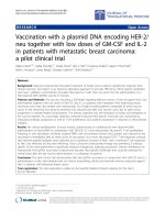
Administration of gelsolin can improve the survival rate
of burn mice
The survival of gelsolin-treated mice at low (Gsn-L) or
high doses (Gsn-H), as well as o f placebo-treated mice,
was asses sed over a 168 h period after burn injur y (Fig-
ure 1). All the mice exposed to sham injury survived the
entire period (n = 10). Placebo-treated mice had a
higher mortality than Gsn-H mic e (70% versus 43.33%,
p < 0.05) within 72 h after burn injury, and no further
mortality occurred after that observation period. Mean
survival time was prolonged in the Gsn-H group (51.17
± 4.7 h, p = 0.0258 versus placebo) and the Gsn-L
group (43.13 ± 4.46 h, p = 0.4875 versus placebo) in
comparison with the placebo group (35.5 ± 4.96 h).
Nevertheless, there was no significant difference in
mean survival time between Gsn-L and Gsn-H groups
(P = 0.0791).
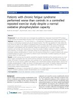
Treatment with gelsolin obviously ameliorated burn-
induced brain damage
As compared with sham-injured mice (Figure 2A), the
brains of mice subjected to thermal injury exhibited
typical pathological lesions. There was invasion of dis-
persed, or even clustered leukocytes in the cortex
Figure 1 Survival rates in burn-injured mice after treatment
with exogenous gelsolin at low (Gsn-L) or high dose (Gsn-H).
There was greater mortality for placebo (burn, 21 of 30) than for
Gsn-H-treated (13 of 30) mice after thermal injury, and survival time
was significantly shorter in placebo-injected mice than in Gsn-L or
Gsn-H-treated mice.
Zhang et al. Journal of Neuroinflammation 2011, 8:118
/>Page 4 of 18
(Figure 2B) and the striatum (Figure 2C) as early as 8 h
pb. Concurrently, neurons were shrunken with con-
densed nuclei, suggesting an early stage of apoptosis
(Figure 2D). As late as 24 h pb, a dispersed infiltration
of leukocytes (Figure 2E) and even microabscesses (Fig-
ure 2F) were seen in the cortex of the mice, indicatin g a
progressive infiltration of inflammatory cells in brain
over this time period. At 24 h pb, dispersed leukocytes
were still observed in the cortex of Gsn-L mice, suggest-
ing that treatment with gelsolin at low dose fails to ame-
liorate the burn-induced brain injury (Figure 2G). In
contrast, administration of gelsolin at high dose could
protect the brain from undergoing the pathological
changes described above (Figure 2H). Similar results
were also obtained for Gsn-H mice at other time points
(data not shown).
Figure 2 Representative images of H&E-stained sections, highlighting cerebral sparing by high dose gelsolin in burn-injured mice.
Cortex of control mice (A) and burned mice at 8 h postburn (B, C, D), 24 h postburn (E, F) and 24 h following low dose (G) and high dose (H)
gelsolin treatment. Images of the lateral ventricles, including the choroid plexus, at 8 h (I), 24 h (J) postburn, and 24 h after gelsolin treatments
at low dose (K) and high dose (L) respectively. ®: leukocyte, ➤: neurosis, *: microabscess.
Zhang et al. Journal of Neuroinflammation 2011, 8:118
/>Page 5 of 18
Strikingly, the leukocyte infiltration occurred in the
ependymal layer of the lateral ventricle as early as 8 h
pb (Figure 2I). In the worst situation, the lateral ventri-
cle was filled with inflammatory exudates at 24 h pb
(Figure 2J). Moreover, a few leukocytes were observed
to accumulate in the choroid plexus in brain of Gsn-L
mice (Figure 2K), but seldom in Gsn-H mice (Figure
2L) at 24 h pb.
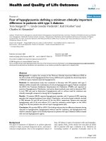
Consistent with the morphological observations, the
neural injury markers cerebral S100 and NSE content
were reduced by high dose gelsolin treatment at 24 h
pb, while these remained at levels similar to control or
sham-burned mice at 8 h pb (Figure 3). It is noteworthy
that both S100 and NSE showed a small trend of
increase at 48 h pb, which could also be slightly reduced
by gelsolin infusion at high dose.
Figure 3 Gelsolin at high dose (Gsn-H) reduces brain-specific proteins, S100 (A) and NSE (B), in mice following burn injury. All data are
expressed as mean ± SD of the mean (n = 6). *P < 0.05 vs. intact control, #p < 0.05 vs. sham-injured, +p < 0.05 vs. placebo mice.
Zhang et al. Journal of Neuroinflammation 2011, 8:118
/>Page 6 of 18
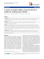
Treatment with gelsolin decreased burn-induced
proinflammatory cytokines in the brain
To further validate and explore the above findings, we
nextinvestigatedthetimecourseofmRNAexpression
of proinflammato ry cytokines by real-time PCR in brain
of burned mice. On account of the lack of significant
improvement in pathology in Gsn-L mice, only the gene
expression of proinflammatory cytokines in the brains of
Gsn-H mice was determined.
Significant reductions in brain levels of early cyto-
kines, including IL-1b and IL-6 mRNA expression, a nd
late cytokine h igh mobility group b ox-1 protein
(HMGB1), were found in the gelsolin-treated group
compared to the placebo group at all time points (Figure
4). Most strikingly, IL-1b mRNA expression in the pla-
cebo mice spiked r apidly, and continued to increase at
various t ime points (Figure 4A). IL-6 mRNA expression
in brain tissue was increased by approximately 1.5- to 2-
fold that of the placebo group compared to normal con-
trols following thermal injury (Figure 4B). Gelsolin
injection resulted i n marked down-regulation of IL-1b
mRNA expression compared wit h the placebo group.
Similarly, IL-6 mRNA levels in the brain were sup-
pressed by approximately 70% in the gelsolin-treated
group compared with the placebo group, close to that of
the sham-injured group (Figure 4B).
HMGB1 is a non-histone DNA bin ding protein that is
secreted by activated monocytes and macrophages [33],
and passively released by necrotic or damaged tissues
[33-35] including brain [36]. Thus, HMGB1 acts as an
immediate trig ger of infl ammation [37] as well as a late
mediator of inflammation [33]. We found that HMGB1
levels were significantly elevated in the brain at 24 and
48 h pb, while they were markedly decreased by gelsolin
treatment at both dosages (Figure 4C).
In addition, we did not find changes in IL-17A or IL-
10 mRNA in the brain tissue, implying that there might
be no T cell infiltration in brain secondary to acute
burns. Similarly, there was no expression of anti-inflam-
matory cytokines, including IL-10 mRNA, induced b y
gelsolin infusion (data not shown).
Administration of gelsolin suppressed burn-induced
microglial activation in the brain
Microgliosis is a common feature of central nervous sys-
tem (CNS) injury and disease, and this involves micro-
glial cell division, hypertrophy, and alterations in
immunophenotype as well as secretory activity [38]. The
augmented neuroinflammation may lead to dysregula-
tion of microglial number and/or microglial activation
intheCNS.Totestsuchahypothesis,westained
microglial populations in brains of burned mice with the
microglial marker (Iba-1) at different intervals.
We observed kinetic changes of Iba-1-immunoreactive
cells in striatum and cortex after thermal injury. In
brief, Iba-1-immun oreactive cells showed morphological
changes and altered immunoreactivity in cortex, stria-
tum and CA1 region with time after acute insults, peak-
ing at 72 h pb in the striatum region (Figure 5A). Iba-1
+
cells were well ramified in burned mice in contrast with
a highly ramified ‘ resting’ morphology in sham-injured
brain (Figure 5B). These alterations might be associated
with delayed neuronal death of striatum cells in burned
mice. In contrast, gelsolin administratio n at either
dosage could suppress activation of Iba-1
+
microglia in
cortex and striatum as exemplified by mice at 72 h pb,
correlating with its anti-inflammatory effect in brain
(Figure 5C).
Taken together, immunohistochemistry analyses
revealed enhanc ed microglial density and activation sta-
tus in brain at 72 h pb, implicating delayed activation of
microglial proliferation and/or ac tivation responses after
thermal injury.
Caspase-3 activation in the brain was inhibited by
gelsolin infusion after burn injury
Caspase-3-positive cells were detected in striatum of
burned mice by immunofluorescence (Figure 6A).
Immunohistochemistry analysis also verified reduced
caspase-3-positive cells in both cortex and hippocampus
by gelsolin treatment (Figure 6B). To determine if gelso-
lin could inhibit caspase-3 activation in our model, we
measured levels of caspase-3 activity in the brain tissue.
We found that there was an approximately 2-fold
increase in caspase-3 activity in the placebo group in
comparison to the sham group at 24 h and 48 h pb.
However, at early 8 h and later 72 h time points, there
were no marked differences in caspase-3 activity
between the placebo and sham groups. As expected, gel-
solin injection either at low or high dosage could reduce
the elevated caspase-3 activity to levels comparable to
sham-injured mice at 24 h pb, while at later time points
such as 48 h pb, only the high dose of gelsolin could
exert a similar effect (Figure 6C).
Gelsolin enhanced CD11b and CD45 monocyte/
macrophage recruitment into brain following burn injury
CD11b is expressed by mature monocytes [16] and by
monocyte-deri ved microglia-like cells [39], and CD45 is
a pan-leukocyte marker. Unexpectedly, an increase in
absolute numbers of macrophage/microglial cells
(CD11b
+
CD45
+
) [40] was found in the gelsolin-treated
groups. It is intriguing that numerous CD11b
+
infiltrat-
ing monocytes and resident microglial cells with promi-
nent amoeboid morphology were noted in the
periventricular regions at 24 h pb (Figure 7A). This
Zhang et al. Journal of Neuroinflammation 2011, 8:118
/>Page 7 of 18
Figure 4 Gelsolin administration protects against burn-induced proinflammatory cytokine expression in brain . Elevated levels of IL-1b
(A) and IL-6 (B) mRNA, as well as HMGB1 content (C) were found in cortex after burn injury. Data are shown as mean ± SD for n = 6. *P < 0.05
and **P < 0.01 vs. sham-injured mice; #p < 0.05, ##p < 0.01, ### p < 0.001 vs. placebo mice; +p < 0.05 and ++p < 0.01 vs. Gsn-L by ANOVA,
Newman-Keuls post-hoc test.
Zhang et al. Journal of Neuroinflammation 2011, 8:118
/>Page 8 of 18
morphology is generally associated with activated micro-
glia or macrophages. Since gelsolin is known as a strong
chemoattractant [22], we further investigated the invol-
vement of gelsolin in t he migration of myeloid-origin
cells into the b rain. To our surprise, the numbers of
CD11b
+
cells were increased in sham and burn-injured
groups as late as 72 h pb, implying that activation of
CD11b
+
cells was delayed. By contrast, the number of
CD11b
+
cells was decreased by treatment with gelsolin
at both dosages (Figure 7B). However, CD45
+
macro-
phages accumulated in the perivascular regions at 8 h
pb in the Gsn-H group (Figure 8A). At both 8 h and 24
h pb, numbers of CD45
+
cells were arrested in the peri-
ventricular region by both doses of gelsolin administra-
tion with differential effects (Figure 8B).
Gelsolin down-regulated burn-mediated ERK1/2
phosphorylation in brain
Western immunoblotting for the active, dually phos-
phorylated form of p44/42 mitogen-activated protein
kinase (MAPK) (ERK1/2) revealed that thermal injury
per se resulted in activation of this signal pathway in
braintissue(Figure9).Anincreaseinphosphorylation
ofp44/42MAPKwasobservedat8hpb,andthiswas
remarkably increased at 24 h pb. ERK1 (44 kDa) density
of the sham group was 18, 207 ± 829, while it reached
25, 564 ± 914 and 30, 546 ± 1077 in burned mice at 8 h
and 24 h, respectively (P < 0.05). Exogenous infusion of
gelsolin could m arkedly down-regulate ERK1/2 phos-
phorylation at 24 h pb (12, 883 ± 877).
Gelsolin improved the suppressed T lymphocytes
functions induced by burn injury
As expected, burn injury resulted in dramatic suppres-
sion of T cell function, as shown by decreased prolifera-
tion (Figure 10A) and IL-2 secretion (Figure 10B),
compared with either intact control or sham-burned
mice. Although inf usion of gelsolin at high dose could
partially prevent the decline, gelsolin at low dose failed
to exert any effect on T cell function.
Figure 5 Treatment with gelsolin reduces microglial activation, as assessed by ionized calcium binding adaptor molecule 1 (Iba-1)
expression after burn-induced neuroinflammatory responses in the cortex 24 h postburn shown at low (A, × 200) and high (B, × 400)
magnifications. Cell counting in cortex and striatum was performed to show that the increased Iba-1 levels after thermal injury were
suppressed by a high dose of gelsolin 72 h postburn (C). All pictures are representative of brain sections from 3 mice for each time point. *P <
0.05 and **P < 0.01 vs. sham-injured mice; ##p < 0.01 vs. placebo mice; ++p < 0.01 vs. Gsn-L mice by ANOVA, Newman-Keuls post-hoc test.
Data are means ± SD for n = 6.
Zhang et al. Journal of Neuroinflammation 2011, 8:118
/>Page 9 of 18
Figure 6 Gelsolin decreases caspase-3 activities in brain of mice following burn injury. A. Immunofluorescen t staining of ca spase-3 in
striatum of brain 24 h postburn; B. Immunohistochemistry of caspase-3 positive cells in the cortex and hippocampus (hippo) from mice under
gelsolin treatments. C. Time course of caspase-3 activity in brain as assayed by colorimetry. *P < 0.05 and **P < 0.01 vs. sham-injured mice; #p <
0.05 and ##p < 0.01 vs. placebo mice by ANOVA, Newman-Keuls post-hoc test. Data are means ± SD for n = 6-8.
Zhang et al. Journal of Neuroinflammation 2011, 8:118
/>Page 10 of 18
Figure 7 Gelsolin affects migration of myeloid-derived cells into brain. CD11b+ cells from mice 72 h postburn (A) and quantification of
infiltrating CD11b+ (B) cells in 10 high power fields (HPF) of the periventricle region following gelsolin treatment. Gelsolin positive cells were
seen in medial habenular nucleus (MHb, a), hippocampal CA field (CA2, b), corpus callosum (cc, c), bed nucleus striatum terminal (BST, d),
choroid plexus (e), cortex (f), lateral ventricle (g, h) and amplified lateral ventricle (g’,h’). Magnifications for “a-f” and “g’-h’” are × 400, “g-h” are ×
200. *P < 0.05, **P < 0.01, and ***P < 0.001 vs. sham-injured mice; #p < 0.05, ## p < 0.01, ### p < 0.001 vs. placebo mice; ++p < 0.01, and +++p
< 0.001 vs. Gsn-L mice by ANOVA, Newman-Keuls post-hoc test. Data are means ± SD for n = 6-8.
Zhang et al. Journal of Neuroinflammation 2011, 8:118
/>Page 11 of 18
Figure 8 Gelsolin affects migrat ion of myel oid-derived cells into brain .CD45
+
cells from gelsolin-treated mice 8 h postburn (A) and
quantification of infiltrating CD45
+
(B) cells in 10 high power fields (HPF) of the periventricle region following gelsolin treatment. Gelsolin
positive cells were seen in medial habenular nucleus (MHb), stria medullaris (sm), hippocampal CA field (CA2) and blood vessel (BV).
Magnifications are × 400. *P < 0.05, **P < 0.01, and ***P < 0.001 vs. sham-injured mice; #p < 0.05, ##p < 0.01, ###p < 0.001 vs. placebo mice; +
+p < 0.01, and +++p < 0.001 vs. Gsn-L mice by ANOVA, Newman-Keuls post-hoc test. Data are means ± SD for n = 6-8.
Zhang et al. Journal of Neuroinflammation 2011, 8:118
/>Page 12 of 18
Kinetic changes in plasma gelsolin concentrations
To evaluate kinetic changes in circulating gelsolin in
various groups in the study, we measured plasma gelso-
lin concentrations to determine its bioavailability. We
found t hat in animals receiving gelsolin, there were not
dramatic changes in circulating gelsolin concentrations.
As exemplifi ed in the Gsn-H group , gelsoli n concent ra-
tions ranged from 851 ± 32 pg/ml at 8 h pb to 844 ±
128 pg/ml at 72 h pb. Baseline values of plasma gelsolin
in both the intact control and placebo groups was
approxi mately 1300 pg/ml, and they remained relatively
constant at all observed time points (Table 1). Following
burn injury, plasma gelsolin levels dropped rapidly to
only 1/3 of the sham group within 8 h (407 ± 57 versus
1273 ± 145, P < 0.001) and r emained at l ow levels for
up to 3 days. Although plasma gelsolin levels in the
Gsn-H group were almost twice that of the placebo
group at most time points, gelsolin administration in
both dosages could only prevent part of the reduction in
plasma gelsolin levels that accompanied burn injury
compared with the sham-burned group.
Discussion
A successful therapeut ic strategy for brain injury should
include inhibition of proinflammatory cytokines, promo-
tion of anti-inflammatory cytokines, suppression of
autoimmunity to CNS antigens and reduction in recruit-
ment of inflammatory cells, etc. In the present study, we
report protective effects of gelsolin in brain of mice sub-
jected to burn injury, characterized by amelioration of
pathological lesions and suppression of microglial activa-
tion, which might be associated with enhanced recruit-
ment of CD11b
+
as well as CD45
+
cells. Likewise,
gelsolin could substantially down-regulate the marked
expression of both early (IL-1b, IL-6) and late proin-
flammatory cytokines (HMGB1) in the brain. In addi-
tion, treatment with gelsolin significantly reduced
caspase-3 activity and inhibi ted ERK phosphorylation in
the brain secondary to severe burns.
As a 90 kDa protein, it is not likely that gelsolin easily
penetrates into brain to perform its effects. Yet a pio-
neer study has demonstrated that peripherally expressed
plasma gelsolin can affect amyloid-b dynamics in the
CNS in two mouse models of Alzheimer’s disease ( AD)
[41]. The authors suggested that one possible clearance
mechanism might be via plasma gelsolin entrance into
brain parenchyma across the BBB, as reports have indi-
cated that the BBB is compromised in mouse models of
AD [42]. Similarly, an increase in the permeability of the
BBB is a common event in thermally injured animals
[8], as also shown in our study by the filling of the lat-
eral ventricles with inflammatory exudates, so it is rea-
sonable to speculate that intravenous infusion of
gelsolin could penetrate the BBB into brain parenchyma
to attenuate neuroinflammation.
Inflammatory mediators are able to alter cellular
metabolism by inducing oxidative stress and mitochon-
drial dysfunction [43], resulting in pathologic abnormal-
ities [44]. Abnormally high levels of cytokines in brain
have been found to correlate with both morbidity and
mortality in patients with extensive burn injury. In our
study, cerebral IL-1b and IL-6 mRNA were up-regulated
around 8 h pb and kept increasing throughout the entire
period. It is likely that HMGB1 levels were significantly
elevated in brain at both 24 and 48 h pb. Gelsolin treat-
ment could significantly reduce e xpression and release
of early as well as late proinflammatory cytokines. This
down-regulation of the inflammatory response would
lead to less damage and cell loss in the brain, which
might, in the future, allow preservation of cognition in
patients with severe burn injury. IL-10 is an immuno-
suppressant that is mainly secreted by regulatory T cells.
It is well known for its positive effects in cerebral ische-
mia in rats [45]. We did not detect IL-10 gene expres-
sion in brain during the entire observation period,
suggesting that administration of gelsolin only inhibits
proinflammatory cytokine transcription, without aug-
menting expression of anti-inflammatory cytokines.
Microglial cells are the primary immune effector cells
in brain and play a pivotal role in neuro inflammatory
processes associated with a variety of neurological as
well as pathological disorders. Microgliosis is a common
feature of CNS injury and disease [38]. Iba-1 is specifi-
call y expressed in microglia and plays an important role
in regulation of microglial function. Increased Iba-1
immunoreactivity is a hallmark of burn-induced inflam-
mation. It has been proposed that microglial activation
induced by sepsis is involved in the pathogenesis of
delirium [46]. There are n o reports to date dealing with
direct investigation of the activation of microglia and
glial scarring following severe burns. We observed
kinetic changes in Iba-1
+
cells in striatum and cortex
after thermal injury, indicating a highest level of
Figure 9 Gelsolin down-regulates burn-induced phospho-ERK1/
2 activity in hippocampus. Lane 1: sham control, Lane 2: 8 h
postburn, Lane 3: 24 h postburn, Lanes 4-5: Gsn-H 24 h postburn.
The ERK1/2 blotting displayed equal loading between wells.
Zhang et al. Journal of Neuroinflammation 2011, 8:118
/>Page 13 of 18
Figure 10 Gelsolin restores suppressed T lymphocyte function after burn injury. Splenic mononuclear cells harvested from the mice under
different treatments were cultured in the presence of the T-cell mitogen concanavalin A (5 mg/L) for 48 h. T-cell viability was determined by
MTT methods (A) and the supernatants were collected for IL-2 analysis (B). Data are shown as mean ± SD for n = 6-8. *P < 0.05 vs. intact
control; #p < 0.05 vs. sham-injured mice; &p < 0.05 vs. placebo mice by ANOVA, Newman-Keuls post-hoc test.
Zhang et al. Journal of Neuroinflammation 2011, 8:118
/>Page 14 of 18
activation as late as 72 h pb. A number of s tudies have
demonstrated that activated glial cells participate in the
degeneration of dopamine neurons [47]. Our data sug-
gest that burn injury per se might result in microgliosis
and loss of vulnerable neuronal populations from
inflammation-induced cell death.
Inflammation and apoptosis are two of the most
important underlying causes of septic encephalopathy
[48]. Because local accumulation of cytokines may
induce apoptosis and significantly extend the initial
injury, we also wanted to clarify whether the ability of
gelsolin to down-regulate cytokine signaling coul d lead
to decreased activation of apoptotic proteins in brain.
Previous investigations have documented that severe
burn injury is associated with a significant increase in
apoptosis in remote organs [30,49,50] including brain
[47]. A number of markers such as S100B and NSE can
serve as general markers of brain injury. Consistent with
our observation of morphological improvement, cerebral
S100B and NSE levels were diminished by gelsolin infu-
sion. Our study further proves that gelsolin administra-
tion immediately following burn injury can reduce
caspase-3 activity in brain, confirming a neuroprotective
effect of gelsolin.
In inflammatory diseases either inside or outside the
CNS, communication between the periphery and the
brain via humoral and/or neural routes results in central
neural changes and related behavioral alterations.
Monocytes are circulating antigen-presenting leukocytes
that play critical roles in inf lammation, T-cell differen-
tiation, phagocytosis, and innate immunity [51,52]. Pre-
vious studies have reported significant infiltration of
activated monocytes into the brain of mice with hepatic
inflammation [18], stroke [19], ischemia-reperfusion [15]
and bacterial meningitis during the post-inflammatory
period [16]. Importantly, these newly recruited mono-
cytes became an integral part of the pool of parenchyma
microglia and contribute to the clearance of damaged
tissue [17]. CD11b is expressed b y mature monocytes
[16] and monocyte-derived microglia-like cells [39],
whereas CD45 is a pan-leukocyte marker. Resoluti on of
CNS infection is often the result of a balance between
immune-mediated pathogen clearance and the
deleterious effects of inflammation. Indeed, in a murine
model of rabies encephalitis, administration of a sex
steroid enhanced permeability of the BBB, promoted
immune cell penetration into the CNS, and improved
survival [53]. It has also been reported that gelsolin is
necessary for rapid motile responses in cell types
involved in stress responses such as hemostasis, inflam-
mation and wound healing [54]. In gelsolin-mutant
mice, macrophage motility was impaired and this contri-
butes to a reduced inflammatory response [54] and a
reduced capacity to recruit macrophages to the injury
site, which in turn, slows the clearance of myelin debris
and consequently remyelination [22]. Consistent with
these findings, we noticed that gelsolin infusion could
accelerate the recruitment of CD11b
+
and CD45
+
cells
into the periventricular region of brain e arly after burn
injury, but could still exert a suppressive effect on their
recruitment at 72 h pb, indicating an early recruitment
of monocyte/macrophage by gelsolin. The increased
penetration of CD11b+ cells and the enhanced micro-
glial activation in gelsolin-treated animals were found to
be associated with down-regulation of proinflammatory
cytokines and caspase-3 activities. Taken together, these
results indicate that treatment with gelsolin could ame-
liorate inflammatory responses in brain and apoptosis of
cerebral cells after burn injury.
To elucidate the potential signaling mechanism under-
lying gelsolin-mediated neuroprotective activity, we
examined expression levels of phospho-ERK in burn
mice. Western blotting experiments using anti-phospho-
p44/42 MAPK (ERK1/2) mouse antibodies revealed acti-
vation of phospho-ERK in brain following thermal
injury, which is consistent with previous reports [7,13].
ERK activation may be downstream of free radicals for-
mation, based on the find ing that do paminergic cells
exposed to 6-hydroxydopamine, a reactive oxygen spe-
cies generating neurotoxin, exhibit a distinct temporal
pattern of ERK1/2 activation and caspase-3 activity [55].
It has been demonstrated that neurons are damaged fol-
lowing prolonged exposure to high concentrations of
corticosterone, with activation of p38 MAPK, ERK1/2,
and c-jun N-terminal protein kinase 1 [56], particularly
in chronic inflammatory and immune diseases. The
Table 1 Circulating levels of gelsolin (pg/ml) after exogenous gelsolin administration in mice after burn injury.
group 0 h 8 h 24 h 48 h 72 h
control 1294 ± 113 1295 ± 81 1276 ± 77 1297 ± 69 1229 ± 50
sham 1295 ± 111 1273 ± 145 1284 ± 96 1290 ± 135 1292 ± 68
burn+placebo 1136 ± 52 407 ± 57* 484 ± 117* 559 ± 106* 529 ± 169*
burn+Gsn-L 1216 ± 52 662 ± 67*
#
862 ± 67*
#
803 ± 197*
#
817 ± 125*
#
burn+Gsn-H 1193 ± 104 851 ± 32*
#+
923 ± 63*
#
956 ± 87*
#+
844 ± 128*
#
*P < 0.05 vs. the sham-injured group; #p < 0.05 vs. the placebo group; +p < 0.05 vs. the Gsn-L group by ANOVA, Newman-Keuls post-hoc test. Data are means ±
SD for n = 6-8.
Zhang et al. Journal of Neuroinflammation 2011, 8:118
/>Page 15 of 18
increased phospho-ERK levels in brain following burn
injury might be a consequence of multiple f actors,
including proinflammatory mediators, ischemia, and oxi-
dative stress.
We further found that gelsolin treatment dramatically
inhibits expression of ph ospho-ERK1/2 in brain of
burned mice. These biochemical results are not in
agreement with a previous observation that the neuro-
protective effects of estrogen could be attributed to
increased phospho-ERK in brain [13]. However, other
authors have reported that administration of neuropro-
tective reagents reduces phospho-ERK1/2 activity
[57,58], and inhibition of ERK1/2 can protect against
brain damage resulting from focal cerebral ischemia
[59]. Furthermore, it has been demonstrated that gelso-
lin overexpression inhibits ERK1/2 phosphorylation,
nuclear factor of activated T-cell activation, and IL-2
production [23]. Thus, the exogenous supply of gelsolin
in our experiments might protect the brain from expo-
sure to pro-apoptotic stimuli, which in turn might
down-regulate ERK1/2 phosphorylation.
Although cerebral complications have been related to
increased mortality in severe sepsis [60] and major burn
victims [10], attenuation of neuroinflammation might
not account for all of the benefit of gelsolin in reducing
burn-induced mortality in our study. Considering that
burn injury could result in severe suppression of the
immune system, which plays an important ro le in the
development of subsequent sepsis, multiple organ failure
and even death, we examined t he dynamic changes in
immune function of splenic T cells as well. We found
an immunosuppressiv e effect involving T cells following
burn injury, which is consistent with a previous r eport
of perturbed T cell homeostasis after burn injury [61]. It
was encouraging to find that gelsolin infusion could
markedly enhance cell-mediated immunity of splenic T
cells, which might also contr ibute to reduced post-b urn
mortality.
Whilethesestudiesareintriguing,thereareseveral
limitations that should be addressed in future investiga-
tions. The first shortcoming is that clinical outcome
variables were not obtained. For clinical relevance,
multi-organ dysfunction which may be the root cause of
burn-induced mortality should be evaluated in further
studies. Secondly, neurological outcomes like edema,
BBB penetrability and cognitive function were not
assessed in the current study. Be tter understanding o f
the improvement of neurological outcome with gelsolin
may allow an in-depth understanding of the mechan-
isms by which gelsolin attenuates the acute response,
and to what extent neurological damage contributes to
post-burn mortality. Although we initiated this study to
observe the acute effects of gelsolin on neuroinflamma-
tion following burn injury, it may be possible to solidify
our current observation s in a further study by also eval-
uating the effects of ge lsolin on these neurological com-
plications which are frequently seen in our clini cal
patients. Thirdly, with regard to the time-window of gel-
solin delivery, intravenous infusion of gelsolin immedi-
ately after burn injury resulted in significantly reduce d
mortality. However, the interventional time could be
postponed to later intervals to more c losely simulate a
clinical setting for this therapeutic strategy. The final,
but foremost concern regards the pharmacokinetics of
gelsolin in this model. With a half life as long as 2.3
days [62], a single administration of gelsolin could pro-
duce considerably elevated gelsolin levels as early as 8 h
and remained high at 72 h pb. As BBB disruption may
occur as early as 7 h after burn injury [8], while gelsolin
might not penetrate the BBB directly within the first
hours, and it is reasonable to speculate that gelsolin
could breach the BBB to perform its effect directly in
the brain at later time points. Nevertheless, the precise
mechanism of our observed gelsolin effect on response
to thermal injury and immunomodulation in both the
brain and the periphery requires further studies.
Conclusion
Despite these limitations, we conclude that, following
severe burn injury in a rodent model, an early, single
dose of gelsolin can dramatically reduce mortality by
ameliorating cerebral inflammatory lesions and apopto-
sis via acceleration of recruitment of monocytes and
down-regulation of phospho-ERK1/2 expression, and
also via improvement of peripheral T cell functions as
well. Although further studies are warrant ed, these find-
ings might be of importance in the near future in the
development of a safe and effective new therapy aimed
at significantly improving the outcome of patients with
severe burns.
Abbreviations
Gsn: gelsolin; TBSA: total body surface area; pb: post burn; caspase-3:
cysteinyl aspartate-specific protease (caspase)-3; BSA: bovine serum albumin;
H&E: hematoxylin and eosin; H
2
O
2
: hydrogen peroxide; Abs: antibodies; DAB:
diamino-benzidine; Iba-1: ionized calcium-binding adapter molecule 1; RIPA:
radio-immunoprecipitation assay, BCA: bicinchoninic acid; CNS: central
nervous system; BBB: blood brain barrier; MAPK: mitogen activated protein
kinase; ERK1/2: extracellular regulated kinase 1/2; kDa: kilo Dalton; MTT: 3-(4,
5-dimethylthiazol-2-yl)-2, 5-diphenyltetrazolium bromide; JNK1: c-jun N-
terminal protein kinase 1; NF-AT: nuclear factor of activated T-cells.
Acknowledgements
This study was supported, in part, by grants from the National Natural
Science Foundation (30973120, 81130035), the National Basic Research
Program of China (2012CB518102), and Eleven-Five Plan for Military Scientific
Foundation (10MA007).
Author details
1
Department of Microbiology and Immunology, Burns Institute, First Hospital
Affiliated to the Chinese PLA General Hospital, Beijing 100048, PR China.
2
Department of Pathology, First Hospital Affiliated to the Chinese PLA
Zhang et al. Journal of Neuroinflammation 2011, 8:118
/>Page 16 of 18
General Hospital, Beijing 100048, PR China.
3
Undergraduate Medical School,
4th Military Medical University, Xi’an, Shaanxi, 710032, PR China.
4
State key
laboratory of kidney disease, the Chinese PLA General Hospital, Beijing
100853, PR China.
Authors’ contributions
QHZ participated in the design of the study; personally conducted a
significant portion of the experiments presented in the manuscript, and
participated in the writing of the manuscript. QC participated in the design
of the study and the preparation of the animal model. JRK prepared all the
cryostat sections. LC and XMZ did the cell counting of the brain. ND
conducted the QPCR detection. ZYS supervised and edited the manuscript.
YMY participated in the design of the experiments, funding of the projects,
and preparation of the manuscript. All authors have read and approved the
final version of the manuscript.
Competing interests
The authors declare that they have no competing interests.
Received: 13 May 2011 Accepted: 21 September 2011
Published: 21 September 2011
References
1. Zhou H, Andonegui G, Wong CH, Kubes P: Role of endothelial TLR4 for
neutrophil recruitment into central nervous system microvessels in
systemic inflammation. J Immunol 2009, 183:5244-5250.
2. Mora AG, Ritenour AE, Wade CE, Holcomb JB, Blackbourne LH, Gaylord KM:
Posttraumatic stress disorder in combat casualties with burns sustaining
primary blast and concussive injuries. J Trauma 2009, 66:S178-185.
3. Rosenberg M, Robertson C, Murphy KD, Rosenberg L, Mlcak R, Robert RS,
Herndon DN, Meyer WJ: Neuropsychological outcomes of pediatric burn
patients who sustained hypoxic episodes. Burns 2005, 31:883-889.
4. Wollgarten-Hadamek I, Hohmeister J, Demirakca S, Zohsel K, Flor H,
Hermann C: Do burn injuries during infancy affect pain and sensory
sensitivity in later childhood? Pain 2009, 141:165-172.
5. Li H, Ying D, Sun J, Bian X, Zhang Y, He B: Comparative observation with
MRI and pathology of brain edema at the early stage of severe burn.
Chin J Traumatol 2001, 4:226-230.
6. Li HT, Ying DJ, He XC, Sun JS, Chen L: Stereoscopic study on capillary
density of early brain oedema in a dog postburn model. Injury 2009,
40:835-839.
7. Zhang Q, Carter EA, Ma B, Fischman AJ, Tompkins RG: Burn-related
metabolic and signaling changes in rat brain. J Burn Care Res 2008,
29:346-352.
8. Patel TH, Sprague S, Lai Q, Jimenez DF, Barone CM, Ding Y: Blood brain
barrier (BBB) dysfunction associated with increased expression of tissue
and urokinase plasminogen activators following peripheral thermal
injury. Neurosci Lett 2008, 444:222-226.
9. Reyes R, Guo M, Swann K, Shetgeri SU, Sprague SM, Jimenez DF,
Barone CM, Ding Y: Role of tumor necrosis factor-alpha and matrix
metalloproteinase-9 in blood-brain barrier disruption after peripheral
thermal injury in rats. J Neurosurg 2009, 110:1218-1226.
10. Flierl MA, Stahel PF, Touban BM, Beauchamp KM, Morgan SJ, Smith WR,
Ipaktchi KR: Bench-to-bedside review: Burn-induced cerebral
inflammation–a neglected entity? Crit Care 2009, 13:215.
11. Bloemsma GC, Dokter J, Boxma H, Oen IM: Mortality and causes of death
in a burn centre. Burns 2008, 34:1103-1107.
12. Chipp E, Milner CS, Blackburn AV: Sepsis in burns: a review of current
practice and future therapies. Ann Plast Surg 2010, 65:228-236.
13. Gatson JW, Maass DL, Simpkins JW, Idris AH, Minei JP, Wigginton JG:
Estrogen treatment following severe burn injury reduces brain
inflammation and apoptotic signaling. J Neuroinflammation 2009, 6:30.
14. Reyes R Jr, Wu Y, Lai Q, Mrizek M, Berger J, Jimenez DF, Barone CM, Ding Y:
Early inflammatory response in rat brain after peripheral thermal injury.
Neurosci Lett 2006, 407
:11-15.
15.
Amantea D, Nappi G, Bernardi G, Bagetta G, Corasaniti MT: Post-ischemic
brain damage: pathophysiology and role of inflammatory mediators.
Febs J 2009, 276:13-26.
16. Barbizan R, Oliveira AL: Impact of acute inflammation on spinal
motoneuron synaptic plasticity following ventral root avulsion.
J Neuroinflammation 2010, 7:29.
17. Djukic M, Mildner A, Schmidt H, Czesnik D, Bruck W, Priller J, Nau R, Prinz M:
Circulating monocytes engraft in the brain, differentiate into microglia
and contribute to the pathology following meningitis in mice. Brain
2006, 129:2394-2403.
18. D’Mello C, Le T, Swain MG: Cerebral microglia recruit monocytes into the
brain in response to tumor necrosis factoralpha signaling during
peripheral organ inflammation. J Neurosci 2009, 29:2089-2102.
19. Vendrame M, Gemma C, de Mesquita D, Collier L, Bickford PC, Sanberg CD,
Sanberg PR, Pennypacker KR, Willing AE: Anti-inflammatory effects of
human cord blood cells in a rat model of stroke. Stem Cells Dev 2005,
14:595-604.
20. Yin HL, Stossel TP: Control of cytoplasmic actin gel-sol transformation by
gelsolin, a calcium-dependent regulatory protein. Nature 1979,
281:583-586.
21. Janmey PA, Lind SE: Capacity of human serum to depolymerize actin
filaments. Blood 1987, 70:524-530.
22. Goncalves AF, Dias NG, Moransard M, Correia R, Pereira JA, Witke W,
Suter U, Relvas JB: Gelsolin is required for macrophage recruitment
during remyelination of the peripheral nervous system. Glia 2010,
58:706-715.
23. Morley SC, Sung J, Sun GP, Martelli MP, Bunnell SC, Bierer BE: Gelsolin
overexpression alters actin dynamics and tyrosine phosphorylation of
lipid raft-associated proteins in Jurkat T cells. Mol Immunol 2007,
44:2469-2480.
24. Jordan JR, Moore EE, Damle SS, Eckels P, Johnson JL, Roach JP, Redzic JS,
Hansen KC, Banerjee A: Gelsolin is depleted in post-shock mesenteric
lymph. J Surg Res 2007, 143:130-135.
25. Osborn TM, Verdrengh M, Stossel TP, Tarkowski A, Bokarewa M: Decreased
levels of the gelsolin plasma isoform in patients with rheumatoid
arthritis. Arthritis Res Ther 2008, 10:R117.
26. Lee PS, Patel SR, Christiani DC, Bajwa E, Stossel TP, Waxman AB: Plasma
gelsolin depletion and circulating actin in sepsis: a pilot study. PLoS One
2008, 3:e3712.
27. Wang H, Cheng B, Chen Q, Wu S, Lv C, Xie G, Jin Y, Fang X: Time course
of plasma gelsolin concentrations during severe sepsis in critically ill
surgical patients. Crit Care 2008, 12:R106.
28. Lee PS, Waxman AB, Cotich KL, Chung SW, Perrella MA, Stossel TP: Plasma
gelsolin is a marker and therapeutic agent in animal sepsis. Crit Care
Med
2007, 35:849-855.
29.
Christofidou-Solomidou M, Scherpereel A, Solomides CC, Christie JD,
Stossel TP, Goelz S, DiNubile MJ: Recombinant plasma gelsolin diminishes
the acute inflammatory response to hyperoxia in mice. J Investig Med
2002, 50:54-60.
30. Carlson DL, Maass DL, White J, Sikes P, Horton JW: Caspase inhibition
reduces cardiac myocyte dyshomeostasis and improves cardiac
contractile function after major burn injury. J Appl Physiol 2007,
103:323-330.
31. Rothenbach PASJ, Dahl B, O’Keefe E, Yamamoto Y, Lee WM, Horton JWYH,
Turnage RH: Recombinant plasma gelsolin infusion attenuates burn-
induced pulmonary microvascular dysfunction. J Appl Physiol 2004,
96:23-31.
32. Livak KJ, Schmittgen TD: Analysis of relative gene expression data using
real-time quantitative PCR and the 2(-Delta Delta C(T)) Method. Methods
2001, 25:402-408.
33. Wang H, Bloom O, Zhang M, Vishnubhakat JM, Ombrellino M, Che J,
Frazier A, Yang H, Ivanova S, Borovikova L, et al: HMG-1 as a late mediator
of endotoxin lethality in mice. Science 1999, 285:248-251.
34. Abraham E, Arcaroli J, Carmody A, Wang H, Tracey KJ: HMG-1 as a
mediator of acute lung inflammation. J Immunol 2000, 165:2950-2954.
35. Bonaldi T, Talamo F, Scaffidi P, Ferrera D, Porto A, Bachi A, Rubartelli A,
Agresti A, Bianchi ME: Monocytic cells hyperacetylate chromatin protein
HMGB1 to redirect it towards secretion. Embo J 2003, 22:5551-5560.
36. Yang QW, Lu FL, Zhou Y, Wang L, Zhong Q, Lin S, Xiang J, Li JC, Fang CQ,
Wang JZ: HMBG1 mediates ischemia-reperfusion injury by TRIF-adaptor
independent Toll-like receptor 4 signaling. J Cereb Blood Flow Metab
2010, 31:593-605.
37. Scaffidi P, Misteli T, Bianchi ME: Release of chromatin protein HMGB1 by
necrotic cells triggers inflammation. Nature 2002, 418:191-195.
38. Streit WJ, Conde JR, Fendrick SE, Flanary BE, Mariani CL: Role of microglia
in the central nervous system’s immune response. Neurol Res 2005,
27:685-691.
Zhang et al. Journal of Neuroinflammation 2011, 8:118
/>Page 17 of 18
39. Hara H, Kataoka S, Anan M, Ueda A, Mutoh T, Tabira T: The therapeutic
effects of the herbal medicine, Juzen-taiho-to, on amyloid-beta burden
in a mouse model of Alzheimer’s disease. J Alzheimers Dis 2010,
20:427-439.
40. Shi FD, Piao WH, Kuo YP, Campagnolo DI, Vollmer TL, Lukas RJ: Nicotinic
attenuation of central nervous system inflammation and autoimmunity.
J Immunol 2009, 182:1730-1739.
41. Hirko AC, Meyer EM, King MA, Hughes JA: Peripheral transgene expression
of plasma gelsolin reduces amyloid in transgenic mouse models of
Alzheimer’s disease. Mol Ther 2007, 15:1623-1629.
42. Dickstein DL, Biron KE, Ujiie M, Pfeifer CG, Jeffries AR, Jefferies WA: Abeta
peptide immunization restores blood-brain barrier integrity in Alzheimer
disease. Faseb J 2006, 20:426-433.
43. Messaris E, Memos N, Chatzigianni E, Konstadoulakis MM, Menenakos E,
Katsaragakis S, Voumvourakis C, Androulakis G: Time-dependent
mitochondrial-mediated programmed neuronal cell death prolongs
survival in sepsis. Crit Care Med 2004, 32:1764-1770.
44. Sharshar T, Gray F, Lorin de la Grandmaison G, Hopkinson NS, Ross E,
Dorandeu A, Orlikowski D, Raphael JC, Gajdos P, Annane D: Apoptosis of
neurons in cardiovascular autonomic centres triggered by inducible
nitric oxide synthase after death from septic shock. Lancet 2003,
362:1799-1805.
45. Spera PA, Ellison JA, Feuerstein GZ, Barone FC: IL-10 reduces rat brain
injury following focal stroke. Neurosci Lett 1998, 251:189-192.
46. van Gool WA, van de Beek D, Eikelenboom P: Systemic infection and
delirium: when cytokines and acetylcholine collide. Lancet 2010,
375:773-775.
47. Depino AM, Earl C, Kaczmarczyk E, Ferrari C, Besedovsky H, del Rey A,
Pitossi FJ, Oertel WH: Microglial activation with atypical proinflammatory
cytokine expression in a rat model of Parkinson’s disease. Eur J Neurosci
2003, 18:2731-2742.
48. Zudaire E, Martinez A, Cuttitta F: Adrenomedullin and cancer. Regul Pept
2003, 112:175-183.
49. Duan H, Chai J, Sheng Z, Yao Y, Yin H, Liang L, Shen C, Lin J: Effect of burn
injury on apoptosis and expression of apoptosis-related genes/proteins
in skeletal muscles of rats. Apoptosis 2009, 14:52-65.
50. Zhang JP, Ying X, Liang WY, Luo ZH, Yang ZC, Huang YS, Wang WC:
Apoptosis in cardiac myocytes during the early stage after severe burn.
J Trauma 2008, 65:401-408, discussion 408.
51. Geissmann F, Manz MG, Jung S, Sieweke MH, Merad M, Ley K:
Development of monocytes, macrophages, and dendritic cells. Science
2010, 327:656-661.
52. Nahrendorf M, Pittet MJ, Swirski FK: Monocytes: protagonists of infarct
inflammation and repair after myocardial infarction.
Circulation
121:2437-2445.
53. Roy A, Hooper DC: Lethal silver-haired bat rabies virus infection can be
prevented by opening the blood-brain barrier. J Virol 2007, 81:7993-7998.
54. Witke W, Sharpe AH, Hartwig JH, Azuma T, Stossel TP, Kwiatkowski DJ:
Hemostatic, inflammatory, and fibroblast responses are blunted in mice
lacking gelsolin. Cell 1995, 81:41-51.
55. Lin ECJ, Leak RK, Perez RG, Zigmond MJ: Rapid activation of ERK by 6-
hydroxydopamine promotes survival of dopaminergic cells. J Neurosci
Res 2008, 86:108-117.
56. Liu B, Zhang H, Xu C, Yang G, Tao J, Huang J, Wu J, Duan X, Cao Y, Dong J:
Neuroprotective effects of icariin on corticosterone-induced apoptosis in
primary cultured rat hippocampal neurons. Brain Res 2010, 1375:59-67.
57. Wakade C, Khan MM, De Sevilla LM, Zhang QG, Mahesh VB, Brann DW:
Tamoxifen neuroprotection in cerebral ischemia involves attenuation of
kinase activation and superoxide production and potentiation of
mitochondrial superoxide dismutase. Endocrinology 2008, 149:367-379.
58. Zhang JZ, Jing L, Ma Y, Guo FY, Chang Y, Li PA: Monosialotetrahexosy-1
ganglioside attenuates diabetes-enhanced brain damage after transient
forebrain ischemia and suppresses phosphorylation of ERK1/2 in the rat
brain. Brain Res 2010, 1344:200-208.
59. Alessandrini A, Namura S, Moskowitz MA, Bonventre JV: MEK1 protein
kinase inhibition protects against damage resulting from focal cerebral
ischemia. Proc Natl Acad Sci USA 1999, 96:12866-12869.
60. Nguyen DN, Spapen H, Su F, Schiettecatte J, Shi L, Hachimi-Idrissi S,
Huyghens L: Elevated serum levels of S-100 beta protein and neuron-
specific enolase are associated with brain injury in patients with severe
sepsis and septic shock. Crit Care Med 2006, 34:1967-1974.
61. Patenaude J, D’Elia M, Hamelin C, Garrel D, Bernier J: Burn injury induces a
change in T cell homeostasis affecting preferentially CD4+ T cells.
J Leukoc Biol 2005, 77:141-150.
62. Smith DBJP, Herbert TJ, Lind SE: Quantitative measurement of plasma
gelsolin and its incorporation into fibrin clots. J Lab Clin Med 1987,
110:189-195.
doi:10.1186/1742-2094-8-118
Cite this article as: Zhang et al.: Treatment with gelsolin reduces brain
inflammation and apoptotic signaling in mice following thermal injury.
Journal of Neuroinflammation 2011 8:118.
Submit your next manuscript to BioMed Central
and take full advantage of:
• Convenient online submission
• Thorough peer review
• No space constraints or color figure charges
• Immediate publication on acceptance
• Inclusion in PubMed, CAS, Scopus and Google Scholar
• Research which is freely available for redistribution
Submit your manuscript at
www.biomedcentral.com/submit
Zhang et al. Journal of Neuroinflammation 2011, 8:118
/>Page 18 of 18