Báo cáo hóa học: " Vaccination with a plasmid DNA encoding HER-2/ neu together with low doses of GM-CSF and IL-2 in patients with metastatic breast carcinoma: a pilot clinical trial" pptx
Bạn đang xem bản rút gọn của tài liệu. Xem và tải ngay bản đầy đủ của tài liệu tại đây (614.07 KB, 11 trang )
RESEA R C H Open Access
Vaccination with a plasmid DNA encoding HER-2/
neu together with low doses of GM-CSF and IL-2
in patients with metastatic breast carcinoma:
a pilot clinical trial
Håkan Norell
1,2†
, Isabel Poschke
1†
, Jehad Charo
3
, Wei Z Wei
4
, Courtney Erskine
5
, Marie P Piechocki
4
,
Keith L Knutson
5
, Jonas Bergh
1
, Elisabet Lidbrink
1†
, Rolf Kiessling
1*†
Abstract
Background: Adjuvant trastuzumab (Herceptin) treatment of breast cancer patients significantly improves their
clinical outcome. Vaccination is an attractive alternative approach to provide HER-2/neu (Her2)-specific antibodies
and may in addition concomitantly stimulate Her2-reactive T-cells. Here we report the first administration of a
Her2-plasmid DNA (pDNA) vaccine in humans.
Patients and Methods: The vaccine, encoding a full-length signaling-deficient version of the oncogene Her2, was
administered together with low doses of GM-CSF and IL-2 to patients with metastatic Her2-expressing breast
carcinoma who were also treated with trastuzumab. Six of eight enrolled patients completed all three vaccine
cycles. In the remaining two patients treatment was discontinued after one vaccine cycle due to rapid tumor
progression or disease-related complications. The primary objective was the evaluation of safety and tolerability of
the vaccine regimen. As a secondary objective, treatment-induced Her2-specific immunity was monitored by
measuring antibody production as well as T-cell proliferation and cytokine production in response to Her2-derived
antigens.
Results: No clinical manifestations of acute toxicity, autoimmunity or cardiotoxicity were observed after
administration of Her2-pDNA in combination with GM-CSF, IL-2 and trastuzumab. No specific T-cell proliferation
following in vitro stimulation of freshly isolated PBMC with recombinant human Her2 protein was induced by the
vaccination. Immediately after all three cycles of vaccination no or even decreased CD4
+
T-cell responses towards
Her2-derived peptide epitopes were observed, but a significant increase of MHC class II restricted T-cell responses
to Her2 was detected at long term follow-up. Since concurrent trastuzumab therapy was permitted, l-subclass
specific ELISAs were performed to specifically measure endogenous antibody production without interference by
trastuzumab. Her2-pDNA vaccination induced and boosted Her2-specific antibodies that could be detected for
several years after the last vaccine administration in a subgroup of patients.
Conclusion: This pilot clinical trial demonstrates that Her2-pDNA vaccina tion in conjunction with GM-CSF and IL-2
administration is safe, well tolerated and can induce long-lasting cellular and humoral immune responses against
Her2 in patients with advanced breast cancer.
Trial registration: The trial registration number at the Swedish Medical Products Agency for this trial is
Dnr151:785/2001.
* Correspondence:
† Contributed equally
1
Department of Oncology and Pathology, Cancer Center Karolinska,
Karolinska Institutet, Stockholm, Sweden
Norell et al. Journal of Translational Medicine 2010, 8:53
/>© 2010 Norell et al; licensee BioMed Central Ltd. This is an Open Access article distributed under the terms of the Creative Commons
Attribution License ( which permits unrestricted us e, distr ibution, and reproduction in
any medium, provided the original work is properly cited.
Background
The proto-oncogene HER-2/neu (Her2) is overexpressed
in a number of malignancies including breast, o varian,
cervical and renal carcinoma [1,2] and represe nts an
attractive therapeutic target. Tra stuzumab (Herceptin), a
recombinant humanized monoclonal antibody binding
Her2, induces durable objective clinical responses and/
or improved time to relapse when administered in the
adjuvant setting in women with Her2-expressing breast
cancer as a single agent or in combination with chemo-
therapy [3-7]. However, trastuzumab was shown to be
therapeutically ineffective in a proportion of patients
and alternative strategies targeting their tumors are
urgently needed [8,9].
Active specific immunotherapy, such as plasmid DNA
(pDNA) vaccination, is an alternative approach to anti-
body therapy and several properties make Her2 a promis-
ing tumor vaccine candidate [10,11]. While trastuzumab
seems to be effective only against breast cancer with
amplified Her2 gene copy numbers and/or high Her2
surface expression, T-cells activated by tumor vaccines
could potentially recognize tumors with intermediate or
low levels o f this molecule. Moreover, there is evidence
that trastuzumab may synergize with specific T-cells [12],
making a combinatorial approach with vaccination and
trastuzumab an attractive clinical treatment modality.
pDNA immunization has several advantages as com-
pared to other vaccination strategies; while immuniza-
tion with proteins primarily induces antibody responses,
pDNA vaccination efficiently promotes generation o f
antigen specific T-cells as well as antibody production
[13]. Similarly, whereas peptide injections only activate
the limited number of T-cells expressing corresponding
T-cell receptors, pDNA immunization may activate
immune responses to a broad repertoire of epitopes.
Also, while peptide immunization could induce T-cell
tolerance and thus enhance d tumor growth if not given
with an efficient adjuv ant, pDNA immunization ensures
antigen-presentation by potent antigen presenting cells
(APCs) [14]. Notably, the nucleotide sequences of
pDNAs can themselves act as adjuvants [15], but the
drawback of competing vector specific immunity asso-
ciated with viral vaccines is c ircumvented [16]. More-
over, Her2-pDNA vaccination has been applied
extensively in experimental models, where it induced
protective immunity against transplantable tumors as
well as against spontaneous tumor development in
Her2-transgenic mice [11,17].
Since immunization of dogs with a human tyrosinase
DNA vaccine produced clinically significant and durable
responses [18,19], a conditional license has been issued
for canin e melanoma therapy by USDA - the regulato ry
agency of animal vaccines - as the first anti-cancer DNA
vaccine strategy approved in any species in the USA
[20]. Nevertheless, pDNA vaccination is often consid-
ered an ineffective approach for immunization in
humans. Notably, vaccine efficacy in animal models has
been improved by including cytokines or plasmids
coding for these as adjuvants [21-24].
Here we present a pilot clinical trial to evaluate the
safety and tolerability of a pDN A coding for a full-
length Her2 molecule administered together with low-
doses of the cytokines granulocyte macrophage colony
stimulating factor (GM-CSF) and interleukin (IL)-2 in
eight patients with metastatic breast carcinoma over-
expressing Her2. All but one patient received concomi-
tant trastuzumab treatment during the study period.
This is the first report on administration of a
Her2-pDNA vaccine in humans. We demonstrate that
injection of the pDNA vaccine and cytokines during
concurrent trastuzumab treatment was safe, well toler-
ated and induced specific endogenous antibody
responses as well as late-onset CD4
+
T-cell responses in
patients with advanced breast cancer.
Patients, Materials and Methods
Patient characteristics
The study was performed at the O ncology clinic,
Radiumhemmet, Karolinska University Hospital, Stock-
holm, and was approved by the local ethics committees
in Uppsala and Stockholm and the Swedish Medical
Product Agency. Eight patients with histologically veri-
fied breast cancer with advanced/metastatic disease were
included in the study, but only six completed three full
vaccination cycles (see table 1 for summary of patient
information). All patients received verbal and written
information and were included after informed consent
in accordance with the Declaration of Helsiniki. Eligibil-
ity criteria included a Zubrod/ECOG performance status
of three or less and an expected survival of more than
three months. Patients were receiving or had been
offered standard-of-care therapy for Her2-overexpres-
sing, locally advanced or metastatic breast carcinoma at
the time of accrual. Her2 status was routinely deter-
mined by immunohistochemistry using the antibodies
CB11 (Ventana, and from 2007 Novocastra Leica, Wet-
zal, Germany), A485 (DAKO, Glostrup, Denmark) and
AB17 (Neomarkers, LabVision Freemont, CA, USA)
between the year 2000 and March 2005, and only CB11
and A485 thereafter. The in ternal control constituted of
four breast cancer cell lines exhibiting different Her2
positivity: BT474 (3+), MDA453 (2+), RT4 (1+), and
5637 (0). Moderate to strong Her2 stainings were veri-
fied by fluorescence in situ hybridization (FISH) to
exclude false-positives, and gene amplification was
demonstrated by inclusion of a centromere probe,
Norell et al. Journal of Translational Medicine 2010, 8:53
/>Page 2 of 11
according to the standard routines at Karolinska Univer-
sity Hospital.
Of the six patients that completed the study, five were
treated with trastuzumab throughout all three vaccina-
tion cycles and the remaining patient (patient #1)
received trastuzumab prior to and again four months
following vaccination. This variation in treatment was
due to the fact that conco mitant trastuzumab admi nis-
tration was allowed, but not an integrated part of the
experimental treatment. Exclusion criteria included a
significant history or evidence of cardiac disease includ-
ing congestive heart failure, coronary artery disease,
uncontrolled hypertension, serious arrhythmia or evi-
dence of prior myocardial infarction on ECG, absence of
measurable disease or evidence of current serious medi-
cal or psychiatric conditions, which would hinder
informed consent or treatment.
Design, construction and production of Her2-pDNA
vaccine
To minimize the risk of malignant transformation of
cells at the site of injection a kinase deficient Her2
DNA sequence (E2A) containing a m utation in codon
753 to convert a lysine (AAA) to an alanine (GCA) resi-
due in the ATP binding site [25,26] was used. From the
pCMV-E2A vector the E2A insert was subcloned into
pVax1 (Invitrogen, Leek, The Netherlands) to generate
pVaxE2A (Her2-pDNA) for clinical use. The correct
sequence of pVaxE2A was verified by DNA sequencing.
The pVax1 vector complies with the Food and Drug
Administration , Center for Biologics Evaluation and
Research (FDA CBER) regulations for vectors to be used
in human DNA vaccination protocols. The vaccine was
produced by the “Gene Therapy Center” at Karolinska
University Hospital Huddinge, Stockholm, under Good
Manufacturing Practice (GMP) conditions with endo-
toxin content less than or equal to 10 EU/mg, >85%
supercoiled plasmid DNA, protein content <10 μg/mL
of plasmid and chromosomal DNA content <30 μg/mL
of plasmid. The vaccine was aliquoted in saline solution,
stored at -80°C and thawed immediate ly prior to admin-
istration. All handling of the DNA vaccine was per-
formed according to national institute of health
guidelines for research involving recombinant DNA
molecules. Her2 protein expression was verified by flow
cytometry after transfection of Cos7 cells with Her2-
pDNA and by immunohistochemistry after intramuscu-
lar (i.m.) injection in mice (data not shown).
Administration of the Her2-pDNA vaccine
The immunization protocol using the cytokines GM-
CSF and IL-2 as adjuvants, was selected based on
encouraging immunological responses in our previous
pDNA trial us ing a similar administration schedule to
Table 1 Patient characteristics
Patient
#
Age
[years]
ǂ
Disease
status
Site of
metastasis
Previous treatments
(abbreviations
explained below)
ER/
PR
▫
Vaccine
cycles
Side
effects
Trastuzumab
and pDNA
vaccine
concurrence
Survival
[month] from
diagnosis*,
+
Survival
[month]
after first
vaccine
Alive/
dead at
last
follow
up*
1 60 PD
Δ
bone Surgery, FEC, DO/T, T,
RT, PA/T, VI/T, RT
-/- 3 - No 46 9 Dead
2 44 PD skin FEC, surgery, FEC, RT,
PA/T, DO/T, VI/T, CP/T,
surgery, T
-/- 1 - Yes 90 42 Dead
3 53 PD Bone, LN° surgery, T, CA/T, T -/- 3 - Yes 80 58.5 Alive
4 64 PD LN (surgery, 5-FU, OP, RT,
FEC, DO, Platinol/CA,
VI/T, PA/T, surgery
? 3 - Yes 96 55 Alive
5 61 PD Bone, lung,
liver
Surgery, FEC, RT, TA,
CA/T, PA/T, RT, T
+/+ 1 - Yes 62 14 Dead
6 64 PD LN, liver FEC, DO, surgery, RT,
surgery, VI/T, CA/T
-/-
+/+
∞
3 - Yes 72 21.5 Dead
7 47 PD Liver, lung Surgery, FEC, RT, TA/
GHRH analog, DO/T,
CA/T, IX, VI/T, T
-/+ 3 - Yes 58 6.5 Dead
8 67 PD LN Surgery, FEC, RT, TA,
PA, DO, CA, CM/T/MT,
CM/BV, VI/T
-/+ 3 - Yes 92.5 28 Dead
BV – Bevacizumab, CA – Capecitabine, CM – Cyclophosphamide, CP – Carboplatin, DO – Docetaxel, 5-FU – 5-Fluorouracil, FEC – epirubicine, cyclosphamide, 5-FU,
IX – Ixabepilon, MT – Methotrexat, OP – Oxaliplatin, PA – Paclitaxel, RT – radiotherapy, T – Trastuzumab, TA – Tamoxifen, VI – Vinorelbine
ǂ
at enrolment,
Δ
PD – progressive disease, ° LN – lymph node,
▫
ER/PR - estrogen/progesteron receptor,
∞
expression site dependent,
*latest follow up July 2009, 87 month after study initiation,
+
median survival 76 month
Norell et al. Journal of Translational Medicine 2010, 8:53
/>Page 3 of 11
target the prostate cancer antigen PSA in patients with
hormone-refractory prostate cancer [27]. Also, we h ave
shown that the same pVaxE2A Her2-pDNA construct
as used in the vacc ine can induce protective i mmunity
in mice when co-injected with a GM-CSF e ncoding
plasmid [28].
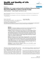
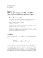
The clinical protocol comprised three pDNA vaccina-
tion cycles per patient. In each cycle, Her2 plasmid was
administered both i.m. (270 μg) and intra cutaneously
(i.c.) (30 μg). Patients also received 3 daily i.c. injections
of GM-CSF (40 μg Leukomax, Novartis, Basel, Switzer-
land) at the same location a s the i.c. vaccine injection,
starting two days prior to Her2-pDNA vaccine adminis-
tration. Injections of low-dose IL-2 (1 μg/kg Proleukin,
Prometheus Laboratories, San Diego, CA, USA) were
given subcutaneously (s.c.) in the abdominal region for
four consecutive days, starting 24 hours after the pDNA
vaccination. A tetanus toxoid (TT) vaccination prior to
Her2-pDNA vaccination was used as a control for
immunomonitoring. Figure 1 provides an overview of
the treatment schedule.
Collection of blood samples and isolation of peripheral
blood mononuclear cells (PBMC)
Blood and serum samples were collected by venipuncture
from the patients immediately before the first and approxi-
mately two weeks after the last vaccine cycle. Three
patients (patient #3, 4 and 8) were long term survivors and
were followed up at a later time point (22, 38.5 and 41
months after last v accination, respectively). PBMC were
isolated by Ficoll-Hypaque (Amersham Biosciences,
Uppsala, Sweden) density gradient centrifugation.
Proliferation assay
T-cell proliferation was assessed using a mo dified limit-
ing dilution assay shown to be useful for evaluation of
low frequency T-cell responses [29]. Freshly isolated
PBMC from patients at every time point and thawed
PBMC from a healthy donor known to be reactive to
TT and phytohemagglutinin (PHA) stimulation as an
inter-experimental control were plated in 12-24 identical
wells per stimuli in med ium alone, or with 1 μg/mL
recombinant human Her2 pro tein (a kind gift from
Dr. Catherine Gerard, GlaxoSmithKline Biologicals,
Belgium), 5 μg/mL TT (Tetravac, Sanofi Pasteur MSD,
Brussels, Belgium) or 5 μg/mL PHA (Sigma-Aldrich,
Irvine,UK).Ondayfour1μCi [methyl-3H]-Thymidine
(Amersham Biosciences, Freiburg, Germany) per well
was added. 24 h later plates were harvested and mea-
sured using a scintillation counter (1450 MicroBeta,
Trilux, Wallac, Turku, Finland).
A standard stimulat ion index (SSI) ≥ 2, defined as at
least twice the mean cpm in stimulated wells compared
to the mean cpm of control wells, was considered as
antigen specific proliferation. The percentage of wells
exhibiting [methyl-
3
H]-Thymidine uptake greater than
the mean plus three standard deviations of the corre-
sponding wells cultured with media alone served as an
additional semi-quantitative measure of re sponding
T-cells [30].
Her2-specific interferon (IFN)-g ELISpot
Four Her2-derived peptides were used t o detect CD4
+
T-cell responses in e nzyme-linked immunospot (ELI-
Spot) assays (Mabtech, Nacka Strand, Sweden) as pre-
viously described [31,32]. Each of these recently
identified 15-mer peptides p59, p88, p422 and p885 [33]
(designated by the position of the first amino acid in the
Her2 p rotein) were in computer modelling predicted to
bind multiple human leukocyte antigen (HLA)-DR
molecules and indeed found to exhibit high-affinity
binding to a variety of major histocompatibility complex
(MHC) class II [33,34]. Pooled cytomegalovirus, Epstein-
Barr virus, and Influenza viral peptide epitopes (CEF,
Figure 1 Schematic overview of the Her2-pDNA vaccination schedule.
Norell et al. Journal of Translational Medicine 2010, 8:53
/>Page 4 of 11
Mabtech, N acka Strand, Sweden) were used as positive
control.
A positive response was defined as the peptide-specific
spot number that was significantly higher (triplicates)
than control wells using a two-tailed t test (P < 0.05).
Counts for each peptide were tallied and reported as the
total number of Her2-specific T-cells assessed at each
time point. It may be possible that while the peptides
bound multiple HLA-DR alleles, some of them could
additionally contain embedded motifs that could stimu-
late CD8
+
T-cells. However, Her2-specific CD8
+
T-cell
responses are typically lower by at least one order of
magnitudeeveninvaccinated patients [31]. Putative
CD8
+
responses against p369, p435 and p689 9-mer
peptides known to bind to HLA-A2 were tested (data
not shown), but are of limited value since patients were
not HLA-typed. Changes between pre- and post-immu-
nization responses were considered significant if there
was at least a two-fold increase or a 50% decrease.
Enzyme-linked immunosorbent assay (ELISA)
ELISAs measuring amounts of Her2-specific Ig l anti-
bodies have been previously described [35]. TT ELISAs
served as internal controls.
All serologic assays were repeated at least twice for
each individual patient . A humoral response was consid-
ered positive by a relative A450 index of >2 or a titer
<1/100.
Statistical analysis
Statistical analyses were performe d using Excel, Graph-
Pad, InStat or Prism Software (GraphPad Software, Inc,
La Jolla, CA USA). Data were analyzed using two-tailed
Mann-Whitney (nonparametric data) or Student’s t tests
unless otherwise stated, and the results were considered
statistically significant if p < 0.05.
Results
Patient characteristics and clinical observations
Eight women with a mean age of 57.5 years were
accrued in this study. Patient chara cteristics are sum-
marized in Table 1. All patients had advanced breast
cancer treated with extensive prior therapy, including
trastuzumab. All patients except one (patient #1) were
on trastuzumab treatment during the study period.
Of the eight patients entering the trial, six completed
all three vaccination cycles. Patient #2 was withdrawn
after one cycle due to severe erysipelas at the location of
a skin metastasi s and patient #5 due to disease progres-
sion. No significant side effects associated with the
vaccination or cytokine administration were observed
in any p atient. There were no manifestations of auto-
immunity or cardiotoxicity, nor was any acute toxicity
observed.
Of the six patients that completed all three cycles of
vaccination, two were long term survivors, still alive
more than 4 years after the last vaccination (in July
2009>56monthsforpatient#3and>53monthsfor
patient #4). Patient #8 lived until 25 month post vacci-
nation. T he median survival time from diagnosis to lat-
est follow up for all 8 enrolled patients was 76 months
with a range of 46-96 months.
Evaluation of Her2-specific T-cell responses
Lymphocyte proliferation assays were performed with
freshly isolated PBMC from pre- and post-vaccination
blood samples of all patients. As expected, PHA induced
significant proliferation in all tested wells with an aver-
age SSI of 69.6 across pre- and post-Her2 vaccination
assays. Importantly, vaccine-induced TT-specific T-cells
proliferated upon stimulation with cognate antigen in
100% of the wells. The average SSI was similar in pre-
Her2-vaccination (15.8) and post-Her2-vaccination
(13.9) samples, indicating that the TT booster vaccina-
tion resulted in stable cellular immunity to TT over the
treatment period. In contrast, the overall proliferative
responses to Her2 protein were minimal as average SSIs
were negative and the percentage of wells with positive
proliferative responses were very low in both pre- (SSI
1.0, range 0.8-1.3; 1.8% of wells exhibiting Her2-speci fic
proliferation) and post- (SSI 1.0, range 0.9-1.1; 3.3% o f
wells exhibiting Her2-specific proliferation) Her2 vacci-
nation samples. The cut offs in mean stimulation i ndex
(SI) for scoring individual experimental wells as
responding or non-responding was on average 1.91
(range 1.4 - 2.4), which is in line with previous reports
[29,36]. Although the frequency of wells exhibiting sig-
nificant proliferation to Her2 protein was almost twice
as high after the treatment regimen, the average SIs of
the positive wells in the post-vaccination samples (2.1)
was only about half that of the pre-vaccine samples
(4.0). Thus, weak and rare pre-existing Her2 protein
specific proliferative responses were observed in fresh
PBMC, but these responses were not significantly
enhanced after the Her2-vaccination regimen.
For four of the patients that completed all three vac-
cine cycles, sufficient amounts of PBMC were available
to evaluate Her2-specific cellular immunity towards a
panel of HLA-DR restricted peptides by ELISpot. Two
of three evaluable patients (patients #4 and 7) demon-
strated pre-vaccination CD4
+
T-cell mediated immunity
to all four Her2-derived peptides, while no pre-vaccine
immunity to these epitopes could be detected in patient
#8(Figure2).Nopre-vaccineELISpotcouldbeper-
formed for patient #3 due to paucity of PBMC.
For the patients who had samples permitting pre- ver-
sus post-vaccination comparison (patients #4, 7 and 8),
there was no consistent change in peptide specific
Norell et al. Journal of Translational Medicine 2010, 8:53
/>Page 5 of 11
responses resulting from immunization when tested
10 days after the last vaccination. Intra-patient compari-
son of pre- and post-vaccination responses to ind ividual
peptides showed that both boosting and reduction
of pre-existing responses occurred and also that new
T-cell specificities could be induced by the treatment
(Figure 2A-D). Interestingly, increased, decreased and
unchanged responses to individual peptides could be
observed in the same patient, e.g. patient #7, after three
cycles of Her2-pDNA vaccination (Figure 2C).
Three of the four patients (patients # 3, 4 and 8) sur-
vived more than two years after the last v accination and
an additional blood sample was collected from each of
these subjects at a later time point. Strikingly, PBMC
from all three patients e xhibited strong Her2-specific
immune response against all tested peptides at this late
follow up. The frequency of Her2-specific T-cells was
significantly increased compared to both pre- and
post-vaccination samples in all patients and newly
induced responses as well as recovery of responses lost
at post-vaccination evaluation were observed. Individual
results and pooled responses f rom patients evaluable at
all time points (Figure 2E) show a significant increase of
MHC class II restricted T-cell responses to Her2-
derived epitopes at long term follow-up, while there was
a transient decrease in Her2-specific immunity immedi-
ately after three cycles of Her2-pDNA plus GM-CSF
and IL-2.
Evaluation of Her2-specific antibody responses
Pre- and post-vaccination sera from all patients were
analyzed for the presence of anti-Her2 antibodies. Since
most patients received concurrent trastuzumab treat-
ment during the Her2-pDNA vaccinations, l-subclass
specific ELISAs were performed. The specific detection
of l-subclass anti-Her2 antibodies allowed measure-
ment of endogenous antibody production without
detection of trastuzumab, an I gG1 antibody present at
high serum conc entrations during the rapeutic adminis-
tration [37]. Comparison of pre- and post-Her2-pDNA
vaccine responses in patients evaluable at a ll time points
showed a trend towards higher mean binding activity o f
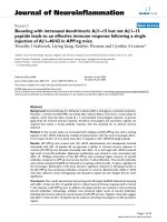
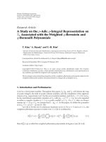
Figure 2 MHC class II restricted T-cell responses to Her2 before and after Her2-pDNA vaccination. A-D. Her2-specific IFN-g production by
T-cells from patients #3, 4, 7 and 8, before (3 days pre-) and after (10 days post-) Her2-pDNA vaccination and at long term follow-up (41, 38.5
and 22 month after last vaccination for patients #3, 4, 8, respectively). Bars show mean (± s.e.m.) frequency of IFN-g producing T-cells (spot
forming units) per 2.5 × 10
5
PBMC responding to a panel of 4 degenerate Her2-derived HLA-DR epitopes (p59, p88, p422 and p885). No pre-
bleeding ELISpot was performed for patient 3 due to insufficient numbers of available PBMC. E. Mean ± s.e.m. Her2-specific T-cell frequency per
2.5 × 10
5
PBMC in patients evaluable at all time points. Bars show pooled responses of patients #4 and 8 pre, post and late. *: p ≤ 0.05.
Norell et al. Journal of Translational Medicine 2010, 8:53
/>Page 6 of 11
post- versus pre-vaccination sera against Her2 (Figure
3A). Notably, the Her2-specific binding activity in the
responding patients reached levels comparable to those
of the TT-specific antibodies following TT vaccine
administered as a control before the Her2-pDNA vacci-
nation schedule (Figure 3B, C). One of eight (12.5%)
patients enrolled in the study had a pre-existing anti-
bodyresponseagainstHer2,asdefinedbyabinding
activity >2 . The majority of evaluable patients (3/5)
showed an increased Her2-specific binding activity aft er
completion of three vaccination cycles (Figure 3C).
Two out of three patients that were ava ilable for long
term monitoring could sustain their positive post-vacci-
nation antibody levels for several years after the last vac-
cination. These two patients were also the ones that
reached positive anti-Her2 binding activity that could be
measured after 3 cycles of vaccination. The third long
term surviving patient (patient #8) never exhibited any
Her2-specific humoral immunity (Figure 3C).
Discussion
From this small pilot study we can conclude that our
full length Her 2-pDNA, administered together with
GM-CSF and IL-2, is safe, well tolerated and can induce
both antibody and T-cell responses in advanced stage
cancer patients. Since our gro up and others have shown
that Her2 can down-modulate MHC class I expression
[38-40], tumor vaccine strategies such as pDNA admin-
istration that are not solely dependent on CTLs but
induce an integrated immune response involving also
antibodies and CD4
+
T-cells should be advantageous.
Bolstering this hypothesis is the observation that the
same pDNA vaccine as used in the present trial can effi-
ciently induce Her2-specific antibodies as well as a
CD8
+
T-cell response and protection from tumor chal-
lenge in conventional and human Her2-transgenic
BALB/c and HLA-A2 transgenic B6 mice [25,38].
The present clinical trial is the first to combine a
Her2-pDNA vaccine with trastuzumab treatment. In
light of preclinical studies demonstrating that tumor
cells binding trastuzumab were more efficientl y recog-
nized by Her2 reactive T-cells [12], conco mitant admin-
istration of trastuzumab and H er2 vaccines may cause
substantial synergies and represents a promising treat-
ment strategy. Combination therapy with trastuzumab
and a peptide (E75) vaccine was recently applied in a
subset of seven strongly Her2-positive cancers where
this combination proved to be safe and immunologically
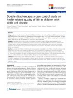
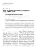
Figure 3 Her2-pDNA vaccination generates Her2-specific humoral immunity. A-B. Mean binding activity derived from A. Her2-specific or
B. tetanus toxoid Ig l-subclass specific ELISAs. Bars show the mean (± s.e.m.) binding activity of patients evaluable at all time points (patient #3,
4, 8, pre, post and late). C. Binding activity in the serum of all patients at all available individual time points (pre- and post-immunization as well
as long-term follow up at 22-41 months following the last immunization).
Norell et al. Journal of Translational Medicine 2010, 8:53
/>Page 7 of 11
beneficial [41]. A similar conclusion was reached for a
Her2 T-helper peptide-based vaccine in combination
with trastuzumab [42].
The combinatorial treatment complicated our
attempts to detect vaccine-induced Her2-specific anti-
bodies in t he vaccinated patients. Howeve r, a r ecently
established l-subclass specific ELISA allowed evaluation
of endogenous Her2-specific antibody responses without
detection of or interference by the IgG1 antibody tras-
tuzumab [35]. Notably, the majority of evaluable
patients demonstrated increased antibody binding activ-
ity after completion of the vaccine trial and in most of
the long term survivors these endogenous Her2-specific
antibodies persisted or were increased in samples
obtained several years after the last vaccine administra-
tion. Due to co-administration of trastuzumab we were
not able to evaluate the contribution of endogenous
Her2-specific antibodies of t he -subclass to the overall
humoral immune response. Considering previous vac-
cine trials [43] it is likely that IgG antibodies were
also induced.
The ability of our vaccine to trigger Her2-specific anti-
body responses has significant therapeutic implications,
as a broader repertoire of Her2-reactivities and antibody
isotypes may lead to enhanced tumor specific antibody
dependent cellular cytotoxicity or enhanced antibody-
induced perturbation of Her2 signaling. Similarly, it is
possible tha t the endogenously induced antibodies
synergize with trastuzumab or are more efficient in
opsonizing Her2 expressing tumor cells or fragments of
these, leading to better uptake by APCs and thus
improved activation of endogenous T-cells. Also,
numerous mouse models have implicated vaccine
induced antibodies as a majorfactorinconferringpro-
tection against transplantable and spontaneous Her2
expressing tumors [44-46].
Mainly non-professional APC in PBMC were available
to process and present epitopes to T-cells in our prolif-
eration assays. This m ay have prevented detection of
rare and/or weak autologous T-cell responses to the
recombinant Her2 protein, while allowing strong T T-
peptide specific T-cell responses to be readily detected.
Indeed, most evaluated patients showed pre-vaccination
CD4
+
T-cell reactivity to all tested peptides in IFN-g
ELISpot assays against Her2-derived 15-mer peptides
known to bind several different HLA-DR allotypes [34].
However, for the three patients who had samples that
allowed a pre- versus post-vaccination comparison, we
failed to observe a consistent increase in peptide specific
CD4
+
T-cell responses. In the event that Her2-specific
immune responses were induced or b oosted, activated
T-cells may have homed to t he site of the tumor, ham-
pering their detection in peripheral blood. Alternatively,
one may speculate whether the induction of regulatory
T-cells by the IL-2 [47], and/or induction of myeloid
derived suppressor cells by the GM-CSF [48] in our
treatment regimen may account for the lack o r decrease
in immune responsiveness and the almost complete dis-
appearance of pre-vaccination immunity against all four
tested epitopes in one patient. Regrettably, the reason
could not be experimentally established due to paucity
of patient PBMC.
In contrast to the absence of CD4
+
T-cell responses
early after vaccination, the three patients who survived
more than two ye ars after the last vaccination all exhib-
ited strong immunity to all of the tested Her2-derived
peptides when re-evaluated at a late time point. This
late immune response to Her2 following vaccination is
not without precedence. Morse et al. [49] provided evi-
dence that the peak response to a DC vaccine loaded
with Her2 intracellular domain could occur more t han
5 years after c oncluding vaccine therapy, and Disis and
colleagues [43] showed that anti-Her2 T-cell responses
could persist for at least 1 year after vaccination with T-
helper epitope derived peptides mixed with GM-CSF
had ended. Since Her2-specific antibody and T-cell
responses have also been detected in non-vaccinated
patients [35,50], and were further confirmed in the pre-
treatment samples in the present study, we cannot
exclude that this late response i s unrelated to the va c-
cine administration and instead induced by the trastuzu-
mab therapy [35] or by patients’ Her2 expressing
tumors.
Although Her2 is overexpressed in a broad range of
carcinomas, low levels are also present in normal epithe-
lial surfaces [51]. The concern is therefore that induc-
tion of an immune response to this “self-antigen” should
lead to autoimmune manifestations. Alternatively, since
trastuzumab can induce cardiac toxicity in a small but
significant proportion of treated patients [52], one may
consider whether the endogenously-induced Her2-speci-
fic antibodies reported in this study and by others may
contribute to o r worsen this side effect. It is therefore
important to note that none of the eight patients who
received the Her2 vaccine had any manifestations of
autoimmunity or cardiac toxicity. This is in concordance
with observations in other Her2 vaccine trials in which
no adverse effects have been reported [43,49]. This
includes a trial based on the E75 peptide derived from
the extracellular domain of Her2 and GM-CSF, which
resulted in a decreased disease recurrence rate [53].
Since this trial was a small phase I clinical study with
only six patients completing all three cycles of vaccine
and cytokine administration, this precludes any conclu-
sion regarding the cli nical efficacy. Further complicating
interpretations of clinical efficacy, all patients suffered
from advanced disease and had undergone prior chemo-
therapy and most were on concomitant trastuzumab
Norell et al. Journal of Translational Medicine 2010, 8:53
/>Page 8 of 11
treatment. Nevertheless, it is noteworthy that three of
the s ix patients who received all three cycles of vaccine
treatment were long-te rm survivors. The median overall
surviv al from start of vaccination was 24.8 months, with
a range of 6.5 to 58.5 months, but as mentioned the sig-
nificance of these data must be interpr eted with caution
because of the small patient number.
The median survival for patients in a randomized
study failing first line trastuzumab therapy was 25.5
months for patients receiving continuous trastuzumab
combined with capecitabine [54]. In another randomized
study patients who failed conventional chemotherapy-
trastuzumab combinations had an estimated median
survival of about 58 weeks on the combination of lapa-
tanib and capacitabine [55].
The relatively long survival from the start of vaccina-
tion for patients #3 and #4, 58.5 and 55 months, respec-
tively, is obviously an interesting observation, especially
as broad Her2-specific immunity was d etected in these
patients. However, these two patients had disease burden
limited to ly mph nodes and skeleton when entering the
study and long term survival in this category of patients
is not unusual. Patient #4 nevertheless had failed several
lines of therapy before inclusion, indicative of treatment-
refractory disease, but continued to be treated with tras-
tuzumab as single agent after the end of vaccination.
Conclusion
Our pilot study demonstrates the feasibility, safety and
tolerability of Her2-pDNA vaccination in combination
with GM-CSF and IL-2 in a small number of advanced
breast cancer patients who are on concurrent trastuzu-
mab treatment with findings warranting further explora-
tion of this concept. The induc tion of long-lasting
cellular and humoral immune responses against Her2
are encouraging and occasional patients appear to draw
clinical benefit from t his treatment, although this must
be confirmed in further studies, at best with a rando-
mized design. Her2-pDNA vaccines already provide a
promising strategy by broadening or potentiating the
response to trastuzumab administration, which is now a
standard adjuvant therapy for women with Her2 over-
expressing breast cancer. If our and similar vaccine stra-
tegies efficiently generate humoral Her2-specific
responses, trastuzumab may later become obsolete and
vaccines alone successful against early and metastatic
breast cancer. This would facilitate the practical man-
agement of Her2 positive carcinoma s, since trastuzumab
based strategies are expensive and require time-consum-
ing three-wee kly intravenous administrations. If demon-
strated to have a favorable benefit-risk ratio the
vaccination approach should also be studied as a
preventive strategy in high risk individuals.
Acknowledgements
Kiessling’s research group is supported by grants from the Swedish Cancer
Society, the Swedish Medical Research Council, the Cancer Society of
Stockholm, the European Union (Grants “EUCAAD” and “DC-THERA” ), the
Karolinska Institutet, and an “ALF-Project” grant from the Stockholm City
Council. Bergh’s research group is supported by grants from the Swedish
Cancer Society, Swedish Research council, the funds at Radiumhemmet,
ALF/FOU grants by the Stockholm County Council, Sweden and Merck Inc,
USA. Wei’s research group is supported by NIH grant CA76340. Knutson’s
research group is supported by NIH/NCI Howard Temin Award K01-
CA100764. The authors thank Dr. Raphael Clynes (Columbia University,
New York, NY) for assistance with the Her2 ELISAs.
Author details
1
Department of Oncology and Pathology, Cancer Center Karolinska,
Karolinska Institutet, Stockholm, Sweden.
2
Department of Surgery, Hollings
Cancer Center, Medical University of South Carolina, Charleston, SC, USA.
3
Max-Delbrück Center for Molecular Medicine, Berlin, Germany.
4
Karmanos
Cancer Institute, Wayne State University, Detroit, MI, USA.
5
Department of
Immunology, College of Medicine, Mayo Clinic, Rochester, MN, USA.
Authors’ contributions
HN designed and performed research, analyzed data and performed
statistical analysis. He was together with IP responsible for collecting and
handling patient samples and for performing the T-cell proliferation assays
and early attempts to measure specific antibody responses. IP performed
research, analyzed data and drafted the manuscript. She was responsible for
collecting and handling the patient samples after HN had departed from
CCK and co-ordinating the collaboration with KLK’s laboratory. Together with
EL she also summarized and processed the patient data and together with
RK she wrote the manuscript. JC designed research. He was involved in
writing the clinical protocol and the early phases of the study. WZW
contributed new reagents/analytic tools. She provided the vaccine construct
and was responsible for the pre-clinical testing of this vaccine in mouse
models. CE performed research by being responsible for performing the
ELISA and ELISPOT assays. MPP contributed new reagents/analytic tools by
collaborating with WZW in the pre-clinical testing of the vaccine in mouse
models. KLK designed research, analyzed data and performed statistical
analysis. He was responsible for the design of the ELISA and ELISPOT assays
and the testing of patient samples in these assays was carried out and
interpreted in his laboratory with the assistance of CE. JB designed research
and provided expert opinion for the study. He was the principal investigator
of the clinical study and responsible for the contact with the regulatory
agents. EL performed clinical research. She was the physician who had all
patient contact and thus carried out all vaccination procedures and also
summarized the patient information for the manuscript. RK designed
research, analyzed data and wrote the paper. He initiated and designed the
study and wrote the clinical protocol. He funded all costs involved and was
responsible for the immune monitoring, with input also from the lab of KLK.
All authors read and approved the final manuscript.
Competing interests
The authors declare that they have no competing interests.
Received: 19 January 2010 Accepted: 7 June 2010
Published: 7 June 2010
References
1. Seliger B, Rongcun Y, Atkins D, Hammers S, Huber C, Storkel S, Kiessling R:
HER-2/neu is expressed in human renal cell carcinoma at heterogeneous
levels independently of tumor grading and staging and can be
recognized by HLA-A2.1-restricted cytotoxic T lymphocytes. Int J Cancer
2000, 87:349-359.
Norell et al. Journal of Translational Medicine 2010, 8:53
/>Page 9 of 11
2. Pegram MD, Konecny G, Slamon DJ: The molecular and cellular biology of
HER2/neu gene amplification/overexpression and the clinical
development of herceptin (trastuzumab) therapy for breast cancer.
Cancer Treat Res 2000, 103:57-75.
3. Slamon DJ, Leyland-Jones B, Shak S, Fuchs H, Paton V, Bajamonde A,
Fleming T, Eiermann W, Wolter J, Pegram M, et al: Use of chemotherapy
plus a monoclonal antibody against HER2 for metastatic breast cancer
that overexpresses HER2. N Engl J Med 2001, 344:783-792.
4. Marty M, Cognetti F, Maraninchi D, Snyder R, Mauriac L, Tubiana-Hulin M,
Chan S, Grimes D, Anton A, Lluch A, et al: Randomized phase II trial of the
efficacy and safety of trastuzumab combined with docetaxel in patients
with human epidermal growth factor receptor 2-positive metastatic
breast cancer administered as first-line treatment: the M77001 study
group. J Clin Oncol 2005, 23:4265-4274.
5. Romond EH, Perez EA, Bryant J, Suman VJ, Geyer CE Jr, Davidson NE,
Tan-Chiu E, Martino S, Paik S, Kaufman PA, et al: Trastuzumab plus
adjuvant chemotherapy for operable HER2-positive breast cancer. N Engl
JMed2005, 353:1673-1684.
6. Smith I, Procter M, Gelber RD, Guillaume S, Feyereislova A, Dowsett M,
Goldhirsch A, Untch M, Mariani G, Baselga J, et al: 2-year follow-up of
trastuzumab after adjuvant chemotherapy in HER2-positive breast
cancer: a randomised controlled trial. Lancet 2007, 369:29-36.
7. Baselga J: Novel agents in the era of targeted therapy: what have we
learned and how has our practice changed? Ann Oncol 2008, 19(Suppl 7):
vii281-288.
8. Park BH, Davidson NE: PI3 kinase activation and response to Trastuzumab
Therapy: what’s neu with herceptin resistance? Cancer Cell 2007,
12:297-299.
9. Berns K, Horlings HM, Hennessy BT, Madiredjo M, Hijmans EM, Beelen K,
Linn SC, Gonzalez-Angulo AM, Stemke-Hale K, Hauptmann M, et al: A
functional genetic approach identifies the PI3K pathway as a major
determinant of trastuzumab resistance in breast cancer. Cancer Cell 2007,
12:395-402.
10. Kiessling R, Wei WZ, Herrmann F, Lindencrona JA, Choudhury A, Kono K,
Seliger B: Cellular immunity to the Her-2/neu protooncogene. Adv Cancer
Res 2002, 85:101-144.
11. Wei WZ, Jacob J, Radkevich-Brown O, Whittington P, Kong YC: The “A, B
and C” of Her-2 DNA vaccine development. Cancer Immunol Immunother
2008, 57:1711-1717.
12. zum Buschenfelde CM, Hermann C, Schmidt B, Peschel C, Bernhard H:
Antihuman Epidermal Growth Factor Receptor 2 (HER2) Monoclonal
Antibody Trastuzumab Enhances Cytolytic Activity of Class I-restricted
HER2-specific T Lymphocytes Against HER2-overexpressing Tumor Cells.
Cancer Res 2002, 62
:2244-2247.
13. Donnelly JJ, Ulmer JB, Liu MA: DNA vaccines. Life Sci 1997, 60:163-172.
14. Corr M, Lee DJ, Carson DA, Tighe H: Gene vaccination with naked plasmid
DNA: mechanism of CTL priming. J Exp Med 1996, 184:1555-1560.
15. Roman M, Martin-Orozco E, Goodman JS, Nguyen MD, Sato Y, Ronaghy A,
Kornbluth RS, Richman DD, Carson DA, Raz E: Immunostimulatory DNA
sequences function as T helper-1-promoting adjuvants. Nat Med 1997,
3:849-854.
16. Rice J, Ottensmeier CH, Stevenson FK: DNA vaccines: precision tools for
activating effective immunity against cancer. Nat Rev Cancer 2008,
8:108-120.
17. Quaglino E, Mastini C, Forni G, Cavallo F: ErbB2 transgenic mice: a tool for
investigation of the immune prevention and treatment of mammary
carcinomas. Curr Protoc Immunol 2008, Chapter 20, Unit 20 29 21-20
29-10.
18. Liao JC, Gregor P, Wolchok JD, Orlandi F, Craft D, Leung C, Houghton AN,
Bergman PJ: Vaccination with human tyrosinase DNA induces antibody
responses in dogs with advanced melanoma. Cancer Immun 2006, 6:8.
19. Bergman PJ, Camps-Palau MA, McKnight JA, Leibman NF, Craft DM,
Leung C, Liao J, Riviere I, Sadelain M, Hohenhaus AE, et al: Development of
a xenogeneic DNA vaccine program for canine malignant melanoma at
the Animal Medical Center. Vaccine 2006, 24:4582-4585.
20. Kutzler MA, Weiner DB: DNA vaccines: ready for prime time? Nat Rev
Genet 2008, 9:776-788.
21. Chen Y, Hu D, Eling DJ, Robbins J, Kipps TJ: DNA Vaccines Encoding Full-
Length or Truncated Neu Induce Protective Immunity against Neu-
expressing Mammary Tumors. Cancer Res 1998, 58:1965-1971.
22. Charo J, Ciupitu AM, Le Chevalier De Preville A, Trivedi P, Klein G, Hinkula J,
Kiessling R: A long-term memory obtained by genetic immunization
results in full protection from a mammary adenocarcinoma expressing
an EBV gene. J Immunol 1999, 163:5913-5919.
23. Charo J, Sundback M, Geluk A, Ottenhoff T, Kiessling R: DNA immunization
of HLA transgenic mice with a plasmid expressing mycobacterial heat
shock protein 65 results in HLA class I- and II-restricted T cell responses
that can be augmented by cytokines. Hum Gene Ther 2001, 12:1797-1804.
24. Lin C-C, Chou C-W, Shiau A-L, Tu C-F, Ko T-M, Chen Y-L, Yang B-C, Tao M-H,
Lai M-D: Therapeutic HER2/Neu DNA Vaccine Inhibits Mouse Tumor
Naturally Overexpressing Endogenous Neu. Mol Ther 2004, 10:290-301.
25. Wei WZ, Shi WP, Galy A, Lichlyter D, Hernandez S, Groner B, Heilbrun L,
Jones RF: Protection against mammary tumor growth by vaccination
with full-length, modified human ErbB-2 DNA. Int J Cancer 1999,
81:748-754.
26. Messerle K, Schlegel J, Hynes NE, Groner B: NIH/3T3 cells transformed with
the activated erbB-2 oncogene can be phenotypically reverted by a
kinase deficient, dominant negative erbB-2 variant. Mol Cell Endocrinol
1994, 105:1-10.
27. Pavlenko M, Roos AK, Lundqvist A, Palmborg A, Miller AM, Ozenci V,
Bergman B, Egevad L, Hellstrom M, Kiessling R, et al: A phase I trial of DNA
vaccination with a plasmid expressing prostate-specific antigen in
patients with hormone-refractory prostate cancer. Br J Cancer 2004,
91:688-694.
28. Lindencrona JA, Preiss S, Kammertoens T, Schuler T, Piechocki M, Wei WZ,
Seliger B, Blankenstein T, Kiessling R: CD4+ T cell-mediated HER-2/neu-
specific tumor rejection in the absence of B cells. Int J Cancer 2004,
109:259-264.
29. Disis ML, Grabstein KH, Sleath PR, Cheever MA: Generation of immunity to
the HER-2/neu oncogenic protein in patients with breast and ovarian
cancer using a peptide-based vaccine. Clin Cancer Res 1999, 5:1289-1297.
30. Reece JC, Geysen HM, Rodda SJ: Mapping the major human T helper
epitopes of tetanus toxin. The emerging picture. J Immunol 1993,
151:6175-6184.
31. Knutson KL, Schiffman K, Disis ML: Immunization with a HER-2/neu helper
peptide vaccine generates HER-2/neu CD8 T-cell immunity in cancer
patients. J Clin Invest 2001, 107:477-484.
32. Knutson KL, Krco CJ, Erskine CL, Goodman K, Kelemen LE, Wettstein PJ,
Low PS, Hartmann LC, Kalli KR: T-cell immunity to the folate receptor
alpha is prevalent in women with breast or ovarian cancer. J Clin Oncol
2006, 24:4254-4261.
33. Karyampudi L, Formicola C, Erskine CL, Maurer MJ, Ingle JN, Krco CJ,
Wettstein PJ, Kalli KR, Fikes JD, Beebe M, et al: A degenerate HLA-DR
epitope pool of HER-2/neu reveals a novel in vivo immunodominant
epitope, HER-2/neu88-102. Clin Cancer Res 2010, 16:825-834.
34. Knutson K, Beebe M, Vielhauer G, Disis ML, Ishioka G: High affinity MHC
class II epitopes can be accurately predicted with publicly available
algorithms [abstract #5165]. Proc Amer Assoc Cancer Res 2005, 46.
35. Taylor C, Hershman D, Shah N, Suciu-Foca N, Petrylak DP, Taub R, Vahdat L,
Cheng B, Pegram M, Knutson KL, Clynes R: Augmented HER-2 specific
immunity during treatment with trastuzumab and chemotherapy. Clin
Cancer Res 2007, 13:5133-5143.
36. Disis ML, Gooley TA, Rinn K, Davis D, Piepkorn M, Cheever MA, Knutson KL,
Schiffman K: Generation of T-cell immunity to the HER-2/neu protein
after active immunization with HER-2/neu peptide-based vaccines. J Clin
Oncol 2002, 20:2624-2632.
37. Leyland-Jones B, Gelmon K, Ayoub JP, Arnold A, Verma S, Dias R,
Ghahramani P: Pharmacokinetics, safety, and efficacy of trastuzumab
administered every three weeks in combination with paclitaxel. J Clin
Oncol 2003, 21
:3965-3971.
38. Vertuani S, Triulzi C, Roos AK, Charo J, Norell H, Lemonnier F, Pisa P,
Seliger B, Kiessling R: HER-2/neu mediated down-regulation of MHC class
I antigen processing prevents CTL-mediated tumor recognition upon
DNA vaccination in HLA-A2 transgenic mice. Cancer Immunol Immunother
2009, 58:653-664.
39. Herrmann F, Lehr HA, Drexler I, Sutter G, Hengstler J, Wollscheid U,
Seliger B: HER-2/neu-mediated regulation of components of the MHC
class I antigen-processing pathway. Cancer Res 2004, 64:215-220.
40. Choudhury A, Charo J, Parapuram SK, Hunt RC, Hunt DM, Seliger B,
Kiessling R: Small interfering RNA (siRNA) inhibits the expression of the
Norell et al. Journal of Translational Medicine 2010, 8:53
/>Page 10 of 11
Her2/neu gene, upregulates HLA class I and induces apoptosis of Her2/
neu positive tumor cell lines. Int J Cancer 2004, 108:71-77.
41. Benavides LC, Gates JD, Carmichael MG, Patel R, Holmes JP, Hueman MT,
Mittendorf EA, Craig D, Stojadinovic A, Ponniah S, Peoples GE: The impact
of HER2/neu expression level on response to the E75 vaccine: from U.S.
Military Cancer Institute Clinical Trials Group Study I-01 and I-02. Clin
Cancer Res 2009, 15:2895-2904.
42. Disis ML, Wallace DR, Gooley TA, Dang Y, Slota M, Lu H, Coveler AL,
Childs JS, Higgins DM, Fintak PA, et al: Concurrent trastuzumab and HER2/
neu-specific vaccination in patients with metastatic breast cancer. J Clin
Oncol 2009, 27:4685-4692.
43. Disis ML, Schiffman K, Guthrie K, Salazar LG, Knutson KL, Goodell V, dela
Rosa C, Cheever MA: Effect of dose on immune response in patients
vaccinated with an her-2/neu intracellular domain protein–based
vaccine. J Clin Oncol 2004, 22:1916-1925.
44. Reilly RT, Machiels JP, Emens LA, Ercolini AM, Okoye FI, Lei RY, Weintraub D,
Jaffee EM: The collaboration of both humoral and cellular HER-2/neu-
targeted immune responses is required for the complete eradication of
HER-2/neu-expressing tumors. Cancer Res 2001, 61:880-883.
45. Nanni P, Landuzzi L, Nicoletti G, De Giovanni C, Rossi I, Croci S, Astolfi A,
Iezzi M, Di Carlo E, Musiani P, et al: Immunoprevention of mammary
carcinoma in HER-2/neu transgenic mice is IFN-gamma and B cell
dependent. J Immunol 2004, 173:2288-2296.
46. Park JM, Terabe M, Sakai Y, Munasinghe J, Forni G, Morris JC, Berzofsky JA:
Early role of CD4+ Th1 cells and antibodies in HER-2 adenovirus vaccine
protection against autochthonous mammary carcinomas. J Immunol
2005, 174:4228-4236.
47. Lemoine FM, Cherai M, Giverne C, Dimitri D, Rosenzwajg M, Trebeden-
Negre H, Chaput N, Barrou B, Thioun N, Gattegnio B, et al: Massive
expansion of regulatory T-cells following interleukin 2 treatment during
a phase I-II dendritic cell-based immunotherapy of metastatic renal
cancer. Int J Oncol 2009, 35:569-581.
48. Parmiani G, Castelli C, Pilla L, Santinami M, Colombo MP, Rivoltini L:
Opposite immune functions of GM-CSF administered as vaccine
adjuvant in cancer patients. Ann Oncol 2007, 18:226-232.
49. Morse MA, Hobeika A, Osada T, Niedzwiecki D, Marcom PK, Blackwell KL,
Anders C, Devi GR, Lyerly HK, Clay TM: Long term disease-free survival
and T cell and antibody responses in women with high-risk Her2+
breast cancer following vaccination against Her2. J Transl Med 2007, 5:42.
50. Disis ML, Pupa SM, Gralow JR, Dittadi R, Menard S, Cheever MA: High-titer
HER-2/neu protein-specific antibody can be detected in patients with
early-stage breast cancer. J Clin Oncol 1997, 15:3363-3367.
51. Press MF, Cordon-Cardo C, Slamon DJ: Expression of the HER-2/neu proto-
oncogene in normal human adult and fetal tissues.
Oncogene 1990,
5:953-962.
52. Ewer MS, Lenihan DJ: Is trastuzumab associated with adverse cardiac
effects in patients with breast cancer? Nat Clin Pract Oncol 2008,
5:192-193.
53. Peoples GE, Holmes JP, Hueman MT, Mittendorf EA, Amin A, Khoo S,
Dehqanzada ZA, Gurney JM, Woll MM, Ryan GB, et al: Combined clinical
trial results of a HER2/neu (E75) vaccine for the prevention of
recurrence in high-risk breast cancer patients: U.S. Military Cancer
Institute Clinical Trials Group Study I-01 and I-02. Clin Cancer Res 2008,
14:797-803.
54. von Minckwitz G, du Bois A, Schmidt M, Maass N, Cufer T, de Jongh FE,
Maartense E, Zielinski C, Kaufmann M, Bauer W, et al: Trastuzumab Beyond
Progression in Human Epidermal Growth Factor Receptor 2-Positive
Advanced Breast Cancer: A German Breast Group 26/Breast International
Group 03-05 Study. J Clin Oncol 2009, 27:1999-2006.
55. Geyer CE, Forster J, Lindquist D, Chan S, Romieu CG, Pienkowski T, Jagiello-
Gruszfeld A, Crown J, Chan A, Kaufman B, et al: Lapatinib plus
capecitabine for HER2-positive advanced breast cancer. N Engl J Med
2006, 355:2733-2743.
doi:10.1186/1479-5876-8-53
Cite this article as: Norell et al.: Vaccina tion with a plasmid DNA
encoding HER-2/neu together with low doses of GM-CSF and IL-2 in
patients with metastatic breast carcinoma: a pilot clinical trial. Journal of
Translational Medicine 2010 8:53.
Submit your next manuscript to BioMed Central
and take full advantage of:
• Convenient online submission
• Thorough peer review
• No space constraints or color figure charges
• Immediate publication on acceptance
• Inclusion in PubMed, CAS, Scopus and Google Scholar
• Research which is freely available for redistribution
Submit your manuscript at
www.biomedcentral.com/submit
Norell et al. Journal of Translational Medicine 2010, 8:53
/>Page 11 of 11