báo cáo hóa học: " Strain-dependent variation in the early transcriptional response to CNS injury using a cortical explant system" doc
Bạn đang xem bản rút gọn của tài liệu. Xem và tải ngay bản đầy đủ của tài liệu tại đây (827.74 KB, 8 trang )
RESEARCH Open Access
Strain-dependent variation in the early
transcriptional response to CNS injury using
a cortical explant system
David J Graber
1*
, Brent T Harris
2,3
and William F Hickey
1
Abstract
Background: While it is clear that inbred strains of mice have variations in immunological responsiveness, the
influence of genetic background following tissue damage in the central nervous system is not fully understood.
A cortical explant system was employed as a model for injury to determine whether the immediate transcriptional
response to tissue resection revealed differences among three mouse strains.
Methods: Immunological mRNAs were measured in cerebral cortex from SJL/J, C57BL/6J, and BALB/cJ mice using
real time RT-PCR. Freshly isolated cortical tissue and cortical sections incubated in explant medium were examined.
Levels of mRNA, normalized to b-actin, were compared using one way analysis of variance with pooled samples
from each mouse strain.
Results: In freshly isolated cerebral cortex, transcript levels of many pro-inflammatory mediators were not significantly
different among the strains or too low for comparison. Constitutive, baseline amounts of CD74 and antisecretory factor
(ASF) mRNAs, however, were higher in SJL/J and C57BL/6J, respectively. When sections of cortical tissue were incubated
in explant medium, increased message for a number of pro-inflammatory cytokines and chemokines occurred within
five hours. Message for chemokines, IL-1a, and COX-2 transcripts were higher in C57BL/6J cortical explants relative to
SJL/J and BALB/cJ. IL-1b, IL-12/23 p40, and TNF-a were lower in BALB/cJ explants relative to SJL/J and C57BL/6J. Similar
to observations in freshly isolated cortex, CD74 mRNA remained higher in SJL/J explants. The ASF mRNA in SJL/J
explants, however, was now lower than levels in both C57BL/6J and BALB/cJ explants.
Conclusions: The short-term cortical explant mode l employed in this study provides a basic approach to evaluate
an early transcriptional response to neurological damage, and can identify expression differences in genes that are
influenced by genetic background.
Keywords: neuroimmunology, cytokine, chemokine, cerebral cortex, CD74, antisecretory factor, explant
Background
Inbred strains of mice with identical or nearly identical
genotypes have been developed and used extensively in
experimental research. They provide a valuable means
to study genetic influence on various biological determi-
nants. Susceptibility to, or the severity of, experimental
models of neurological disease and injury is often strain-
dependent [1-3]. In such systems, inflammatory media-
tors and immun ological activation are recognized as key
factors. Gene linkage analysis of hybrids from two
strains of mice and rats have implicated many immuno-
logically relevant genes that may regulate in clinical sus-
ceptibility or severity [2,4-15].
SJL/Jmiceareastraincommonlyusedinanimalmod-
els of neurological disease. Variations in immune respon-
siveness have also been well defined between C57BL/6J
and BALB/cJ mice in non-CNS tissues. It is not fully
known whether the immediate response to injury in CNS
tissue differs among these strains. A simple ex vivo sys-
tem was devised to address this. The transcriptional
response of inflammatory- related genes was measured in
cerebral cortical tissue that was incubated in explant
medium for less than five hours from resection. The
* Correspondence:
1
Dept. Pathology, Dartmouth Medical School, Lebanon, New Hampshire, USA
Full list of author information is available at the end of the article
Graber et al. Journal of Neuroinflammation 2011, 8:122
/>JOURNAL OF
NEUROINFLAMMATION
© 2011 Graber et al; licensee BioMed Central Ltd. This is an Open Access article dis tributed under the terms of the Cre ative Commons
Attribution License (http://c reativecommons.org/licenses/by/2.0), which permits unrestricted use, distribution, and reproduction in
any medium, provided the original work is properly cited.
inflammation-related transcriptional targets selected for
analysis were base d on their previously documented
involvement in models of neurological disease and injury
[16-18]. This included pro-inflammatory cyto kines,
chemokines, CD74, and antisecretory factor. CD74 is
differentially regulated among inbred strains following
CNS injury [4,19]. Antisecretory factor is an understu-
died molecule with anti-inflammatory activity that has
been imp licated in severity of experimental autoim mune
encephalomyelitis [20], a model system known to exhibit
well established strain-dependent variability [21,22]. In
this study, the levels of mRNAs were compared in freshly
isolated cerebral cortex and cortical explants among
three mouse strains. A classic injury response of pro-
inflammatory mediators was observed in cortical
explants, yet differences based on gen etic background
were also observed.
Methods
Animals
The Institutional Animal Care and Use Comm ittee at
Dartmouth College approved all experimental protocols.
All mice were obtained from Jackson Laboratory (Bar
Harbor, ME). SJL/J (n = 11), C57BL/6J (7), and BALB/cJ
(11) strains were housed at Borwell Animal Facility for
several weeks before use in cortical explant experiments.
All mice were female w ith an average age of 3.9 ± 0.6
months. Only female mice were used in this study to
avoid gender difference s that are well documented in
the SJL/J strain, for which a polymorphism on the Y
chromosome has been implicated [23].
Cortical Explants
Mice were euthanized via halothane over-exposure and
then decapitated. Brains were re moved and set in an
acrylic brain matrices (Braintree Scientific, Braintree,
MA) where two 1-mm-thick coronal sections positioned
within 2 mm from either side of bregma were cut using
a razor blade. Cortex was dissected at the corpus callo-
sum and the midline producing four sections per mouse
brain. One section of cortex was processed for RNA iso-
lation immediately to determine basal mRNA levels.
Other sections of cortical tissue was placed in individual
wells of a 48-well Falcon tissue culture plate containing
0.5mlofpre-warmedDMEM/HighGlucosemedium
(Thermo Scienti fic HyClone, Rockf ord, IL ) supplemen-
ted with fetal bovine serum (FBS; 10%; Thermo Scienti-
fic HyClone), L-glutamine (2 mM), and penicillin (100
units/ml)/streptomycin (100 ug/ml). Explants were
placed in a humidified incubator at 37°C with 5% CO
2
for designated times. The el apsed time from euthanasia
to commencement of incubation of cortical explants
was less than ten minutes.
Quantitative real time reverse transcription (RT)-PCR
Cortical expl ants were stored immediately in RNAlater
solution (Invitrogen, Carlsbad, CA) for one day at 4°C and
then stored at -80°C until RNA isolation. RNA was
extracted using TRIzol Reagent (Invitrogen). Eluted RNA
was quantified by spectrophotometry and 1 ug was
reverse-transcribed using qScript cDNA SuperMix
(Quanta Biosciences, Gaithersburg, MD). Quantitative
real-time PCR was performed using PerfeCTa SYBR
Green FastMix with low ROX (Quanta Biosciences), 4 ng
sample cDNA, and 300 nM of a RT-PCR primer set (IDT,
San Jose, CA) listed in Table 1. Settings for analysis using
an ABI 7500 machine were as follows: initial denaturation
(95°C/3 min) was followed by 50 cycles of denaturation
(95°C/15 s) and primer annealing (60°C/45 s). A melt
curve was performed on all samples for quality control.
The relati ve quantity of gene expression was analyzed by
the 2
(-ΔΔCt)
method with normalization to the endogenous
control b-actin.
Results
Baseline levels of immunological mRNA in SJL/J cerebral
cortex
Freshly isolated sections of cortical tissue from SJL/J mice
were immediately processed for RNA to determine base-
line levels of fourtee n immunological transcripts. b-actin
mRNA tissue levels served as a reference amount. All
were lower with messages for chemokines, IL-1a and b,
IL-6, and IL-23 less than 0.1% of b-actin levels (Table 2).
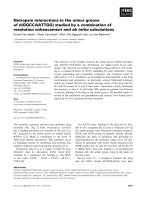
Comparison of constitutive mRNA levels in cerebral
cortex from three mouse strains
Levels of immunological transcripts in freshly isolated
SJL/J cerebral cortex were compared to baseline levels in
freshly isolated C57BL/6J and BALB/cJ cortices.
Amounts of CD74 and antisecretory factor (ASF) mRNA
differed (Figure 1). CD74 i n C57BL/6J and BALB/cJ were
lower than 50% of that in SJL/ J tissue. ASF was 30%
higher in C57BL/6 relative to SJL/J and BALB/cJ tissues.
No significant differences in constitutive expression of
COX-2 (one-way analysis of variance; P =0.7),TNF-a
(0.1), IL-12 p35 (0.08), or CiiTA (0.6) were found. Base-
line levels of chemokines, IL-1a and b, IL-6, and IL-23
were negligible (see Table 2) and considered too low to
reliably compare among strains based on the detection
limits for real-time RT-PCR.
Immunological transcriptional response in SJL/J cortical
explants
Tissue levels of mRNA in sections of cortical tissue from
SJL/J m ice incubated in explant medium were expressed
as a fol d difference relative to baseline levels in freshly
isolated SJL/J cerebral cortex. The quantity of eleven
Graber et al. Journal of Neuroinflammation 2011, 8:122
/>Page 2 of 8
transcripts changed within five hours of incubation
(Table 3). Chemokines and pro-inflammatory cytokine s
increased in cortical explants with the exception to the
p35 subunit of IL-12. Message for ASF decreased in a
time-dependent manner (Figure 2).
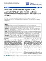
Comparison of mRNA levels in cortical explant from three
mouse strains
Sections of cortical tissue from SJL/J, C57BL/6J, and
BALB/cJ mice were incubated in explant medium for 4.5
hours. Immunological transcripts in cortical explants from
these strains were determined and expressed as a percent
of the amount in SJ L/J - i.e., for this interstrain compari-
son the transcript amount for the moieties studied were
calculated using the levels found in SJL/J explants as the
standard. Transcripts for many pro-inflammatory media-
tors and antisecretory factor revealed differential tissue
levels among strains (Figure 3). C57BL/6J explants had
higher mRNA amounts for chemokines and IL-1a relative
SJL/J and BALB/cJ explants. IL-1b, TNF-a, IL-12/23 p40,
and COX-2 revealed a similar profile with SJL/J and
C57BL/6J having similar amounts that were higher than in
BALB/cJ explants. No significant difference in abundance
of IL-6 (one-way analysis of variance; P =0.6),IL-12p35
(0.07), IL-23 p19 (0.4), CiiTA (0.1) mRNA were observed
among these strains. Since strain differences in CD74 and
ASF mRNA were found in freshly i solated cortex and in
cortical explants, fold differences within each strain was
evaluated. CD74 mRNA was down-regulated in C57BL/6J
and BALB/cJ, but not in SJL/J explants relative to baseline
levels, while ASF was down-regulated by varying degrees
in all three strains (Figure 4).
Discussion
The data reported in this study establish that differences
in the immediate gene response to damage of central
Table 1 Oligonucleotide primer sets used in quantitative real time RT-PCR analysis
Sense Primer Sequence Amplicon
Size
Assession# Name
b-Actin Forward GGCTGTATTCCCCTCCATC 141 bp NM_007393.2 actin, beta, cytoplasmic
Reverse ATGCCATGTTCAATGGGGTA
ASF Forward CAGATCGCCTACGCCATGCAGA 81 bp NM_008951.1 antisecretory factor
Reverse GGCTGAGCTGGCATCCATGTCA
CCL2 Forward ACCACCATGCAGGTCCCTGTCAT 75 bp NM_011333.3 chemokine (C-C motif) ligand 2 (MCP-1)
Reverse AGCCAACACGTGGATGCTCCAG
CCL3 Forward ACCAGCAGCCTTTGCTCCCA 141 bp NM_011337.2 chemokine (C-C motif) ligand 3 (MIP-1alpha)
Reverse TCCTCGCTGCCTCCAAGACTCT
CCL4 Forward TGCTCGTGGCTGCCTTCTGT 99 bp NM_013652.2 chemokine (C-C motif) ligand 4 (MIP-1beta)
Reverse TGTGAAGCTGCCGGGAGGTGTA
CD74 Forward CATGGATGACCAACGCGAC 101 bp NM_010545.3 invariant polypeptide of major histocompatibility complex, class
II antigen-associated
Reverse TGTACAGAGCTCCACGGCTG
CiiTA Forward GCATGTTGCACACCAGCTCCCT 135 bp NM_007575.2 major histocompatibility complex class II transactivator
Reverse ACGCCAGTCTGACGAAGGTCCA
COX-2 Forward CAGACAACATAAACTGCGCCTT 71 bp NM_011198.3 prostaglandin-endoperoxide synthase 2 (Ptgs2)
Reverse GATACACCTCTCCACCAATGACC
IL-1a Forward TACTCGTCGGGAGGAGACGACTCT 107 bp NM_010554.4 interleukin 1 alpha
Reverse TCCTTCAGCAACACGGGCTGGT
IL-1b Forward CCTTCCAGGATGAGGACATGA 71 bp NM_008361.3 interleukin 1 beta
Reverse TGAGTCACAGAGGATGGGCTC
IL-6 Forward GAGGATACCACTCCCAACAGACC 141 bp NM_031168.1 interleukin 6
Reverse AAGTGCATCATCGTTGTTCATACA
IL-12 P35 Forward GCATGCTGGTGGCCATCGATGA 130 bp NM_008351.1 interleukin 12, alpha subunit p35
Reverse GCGTGAAGCAGGATGCAGAGCT
IL-12/23
P40
Forward TGTGCTCGTGGCCTGATCCACT 91 bp NM_008352.2 interleukin 12,23, beta subunit p40
Reverse CGCAGCCCTGATTGAAGAGCTGT
IL-23 P19 Forward TATGGCTGTTGCCCTGGGTCACT 118 bp NM_031252.2 interleukin 23, alpha subunit p19
Reverse GCATGTGCGTTCCAGGCTAGCA
TNF-a Forward CAAGGGACAAGGCTGCCCCG 109 bp NM_013693.2 tumor necrosis factor alpha
Reverse GCAGGGGCTCTTGACGGCAG
Graber et al. Journal of Neuroinflammation 2011, 8:122
/>Page 3 of 8
nervous system (CNS) tissue occur among mouse
strains. Message for several genes involved in CNS
injury and neurological diseases with autoimmunity or
chronicinnateimmuneactivationwerestrain-depen-
dently altered. Short-term explants of cortical sections
providedareliablesystemfor defining immunologically
relevant transcriptional c hanges in CNS tissue and the
model may serve as a cost-effective method to test novel
immunomodulating pharmaceuticals.
One m illimeter-thick explants of adult cerebral cortex
were used in this study. Acute CNS explants of this
thickness have been previously demonstrated to induce
pro-inflammatory mRNA expression consistent to the
temporal profile observed following injury in vivo [24,25].
The production of immunologically relevant mRNAs is
likely caused by a combination of tissue damage at the
periphery of the explant due to tissue sectioning and
axotomy of projection neurons throughout the explant,
and some undefined amount of ischemia in the center of
the explant. Pro-inflammatory mRNA increase occurs in
cortex within hours following lesioning [26] or ischemia
[27,28]in vivo. Since there were no established foci of
inflammation in the tissue prior to sectioni ng in our
explant model, the influence of the miniscule number of
leukocytes in the vasculature would contribute only mar-
ginally and is limited to the residual cells in the vascula-
ture at the time of sectioning. Therefore, the extent of
the measured pro-inflammatory response is predomi-
nantly by resident CNS cells.
Pro-inflammatory cytokines and chemokines expres-
sion are activated in cells of the monocyte/macrophage
lineage in the innate response to injury or infections.
Microglia are considered the primary cell type within the
CNS parenchyma that carry out this function. Pattern
recognition receptors recognize specific molecules
released from damaged host cells or foreign microbes
leading to the activation of transcription factors that
induce transcription of certain inflammatory genes. Sec-
tions of cerebral cortex incubated ex vivo in explant
medium were demonstrated to increase mRNA for pro-
inflammatory cytokines and chemokines within five
hours. The constitutive amounts in uninjured cortical tis-
sue were mostly very low and transcripts that could be
confidently q uantified did not differ significantly among
mouse strains. How ever the amount measured in cortical
explants from these strains was different for several of
the transcripts suggesting st rain-related alterations in
their induction. The chemokines evaluated were CCL2
(MCP-1), CCL3 (MIP-1a), and CCL4 (MIP-1 b). These
Table 2 Baseline levels of mRNA in SJL/J cerebral cortex
mRNA Relative amount (% b-actin)
b-actin 100
ASF 14
COX-2 2
CD74 1
TNF 0.4
IL-12 p35 0.4
CiiTA 0.4
CCL4 0.09
IL-1a 0.08
CCL3 0.06
CCL2 0.02
IL-1b 0.02
IL-6 0.01
IL-23 p19 0.009
IL-12/23 p40 0.002
Freshly isolated cortical tissues were processed for RNA. Pooled cDNA samples
from eleven SJL/J mice were measured in replicates of at least three. Amount
of each mRNA was expressed as a percentage compared the average b-actin
mRNA levels.
Figure 1 Comparison of baseline mRNA levels in cortex among mice. Differential expression of baseline, constitutive CD74 and antisecretory
factor (ASF) mRNAs in cerebral cortex among mouse strains. RNA was isolated from resected cortical tissue immediately. b-actin was used as a
reference mRNA and values were expressed as percent amount relative to SJL/J cortex + SEM. Pooled cDNA from SJL/J (n = 11), C57BL-6J (7),
and BALB/cJ (11) mice were measured in replicates of at least three. Thinner line = P < 0.01 and thicker line = P < 0.001 between strains,
Newman-Keuls multiple comparison test.
Graber et al. Journal of Neuroinflammation 2011, 8:122
/>Page 4 of 8
transcripts were found in increasing abundance in BALB/
cJ, SJL/J, and C57BL/6J, respectively. Interestingly, the
abundance of IL-1a and COX-2 mRNA revealed a similar
pattern to the chemokines suggesting these inflammatory
mediators could have a common regulatory mechanism
that is distinct am ong these mouse strains. Messages for
IL-1b and TNF-a were similar in SJL/J and C57BL/6
explants, but higher than BALB/cJ. Taken together, BALB/
cJ appears to have a dampened immunological response to
tissue damage in cerebral cortex.
Expression of genes associated with autoimmunity was
also examined in this study. Experimental autoimmune
encephalomyelitis is a widely studied autoimmune model,
and involves infiltration of CD4+ lymphocytes and immu-
nological activation of microglia within the CNS
[21,22,29]. Although MHC class II haplotype and its bind-
ing to specific myelin autoantigen play a pivotal role in
this model, non-MHC class II effectors are also implicated
[30]. BALB/cJ is EAE resistant while SJL/J and C57BL/6J
are susceptible [2,3,31]. Cytokines IL-12 and IL-23 are
implicated in the pathogenesis of autoimmune diseases
including EAE [17]. The p35 subunit of IL-12 and p19
subunit of IL-23 form respective cytokines with a common
p40 subunit. Blocking the IL-12/23 p40 subunit with inhi-
biting antibodies is effective in non-CNS autoimmune dis-
eases such as psoriasis [32]. In cortical explants, the p40
mRNA was upregulated. Its levels were considerably lower
in the resistant BALB/cJ explants. This suggests that
inherent difference within the CNS tissues may contribute
to strain susceptibility to autoimmunity.
Antisecretory factor (ASF) has been shown to affect the
severity of EAE. Blocking its activity with an inhibiting
antibody increases clinical severity implying it has an
anti-inflammatory property [20]. Our results showed that
differences in ASF mRNA expression occurred in normal
cortical tissue and in cultured cortical explants. C57BL/6J
Table 3 Change in mRNA levels in SJL/J cortical tissue after incubation in explant medium
mRNA Fold difference in explants at 4.5 hrs (relative to freshly isolated cortex) Significance (unpaired t test)
CCL4 912 P < 0.001
CCL3 518 P < 0.001
IL-12/23 p40 159 P < 0.001
IL-1b 98 P < 0.0001
IL-6 55 P < 0.0001
IL-1a 50 P < 0.001
CCL2 15 P < 0.01
TNF-a 13 P < 0.001
COX-2 3.9 P < 0.0001
IL-23 p19 3.3 P < 0.01
IL-12 p35 1.1 NS
CD74 0.90 NS
CiiTA 0.61 NS
ASF 0.58 P < 0.001
Freshly isolated cortical tissue and cortical tissue incubated in explant medium for 4.5 hours were processed for RNA. Transcript levels were referenced to b-actin
mRNA levels. Values were expressed as fold difference in explants relative to freshly isolated cortex. Pooled cDNA from eleven SJL/J mice were measured in
replicates of at least three. NS, not significant.
Figure 2 Change in mRNA expression in cortical explants over
time. Time-dependent change in CCL4 and antisecretory factor (ASF)
mRNA expression in SJL/J cortical explants. RNA was isolated from
resected cortical tissue after incubation in explant medium for 0, 0.5,
2, and 4.5 hours. Transcript levels were referenced to b-actin mRNA
levels. Values were expressed as fold difference relative to freshly
isolated cortex (baseline) + SEM. Pooled cDNA from SJL/J mice (n =
4) were measured in replicates of four. * = P < 0.05 and ** = P < 0.01,
relative to 0 hours, Dunnett’s multiple comparison test.
Graber et al. Journal of Neuroinflammation 2011, 8:122
/>Page 5 of 8
mice had high constitutive ASF mRNA. Its levels
decreased after injury in cortical explants in each strain,
but to a lesser degree in BALB/cJ. This supports the
hypothesis that higher amounts of ASF due to genetic
background may contribute to EAE resistance.
Antigen presentation by MHC class II is critical for
many autoimmune diseases. CD74 (invariant chain, Ii)
acts as an MHC class II chaperone [33,34]. CD74 mRNA
was higher in SJL/J cortex relative to BALB/cJ and C57BL/
6. A similar trend among these strains was reported in
Figure 3 Comparison of mRNA levels in cortical explant among mice. Differential expression of immunological mR NAs in corti cal explants
among mouse strains. RNA was isolated from resected cortical tissue after incubation in explant medium for 4.5 hours. b-actin was used as a
reference mRNA and values were expressed as percent amount relative to SJL/J cortical explants + SEM. Pooled cDNA from SJL/J (n = 11),
C57BL/6J (7), and BALB/cJ (11) mice were measured in replicates of at least four. Dotted line = P < 0.05, thinner line = P < 0.01, and thicker line
= P < 0.001 between strains, Newman-Keuls multiple comparison test.
Figure 4 Change in mRNA expression in cortical explants among mice. Change in CD74 and antisecre tory factor (ASF) mRNA in cort ical
explants in mouse strains. RNA was isolated from freshly resected cortical tissue (baseline) and cortical explants after incubation for 4.5 hours.
Transcript levels were referenced to b-actin mRNA levels. Values were expressed as fold difference relative to baseline + SEM. Pooled cDNA from
SJL/J (n = 11), C57BL/6J (7), and BALB/cJ (11) mice were measured in replicates of at least three. * = P < 0.05 and ** = P < 0.01, relative to
baseline, unpaired t test.
Graber et al. Journal of Neuroinflammation 2011, 8:122
/>Page 6 of 8
facial nucleus two weeks following facial nerve axotomy
[19]. Our study revealed that higher levels were found in
uninjured cortex and that these differences were increased
furtherinexplantsduetodown-regulationinC57BL/6J
and BALB/cJ tissues. CD74 also mediates transcription by
NF-B [35,36] and regulates dendritic cell migration [37].
This suggests its altered expression among strains could
influence a wide range of effects.
Now, identifying the inherent genetic polymorphisms
that control the variations i n transcr iptional response to
an injury stimulus among strai ns is important for under-
standing genetically variable responses to a spectrum of
neurological disorders. Approaches such as quantitative
trait locus analysis and/or haplotype-based computational
genetic mapping can be ut ilized with the cortical explant
model. Haplotype-based co mputational genetic mapping
has recently pinpointed genetic variation of Nalp1 as a
contributor to in terstrain differences in the inflammatory
response to injured skin [38]. It is important to recognize
that the involvement of multiple genes may be required
and that such analyses wil l likely require data from addi-
tional strains and transcripts.
Conclusions
The genes expressed differentially in co rtical explants
derived from disparate strains of mice reveal t hat
genetic background can influence immediate response
to neurological damage within the CNS. The straightfor-
ward approach described in this study may help uncover
the inherent regulatory me chanism that control altered
immunological responsiveness and perhaps neurological
disease susceptibility in future studies.
List of abbreviations
ASF: antisecretory factor; CCL: CC chemokine ligand; CiiTA: class II
transactivator; COX: cyclooxygenase; CNS: central nervous system; FBS: fetal
bovine serum; IL: interleukin; RT-PCR: reverse transcription- polymerase chain
reaction; TNF: tumor necrosis factor
Acknowledgements
DJG and WFH acknowledge support from the Department of Pathology,
Dartmouth Medical School. DJG and BTH were supported in part by grants
from Reata Pharmaceuticals and the ALS Center of Dartmouth-Hitchcock
Medical Center.
Author details
1
Dept. Pathology, Dartmouth Medical School, Lebanon, New Hampshire,
USA.
2
Dept. Pathology, Georgetown University School of Medicine,
Washington D.C., USA.
3
Dept. Neurology, Georgetown University School of
Medicine, Washington D.C., USA.
Authors’ contributions
DJG carried out the experiments and evaluated the data. DJG, BTH, and
WFH participated in the design and assisted with the preparation of the
manuscript. All authors have read and approved the final version of the
manuscript.
Competing interests
The authors declare that they have no competing interests.
Received: 21 July 2011 Accepted: 26 September 2011
Published: 26 September 2011
References
1. Heiman-Patterson TD, Deitch JS, Blankenhorn EP, Erwin KL, Perreault MJ,
Alexander BK, Byers N, Toman I, Alexander GM: Background and gender
effects on survival in the TgN(SOD1-G93A)1Gur mouse model of ALS. J
Neurol Sci 2005, 236:1-7.
2. Encinas JA, Lees MB, Sobel RA, Symonowicz C, Greer JM, Shovlin CL,
Weiner HL, Seidman CE, Seidman JG, Kuchroo VK: Genetic analysis of
susceptibility to experimental autoimmune encephalomyelitis in a cross
between SJL/J and B10. S mice. J Immunol 1996, 157:2186-92.
3. Royle SJ, Collins FC, Rupniak HT, Barnes JC, Anderson R: Behavioural
analysis and susceptibility to CNS injury of four inbred strains of mice.
Brain Res 1999, 816:337-49.
4. Diez M, Abdelmagid N, Harnesk K, Ström M, Lidman O, Swanberg M,
Lindblom R, Al-Nimer F, Jagodic M, Olsson T: Identification of gene
regions regulating inflammatory microglial response in the rat CNS after
nerve injury. J Neuroimmunol 2009, 212:82-92.
5. Piehl F, Swanberg M, Lidman O: The axon reaction: Identifying the genes
that make a difference. Physiol Behav 2007, 92:67-74.
6. Swanberg M, Harnesk K, Ström M, Diez M, Lidman O, Piehl F, Linden R: Fine
mapping of gene regions regulating neurodegeneration. PLoS One 2009,
4:689-700.
7. Butterfield RJ, Sudweeks JD, Blankenhorn EP, Korngold R, Marini JC,
Todd JA, Roper RJ, Teuscher C: New genetic loci that control
susceptibility and symptoms of experimental allergic encephalomyelitis
in inbred mice. J Immunol 1998, 161:1860-7.
8. Baker D, Rosenwasser OA, O’Neill JK, Turk JL: Genetic analysis of
experimental allergic encephalomyelitis in mice. J Immunol 1995,
155:4046-51.
9. Sundvall M, Jirholt J, Yang HT, Jansson L, Engström Å, Pettersson U,
Holmdahl R: Identification of murine loci associated with susceptibility to
chronic experimental autoimmune encephalomyelitis. Nat Genet 1995,
10:313-7.
10. Croxford JL, O’Neill JK, Baker D: Polygenic control of experimental allergic
encephalomyelitis in Biozzi ABH and BALB/c mice. J Neuroimmunol 1997,
74:205-11.
11. Teuscher C, Butterfield RJ, Ma RZ, Zachary JF, Doerge RW, Blankenhorn EP:
Sequence polymorphisms in the chemokines Scya1 (TCA-3), Scya2
(monocyte chemoattractant protein (MCP)-1), and Scya12 (MCP-5) are
candidates for eae7, a locus controlling susceptibility to monophasic
remitting/nonrelapsing experimental allergic encephalomyelitis. J
Immunol 1999, 163:2262-6.
12. Marta M, Stridh P, Becanovic K, Gillett A, Öckinger J, Lorentzen JC,
Jagodic M, Olsson T: Multiple loci comprising immune-related genes
regulate experimental neuroinflammation. Genes Immun 2009, 11:21-36.
13. Teuscher C, Doerge RW, Fillmore PD, Blankenhorn EP: eae36, a locus on
mouse chromosome 4, controls susceptibility to experimental allergic
encephalomyelitis in older mice and mice immunized in the winter.
Genetics
2006, 172:1147-53.
14.
Butterfield RJ, Blankenhorn EP, Roper RJ, Zachary JF, Doerge RW,
Teuscher C: Identification of genetic loci controlling the characteristics
and severity of brain and spinal cord lesions in experimental allergic
encephalomyelitis. Am J Pathol 2000, 157:637-45.
15. Spach KM, Case LK, Noubade R, Petersen CB, McElvany B, Zalik N,
Hickey WF, Blankenhorn EP, Teuscher C: Multiple linked quantitative trait
loci within the Tmevd2/Eae3 interval control the severity of
experimental allergic encephalomyelitis in DBA/2J mice. Genes Immun
2010, 11:649-59.
16. Wang CX, Shuaib A: Involvement of inflammatory cytokines in central
nervous system injury. Prog Neurobiol 2002, 67 :161-72.
17. Kroenke MA, Carlson TJ, Andjelkovic AV, Segal BM: IL-12-and IL-23-
modulated T cells induce distinct types of EAE based on histology, CNS
chemokine profile, and response to cytokine inhibition. J Exp Med 2008,
205:1535-41.
18. Fischer FR, Santambrogio L, Luo Y, Berman MA, Hancock WW, Dorf ME:
Modulation of experimental autoimmune encephalomyelitis: effect of
altered peptide ligand on chemokine and chemokine receptor
expression. J Neuroimmunol 2000, 110:195-208.
Graber et al. Journal of Neuroinflammation 2011, 8:122
/>Page 7 of 8
19. Harnesk K, Swanberg M, Diez M, Olsson T, Piehl F, Lidman O: Differential
nerve injury-induced expression of MHC class II in the mouse correlates
to genetic variability in the type I promoter of C2ta. J Neuroimmunol
2009, 212:44-52.
20. Davidson TS, Hickey WF: Antisecretory factor expression is regulated by
inflammatory mediators and influences the severity of experimental
autoimmune encephalomyelitis. J Leukoc Biol 2004, 76:835-44.
21. Furlan R, Cuomo C, Martino G: Animal models of multiple sclerosis.
Methods Mol Biol 2009, 549:157-73.
22. Gold R, Linington C, Lassmann H: Understanding pathogenesis and
therapy of multiple sclerosis via animal models: 70 years of merits and
culprits in experimental autoimmune encephalomyelitis research. Brain
2006, 129:1953-71.
23. Spach KM, Blake M, Bunn JY, McElvany B, Noubade R, Blankenhorn EP,
Teuscher C: Cutting edge: the Y chromosome controls the age-
dependent experimental allergic encephalomyelitis sexual dimorphism
in SJL/J mice. J Immunol 2009, 182:1789-93.
24. Pan JZ, Ni L, Sodhi A, Aguanno A, Young W, Hart RP: Cytokine activity
contributes to induction of inflammatory cytokine mRNAs in spinal cord
following contusion. J Neurosci Res 2002, 68.
25. Rice T, Larsen J, Rivest S, Yong VW: Characterization of the early
neuroinflammation after spinal cord injury in mice. J Neuropathol Exp
Neurol 2007, 66:184.
26. Rostworowski M, Balasingam V, Chabot S, Owens T, Yong VW: Astrogliosis
in the neonatal and adult murine brain post-trauma: elevation of
inflammatory cytokines and the lack of requirement for endogenous
interferon-γ. J Neurosci 1997, 17:3664.
27. Wang X, Yue TL, Barone FC, White RF, Gagnon RC, Feuerstein GZ:
Concomitant cortical expression of TNF-α and IL-1β mRNAs follows early
response gene expression in transient focal ischemia. Mol Chem
Neuropathol 1994, 23:103-14.
28. Berti R, Williams AJ, Moffett JR, Hale SL, Velarde LC, Elliott PJ, Yao C,
Dave JR, Tortella FC: Quantitative real-time RT-PCR analysis of
inflammatory gene expression associated with ischemia-reperfusion
brain injury. J Cereb Blood Flow Metab 2002, 22:1068-79.
29. Murphy ÁC, Lalor SJ, Lynch MA, Mills KHG: Infiltration of Th1 and Th17
cells and activation of microglia in the CNS during the course of
experimental autoimmune encephalomyelitis. Brain Behav Immun 2010,
24:641-51.
30. Happ M, Wettstein P, Dietzschold B, Heber-Katz E: Genetic control of the
development of experimental allergic encephalomyelitis in rats.
Separation of MHC and non-MHC gene effects. J Immunol 1988,
141:1489-94.
31. Teuscher C, Hickey WF, Grafer CM, Tung KSK: A common
immunoregulatory locus controls susceptibility to actively induced
experimental allergic encephalomyelitis and experimental allergic
orchitis in BALB/c mice. J Immunol 1998,
160:2751-9.
32. Leonardi CL, Kimball AB, Papp KA, Yeilding N, Guzzo C, Wang Y, Li S,
Dooley LT, Gordon KB: Efficacy and safety of ustekinumab, a human
interleukin-12/23 monoclonal antibody, in patients with psoriasis: 76-
week results from a randomised, double-blind, placebo-controlled trial
(PHOENIX 1). The Lancet 2008, 371:1665-74.
33. Roche PA, Teletski CL, Stang E, Bakke O, Long EO: Cell surface HLA-DR-
invariant chain complexes are targeted to endosomes by rapid
internalization. Proc Natl Acad Sci USA 1993, 90:8581-5.
34. Cresswell P: Assembly, transport, and function of MHC class II molecules.
Annu Rev Immunol 1994, 12:259-91.
35. Teo BHD, Wong SH: MHC class II-associated invariant chain (Ii) modulates
dendritic cells-derived microvesicles (DCMV)-mediated activation of
microglia. Biochem Biophys Res Commun 2010, 400:673-8.
36. Matza D, Kerem A, Medvedovsky H, Lantner F, Shachar I: Invariant chain-
induced B cell differentiation requires intramembrane proteolytic release
of the cytosolic domain. Immunity 2002, 17:549-60.
37. Faure-Andre G, Vargas P, Yuseff MI, Heuze M, Diaz J, Lankar D, Steri V,
Manry J, Hugues S, Vascotto F: Regulation of dendritic cell migration by
CD74, the MHC class II-associated invariant chain. Science 2008,
322:1705-10.
38. Hu Y, Liang D, Li X, Liu HH, Zhang X, Zheng M, Dill D, Shi X, Qiao Y,
Yeomans D, Carvalho B, Angst MS, Clark JD, Peltz G: The Role of
Interleukin-1 in Wound Biology. Part I: Murine In Silico and In Vitro
Experimental Analysis. Anesth Analg 2010, 111:1525-33.
doi:10.1186/1742-2094-8-122
Cite this article as: Graber et al.: Strain-dependent variation in the early
transcriptional response to CNS injury using a cortical explant sy stem.
Journal of Neuroinflammation 2011 8:122.
Submit your next manuscript to BioMed Central
and take full advantage of:
• Convenient online submission
• Thorough peer review
• No space constraints or color figure charges
• Immediate publication on acceptance
• Inclusion in PubMed, CAS, Scopus and Google Scholar
• Research which is freely available for redistribution
Submit your manuscript at
www.biomedcentral.com/submit
Graber et al. Journal of Neuroinflammation 2011, 8:122
/>Page 8 of 8