báo cáo hóa học: " Comparing the immunosuppressive potency of naïve marrow stromal cells and Notch-transfected marrow stromal cells" docx
Bạn đang xem bản rút gọn của tài liệu. Xem và tải ngay bản đầy đủ của tài liệu tại đây (1.3 MB, 14 trang )
RESEARC H Open Access
Comparing the immunosuppressive potency of
naïve marrow stromal cells and Notch-transfected
marrow stromal cells
Mo A Dao
1*
, Ciara C Tate
1
, Irina Aizman
1
, Michael McGrogan
2
and Casey C Case
1
Abstract
Background: SB623 cells are expanded from marrow stromal cells (MSCs) transfected with a Notch intracellular
domain (NICD)-expressing plasmid. In stroke-induced animals, these cells reduce infarct size and pro mote
functional recovery. SB623 cells resemble the parental MSCs with respect to morphology and cell surface markers
despite having been in extended culture. MSCs are known to have immunosuppressive properties; whether long-
term culture of MSCs impact their immunomodulatory activity has not been addressed.
Methods: To assess the possible senescent properties of SB623 cells, we performed cell cycle related assays and
beta-galactosidase staining. To assess the immunomodulatory activity of these expanded NICD-transfected MSCs,
we performed co-cultures of SB623 cells or MSCs with either enriched human T cells or monocytes and assessed
cytokine production by flow cytometry. In addition, we monitored the immunosuppressive activity of SB623 cells in
both allogenic and xenogenic mixed lymphocyte reaction (MLR).
Results: Compared to MSCs, we showed that a small number of senescent-like cells appear in each lot of SB623
cells. Nevertheless, we demonstrated that these cells suppress human T cell proliferation in both the allogeneic
and xenogeneic mixed lymphocyte reaction (MLR) in a manner comparable to MSCs. IL-10 producing T cells were
generated and monocyte-dendritic cell differentiation was dampened by co-culture with SB623 cells. Compared to
the parental MSCs, SB623 cells appear to exert a greater inhibitory impact on the maturation of dendritic cells as
demonstrated by a greater reduction in the surface expression of the co-stimulatory molecule, CD86.
Conclusion: The results demonstrated that the immunosuppressive activity of the expanded NICD-transfected
MSCs is comparable to the parental MSCs, in spite of the appearance of a small number of senescent-like cells.
Introduction
There is an important need for stromal cell lines that
support neural cells and the mesenchymal stem cell
(MSC) line SB623, transfected with the Notch-intracel-
lular domain (NICD), appear to meet these crite ria. In
cultures of embryonic cortical neurons, SB623 cells pro-
duce extracellular m atrix proteins which enhance and
maintain neurite outgrowth [1]. In neonatal hippocam-
pal organotypic culture, SB623 cell-derived soluble
trophic factors rescue neural cells subjected to oxygen-
glucose deprivation [2]. In experimental Parkinson’s dis-
ease, grafting of SB623 cells efficiently reverses the
degeneration of dopaminergic neurons by promoting
end ogeneous neuronal cell recovery [3,4]. And in stable
stroke animal models, t ransplantation of SB623 cells
reduces infarct size and promotes behavioral improve-
ment [5]. These studies validate one of the therapeutic
applications of SB623 cells - to supply trophic factors
for the endogenous neural cells after injury or disease.
Human marrow stromal cells are attractive for cell
therapy because they can be obtained with minimal
invasiveness and can be expanded in culture. However,
as non-immortalized primary cells, MSCs have limited
regenerative potential, committing to cellular senescence
after extensive ex vivo manipulation [6,7]. A potential
upside of senescent cells is their robust cytokine secre-
tome profile which could be beneficial in tissue regen-
eration. A potential downside is that the senescent-
* Correspondence:
1
Research Department San-Bio Incorporated 231 South Whisman Road,
Mountain View, 94041, USA
Full list of author information is available at the end of the article
Dao et al. Journal of Neuroinflammation 2011, 8:133
/>JOURNAL OF
NEUROINFLAMMATION
© 2011 Dao et a l; licensee BioM ed Central Ltd. This is an Open Access articl e distributed under the terms o f the Creative Commons
Attribution License ( which perm its unrestricted use, distribution, and re production in
any medium, provided the or iginal work is properly cited.
associated-secretome profile is thought to be pro-inflam-
matory [8-10]. To date, intracerebral implantation of
human SB623 cells in stroke-induced animals has not
triggered any immunological adverse e ffect. Neverthe-
less, as SB623 cells are derived from MSCs that have
undergone gene transfection and cell expansion in cul-
ture,weinitiatedthecurrentstudytodetermine
whether SB623 cells display senescent-like properties.
More importantly, we com pare the immunomodulatory
activity b etween SB623 ce lls and the corresp onding par-
ental MSCs. We demonstrate that SB623 cells, currently
in a clinical trial for stable stroke (http://clinicaltrials.
gov/ct2/show/NCT01287936), retain the immunosup-
pressive activity of standard MSCs despite the appear-
ance of a small number of senescent-like cells.
Materials and methods
Production of MSCs and SB623 cells
MSC and SB623 cells were produced as previously
reported [1,2]. Briefly, human adult bone marrow aspi-
rates (Lonza, Walkersville, MD) were plated in growth
medium - aMEM (Mediatech, Ma nassas, VA) supple-
mented with 10% fetal bovine serum (FBS) (Hyclone,
Logan, UT), 2 mM L-glutamine and penicillin/strepto-
mycin (both from Invitrogen, Carlsbad, CA) for three
days to obtain the marrow stromal cell (MSC) mono-
layer. After two passages, a portion of the culture was
cryopreserved as MSCs. The re maining cells (passage 2)
were transfected with the pCMV-hNICD1-SV40- Neo
R
plasmid using Fugene6 (Roche Diagnostic s, Indianapolis,
IN). After 7 days of selection with 100 μg/ml G418
(Invitrogen), the G418-resistant colonies were expanded
and passed twice prior to cryopreservation as SB623
cells. This results in a uniformly transiently transfected
population of MSCs.
qPCR and qRT-PCR
Two days a fter transfection with pN2-NICD plasmid,
cells were lysed and DNA or RNA purified using Qia-
gen’ s QIAAmp DNA or RNeasy mini kits (Qiagen,
Valencia, CA), correspondingly, according to the manu-
facturer’ s protocols. Quantitative real time PCR or RT-
PCR analyses were conducted using QuantiTect Probe
PCR or RT-PCR kits, respectively, on Lightcycler
(Roche).
For exogenous-NICD (eNICD) qP CR analysis, purified
RNA-free DNA samples were used at 65 ng (10000
diploid human genomes) per reaction and eNICD gene
copy numbers were determined using eNICD-DNA-spe-
cific Taqman assay (forward primer: TTGGTC TTACT-
GACATCCACTTTG, reverse primer CAGACACTT
TGAAGCCCTCAG, exo-NICD-specific probe [6-FAM]
CCCAGTTCAATTACAGCTCTTAAGGCTAGAG
[BHQ1a-6FAM])). Amplification signals were compared
to those of pN2-NICD plasmid serially diluted in
human genomic DNA (Clontech, Mountain View, CA);
results expressed in numbers of p lasmids per one
human diploid genome (plasmids/cell). For expression
analysis of a NICD target gene, human Hes1 and
GAPDH (control) Taqman assays (Applied Biosystems,
Carlsbad, CA) were used. Normalized He s1 expression
levels are presented relative to levels in non-transfected
cells.
Phenotypic characterization by flow cytometry
For cell surface phenotyping , MSCs or SB623 cells were
harvested with 0.25% Trypsin/EDTA (Invitrogen),
washed in PBS/2% FBS, and re-suspended in 1 ml of
PBS/2% FBS. Cells were then stained with fluoro-
chrome-conjugated antibodies against CD29, CD31,
CD34, CD44, CD45, CD73, CD90 (all from BD Bios-
ciences, San Jose, CA) and CD105 (Invitrogen, Carlsbad,
CA) for 15 minutes on ice. After one wash in PBS/2%
FBS, cells were acquired using BD FACS Calibur. Ana-
lyses were done to assess the percentage of surface mar-
kers that are positive (CD29, CD44, CD73, CD90, and
CD105) versus negative (CD31, CD34, and CD45) for
mesenchymal cells using CellQuestPro program (BD
Biosciences). To compare the density of specific surface
molecule expression on MSCs versus SB623 cells, the
delta mean fluorescent intensity (dM FI) was calculated -
e.g., dMFI of CD44 = (MFI of CD44) - (MFI of IgG).
For intracellular protein detection of p16Ink4A and
NICD, cells were fixed with 4% paraformaldehyde and
permeabilized with PBS/0.1% TritonX- 100. After two
washes in PBS/2% F BS, cell pelle ts were resuspended in
200 ul of PBS/2% FBS and divided into two tube s, one
for staining with phycoerythrin (PE)-conjugated IgG
(control) and the other for staining with PE-conjugated
p16Ink4A antibody (BD Bios cience) or PE-conjugated
NICD antibody (eBioscience). For intracellular cytokine
detection, cells were treated with BrefeldinA for six
hours prior to harvest. After fixation and permeabiliza-
tion, cells were incubated with fluorochrome-conjugated
antibody against human GM-CSF (BD Bioscience), IL-1a
(eBioscience, San Diego, CA), IL-6 (BD Bioscience),
TGFb1 (RnD Systems, Minneapolis, MN) for one hour
followed by two washes in PBS/2% FBS. Acquisition and
analysis of all samples were performed on BD FACS
Calibur using CellQuestPro software.
Cell proliferation measurement
To quantify viable cell expansion, one million MSCs or
SB623 cells were plated on Day 0 and cell counts by try-
pan blue exclusion were done on Day 3. For cell cycle
profile after culture, one million MSCs or SB623 cells
were fixed in 70% ethanol overnight at 4°C. After two
washes in PBS/2% FBS, cells were incubated in one ml
Dao et al. Journal of Neuroinflammation 2011, 8:133
/>Page 2 of 14
of staining buffer (50 μg/ml propidium iodide, 50 μg/ml
RNAse) (Sigma, St. Lou is, MO) in PBS/2% FBS for 30
mininthedark.Acquisitionandanalysisweredone
using CellQuestPro program on the FL-2 linear channel.
For cell cycle kinetics over 5 days in culture, MSCs and
SB623 cells were labeled with 5 μM of 5-(and-6)-carbox-
yfluorescein diacetate ( CFSE) (Invitrogen) for 2 m in at
room temperature prior to culture. Flow cytometry
acquisition and analysis were done on the FL-1 log
channel.
Generation of monocyte-derived dendritic cells (Mono-
DC)
Peripheral blood was obtained from healt hy donors and
mononuclear cells recove red from buffy coat prepara-
tions by Ficoll Paque (Amersham Pharmacia, Sweden)
gradient separation. Mononuclear cells were re-sus-
pended in RPMI/10%FBS a nd plated in a T-75 flask
overnight. Non-adherent cells w ere discarded and the
flasks were rinsed twice with PBS. Adherent monocytes
were recovered using 0.25% trypsin/2 mM EDTA. Purity
was assessed by staining with FITC-conjugated antibody
against human CD14, a monocyte surface marker (Bec-
ton Dickinson) and was routinely shown to be > 90%.
For monocytic-to-dendritic cell differentiation assays,
monocytes were cultured in R PMI-1640 (Mediatech)
containing 10% FBS, 2 mM glutamine, 2 mM sodium
pyruvat e, 100 U/mL penicillin, 100 μg/mL streptomycin,
40 ng/mL granulocyte-macrophage colony stimulating
factor (GM-CSF) and 20 ng/mL interleukin-4 (IL-4)
(both from Peprotech, Rocky Hill, NJ) in the presence of
MSCs or SB623 cells at a 10:1 monocyte to MSC or
SB623 cell ratio. On Day 5, a subset of cultures were
harvested by 0.25% trypsin/2 mM EDTA and stained
with fluorochrome-conjugated antibodies against CD1a
and C D14 (eBioscience). Data acquisition and analysis
were done on the FACS Calibur using CellQuestPro
software.
To assess the impact of MSCs and SB623 cells on the
maturation of dendritic cells, monocyte-derived dendri-
tic cells were generated in the presence of GM-CS F and
IL-4.OnDay5,humanTNF-a (10 ng/ml; Peprotech)
was added to each well with or without MSCs or SB623
cells. As previous studies confirmed a role of cyclos-
porin A in hindering dendritic cell maturation [11],
addition of cyclosporin A (1 μg/ml; Santa Cruz Biotech-
nology, Santa Cruz, CA) in the absence of either MSCs
or SB623 cells was included as an internal control. On
Day 7, cells were incubated with a fluorochrome- conju-
gated monoclonal antibody against human CD86 (BD
Bioscience), a co-stimulatory molecule for priming and
activating naïve a nd memory T cells and analyzed on
the BD FACS Calibur using CellQuestPro.
Ex vivo culture of human peripheral blood T cells
Human T cells were enriched from peripheral b lood
using the T-cell isolation kit (StemCell Technologies,
Vancouver, Canada) according to the manufacturer’ s
protocol. Enriched T cells were cultured in RPMI-1640/
10% heat-inactivated FBS/pen/strep overnight prior to
use. On Day -1, 10,000 MSCs or SB623 cells were plated
per well o f 96-well U-bottom plates. On Day 0 of the
culture assay, 100,000 enriched T cells were transferred
to each well with a pre-established MSC or SB623 cell
monolayer. As an internal control, T cell cultures were
maintained in the absence of MSCs or SB623 cells. On
Day 7, a sub-optimal dose of 25 ng/ml of phorbol 12-
myristate 13-acetate (PMA)/0.5 μM Ionomycin ( both
from Sigma-Aldrich) was added in the presence of Bre-
feldinA (eBioscience, 1:1000) for 6 hours prior to har-
vest for intracellular detectio nofinterleukin-10(IL-10)
and interferon gamma (IFN-g). For IL-17 producing
TH17 cells, T cells were co-cultured with SB623 cells or
MSCs in the presence of IL-23, or in the presence of IL-
23 alon e. After sub-optimal activation with PMA/Iono-
mycin in the pre sence of BrefeldinA, the ce lls were
stained with fluorochrome-co njugated antibody against
IL-17A (eBioscience) and analyzed by flow cytometry.
For regulatory T cell culture, human enriched T cells
were co-cultured with MSCs or SB623 cells in the pre-
sence of human interleukin-2 (IL-2) ( Peprotech, Rocky
Hill, NJ) at a 10:1 T cells to MSC or SB623 cell ratio for
7 days followed by cell surface staining for CD4, a
helper T cell marker and CD25, the IL-2 receptor alpha
chain. For FoxP3 intracellular staining, cells were fixed
and permeabilized with CytoFix/Perm (eBi oscience). PE-
conjugated antibody against FoxP3 (clone PCH101,
eBioscience) was used at 1:50 dilution and flow cytome-
try analysis was done gating on lymphocytes. For assess-
ment of constitutive IL-10 production, intracellular
staining with fluorochrome conjugated antibody against
IL-10 was performed without PMA/Io stimulation on
Day 7.
Mixed lymphocyte reaction (MLR)
Human allogeneic mixed lymphocyte reaction was
established using peripheral blood from unrelated
healthy volunteers. To obtain responder cells, T cell
enrichment using a commercial T-cell rosette separation
kit (Stem Cell Technologies) was done based on the
manufacturer’s protocol. Enriched T cells (= responders)
were labeled with 5 μM of 5-(and -6)-carboxyflu orescein
diacetate (CFSE) (Invitrogen) for 2 min at room tem-
perature. CFSE-labeled lymphocytes were th en plated in
a 96-well U bottom plate at a concentration of 100,000
cells per 100 μl per well. To obtain stimulator cells, per-
ipheral blood buffy coat mononuclear cells were
Dao et al. Journal of Neuroinflammation 2011, 8:133
/>Page 3 of 14
recovered after Ficoll-density gradient centrifugation and
red blood cell lysis buffer (Sigma-Aldrich) was added for
10 min at 37°C. 100,000 stimulator cells were added to
a tu be containing 10,000 MSCs or SB623 cell s; and the
mixed cells were then centrifuged and re-suspended at
110,000 mixed cells per 100 μl. 100 μl of stimulator/
MSC cell mix or 100 μl of stimulator/SB623 cell mix
was added to each well of CFSE-responder cells. To
assess the activati on state of T cells in the MLR, cells
were harvested on Day 2 and stained with a fluoro-
chrome conjugated antibody against CD69 (BD
Bioscience), an antigen induced on activated T cells. To
monitor cell proliferation kinetics of T cells in the MLR,
cell s were harvested on Day 7 and stained with PE-con-
jugated CD4 antibody (BD Bioscience). Flow cytometry
data acquisit ion was done on BD FACS C alibur, gating
on CD4+ lymphocyte gate, and analysis was done using
CellQuestPro.
Xenogeneic MLRs were established using postnatal
day 9 Sprague-Dawley rat glial mix cells as stimulators
and human periphera l blood T cells as responders.
Briefly, rat b rains were harvested and triturated prior to
treatment with 0.25% t rypsin (Invitrogen) for 30 min.
Cell suspension s were filtered through a 70 μMcell
strainer and overlaid on Ficoll prior to density centrifu-
gation. Glial mix cells were cultured in DMEM/F12
(Mediatech)/10%FBS/pen-s trep for 14 days prior to use
in the MLR. Xenogeneic MLRs were performed at a
similar cell ratio as allogeneic MLRs (100,000 glial mix:
100,000 CFSE-labeled human T cells: 10,000 MSCs or
SB623 cells) over a 5-day period. CFSE dilution of
human CD3-gated T cells was assessed by flow
cytometry.
Statistics
Statistical assessments (SigmaStat, Systat Software, Chi-
cago, IL) were made for SB623 or MSC groups to deter-
mine if there were differences either between those two
groups or in some cases compared to the internal assay
controls. To compare co-cultures with SB623 cells to
those with MSCs (n = 3-6 mat ched lots), Tukey’spair-
wise comparisons were made. To compare co-cultures
with SB623 cells or MSCs to internal experimental con-
trols (when n>1), a general linear model ANOVA, fol-
lowed by Tukey’ s pairwise comparisons was u sed. An
alpha value of 0.05 was used to assess if the means were
significantly different. Data are reported as mean ± stan-
dard deviation.
Results
Comparison of SB623 cells with the corresponding
parental MSCs
SB623 cells were expanded from human MSCs after
transfection with an NICD1-expressing plasmid - a
process t hat takes eight to ten weeks in culture (Addi-
tional File 1A). Morphologically, SB623 cells retained
the mesenchymal appearance similar to the parental
MSCs. However, in each tested culture of SB623 cells,
the frequency of beta-galactosidase-positive cells was
higher than the parental MSC cultures, suggesting the
presence of senescent cells in SB623 cell culture (Addi-
tional File 1B). qRT-PCR for the exogenous NICD1 gene
and Hes1, a downstream target of Notch signaling, vali-
dated the high expression of exogenous NICD1 DNA
and the induction of endogenous Hes1 transcript after
transfection (Additional File 1C). Intracellular flow cyto-
metry analysis of NICD1 confirmed its reduction over
increasing passages consistent with transient transfection
(Additional File 1D).
To measure cell proliferation in culture, we plated
one million MSCs or SB623 cells and used trypan blue
exclusion to count the number of viable cells on Day
3.Theresultsshowedahighernumberofviablecell
counts for MSC culture than for SB623 cell culture,
suggesting a lower proli ferative index for SB623 cells.
We next assessed the cell cycle profile by staining the
cells with propidium iodide, a DNA intercalating dye,
after fixation and permeabilization. We observed a sig-
nificantly higher number of cells in G0/G1 resting
phase (p < 0.05) in SB623 cultures, again suggesting
reduced proliferation in SB623 cells (Figure 1A). Lastly,
to monitor cell division kinetics over a 5-day growth
culture, we opted for the use of CFSE, a cell permeable
dye which is diluted with each cell division (Figure 1B).
We noted a persistence of a small number of CFSE-
high cells within each lot of SB623 cells and this was
not observed in parental MSC where the vast majority
of cells proliferated.
P16Ink4A is a negative cell cycle regulator and has
been shown to be upregulated in human senescent MSC
[6,7]. To determine whether the CFSE-high (low/no pro-
liferating) cells express p16In k4A, intracellular staining
for the p16Ink4 protein was performed in CFSE-labeled
cells after culture. The subpopulation of SB623 cells
expressing p16Ink4A corresponded to the CFSE-high
SB623 cells, consistent with the role of p16ink4A as a
negative regulator of cell cycle entry (Figure 1B). Coll ec-
tively, the results demonstrate that within each final lot
of SB623 cells, there is a small number of non-prolifer-
ating cells.
Phenotypically, SB623 cells expressed all the standard
mesenchymal surface markers (CD73. CD105, CD29,
CD44, CD106). However, CD44 and CD73 were
expressed at significantly higher density per cell as
shown by mean fluorescent intensity (Figure 2A). CD54,
an inter-cellular adhesion molecule not commonly pre-
sent on MSCs, was detectable on a small number of
cells within each lot of SB623 cells.
Dao et al. Journal of Neuroinflammation 2011, 8:133
/>Page 4 of 14
Inapreviousstudyusingcytokinearraytechnology,
Tate et al. characteriz ed the secretome profile of SB623
cells compared to p arental MSCs [2]. Here, using intra-
cellular cytokine detection by flow cytometry, we com-
pared the expression of trophic factors in SB623 cells
and the corresponding parental MSCs by inhibiting pro-
tein secretion with BrefeldinA. The results demonstrate
that although the amount of cytokines (IL-6, GM-CSF,
IL-1a, VEGF-A, and TGFb1) expressed varied between
different lots, there was a general trend towards a small
but detecta ble increase in IL-6 and GM-CSF intracellu-
lar protein expression in lots of SB623 cells compared
to the corresponding lots of parental MSCs (Figure 2B).
SB623 cells suppress T cell activation and proliferation
comparable to parental MSCs in mixed lymphocyte
reaction (MLR)
Senescent cells have been shown t o produce higher
levels of pro-inflammatory factors than their younger
counterparts [9]. Because we observed a small number
of senescent-like cells within each lot of SB623 cells, we
next compared the immunosuppressive activity of SB623
cells and the corresponding parental MSCs in the allo-
geneic mixed lymphocyte reaction. On Day 0, 10,000
SB623 cells or MSCs were added to each well of allo-
geneic mixed lymphocyte reactions (MLR), consisting of
100,000 CFSE-labeled peripheral blood enriched T cells
and 100,000 peripheral blood mononuclear cells from
unrelated donors. On Day 2, we assessed the induction
of CD69, an early T cell activation marker. As shown in
Figure 3A, the T cell activation marker, CD69, was
robustly induced in the allogeneic MLR, validating the
functionality of the assay. In the presence of SB623
cells, the percentage of CD4+ helper T c ells expressing
CD69 was reduced and this was comparable to the per-
cent reduction seen with the parental MSCs. On Day 7,
gating on CD4 immunostained T cells, we evaluated
CFSE-dilution as an indicator of CD4+ helper T cell
proliferationinMLR.AsshowninFigure3B,inthe
absence of MSC or SB623 cells, more than 80% of the
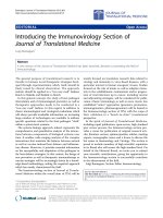
Figure 1 SB623 cells proliferate more slowly and express more p16Ink4A than parental MSCs. A) Cell proliferation assessed by cell counts
(with trypan blue exclusion; left plot) and the percentage of G0/G1 cells measured by propidium iodide staining (middle plot) reveal a significant
reduction in proliferation for SB623 cells compared to parental MSCs (*p < 0.05; n = 6); Far right shows representative FACS data for determining
cell cycle profile. B) Cell division kinetics assessed by CFSE dilution and p16Ink4A protein by intracellular flow cytometry show a significant
increase in p16Ink4A in SB623 cells versus parental MSCs (*p < 0.05); Representative FACS data on left and the mean expression for 4 different
matched lots of MSC and SB623 cells on right.
Dao et al. Journal of Neuroinflammation 2011, 8:133
/>Page 5 of 14
CD4+ cells had proliferated. In the presence of SB623
cells, proliferation was significantly reduced (p < 0.05),
comparable to the parental MSCs.
HLA-DR expression is known to be induced on acti-
vated T ce lls and on antigen presenting cells [12]. We
assessed the percentage of HLA-DR-expressing cells
within each MLR well as an additional measurement of
cell activation. We demonstrated a s ignificant reduction
in HLA-DR-expressing cells when either SB623 cells or
MSCs were included in the MLR (Figure 3C). These
results from allogeneic MLR suggest that SB623 cells
retain immunosuppressive activity comparable to their
parental MSCs.
Transplantation of SB623 cells into rodents following
experimental stroke has been performed via direct injec-
tion into recipient brain along with immunosuppressive
drug administration [3-5]. To determine if SB623 cells
and the parental MSCs can suppress T cell prolife ration
in a xenogeneic MLR, we isolated glial mix cells (astro-
cytes+microglia) to be used as stimulators for human
CFSE-labeled T cells. By flow cytometry analysis gating
on CD3, a mar ker present on all T cell subsets, we
demonstrate that the addition of the parental MSCs as
well as SB623 cells reduced the proliferation of human
T cells in the xenogeneic MLR (Figure 4).
SB623 cells support the generation of peripheral blood
Treg-like cells comparable to parental MSCs
One of the mechanisms by which MSCs suppress
immune activity is t hrough the support for regulatory
T (Treg) cell development [13-15]. IL-2, TGFb1and
Notch ligands have all been shown to enhance regula-
tory T cell (Treg) differentiation [16-21]. To assess
the potential of SB623 cells in supporting regulatory
T cells in culture, we performed a 1:10 peripheral
bloodenrichedTcells-to-MSCsorSB623cellco-cul-
ture in the prese nce and absence of IL-2 over a 7 day
period. CD25 expre ssion on non-activated CD4+ T
cells is commonly used as one of the ide ntity markers
for Tregs [22,23]. We therefore assessed the percen-
tage of CD4+CD25+ cells within each culture and
found significantly more CD4+CD25+ cells in co-cul-
tures with SB623 cells than with MSCs for one of
two blood donors tested (p < 0.05, Figure 5A).
Another marker commonly used to identify Tregs is
the transcription factor, FoxP3. By intracellular
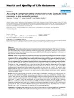
Figure 2 Surface marker and cytokine expression profile of SB623 cells compared to parental MSCs. A)Plotsshowingsurfacemarker
expression on SB623 cells and MSCs. Both SB623 cells and MSCs have >95% expression of CD44, CD73, and CD105, however, there is an
increase in fluorescence intensity (measured by FACS) of these markers in SB623 cells compared to parental MSCs (*p < 0.05, n = 3, left plot);
there is consistently higher expression of CD54 in SB623 cells versus matched lots of parental MSCs (right plot); B) Plots showing mean
fluorescence intensity of IL-6, GM-CSF, IL-1A, TGFb1, and VEGF-A (measured by intracellular antibody staining and flow cytometry) for 3 different
matched lots of MSC and SB623 cells.
Dao et al. Journal of Neuroinflammation 2011, 8:133
/>Page 6 of 14
staining with a fluorochrome conjugated antibody
against FoxP3 (clone PCH101) and analysis by flow
cytometry, we demonstrate that the presence of
MSCs and SB623 cells increased the detection of
FoxP3-expressing T cells in the p resence (>8% F oxP3
+) of IL-2 (Figure 5B). And lastly, Tregs have been
reported to constitutively produce IL-10. Therefore,
by intracellular staining with fluorochrome conjugated
antibody against IL-10, we assessed the percentage of
T cells producing IL-10 in IL-2 treated T cell co-cul-
turedwitheitherMSCsorSB623cells.Theresults
demonstrate that MSCs and SB623 cells both
enhanced the detection of IL-10 expressing T cells
cultured in the presence of IL-2, compared to culture
with only IL-2 (Figure 5C).
SB623 cells alter the activated immune secretome profile
similar to MSCs
Independent studies suggest that MSCs skew the acti-
vated cytokine profile of immune cells, from a pro- to
anti-inflammatory state [24,25]. Receptor ligands such as
Notch ligand and TGFb1 have immunomodulatory
activity and can be presented by various environmental
cells, including MSCs. As SB623 cells express the Notch
ligand Jagged-1 (Figure 6A) and TGFb1(Figure2),we
performed additional cellular immune assays of human
T cells co-cultured with either SB623 cells or MSCs fol-
lowed by sub-optimal doses of PMA/Ionomycin i n the
presence of BrefeldinA on Day 7 to monitor the acti-
vated immune secretome pr ofile by intracellular anti-
body staining for specific cytokines.
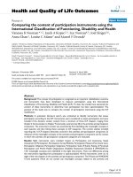
Figure 3 MSC and SB623 attenuate T c ell activat ion and proliferation in human allogeneic mixed lymphocyte reaction. CFSE-labeled
human enriched T cells plus allogeneic PBMCs were cultured with or without MSCs or SB623 cells. A) CD69 on day 2, B) non-dividing T cells
(M1 gating on CFSE-high cells), and C) The percentage of cells expressing HLA-DR were assessed on day 5 by flow cytometry. Representative
FACS data and the mean expression for 3 different matched lots of MSC and SB623 cells are shown. *p < 0.05 versus T cells alone; #p < 0.05
versus MLR.
Dao et al. Journal of Neuroinflammation 2011, 8:133
/>Page 7 of 14
For Th17 cells, enriched T cells were co-cultured with
either SB623 cells or MSCs in the presence or absence
of exogenous IL-23 for 7 days. Intracellular detection of
IL-17A expression by flow cytometry demonstrated a
small percentage of IL-17A expressing T cells, averaging
to less than 1.5% (Figure 6C). For Th1 c ells, enriched T
cells were co-cultured with either SB623 cells or MSCs
in the absence of exogenous cytokines. As Th1 can
secrete bot h IFN-g and IL-10, we performed dual stain-
ing for these two cytokines to determine if the inclusion
of SB623 cells skewed the activated immune secretome.
We demonstrate that the inclusion of either MSCs or
SB623 cells resulted in robust skewing of the activated
immune secretome profile with more than 20% of the
cells expressing IL-10 with less than 0.5% of the cells
expressing IFN- g in cultures that included either MSCs
or SB623 cells.
SB623 cells impede monocyte-to-dendritic cell
differentiation in a manner comparable to parental MSCs
Another immunomodulatory property of MSCs lies in
their ability to block monocyte differentiaton along the
dendriticlineage[26-30].Bycytokinearray,wepre-
viously identified IL-6 and VEGF as being secreted by
SB623 cells [2]. Both o f these cytokines have been
shown to regulate dendritic cell differentiation and
maturation [31-34]. To determine if SB623 cells can
prevent monocytic differentiation to the dendritic line-
age, we performed a 1:10 co-culture of SB623 cells with
peripheral blood monocytes i n the presence of IL-4 and
GM-CSF. In parallel, we established co-cultures with the
parental MSCs. After 7 day s, phase contrast microscopy
pictures were taken and each culture was stained with
fluorochrome conjugated antibodies against CD14, a
surface marker of monocytes, and CD1a, a surface mar-
ker of dendritic cells. As shown in Figure 7A, in the
absence of SB623 cells or MSCs, dendritic cell clusters
were readily visi ble. In contrast, such clusters were
rarely seen in c o-cultures with SB623 cells or MSCs. By
flow cytometry, we noted that in the presence of IL-4
and GM-CSF, there was a conversion of CD14+CD1a+
dendritic cell precursors to predominantly CD14-CD1a+
dendritic cells. In contrast, when SB623 cells or parental
MSCs were included in the monocyte-dendritic cell dif-
ferentiation cultures, the transition was significantly
reduced (Figure 7B). These results demonstrate that
SB623 cells retain the ability to suppress monocytic-den-
dritic cell differentiation.
SB623 cells impede dendritic cell maturation better than
parental MSCs
Studies show that IL-6 can block dendritic cell matura-
tion in vivo [33] while VEGF inhibit s maturation in
response to lypopolysaccharides (LPS) in vitro [34]. As
shown in Figure 2, SB623 cells secrete both IL-6 and
VEGF. To assess the ability of SB623 cells to dampen
dendritic cell maturation, peripheral blood monocy tes
cultured for 7 days with GM-CSF and IL-4 were stimu-
lated with TNF-a for an additional 48 hrs to promote
maturation. SB623 cells or MSCs were added during
this 48 hr stimulation. Two additional conditions -
TNF-a alone or TN F-a + Cyclosporine A - were used
as internal controls. At each endpoint, the expression
levels of the T cell co-stimulatory molecule CD86 were
assessed by flow cytometry (Figure 7C). Consistent with
a previously p ublished report [11], Cyclosporine A
inhibited the induction of co-stimulatory molecule
CD86, compared to conditions with TNF-a alone. Both
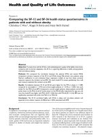
Figure 4 MSCs and SB623 cells attenuate human T cell proliferation in xenogeneic mixed lymphocyte reaction. PKH26 -labeled human
CD3+ T cells plus rat mixed glial cells were co-cultured with or without MSCs or SB623 cells. PKH26 flow cytometry analysis gating on human
CD3+ T cells was performed after 5 day co-culture with M1 gating on non-dividing T cells. Representative FACS data shown on top and the
mean expression for 3 different matched lots of MSC and SB623 cells on bottom.
Dao et al. Journal of Neuroinflammation 2011, 8:133
/>Page 8 of 14
SB623 cells and MSCs attenuated CD86 expression
levels. Notably, SB623 cells were significantly more
effective than MSCs (p < 0.05).
Discussion
Ex vivo manipulation of MSCs has been shown to
induce their cellular sene scence [6,7] and senescent cells
have been described t o have a pro-inflammatory secre-
tome [8-10]. B ecause SB623 cells are derived from ex
vivo manipulated MSCs, we investigated the possible
senescent onset in mul tiple lots of SB623 cells and
compared the immunomodulato ry activity of SB623
cells to that of the parental MSCs.
Morphologically, SB623 cells resemble their parental
MSCs. Phenotypically, SB623 cells expressed all the
standard MSC surface markers (CD90, CD105, CD29,
CD44, CD73), although th ere was an increased density
of CD44 and CD73. A small number of SB623 cells
expressed surface CD54, an inter-cellular adhesion
molecule serving as the ligand for LFA-1, the lympho-
cyte function-associated antigen. SB623 cells displayed a
similar cytokine expression profile as parental MSCs.
The effect of Notch in MSCs has been under much
Figure 5 Detection of CD25, FoxP3, and constitutive IL-10 in T cells co-cultured with MSCs or SB623 cells in the presence of
exogenous IL-2. Human T cells were co-cultured with MSCs or SB623 cells plus hIL-2 for 7 days. A) CD4 and CD25 surface expression with
representative FACS data (left) and the mean expression for 5 different matched lots of MSC and SB623 cells (right); For “Donor 1 PBL”, there is a
significant increase in CD4+CD25+ cells when T cells were co-cultured with SB623 cells versus parental MSCs (*p < 0.05). B) The percentage of
FoxP3-positive T cells as measured by intracellular staining with PE-conjugated antibody against FoxP3 followed by flow cytometry acquisition
and analysis. The bar graphs represent the mean percentage of FoxP3-expressing T cells after co-culture without or with 3 different matched lots
of MSC and SB623 cells. C) To assess the basal constitutive expression of IL-10, BrefeldinA (1:1000) was added during the last 6 hours of culture
to inhibit the secretory pathway. By intracellular staining with a fluorochrome conjugated antibody against IL-10 and analyzed by flow
cytometry. Shown here is a representative flow cytometry data data (left) and the mean expression for 3 different matched lots of MSC and
SB623 cells (right) looking at the percentage of cells staining positive for intracellular IL-10 protein.
Dao et al. Journal of Neuroinflammation 2011, 8:133
/>Page 9 of 14
investigation. One study has implicated a function of
Notch in promoting cellular senescence of rodent cells
[35]. In our system, w e transiently expresse d NICD in
human MSCs by DNA plasmid transfection. Analyzing
for two senescent markers - beta-galactosi dase positivit y
and p16Ink4A expression, we noted a small number of
senescent-like cells within each lot of SB623 cells. From
cell cycle profile and kinetics, we observed reduced pro-
liferation in SB623 cultures compared to the parental
MSCs. This reduced proliferation is most likely not
mediated by exogenous NICD transient expression as
we observed similar reduction in growth for MSCs
transfected with an empty expression vector (data not
shown). Therefore, we suspect that the small number of
senescent-like cells within each lot of SB623 cells is a
reflection of the extended time in culture (~2 months).
As noted above, some studies have highlighted the
pro-inflammatory secretome of senescent cells [8-10].
To date, we did not observe immunological side effects
from SB623 cell implantation in rats. Nevertheless, as
we noted a small population of senescent-like cells
within each lot of SB623 cells, we initiated various cellu-
lar i mmune assays to compare their immunomodulatory
activity to parental MSCs in more detail. In an allo-
geneic mixed lymphocyte reaction (MLR), we d emon-
strated that similar to MSCs, SB623 cells attenuated the
activation of CD4+ T cells as evident by reduction in
CD69 (an early T cell ac tivation marker) and H LA-DR
(an activation marker on both T cells and monocytes).
In experimental rodent stroke, intracerebral implanta-
tion of SB623 cells elicits functional recovery [5]. As the
glial cells are among the common antigen presenting
Figure 6 MSCs and SB623 cells skew the “activated” T cell secretome profile. Human T cells were co-cultured with MSCs or SB623 cells for
7 days in the absence of exogenous cytokines. To measure the activated T cell secretome profile, the cultures were stimulated with suboptimal
doses of PMA/Ionomycin and Brefeldin A for an additional 6 hr prior to intracellular flow cytometry analysis of cytokines. A) Representative FACS
analysis of Jagged-1 surface expression on MSC and SB623 cells prior to co-culture. B) Intracellular detection of IFN-g and IL-10 of T cells after co-
culture with or without MSCs or SB623 cells; Representative FACS data (left) and mean expression for 3 different matched lots of MSC and SB623
cells (right). C) Intracellular detection of IL-7A of T cells after co-culture with MSCs or SB623 cells in the presence or absence of IL-23. Negative
controls include T cells cultured in RPMI/10%FCS alone and T cells cultured in the presence of IL-23 alone.
Dao et al. Journal of Neuroinflammation 2011, 8:133
/>Page 10 of 14
cells in the nervous system, we assessed the efficiency of
SB623 cells in suppressi ng the prolifer ation of human T
cells stimulated by rat glial mix cells. We demonstrate
that SB623 cells elicited immunosuppressive activity i n
this xenogeneic MLR assay, comparable to parental
MSCs.
In the context of immune modulation, MSCs have
been reported to impact both the innate and acquired
immune cells [25]. In a standard mono-dendritic cell
differentiation assay with GM-CSF and IL-4, we demon-
strate that the inclusion of SB623 cells in the mono-
dendritic cell differentiation culture reduced the produc-
tion of dendritic cells (CD1a+CD14-) to similar extent
as the parental MSCs. In a 2-day TNF- a mediated den-
dritic cell maturation assay, the inclusion of SB623 cells
reduced the density of CD86 co-stimulatory molecules,
as measu red by mean fluorescent intensity. Interestingly,
the reduction in CD86 surface expression was signifi-
cantly higher in the presence of SB623 cells than the
parental MSCs. A recent study reported that activated
MSCs secrete soluble TNF-a receptors which, in turn,
attenuate systemic inflammation [36]. As TNF- a is
commonly used to induce dendritic cell m aturation, we
hypothesize that a differ ential expression and/or secre-
tion of TNF- a receptors between SB623 cells and
MSCs could explain our current observations.
Additional studies are warranted to address this possible
underlying mechanism of action.
To assess the impact of SB623 cells on the acquired
immune cells, we performed co-cultures of human
enric hed T cells with SB623 cells or MSCs and assessed
the activated T cell secretome profile following stimula-
tion with sub-optimal dosage of PMA/Ionomycin. By
intracellular staining with antibodies against IL-10 and
IFN-g, we observed a robust skewing in the activated
immune secretome profile with more than 95% of cells
expressing IL-10 and less than 5% expressing IFN-g.
The detection of predominantly IL-10 expressing cells is
in line with previous reports that MSCs pro mote the
anti-inflammat ory secretome of T ce lls. The lack of
IFN-g production was unexpected since Th1 cells are
known to produce both IFN-g and IL-10, not just IL-10
alone. However, IL-10 has been shown to downregulate
IFN-g production [37-39]. It is therefore possible that in
thepresenceofSB623cellsorMSCs,thelevelofIL-10
produced was high enough to form a negative feedback
loop on the production of IFN-g.Afewstudieshave
highlighted the production of IL-10 from Th1 T cells as
a “self-con trol” mechanism [40,41] and Notch activation
has been associated with this process [42]. TGFb1and
IL-6, both of which are known to be produced by
MSCs, have a role in Th17- and Th1- T cell
Figure 7 MSCs and SB623 cells interfere with monocyte-DC differentiation and maturation. Human monocytes were differentiated along
the dendritic lineage with GM-CSF+IL-4, with or without MSCs or SB623 cells. A) Phase contrast images illustrating monocytic cell clustering; B)
CD1A and CD14 staining on day 7. Representative FACS data (above) and mean expression for 3 different matched lots of MSC and SB623 cells
(below); the percentage of cells co-expressing CD14 and CD1A after GM-CSF+IL-4 culture is significantly higher when monocytes were cultured
with either MSCs or SB623 cells versus alone (#p < 0.05); the percentage of CD14-CD1A+ cells is significantly lower when differentiation culture
included either MSCs or SB623 cells versus alone (*p < 0.05); C) CD86 surface expression after 2-day TNF-a treatment; Representative FACS data
(left) and mean fluorescence intensity for 3 different matched lots of MSC and SB623 cells (right); there is a significant decrease in the mean
fluorescent intensity of CD86 co-stimulatory molecule when monocytes were co-cultured with SB623 cells versus parental MSCs (*p < 0.05).
Dao et al. Journal of Neuroinflammation 2011, 8:133
/>Page 11 of 14
development. To determine if SB623 cells impact the
number of Th17-T cells, we performed co-cultures of T
cells with SB623 cells or MSCs in the presence or
absence of IL-23, a cytokine supportive of human Th17-
T cell development. By intrace llular staining with an
antibody against IL-17A, we detected an average of less
than 1.5% Th17-T cells, with no significant differences
between co-cultures with SB623 cells or the parental
MSCs. While it is surprising to see a positive impact of
marrow stromal cells on Th17 cell number in culture,
one study to date has highlighted this property of fetal
marrow stromal cells [43]. I t is also important to note
that the percentage of IL-17-producing T cells is rela-
tively low compared to the percentage of IL-10 produ-
cing T cells and thus, may explain why transplantation
of MSC elicits an overall immunosuppressive outcome
[24,25].
Another immunomdulatory property of TGFb1and
Notch ligands is in the regulation of regulatory T cells
(Tregs) [13,16,21]. As both TGFb and Notch ligand are
expressed by SB623 cells and the parental MSCs, we
next investigated the impact of SB623 cells on T cells
cultured in the presence of IL-2, a cytokine important in
Treg cell development. In th e presence of SB623 cells,
we observed a higher percentage of cells expressing sur-
face CD25, the IL-2 receptor alpha chain. Although
CD25 is commonly used to identify Tregs [22,23], this
surface marker is also present on activated T cells [44].
An alternate common marker for Tregs is the forkhead
family transcription factor FoxP3[45]. By intracellular
staining with an antibody against FoxP3, we demon-
strated a higher percentage of FoxP3+ cells when SB6 23
cells or MSCs were included. As FoxP3 is not exclu-
sively expressed in Tregs [46], we measured the intracel-
lular expression of IL-10, a cytokine constitutively
produced at low levels by Tregs [47,48]. We consistently
detect ed a small but higher percentage of CD4+ T cells
expressing IL-10 in c ultures with SB623 cells or MSCs
compared to the controls. These results suggest that
SB623 cells, like the parental MSCs, may have a role in
supporting T cells having various features of regulatory
T cells.
SB623 cell s are derived from NICD-transfected MSCs
and expanded in the absence of exogenous cytokines. As
such, SB623 cells retained the standard phenotypes and
morphology of conventional MSCs. Compared to the
early passage MSCs, SB623 cells contained a small num-
ber of sene scent-like cells as a result of cel l expansion
in vitro. Nevertheless, we show here that SB623 cells
effectively suppressed T cell proliferation in MLR and
modulated the T cell secretome profile as efficiently as
the parental MSCs. The current study demonstrate that
the transient overexpression of exogenous NICD wi th
subsequent expansion of human MSCs preserved, and
in some cases increased, their immunosuppressive
activity.
Disclosure
All authors are employees of San-Bio Inc.
Additional material
Additional file 1: Production and characterization of SB623 cells.A)
Representative illustration of SB623 cell production. Marrow stromal cells
were established at passage 2, followed by plasmid transfection and
drug selection, and ending with cell expansion for two additional
passages to generate the end product, SB623 cells. B) Beta-galactosidase
staining of MSCs and SB623 cells culture. MSCs of passage 2 and SB623
cells plated for 48 hr in growth medium were stained with a commercial
beta-galactosidase staining kit (Cell Signaling) according to
manufacturer’s protocol. C) Assessment of exogenous NICD DNA and
endogenous Hes1 (downstream target of Notch) transcript in NICD-
transfected MSC. D) Flow cytometric analysis for the percentage of NICD
expressing cells during SB623 cell production. At different passages after
transfection and selection, cells were harvested using 0.25% Trypsin, fixed
with paraformaldehyde, and permeabilized with 0.1% Triton-X100.
Samples were stained either with a fluorochrome-conjugated IgG or a
fluorochrome-conjugated antibody against NICD protein. Cell acquisition
and analyses were done on the BD FACS Calibur using CellQuestPro
software.
Acknowledgements
We are grateful to Dr. Naomi Taylor (Institute Genetique Medicine
Moleculaire/INSERM) for sharing protocols and providing valuable scientific
advice. We thank Rouzbeh Shooshtarian for producing MSCs and SB623 cells
at San-Bio Inc. cell manufacturing facility.
Author details
1
Research Department San-Bio Incorporated 231 South Whisman Road,
Mountain View, 94041, USA.
2
Production Development Department San-Bio
Incorporated 231 South Whisman Road, Mountain View, 94041, USA.
Authors’ contributions
MD conceived and designed the study, performed immunoassays and
flow cytometry, analyzed and interpreted data, and wrote the manuscript .
CCT partici pated in data analysis and interpretation, performed statistical
analysis, and edited the manuscript. IA prepared DNA and RNA samples,
performed PCR, analyzed and interpreted PCR data. MM d esigned and
coo rdinated the production of MSCs and SB623 cells. CCC directed the
study, designed PCR prime rs for the detection of exogenous NICD, and
hel ped with data interpr etation. All authors read and approved the final
manuscript.
Received: 15 March 2011 Accepted: 7 October 2011
Published: 7 October 2011
References
1. Aizman I, Tate CC, McGrogan M, Case CC: Extracellular matrix produced
by bone marrow stromal cells and by their derivative, SB623 cells,
supports neural cell growth. J Neurosci Res 2009, 87:3198-3206.
2. Tate CC, Fonck C, McGrogan M, Case CC: Human mesenchymal stromal
cells and their derivative, SB623 cells, rescue neural cells via trophic
support following in vitro ischemia. Cell Transplant 2010, 19:973-984.
3. Glavaski-Joksimovic A, Virag T, Mangatu TA, McGrogan M, Wang XS,
Bohn MC: Glial cell line-derived neurotrophic factor-secreting genetically
modified human bone marrow-derived mesenchymal stem cells
promote recovery in a rat model of Parkinson’s disease. J Neurosci Res
2010, 88:2669-2681.
4. Glavaski-Joksimovic A, Virag T, Chang QA, West NC, Mangatu TA,
McGrogan MP, Dugich-Djordjevic M, Bohn MC: Reversal of dopaminergic
Dao et al. Journal of Neuroinflammation 2011, 8:133
/>Page 12 of 14
degeneration in a parkinsonian rat following micrografting of human
bone marrow-derived neural progenitors. Cell Transplant 2009, 18:801-814.
5. Yasuhara T, Matsukawa N, Hara K, Maki M, Ali MM, Yu SJ, Bae E, Yu G, Xu L,
McGrogan M, Bankiewicz K, Case C, Borlongan CV: Notch-induced rat and
human bone marrow stromal cell grafts reduce ischemic cell loss and
ameliorate behavioral deficits in chronic stroke animals. Stem Cells Dev
2009, 18:1501-1514.
6. Wagner W, Horn P, Castoldi M, Diehlmann A, Bork S, Saffrich R, Benes V,
Blake J, Pfister S: Eckstein V, Ho AD. Replicative senescence of
mesenchymal stem cells: a continuous and organized process. PLoS One
2008, 3(5):e2213.
7. Jin Y, Kato T, Furu M, Nasu A, Kajita Y, Mitsui H, Ueda M, Aoyama T,
Nakayama T, Nakamura T, Toguchida J: Mesenchymal stem cells cultured
under hypoxia escape from senescence via down-regulation of p16 and
extracellular signal regulated kinase. Biochem Biophys Res Commun 2010,
391(3):1471-6.
8. Rodier F, Coppé JP, Patil CK, Hoeijmakers WA, Munoz DP, Raza SR,
Freund A, Campeau E, Davalos AR, Campisi J: Persistent DNA damage
signalling triggers senescence-associated inflammatory cytokine
secretion. Nat Cell Biol 2009, 11:973-979.
9. Coppé JP, Patil CK, Rodier F, Sun Y, Muñoz DP, Goldstein J, Nelson PS,
Desprez PY, Campisi J: Senescence-associated secretory phenotypes
reveal cell-nonautonomous functions of oncogenic RAS and the p53
tumor suppressor. PLoS Biol 2008, 6(12):2853-68.
10. Freund A, Orjalo AV, Desprez PY, Campisi J: Inflammatory networks during
cellular senescence: causes and consequences. Trends Mol Med 2010,
16(5):238-46, Review.
11. Duperrier K, Farre A, Bienvenu J, Bleyzac N, Bernaud J, Gebuhrer L, Rigal D,
Eljaafari A: Cyclosporin A inhibits dendritic cell maturation promoted by
TNF-alpha or LPS but not by double-stranded RNA or CD40L. J Leukoc
Biol 2002, 72:953-961.
12. Robbins PA, Maino VC, Warner NL, Brodsky FM: Activated T cells and
monocytes have characteristic patterns of class II antigen expression. J
Immunol 1988, 141:1281-1287.
13. English K, Ryan JM, Tobin L, Murphy MJ, Barry FP, Mahon BP: Cell contact,
prostaglandin E(2) and transforming growth factor beta 1 play non-
redundant roles in human mesenchymal stem cell induction of CD4
+CD25(High) forkhead box P3+ regulatory T cells. Clin Exp Immunol 2009,
156:149-160.
14. Poggi A, Zocchi MR: Role of bone marrow stromal cells in the generation
of human CD8+ regulatory T cells. Hum Immunol 2008, 69:755-759.
15. Wang Y, Zhang A, Ye Z, Xie H, Zheng S: Bone marrow-derived
mesenchymal stem cells inhibit acute rejection of rat liver allografts in
association with regulatory T-cell expansion. Transplant Proc 2009,
41:4352-4356.
16. Ou-Yang HF, Zhang HW, Wu CG, Zhang J, Li JC, Hou LH, He F, Ti XY,
Song LQ, Zhang SZ, Feng L, Qi HW, Han H: Notch signaling regulates the
FOXP3 promoter through RBP-J- and Hes1-dependent mechanisms. Mol
Cell Biochem 2009,
320:109-14.
17.
Asano N, Watanabe T, Kitani A, Fuss IJ, Strober W: Notch1 signaling and
regulatory T cell function. J Immunol 2008, 180:2796-2804.
18. Ostroukhova M, Qi Z, Oriss TB, Dixon-McCarthy B, Ray P, Ray A: Treg-
mediated immunosuppression involves activation of the Notch-HES1
axis by membrane-bound TGF-beta. J Clin Invest 2006, 116:996-1004.
19. Wang G, Khattar M, Guo Z, Miyahara Y, Linkes SP, Sun Z, He X,
Stepkowski SM, Chen W: IL-2-deprivation and TGF-beta are two non-
redundant suppressor mechanisms of CD4+CD25+ regulatory T cell
which jointly restrain CD4+CD25- cell activation. Immunol Lett 2010,
132:61-68.
20. Kosiewicz MM, Alard P, Liang S, Clark SL: Mechanisms of tolerance
induced by transforming growth factor-beta-treated antigen-presenting
cells: CD8 regulatory T cells inhibit the effector phase of the immune
response in primed mice through a mechanism involving Fas ligand. Int
Immunol 2004, 16:697-706.
21. Nakamura K, Kitani A, Fuss I, Pedersen A, Harada N, Nawata H, Strober W:
TGF-beta 1 plays an important role in the mechanism of CD4+CD25+
regulatory T cell activity in both humans and mice. J Immunol 2004,
172:834-842.
22. Baecher-Allan C, Brown JA, Freeman GJ, Hafler DA: CD4+CD25high
regulatory cells in human peripheral blood. J Immunol 2001,
167(3):1245-53.
23. Dieckmann D, Plottner H, Berchtold S, Berger T, Schuler G: Ex vivo isolation
and characterization of CD4(+)CD25(+) T cells with regulatory properties
from human blood. J Exp Med 2001, 193(11):1303-10.
24. Kong QF, Sun B, Bai SS, Zhai DX, Wang GY, Liu YM, Zhang SJ, Li R, Zhao W,
Sun YY, Li N, Wang Q, Peng HS, Jin LH, Li HL: Administration of bone
marrow stromal cells ameliorates experimental autoimmune myasthenia
gravis by altering the balance of Th1/Th2/Th17/Treg cell subsets
through the secretion of TGF-beta. J Neuroimmunol 2009, 207:83-91.
25. Tolar J, Le Blanc K, Keating A, Blazar BR: Concise review: hitting the right
spot with mesenchymal stromal cells. Stem Cells 2010, 28:1446-1455.
26. Lai HY, Yang MJ, Wen KC, Chao KC, Shih CC, Lee OK: Mesenchymal stem
cells negatively regulate dendritic lineage commitment of umbilical-
cord-blood-derived hematopoietic stem cells: an unappreciated
mechanism as immunomodulators. Tissue Eng Part A 2010, 16:2987-2997.
27. Spaggiari GM, Abdelrazik H, Becchetti F, Moretta L: MSCs inhibit
monocyte-derived DC maturation and function by selectively interfering
with the generation of immature DCs: central role of MSC-derived
prostaglandin E2. Blood 2009, 113:6576-6583.
28. Ramasamy R, Fazekasova H, Lam EW, Soeiro I, Lombardi G, Dazzi F:
Mesenchymal stem cells inhibit dendritic cell differentiation and
function by preventing entry into the cell cycle. Transplantation 2007,
83:71-76.
29. Jiang XX, Zhang Y, Liu B, Zhang SX, Wu Y, Yu XD, Mao N: Human
mesenchymal stem cells inhibit differentiation and function of
monocyte-derived dendritic cells. Blood 2005, 105:4120-4126.
30. Zhang W, Ge W, Li C, You S, Liao L, Han Q, Deng W, Zhao RC:
Effects of
mesenchymal
stem cells on differentiation, maturation, and function of
human monocyte-derived dendritic cells. Stem Cells Dev 2004, 13:263-271.
31. Chomarat P, Banchereau J, Davoust J, Palucka AK: IL-6 switches
thedifferentiation of monocytes from dendritic cells to macrophages.
Nat Immunol 2000, 1:510-514.
32. Djouad F, Charbonnier LM, Bouffi C, Louis-Plence P, Bony C, Apparailly F,
Cantos C, Jorgensen C, Noel D: Mesenchymal stem cells inhibit the
differentiation of dendritic cells through an interleukin-6-dependent
mechanism. Stem Cells 2007, 25:2025-32.
33. Park SJ, Nakagawa T, Kitamura H, Atsumi T, Kamimura D, Park SJ,
Murakami M, Kitamura Y, Iwakura Y, Hirano T: IL-6 regulates in vivo
dendritic cell differentiation through STAT3 activation. J Immunol 2004,
173:3844-3854.
34. Takahashi A, Kono K, Ichihara F, Sugai H, Fujii H, Matsumoto Y: Vascular
endothelial growth factor inhibits maturation of dendritic cells induced
by lipopolysaccharide, but not by proinflammatory cytokines. Cancer
Immunol Immunother 2004, 53:543-550.
35. Zhang K, Huang L, Sun H, Zhu Y, Xiao Y, Huang M, Liu W: Role of Notch
expression in premature senescence of murine bone marrow stromal
cells. Progress in Natural Science 2009, 19:557-562.
36. Yagi H, Soto-Gutierrez A, Navarro-Alvarez N, Nahmias Y, Goldwasser Y,
Kitagawa Y, Tilles AW, Tompkins RG, Parekkadan B, Yarmush ML: Reactive
bone marrow stromal cells attenuate systemic inflammation via sTNFR1.
Mol Ther 2010, 18(10):1857-64.
37. Shibata Y, Foster LA, Kurimoto M, Okamura H, Nakamura RM, Kawajiri K,
Justice JP, Van Scott MR, Myrvik QN, Metzger WJ: Immunoregulatory roles
of IL-10 in innate immunity: IL-10 inhibits macrophage production of
IFN-gamma-inducing factors but enhances NK cell production of IFN-
gamma. J Immunol 1998, 161:4283-4288.
38. Murray PJ: The primary mechanism of the IL-10-regulated anti-
inflammatory response is to selectively inhibit transcription. Proc Natl
Acad Sci USA 2005, 102:8686-8691.
39. Yamana J, Yamamura M, Okamoto A, Aita T, Iwahashi M, Sunahori K,
Makino H: Resistance to IL-10 inhibition of interferon gamma production
and expression of suppressor of cytokine signaling 1 in CD4+ T cells
from patients with rheumatoid arthritis. Arthritis Res Ther 2004, 6:
R567-R577.
40. Trinchieri G: Interleukin-10 production by effector T cells: Th1 cells show
self control. J Exp Med 2007, 204:239-243.
41. O’Garra A, Vieira P: T(H)1 cells control themselves by producing
interleukin-10. Nat Rev Immunol 2007, 7:425-428.
42. Rutz S, Janke M, Kassner N, Hohnstein T, Krueger M, Scheffold A: Notch
regulates IL-10 production by T helper 1 cells. Proc Natl Acad Sci USA
2008, 105:3497-3502.
Dao et al. Journal of Neuroinflammation 2011, 8:133
/>Page 13 of 14
43. Guo Z, Zheng C, Chen Z, Gu D, Du W, Ge J, Han Z, Yang R: Fetal BM-
derived mesenchymal stem cells promote the expansion of human Th17
cells, but inhibit the production of Th1 cells. Eur J Immunol 2009,
39(10):2840-9.
44. Schuh K, Twardzik T, Kneitz B, Heyer J, Schimpl A, Serfling E: The
interleukin 2 receptor alpha chain/CD25 promoter is a target for nuclear
factor of activated T cells. J Exp Med 1998, 188(7):1369-73.
45. Hori S, Nomura T, Sakaguchi S: Control of regulatory T cell development
by the transcription factor Foxp3. Science 2003, 299(5609):1057-61.
46. Manrique SZ, Correa MA, Hoelzinger DB, Dominguez AL, Mirza N, Lin HH,
Stein-Streilein J, Gordon S, Lustgarten J: Foxp3-positive macrophages
display immunosuppressive properties and promote tumor growth. J
Exp Med 2011, 208(7):1485-99.
47. Annacker O, Pimenta-Araujo R, Burlen-Defranoux O, Barbosa TC, Cumano A,
Bandeira A: CD25+ CD4+ T cells regulate the expansion of peripheral
CD4 T cells through the production of IL-10. J Immunol 2001,
166:3008-2018.
48. Vieira PL, Christensen JR, Minaee S, O’Neill EJ, Barrat FJ, Boonstra A,
Barthlott T, Stockinger B, Wraith DC, O’Garra A: IL-10-secreting regulatory T
cells do not express Foxp3 but have comparable regulatory function to
naturally occurring CD4+CD25+ regulatory T cells. J Immunol 2004,
172:5986-5993.
doi:10.1186/1742-2094-8-133
Cite this article as: Dao et al.: Comparing the immunosuppressive
potency of naïve marrow stromal cells and Notch-transfected marrow
stromal cells. Journal of Neuroinflammation 2011 8:133.
Submit your next manuscript to BioMed Central
and take full advantage of:
• Convenient online submission
• Thorough peer review
• No space constraints or color figure charges
• Immediate publication on acceptance
• Inclusion in PubMed, CAS, Scopus and Google Scholar
• Research which is freely available for redistribution
Submit your manuscript at
www.biomedcentral.com/submit
Dao et al. Journal of Neuroinflammation 2011, 8:133
/>Page 14 of 14