báo cáo hóa học: " Proinflammatory and proapoptotic markers in relation to mono and di-cations in plasma of autistic patients from Saudi Arabia" docx
Bạn đang xem bản rút gọn của tài liệu. Xem và tải ngay bản đầy đủ của tài liệu tại đây (606.2 KB, 9 trang )
RESEARCH Open Access
Proinflammatory and proapoptotic markers in
relation to mono and di-cations in plasma of
autistic patients from Saudi Arabia
Afaf K El-Ansary
1,2,3*
, Abir G Ben Bacha
1,2,3
and Laila Y Al-Ayadhi
2,3,4
Abstract
Objectives: Autism is a developmental disorder characterized by social and emotional deficits, language
impairments and stereotyped behaviors that manifest in early postnatal life. This study aims to clarify the
relationship amongst absolute and relative concentrations of K
+
,Na
+
,Ca
2+
,Mg
2+
and/or proinflammatory and
proapoptotic biomarkers.
Materials and methods: Na
+
,K
+
,Ca
2+
,Mg
2+
,Na
+
/K
+
,Ca
2+
/Mg
2+
together with IL6, TNFa as proinflammatory
cytokines and caspase3 as proapoptotic biom arker were determined in plasma of 25 Saudi autistic male patients
and compared to 16 age and gender matching control samples.
Results: The obtained data recorded that Saudi autistic patients have a remarkable lower plasma caspase3, IL6,
TNFa,Ca
2+
and a significantly higher K
+
compared to age and gender matching controls. On the other hand both
Mg
2+
and Na
+
were non-significantly altered in autistic pat ients. Pearson correlations revealed that plasma
concentrations of the measured cytokines and caspase-3 were positively correlated with Ca
2+
and Ca
2+
/K
+
ratio.
Reciever Operating Characteristics (ROC) analysis proved that the measured parameters recorded satisfac tory levels
of specificity and sensitivity.
Conclusion: Alteration of the selected measure d ions confirms that oxidative stress and defective mitochondrial
energy production could be contributed in the pathogenesis of autism. Moreover, it highlights the relationship
between the measured ions, IL6, TNFa and caspase3 as a set of signalling pathways that might have a role in
generating this increasingly prevalent disorder. The role of ions in the possible proinflammation and proapoptic
mechanisms of autistics’ brains were hypothesized and explained.
Keywords: Ions, Caspase3, IL6, TNFα, Autism
Introduction
Children with Autism Spectrum Disorders (ASD) have
impairments in three core domains: socialization, commu-
nication, and restricted interests and repetitive behaviors
[1-4]. Researchers have reported that psychiatr ic comor-
bidity in ASD ranges from 41% to 70% [5,6].
Although the etiology of the disorder is unknown,
recent studies have suggested that the susceptibility to
autism is clearly attributable to genetic factors [7,8]. In
addition, emerging evidence points t o inflammatory and
apoptoti c mech anisms being responsible for certain neu-
ropsychiatri c disorders including autism. Vargas et al. [9]
suggested neuroinflammatory processes are present in
the autistic brain by showing that transforming growth
factor (TGF)a1, macrophage chemoattractant protein
(MCP) 1, interleukin (IL)6 and IL10 are increased in the
brain of auti stic subjects. A number of studies have also
shown that inf lammatory cytokines including t umor
necrosis factor (TNF)a, interferon (IFN)a,IL1a,IL6,IL8
and IL12 are elevated in blood mononuclear cells, serum,
plasma and cerebrospinal fluid (CSF) of autistic subjects
[9-16].
The mechanisms of apoptosis induction are complex
and not fully known, but some key events are identified
* Correspondence:
1
Biochemistry Department, Science College, King Saud University, P.O box
22452, Zip code 11495, Riyadh, Saudi Arabia
Full list of author information is available at the end of the article
El-Ansary et al. Journal of Neuroinflammation 2011, 8:142
/>JOURNAL OF
NEUROINFLAMMATION
© 2011 El-Ansary et al; licensee BioMe d Central Ltd. This is an Open Access article distributed under the terms of the Creative
Commons Attribution License ( y/2.0), which permits unrestricted use, distribution, and
reproduction in any medium, provided the original work is properly cited.
that appear essenti al for the cell to enter apoptosis. The
role of specific ions in the apoptotic process is slowly
being revealed. Change s in intracellular Ca
2+
have long
been associated with apoptotic neuronal cell de ath. Ca
2+
ionophores have been shown to induce ultrastructural
changes, such as cell shrinkage, chromatin condensation,
and DNA fragmentation, consistent with apoptosis
[17-20]. Increased Ca
2+
has been linked to processes
occurring during apoptosis including caspase activation.
One key event in apoptosis is loss of intracellular potas-
sium ions (K
+
). Depletion of K
+
is necessary for cells to
shrink, activate caspases and degrade DNA [21-23], events
that in turn lead to further characteristic apoptotic c hanges
such as membrane blebbing and formation of apoptotic
bodies. Apoptosis due to forced loss of intracellular K
+
can
be induced by ionophores or K
+
channel activators [24-26].
In addition, Yu et al. [25,27] have also shown that the out-
ward K
+
current that ensues from N-methyl-D-aspartate
receptor activation has also been shown to induce apopto-
tic changes in cultured hippocampal neurons.
Just as with increased Ca
2+
and K
+
efflux, the importance
of sodium (Na
+
) entry in inducing neuronal injury and
death in response to pathophysiologic conditions, such as
hypoxia, has been well estab lished [28-34]. Moreover,
Banasiaketal.[35]provedthatblockingNa
+
entry in
hypoxia-exposed neurons reduced the proportion of DNA
fragmentation and reduced apoptotic cell.
Magnesium (Mg
2+
) has a profound effect on neural
excitability; the most characteristic signs and symptoms of
Mg
2+
deficiency are produced by neural and neuromuscu-
lar hyperexcitability [36]. Iotti and Malucelli [37] clarify
the functional relationship between energy metabolism
and free [Mg
2+
], providing evidence that brain cells cyto-
solic [Mg
2+
] is regulated to equilibrate any changes in
rapidly available free energy. Moreover, it has also been
shown that the measurement of brain Mg
2+
can help in
the differential diagnosis of neurodegenerative diseases
sharing common clinical features.
The immune system has been postulated to play an
important role in the etiology of autism. Investigators have
proposed infectious, autoimmune, and cytokine-related
etiologies.
These information initiate our interest to measure con-
centrations of Na
+
,K
+
,Ca
2+
,Mg
2+
together with caspase3
as a proapoptotic marker, IL6 and TNFa as proinflamma-
tion markers in the plasma of autistic patients from Saudi
Arabia in an attempt to understand the role and relation-
ship of these biochemical parameters in the etiology of
autism and its commonly related psychiatric conditions.
Material and methods
Subjects and methods
The study protocol followed the ethical guidelines of the
most recent Declaration of Helsinki (Edinburgh, 2000). All
subjects enrolled in the study (25 autistic male patients
and 16 age and gender matched controls) had written
informe d consent provided by their parents and assented
to participate if developmentally able. They were enrolled
through the ART Center (Autism Research & Treatment
Center) clinic (Riyadh, Saudi Arabia). The ART Center
clinic sample population consisted of children diagnosed
on the ASD. The diagnosis of ASD was confirmed in all
subjects using the Autism Diagnostic Interview-Revised
(ADI-R) and the Autism Diagnostic Observation Schedule
(ADOS) and 3DI (Developmental, dimensional diagnostic
interview). The ages of all autistic children who partici-
pated were between the ages of 4 and 12 years old. All
were simplex cases. All are negative for fragile × gene
study. The control group recruited from Well baby Clinic
at King Khaled University hospital with mean age 4-11
year old. Subjects were excluded from the investigation if
they had organic aciduria, dysmorphic features, or diagno-
sis of Fragile × or other serious neurological (e.g., sei-
zures), psychiatric (e.g., bipolar disorder) or known
medical conditions. All participants were screened via par-
ental interview for current and past physical illness. Chil-
dren with known endocrine, cardiovascular, pulmonary,
liver, kidney or other medical disease were excluded from
the study. None of the recruited autistic patients were on
special diets or alternative treatments.
Ethics approval and consent
A written consent was obtained from the parents of each
individual case, according to the guideline s of the ethical
committee of King Khalid Hospital, King Saud University.
Blood samples
After overnight fast, 10 ml blood samples were collected
from both groups in test tubes containing sodium
heparin as anticoagulant. Tubes were centrifuged at 3500
rpm at room temperature for 15 minutes, plasma was
obtained and deep freezed (at -80°C) until analysis time.
Measurement of calcium
The UDI (United Diagnostics Industry, Saudi Arabia) Ca
2
+
procedure is based on the reaction of Ocresolphthalein
complexone (O-CPC) wit h Ca
2+
to form a chromogenic
complex that absor bs light which is measured photome-
trically at 575 nm. Mg
2+
interference is prevented by
sequestration with 8-hydroxyquinoline. 2-Ethylami-
noethanol is used to establish the reaction pH at 12.
Dimethyl sulfoxide is used to lower the dielectric con-
stant of the reaction mixture and to repress the ioniza-
tion of cresolphthalein complexone [38].
Measurement of potassium
K
+
reacts with so dium tetra phenyl boron in a protein
free alkaline medium to produce a colloidal suspension
El-Ansary et al. Journal of Neuroinflammation 2011, 8:142
/>Page 2 of 9
[39]. The turbidity which is propor tiona l to the K
+
con-
centration in the range of 2-7 mmol/L was measured
against blank. The concentration was calculated using a
typically treated standard solution of K
+
chloride in
Bovine albumin equivalent to 4 mol/L.
Measurement of sodium
Plasma Na
+
was measured according to the method of
Tietz [40] using a diagnostic kit, a product of UDI in which
Na
+
was determined via Na+ dependent b-galactosidase
activity usin g O-nitrophenyl-b, D-galactopyranoside.
Measurement of magnesium
The UDI method stems from the original work of Lind-
strom and Diehl [41] using calmagite, 1-(1-hydroxy-4-
methyl-2- phenylazo)-2-naphthol-4-sulfonic acid, as the
complexometric reagent. Ca
2+
is masked by sequestration
with strontium ethylene-bis-(oxyethylenenitrilo)-tetra
acetate (EGTA Sr) [42]. A surfactant system has been uti-
lized to overcome protein interference. Mg
2+
form a
colored complex with calmagite in alkaline medium to
produce a red c omplex that absorbs light which is mea-
sured spectrophotometrically at 530 nm. The absorbance
of the red complex is directly proportional to the concen-
tration of Mg
2+
in the sample.
Statistical analysis
A SPSS (Statistical Package for the Social Sciences) com-
puter program was used. Results were expressed as mean
± S.D. and all statistical comparisons were made by
means of independent t-test with P ≤ 0.05 was considered
significant. ROC analysis was performed. Area under the
curve, cutoff values together with degree of specificity
and sensitivity were calculated.
Results
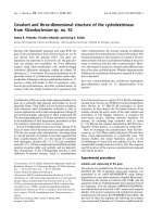
Table1andFigure1demonstrateconcentrationsofthe
measured parameters in plasma of autistic patients com-
pared to control. Concentrations of caspase3, IL6 and
TNFa were significantly lower in children with autism
compared to control. In contrast, K
+
was significantly
raised in plasma samples from children with autism com-
pared to age and gender matching controls recording 2.3
fold higher values. In addition, Ca
2+
,Ca
2+
/Mg
2+
and Na
+
/
K
+
ratio were significantly lower in autistic compared to
control with the latter showing almost 3 fold lower values.
Figure 2 shows the percentage changes of the measured
parameters in autistics relative to control subjects. It could
be easily seen that caspase3, IL6 and TNFa recorded more
or less the average % decrease with values of -27.5,-20.2
and -29.8. Among the measured elements K
+
recorded the
most remarkable percentage increase recording value of
130% higher concentration in autistic compared to control
with concomitant decrease in Na
+
/K
+
ratio of 69.9%
decrease. Ca
2+
/Mg
2+
ratio recorded 63.8% lower values in
control. Absolute values of Na
+
and Mg
2+
recorded the
lowest percentage changes recording 13.1% and 5.9%
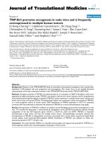
increase, respectively. Table 2 and Figure 3 show the sig-
nificantly positive and negative correlated parameters. Out
of the 27 correlations recorded in table 3, the most signifi-
cantly correlated parameters were selected to be presented
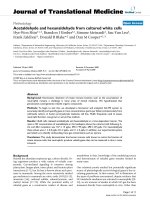
in Figure 3. Table 3 together with Figure 4 show ROC ana-
lysis of the measured parameters. It could be easily noticed
that most of the measured parameters recorde d satisfac-
tory values of sensitivity and specificity with the exception
of Mg
2+
and Na
+
which show low specificity values.
Discussion
Protection of the brain from injury during the fetal, neo-
natal and postnatal periods is of major importance
owing to the significant number of infants who now sur-
vive early brain in jury but develop neurodevelopmental
and motor disabilities.
Table 1 and Figures 1 and 2 show the unexpected
lower concentrations of caspase3, TNF a and IL6. This
could be interpreted on the basis that the etiology of the
fetal brain damage inflammation will involv e many
Table 1 Caspase3, IL6, TNFa,Ca
2+
,Mg
2+
,Na
+
and K
+
concentrations and Ca
2+
/Mg
2+
and Na
+
/K
+
ratios in
plasma of autistic patients (N = 25) compared to age and
gender matching controls (N = 16)
Parameters Groups Min. Max. Mean ± S.D. P value
Caspase3 (ng/ml) Control 135.54 189.47 170.17 ± 13.05 > 0.001
Autistic 81.94 158.28 123.40 ± 23.37
IL6
(pg/ml)
Control 303.18 394.41 343.34 ± 28.16
Autistic 225.42 347.41 273.95 ± 30.82
TNFa
(pg/ml)
Control 306.53 395.66 360.85 ± 29.05
Autistic 129.44 381.28 253.16 ± 64.07
Ca
2+
(mmol/L)
Control 9.49 14.77 12.29 ± 1.53
Autistic 3.17 6.85 4.42 ± 0.87
Mg
2+
(mmol/L)
Control 1.42 2.47 1.86 ± 0.35 0.411
Autistic 1.00 2.76 1.97 ± 0.43
Na
+
(mmol/L)
Control 76.20 139.92 120.92 ± 21.94 0.036
Autistic 65.18 123.69 105.06 ± 17.43
K
+
(mmol/L)
Control 1.20 7.90 4.76 ± 2.04 > 0.001
Autistic 3.60 22.30 10.95 ± 5.26
Ca
2+
/Mg
2+
Control 5.01 8.41 6.74 ± 0.99
Autistic 1.40 6.82 2.44 ± 1.15
Na
+
/K
+
Control 10.45 109.14 34.55 ± 26.01 0.004
Autistic 4.15 19.57 10.41 ± 4.73
El-Ansary et al. Journal of Neuroinflammation 2011, 8:142
/>Page 3 of 9
Figure 1 Mean with the standard error bars of measured Caspase3, IL6 and TNFa (a), Ca
2+
,Mg
2+
and Ca
2+
/Mg
2+
(b), and Na
+
,K
+
and
Na
+
/K
+
(c) in autistic patients (N = 25) compared to age and gender matching controls (N = 16). Caspase3 concentration is expressed as
ng/mL plasma and IL6 and TNFa concentrations are expressed as pg/mL plasma. Na
+
,K
+
,Mg
2+
and Ca
2+
concentrations are expressed in
mmol/L plasma.
Figure 2 Percentage change in caspase3, IL6, TNFa,Ca
2+
,Mg
2+
,Na
+
,K
+
,Ca
2+
/Mg
2+
and Na
+
/K
+
of autistic patients (N = 25) compared
to age and gender matching controls (N = 16).
El-Ansary et al. Journal of Neuroinflammation 2011, 8:142
/>Page 4 of 9
fact ors and is likely to include an increase in circulating
cytokine concentrations. Rees et al. [43] have shown, for
example that TNFa [44] and IL6 concentrations [45]
increase within the early 6 hours of lipopolysaccharide
(LPS) exposure. It has been proposed that circulating
cytokines might act on cere bralendo thelial cells or peri-
ventricular cells to upregulate prostaglandin synthesis,
resulting in increased permeability of the blood-brain
barrier [46]; thus the administration of LPS to fetal
sheep results in the extravasation of plasma proteins
and macrophages into the brain [46].
TNFa and IL6 are cytokines involved in cell-mediated
immune response and their production has been shown
to be associated with tissue inflammation and necrosis
[47]. Based on these information, the recorded lower
plasma concentrations of these two cytokines does n ot
oppose with the neuroinflammatory model recently
proved for autism [48]. This could help us to suggest that
localized inflammation of the central nervous system
may contribute to the pathogenesis of autism and that
elevation of plasma cytokines could be an early event fol-
lowed by infiltration of macrophages, cytokines and proa-
potic factors across the BBB to the brain. The lower
recorded concentration of caspase3 in autistics compared
to control subjects could be easily related to the decrease
in TNFa. This could be supported through considering
the previous report of Mundle et al. [49] which demon-
strated a link between TNFa and the major eff ectors of
its apoptotic signal, i.e. Caspase1 and 3. They identify the
downstream effectors of TNFa apoptotic signalling and
show a positive correlation of TNFa with Caspase3.
A major endogenous antioxidant in mammalian cells is
the enzyme superoxide dismutase (SOD), which catalyzes
the dismutation of the superoxide anion (O
2
-
)into
hydrogen peroxide (H
2
O
2
) and molecular oxygen (O
2
).
Dimayuga et al. [50] show that overexpression of SOD1
in microglial cells leads to significant decreases in super-
oxide concentrations, with corresponding increases in
H
2
O
2
concentrations. They proved that the release of the
proinflammatory cytokines TNFa and IL6 is significantly
attenuated by overexpression of SOD1. With special con-
siderat ion of the effect of population, the recorded lower
concentrations of TNFa and IL6 in autistic patients as
subjects of the present study compared to controls could
be related to the overexpression of SOD previously
reported as metabolic biomarker in Saudi autistic
patients [51].
Table 1 and Figure 2 demonstrate that autistic patients
from Saudi Arabia recorded lower concentrations of
plasma Ca
2+
. This could find a support through consider-
ing the work of Shearer et al. [52] in which they observed
lower Ca
2+
concentrations in the hair of autistic popula-
tion and that of Krey and Dolmetsch [53] in which they
proved that some forms of autism are caused by failures
in activity-dependent regulation of neural development
due to mutations of several voltage-gated and ligand-
gated ion channels that regulate neuronal excitability and
Ca
2+
signalling. On the other hand, the recorded lower
concentration of Ca
2+
is not in accordance with the
recent work of Laura et al. (2011) [54] which reported
higher Ca
2+
concentrations in plasma of Italian autistic
patients compared to age and gender matching controls.
The reduced plasma Ca
2+
concentrations of the present
study could be associated with high intracellular brain
Ca
2+
in autistics compar ed to control subjects. This sug-
gestion could be supported with the recent evidence
from post-mortem studies of autistic brains which points
toward abnormalities in mitochondrial function as possi-
ble downstream consequences of dysreactive immunity
and altered Ca
2+
signalling [55]. Low plasma Ca
2+
and
the speculated high brain Ca
2+
concentration could be
easily correlated to the oxidative stress previously
Table 2 Pearson correlation test between the measured
parameters
Parameters R (Person Correlation) Sig.
Caspase3 ~ IL6 0.627 + > 0.01
Caspase3 ~ TNFa 0.598 +
Caspase3 ~ Ca
2+
0.731 +
Caspase3 ~ Na
+
0.486 +
Caspase3 ~ K+ -0.412 -
Caspase3 ~ Ca
2+
/Mg
2+
0.666 +
Caspase3 ~ Na
+
/K
+
0.459 +
IL6 ~ TNFa 0.469 +
IL6 ~ Ca
2+
0.680 +
IL6 ~ Na
+
0.505 +
IL6 ~ K
+
-0.423 -
IL6 ~ Ca
2+
/Mg
2+
0.691 +
IL6 ~ Na
+
/K
+
0.551 +
TNFa ~Ca
2+
0.633 +
TNFa ~Ca
2+
/Mg
2+
0.521 +
Ca
2+
~K
+
-0.582 -
Ca
2+
~Ca
2+
/Mg
2
+ 0.912 +
Ca
2+
~Na
+
/K
+
0.503 +
Mg
2+
~Na
+
-0.537 -
Mg
2+
~Ca
2+
/Mg
2+
-0.476 -
Na
+
~Ca
2+
/Mg
2+
0.552 +
Na
+
~Na
+
/K
+
0.526 +
Ca
2+
/Mg
2+
~Na
+
/K
+
0.592 +
K
+
~Ca
2+
/Mg
2+
-0.604 -
K
+
~Na
+
/K
+
-0.650 -
Na
+
~K
+
-0.363 - 0.049
Correlation is significant at the 0.01 level (2-tailed).
+
Positive Correlation
-
Negative Correlation
El-Ansary et al. Journal of Neuroinflammation 2011, 8:142
/>Page 5 of 9
recorded in Saudi autistic patients [56] as elevated brain
Ca
2+
is recently related to ROS generation. Mitochondrial
aspartate/glutamate carrier (AGC1), isoform predomi-
nantly expressed in the brain, heart and skeletal muscle,
is known to play a pivo tal role in energy metabolism and
is regulated by neurone intracellular Ca
2+
[57,58]. This
carrier was found to be approximately three-fold higher
in brain homogenates from each of six autistic patients
compared to their matched controls. This could support
the lower plasma Ca
2+
concentrations recorded in the
present study. Moreover, direct fluorimetric measure-
ments of Ca
2+
concentrations in the post-mortem mito-
chondrial supernatant confirmed significantly higher Ca
2+
concentrations in brain of autistics [55].
This suggested increased influx of blood-to-brain Ca
2+
could be easily related to the loss of amyloid beta (Ab)
equilibrium between the brain and blood which may
lead to failure of drawing out Ab from the brain across
the blood brain barrier (BBB) as a mechanism for Ab
accumulation in Saudi autistics [Al-Ayahdi L, Ben Bacha
A, Kotb M, El-Ansary A: Anovelstudyonamyloidb
peptide 40, 42 and 40/42 ratio in Saudi autistics,
Submitted]. Vitamin E which is known to attenuate A
b-induced apoptosis despite Ca
2+
accumulation in brain
cell s is significantly lower in Saudi autistic patients [51].
This could support the suggested mechanism relating
Ab and Ca
2+
- induced apoptosis in brain cells of Saudi
autistics.
Figure 3 Pearson correlations between the measured parameters with best fit line curve: (a) Caspase3 and IL6 (positive correlation); (b):
Caspase3 and TNFa (positive correlation); (c): Caspase3 and Ca
2+
(positive correlation); (d): Caspase3 and K
+
(negative correlation); (e): Caspase3
and Ca
2+
/Mg
2+
(positive correlation); (f): IL6 and Ca
2+
(positive correlation); (g): IL6 and Ca
2+
/Mg
2+
(positive correlation), (h): TNFa and Ca
2+
(positive correlation); (i):Ca
2+
and K
+
(negative correlation).
Table 3 ROC analysis of Ca
2+
/Mg
2+
and Na
+
/K
+
ratios and Caspase3, IL6, TNFa,Ca
2+
,Mg
2+
,Na
+
and K
+
in autistic
groups (N = 25)
Parameter Area under the curve Best Cutoff value Sensitivity % Specificity %
Caspase3 0.968 161.17 100.0% 86.7%
IL6 0.952 301.95 84.0% 100.0%
TNFa 0.915 297.67 76.0% 100.0%
Ca
2+
1.000 8.17 100.0% 100.0%
Mg
2+
0.592 1.76 97.2% 53.3%
Na
+
0.786 124.50 100.0% 71.4%
K
+
0.900 7.00 84.0% 85.7%
Ca
2+
/Mg
2+
0.981 4.41 95.8% 100.0%
Na
+
/K
+
0.888 17.14 93.8% 78.6%
El-Ansary et al. Journal of Neuroinflammation 2011, 8:142
/>Page 6 of 9
Table 1 and Figure 1 demonstrate K
+
concentrations in
plas ma of autistic and control subjects. It could be easily
noticed that autistic patients recorded raised concentra-
tions of K
+
compared to controls. This could be attributed
to the altered Na
+
/K
+
ATPase activity previously reported
by El-Ansary et al. [56], which may represent an important
neurotoxic mechanism for neurons.
The recorded higher plasma concentrations of K
+
which reflect the remarkable higher rate of k
+
efflux from
brain to blood in autistic patients could be easily related
to the significant lower Ca
2+
, the unchanged Na
+
,lower
Ca
2+
/Na
+
ratios and to the speculated higher brain cas-
pase3 activity. Xiao et al. [59] showed previously that
activat ion of the N-methyl-D-aspartic acid (NMDA) sub-
type of glutamate receptors in a l ow Ca
2
and Na
+
condi-
tion induced apoptotic neuronal death, and that the K
+
efflux via NMDA receptor channels was likely a key
event in NMDA-induced apoptosis. This postulation
could be supported by Pigozzi et al. [60] who proved that
entry of Ca
2+
into neuron cells can accelerate apoptosis
by accelerating the express ion of growth arrest and DNA
Damage inducible gene 153 (GADD153) and by inducing
a prolonged efflux of K
+
out of the cell. This is in good
agreement with the elevated K
+
and the reduced Ca
2+
concentrations in plasma of autistic patients compared to
controls as a report of the present study. Moreover, the
significantly impaired Ca
2+
and K
+
concentrations in
plasma of autistic patients could be easily related to the
postulated increase of brain cytok ines (TNFa and IL6)
aft er infiltration from pla sma to brain. Experimental evi-
dence demonstrates that ion channels are targeted by
cytokines, which can specifically modulate their function
[61] and TNFa was associated with the remarkable Ca
2+
influx from blood to brain [62]. These suggested mechan-
ism s of the alteration of the studi ed parameters could be
supported through the obtained Pearson correlations
presented in table 3 and Figure 3.
ROC analysis presented in Figure 4, support the pre-
vious discussion and suggestions which based on the
obtained data. Most of the measured parameters recorded
AUC near 1 and satisfactory levels of specificity and sensi-
tivity and hence they could b e used as biochemical mar-
kers for the early diagnosis of autism in Saudi population.
Acknowledgements
The authors extend their appreciation to the Deanship of Scientific Research
at King Saud University for funding the work through the research group
project No (RGP-VPP-005).
Author details
1
Biochemistry Department, Science College, King Saud Universi ty, P.O box
22452, Zip code 11495, Riyadh, Saudi Arabia.
2
Autism Research and
Treatment Center, Riyadh, Saudi Arabia.
3
Shaik AL-Amodi Autism Research
Chair, King Saud University, Riyadh, Saudi Arabia.
4
Department of Physiology,
Faculty of Medicine, King Saud University, Riyadh, Saudi Arabia.
Authors’ contributions
AE designed the study and drafted the manuscript. ABB helped to draft the
manuscript and performed the statistical analysis. LA provided samples and
participated in the design of the study. All authors have read and approved
the final manuscript.
Competing interests
The authors declare that they have no competing interests.
Received: 28 May 2011 Accepted: 15 October 2011
Published: 15 October 2011
Figure 4 ROC curves showing area under the curves, specificity and sensitivity of caspase3 (a), IL6 (b), TNFa (c), K
+
(d) Ca
2+
(e), Mg
2+
(f),
Na
+
(g) Ca
2+
/Mg
2+
(h) and Na
+
/K
+
(i) in autistic patients (N = 25).
El-Ansary et al. Journal of Neuroinflammation 2011, 8:142
/>Page 7 of 9
References
1. Matson JL, Gonzalez M, Wilkins JP: Validity study of the Autism Spectrum
Disorders-Diagnostic for Children (ASD-DC). Research in Autism Spectrum
Disorders 2009, 3:196-206.
2. Matson JL, LoVullo SV: Trends and topics in autism spectrum disorders
research. Research in Autism Spectrum Disorders 2009, 3:252-257.
3. Matson JL, Neal D: Diagnosing high incidence autism spectrum disorders
in adults. Research in Autism Spectrum Disorders 2009, 3:581-589.
4. Njardvik V, Matson JL, Cherry KE: A comparison of social skills in adults
with autistic disorders, pervasive developmental disorders not otherwise
specified, and metnal retardation. Journal of Autism and Developmental
Disorders 1999, 29:287-295.
5. Morgan CN, Roy M, Chance P: Psychiatric comorbidity and medication use in
autism: A community survey. Psychiatric Bulletin 2003, 27(Suppl 10):378-381.
6. Simonoff E, Pickles A, Charman T, Chandlew S, Loucas T, Biard G:
Psychiatric disorders in children with autism spectrum disorders:
Prevalence, comorbidity, and associated factors in a population-derived
sample. Journal of the American Academy of Child and Adolescent Psychiatry
2008, 47:921-929.
7. Abrahams BS, Geschwind DH: Advances in autism genetics: on the
threshold of a new neurobiology. Nat Rev Genet 2008, 9:341-355.
8. Folstein SE, Rosen-Sheidley B: Genetics of autism: complex aetiology for a
heterogeneous disorder. Nat Rev Genet 2001, 2:943-955.
9. Vargas DL, Nascimbene C, Krishnan C, Zimmerman AW, Pardo CA:
Neuroglial activation and neuroinflammation in the brain of patients
with autism. Ann Neurol 2005, 57:67-81.
10. Jyonouchi H, Sun S, Le H: Proinflammatory and regulatory cytokine
production associated with innate and adaptive immune responses in
children with autism spectrum disorders and developmental regression.
J Neuroimmunol 2001, 120:170-179.
11. Jyonouchi H, Sun S, Itokazu N: Innate immunity associated with
inflammatory responses and cytokine production against common
dietary proteins in patients with autism spectrum disorder.
Neuropsychobiology 2002, 46:76-84.
12. Singh VK: Plasma increase of interleukin-12 and interferon-gamma:
Pathological significance in autism. Journal of Neuroimmunology 1996,
66:143-145.
13. Croonenberghs J, Bosmans E, Deboutte D, Kenis G, Maes M: Activation of
the inflammatory response system in autism. Neuropsychobiology 2002,
45(suppl 1):1-6.
14. Molloy CA, Morrow AL, Meinzen-Derr J, Schleifer K, Dienger K, Manning-
Courtney P, Altaye M, Wills-Karp M: Elevated cytokine levels in children
with autism spectrum disorder. Journal of Neuroimmunology 2006,
172:198-205.
15. Ashwood P, Wakefield AJ: Immune activation of peripheral blood and
mucosal CD3α lymphocyte
cytokine profiles in children with autism and
gastrointestinal symptoms. J Neuroimmunol 2006, 173:126-134.
16. Chez MG, Burton Q, Dowling T, Chang M, Khanna P, Kramer C: Memantine
as adjunctive therapy in children diagnosed with autistic spectrum
disorders: an observation of initial clinical response and maintenance
tolerability. J Child Neurol 2007, 22:574-579.
17. Joseph R, Li W, Han E: Neuronal death, cytoplasmic calcium and
internucleosomal DNA fragmentation: evidence for DNA fragments
being released from cells. Mol Brain Res 1993, 17:70-76.
18. Zhong LT, Sarafian T, Kane DJ, Charles AC, Mah SP, Edwards RH,
Bredesen DE: bcl-2 inhibits death of central neural cells induced by
multiple agents. Proc Natl Acad Sci USA 1993, 90:4533-4537.
19. Takei N, Endo Y: Ca2
+
ionophore-induced apoptosis on cultured
embryonic rat cortical neurons. Brain Res 1994, 652:65-70.
20. Hatanaka Y, Suzuki K, Kawasaki Y, Endo Y, Taniguchi N, Takei N: A role of
peroxides in Ca
2+
ionophore-induced apoptosis in cultured rat cortical
neurons. Biochem Biophys Res Comm 1996, 227:513-518.
21. Hughes FM Jr, Bortner CD, Purdy GD, Cidlowski JA: Intracellular K
+
suppresses the activation of apoptosis in lymphocytes. Journal of
Biological Chemistry 1997, 272:30567-30576.
22. Hughes FM Jr, Cidlowski JA: Potassium is a critical regulator of apoptotic
enzymes in vitro and in vivo. Advances in Enzyme Regulation 1999,
39:157-171.
23. Cain K, Langlais C, Sun XM, Brown DG, Cohen GM: Physiological
concentrations of K
+
inhibit cytochrome c-dependent formation of the
apoptosome. Journal of Biological Chemistry 2001, 276:41985-41990.
24. Yu SP, Yeh CH, Sensi SL, Gwag JB, Canzoniero LMT, Farhangrazi ZS, Ying HS,
Tian M, Dugan LL, Choi DW: Mediation of neuronal apoptosis by
enhancement of outward potassium current. Science 1997, 278:114-117.
25. Yu SP, Yeh C, Strasser U, Tian M, Choi DW: NMDA receptor-mediated K
+
efflux and neuronal apoptosis. Science 1999, 284:336-339.
26. Krick S, Platoshyn O, Sweeney M, Kim H, Yuan JX: Activation of K
+
channels induces apoptosis in vascular smooth muscle cells. American
Journal of Physiology Cell Physiology 2001, 280:C970-C979.
27. Yu SP, Yeh CH, Gottron F, Wang X, Grabb MC, Choi DW: Role of the
outward delayed rectifier K
+
current in ceramide-induced caspase
activation and apoptosis in cultured cortical neurons. J Neurochem 1999,
73:933-941.
28. Jiang C, Agulian S, Haddad GG: O2 tension in adult and neonatal brain
slices under several experimental conditions. Brain Res 1991, 568:159-164.
29. Friedman JE, Haddad GG: Anoxia induces an increase in intracellular
sodium in rat central neurons in vitro. Brain Res 1994, 663:329-334.
30. Friedman JE, Haddad GG: Removal of extracellular sodium prevents
anoxia-induced injury in freshly dissociated rat CA1 hippocampal
neurons. Brain Res 1994, 641:57-64.
31. Waxman SG, Black JA, Ransom BR, Stys PK: Anoxic injury of rat optic
nerve: ultrastructural evidence for coupling between Na
+
influx and Ca(2
+
)-mediated injury in myelinated CNS axons. Brain Res 1994, 644:197-204.
32. Gleitz J, Tosch C, Beile A, Peters T: The protective action of tetrodotoxin
and (+/-)-kavain on anaerobic glycolysis, ATP content and intracellular
Na
2+
and Ca
2+
of anoxic brain vesicles. Neuropharmacology 1996,
35:1743-1752.
33. Chidekel AS, Friedman JE, Haddad GG: Anoxia-induced neuronal injury:
role of Na
+
entry and Na
+
-dependent transport. Exp Neurol 1997,
146:403-413.
34. Stys PK, Lopachin RM: Mechanisms of calcium and sodium fluxes in
anoxic myelinated central nervous system axons. Neuroscience 1998,
82:21-32.
35. Banasiak KJ, Burenkova O, Haddad GG: Activation of voltage-sensitive
sodium channels during oxygen deprivation leads to apoptotic neuronal
death. Neuroscience 2004, 126:31-44.
36. Galland L: Magnesium, stress and neuropsychiatric disorders. Magnes
Trace Elem 1992, 10(Suppl 2-4):287-301.
37. Iotti S, Malucelli E: In vivo assessment of Mg2+ in human brain and
skeletal muscle by 31P-MRS. Magnes Res 2008, 21(Suppl 3):157-62.
38. Faulker WR, Meites S: Selected methods for the small clinical chemistry
laboratory Washington, DC; 1982.
39. Henry RL, Cannon DC, Winklemen JW: Clinical Chemistry Principles and
Techniques. 2 edition. Hagerstown, MD: Harper and Row; 1974.
40. Tietz NW: Fundamentals of Clinical Chemistry WB, Saunder Co, Philadelphia,
PA; 1982, 874.
41. Lindstrom F, Diehl H: Indicator for the titration of calcium plus
magnesium with (ethylenedinitrilo)tetraacetate. Anal Chem 1960, 32:1123.
42. Klein B, Oklander M: Clin Chem 1976, 13:26.
43. Rees S, Harding R, Walker D: The biological basis of injury and
neuroprotection in the fetal and neonatal brain. Int J Devl Neuroscience .
44. Dalitz P, Harding R, Rees SM, Cock ML: Prolonged reductions in placen- tal
blood flow and cerebral oxygen delivery in preterm fetals heep exposed
to endotoxin: possible factors in white matter injury after acute
infection. J Soc Gynecol Investig 2003, 10:283-290.
45. Duncan JR, Cock ML, Suzuki K, Scheerlinck JP, Harding R, Rees SM: Chronic
endo toxin exposure causes brain injury in the ovine fetus in the
absence of hypoxemia. J Soc Gynecol Investig 2006, 13:87-96.
46. Yan E, Castillo-Melendez M, Nicholls T, Hirst J, Walker D: Cerebrovascu- lar
responses in the fetal sheep brain to low-dose endotoxin. Pediatr Res
2004, 55:855-863, 1531.
47. Beutler B, Cerami A: The biology of cachectin/TNFα a primary mediator
of host response. Annu Rev Immunol 1989, 7:625-655.
48. Singh VK: Phenotypic expression of autoimmune autistic disorder (AAD):
A major subset of autism. Annals of Clinical Psychiatry 2009, 21(3):148-161.
49. Mundle SD, Reza S, Ali A, Mativi BY, Shetty V, Venugopal P, Gregory SA,
Rasa A: Correlation
of tumor necrosis factor a (TNFα) with high
Caspase3-like activity in myelodysplastic syndromes. Cancer Letters 1999,
140:201-207.
50. Dimayuga FO, Wang C, Clark JM, Dimayuga ER, Dimayuga VM, Bruce AJ:
SOD1 overexpression alters ROS production and reduces neurotoxic
El-Ansary et al. Journal of Neuroinflammation 2011, 8:142
/>Page 8 of 9
inflammatory signaling in microglial cells Keller. Journal of
Neuroimmunology 2007, 182:89-99.
51. Al-Gadani Y, El-Ansary A, Attas O, Al-Ayadhi L: Oxidative stress and
antioxidant status in Saudi autistic children. Clin Biochem 2009,
42:1032-1040.
52. Shearer T, Larson IC, Neurochwonder J: Minerals in the Hair and Nutrient
Intake of Autistic Children. Journal of Autism and Develop Disorders 1982,
12:25-34.
53. Krey JF, Dolmetsch RE: Molecular mechanisms of autism: a possible role
for Ca
2+
signalling. Opin Neurobiol 2007, 17:112-119.
54. Laura V, Cristina L, Paola R, Luisa AM, Shyti G, Edvige V, Giuseppe M,
Elena G, Laura C, Adriana V: Metals, metallothioneins and oxidative stress
in blood of autistic children. Research in Autism Spectrum Disorders 2011,
5:286-293.
55. Palmieri L, Persico AM: Mitochondrial dysfunction in autism spectrum
disorders: cause or effect? Biochim Biophys Acta 2010, 1797(6-7):1130-1137.
56. El-Ansary A, Al-Daihan S, Al-Dbass A, Al-Ayadhi L: Measurement of selected
ions related to oxidative stress and energy metabolism in Saudi autistic
children. Clinical Biochemistry 2010, 43:63-70.
57. Palmieri L, Pardo B, Lasorsa FM, del Arco A, Kobayashi K, Iijima M,
Runswick MJ, Walker JE, Saheki T, Satrustegui J, Palmieri F: Citrin and
aralar1 are Ca
2+
-stimulated aspartate/glutamate transporters in
mitochondria. EMBO J 2001, 20:5060-5069.
58. Satrústegui J, Contreras L, Ramos M, Marmol P, del Arco A, Saheki T,
Pardo B: Role of aralar, the mitochondrial transporter of aspartate-
glutamate, in brain N-acetylaspartate formation and Ca(
2+
) signaling in
neuronal mitochondria. J Neurosci Res 2007, 85:3359-3366.
59. Xiao AY, Homma M, Wang XQ, Wang X, Yu SP: Role of K(+) efflux in
apoptosis induced by AMPA and kainate in mouse cortical neurons.
Neuroscience 2001, 108(1):61-67.
60. Pigozzi D, Tombal B, Ducret T, Vacher P, Gailly P: Role of store-dependent
influx of Ca2+ and efflux of K+ in apoptosis of CHO cells. Cell Calcium
2004, 36(5):421-30.
61. Viviani B, Gardoni F, Marinovich M: Cytokines and neuronal ion channels
in health and disease. Int Rev Neurobiol 2007, 82:247-63.
62. Leonoudakis D, Zhao P, Beattie EC: Rapid tumor necrosis factor alpha-
induced exocytosis of glutamate receptor 2-lacking AMPA receptors to
extrasynaptic plasma membrane potentiates excitotoxicity. J Neurosci
2008, 28(9):2119-2130.
doi:10.1186/1742-2094-8-142
Cite this article as: El-Ansary et al.: Proinflammatory and proapoptotic
markers in relation to mono and di-cations in plasma of autistic
patients from Saudi Arabia. Journal of Neuroinflammation 2011 8:142.
Submit your next manuscript to BioMed Central
and take full advantage of:
• Convenient online submission
• Thorough peer review
• No space constraints or color figure charges
• Immediate publication on acceptance
• Inclusion in PubMed, CAS, Scopus and Google Scholar
• Research which is freely available for redistribution
Submit your manuscript at
www.biomedcentral.com/submit
El-Ansary et al. Journal of Neuroinflammation 2011, 8:142
/>Page 9 of 9