báo cáo hóa học: " Chitotriosidase as a biomarker of cerebral adrenoleukodystrophy" doc
Bạn đang xem bản rút gọn của tài liệu. Xem và tải ngay bản đầy đủ của tài liệu tại đây (730.13 KB, 9 trang )
RESEARCH Open Access
Chitotriosidase as a biomarker of cerebral
adrenoleukodystrophy
Paul J Orchard
1*
, Troy Lund
1
, Wes Miller
1
, Steven M Rothman
2
, Gerald Raymond
3
, David Nascene
4
, Lisa Basso
1
,
James Cloyd
5
and Jakub Tolar
1
Abstract
Background: Adrenoleukodystrophy (ALD) is an X-linked peroxisomal disorder characterized by the abnormal beta-
oxidation of very long chain fatty acids (VLCFA). In 35-40% of children with ALD, an acute inflammatory process
occurs in the central nervous system (CNS) leading to demyelination that is rapidly progressive, debilitating and
ultimately fatal. Allogeneic hematopoietic stem cell transplantation (HSCT) can halt disease progression in cerebral
ALD (C-ALD) if performed early. In contrast, for advanced patients the risk of morbidity and mortality is increased
with transplantation. To date there is no means of quantitating neuroinflammation in C-ALD, nor is there an
accepted measure to determine prognosis for more advanced patients.
Methods: As cellular infiltration has been observed in C-ALD, including activation of monocytes and macrophages,
we evaluated the activity of chitotriosidase in the plasma and spinal fluid of boys with active C-ALD. Due to
genotypic variations in the chitotriosidase gene, these were also evaluated.
Results: We document elevations in chitotriosidase activity in the plasma of patients with C-ALD (n = 38; median
activity 1,576 ng/mL/hr) vs. controls (n = 16, median 765 ng/mL/hr, p = 0.0004), and in the CSF of C-ALD patients
(n = 38; median activity 4,330 ng/mL/hr) vs. controls (n = 16, median 0 ng/mL/hr, p < 0.0001). In addition, activity
levels of plasma and CSF chitotriosidase prior to transplant correlated with progression as determined by the
Moser/Raymond functional score 1 year following transplantation (p = 0.002 and < 0.0001, respectively).
Conclusions: These findings confirm elevation of chitotriosidase activity in patients with active C-ALD, and suggest
that these levels predict prognosis of patients with C-ALD undergoing transplantation.
Keywords: biomarker, adrenoleukodystrophy, neuroinflammation, chitotriosidase
Introduction
Adrenoleukodystrophy (ALD) is a n X-linked, peroxiso-
mal disorder of very long chain fatty acid (VLCFA)
metabolism, resulting in the accumulation of VLCFA in
the adrenal gland, testes and brain. The disease fre-
quency is approximately 1 in 17,000 males, and has
been reported to be similar in distribution across ethnic
and racial groups [1,2]. The capacity to metabolize
VLCFA, a reaction that normally takes place in the per-
oxisome, is impaired in patients with X-ALD due to
defects in the ABCD1 gene encoding a peroxisomal
membrane protein designated ALDp. A large number of
genetic mutations have been identified as causing dis-
ease, and there is substantial clinical variability within
kindreds despite a conserved genotype [2,3].
The most severe phenotype of ALD is the cerebral
form (C-ALD), which is observed in approximately 40%
of children affected by ALD. The median age of clinical
onset is 7 years. A characteristic finding associated with
C-ALD is inflammation of the white matter of the brain,
with changes suggesting active oxidative damage
thought to be due to the inflammatory process [4]. The
disease is associated with progressive demyelination, and
once initiated, generally leads to a vegetati ve state or
death within several years of onset. The only available
therapy shown to provide long-term stab ilization of C-
ALD is allogeneic hematopoietic stem cell transplanta-
tion, although there is an interest in the development of
* Correspondence:
1
Department of Pediatrics, Program in Blood & Marrow Transplantation,
University of Minnesota, Minneapolis, USA
Full list of author information is available at the end of the article
Orchard et al. Journal of Neuroinflammation 2011, 8:144
/>JOURNAL OF
NEUROINFLAMMATION
© 2011 Orchard et al; licensee BioMed Central Ltd . This is an Open Access article distributed under the terms of t he Creative Commons
Attribution License (http:// creativecommons .org/licenses/by/2.0), which permits unrestricted use, distribution, and reproduction in
any medium, provided the original work is prope rly cited.
gene therapy [5]. At this time, the mechanism by which
transplantation arrests the disease process is incomple-
tely understood. It is thought to be due, at leas t in part,
to modulation of the neuroinflammatory process. Given
the risks associated with transplantation, the current
standard of care for neurologically asymptomatic
patients is to monitor them prospectively for cerebral
involvement by schedul ed MRI imaging . If white matter
changes with gadolinium enhancement are observed,
providing evidence of active inflammation and progres-
sion, transplantation should be expediently performed.
Currently, there is no clear means of determining
which patients with ALD are likely to develop C-ALD.
In addition, for patients with symptomatic disease con-
sidering transplantation, predicting outcome is very
difficult. While these advanced patients may remain
relatively neurologically stable undergoing transplanta-
tion, in many cases dramatic progression is observed
in the peri-transplant period. The Loes MRI severity
scoring system was established to quantify the extent
of white matter changes [6], but this does not closely
correlate with clinical findings. The rate of progression
may be dete rmined with serial MRI scans. However, if
transplantation is being considered for patients with
active, extensive disease it is impractical to wait a per-
iod of months to assess this, as any delay could
increase the risk of transplantation and worsen out-
comes. Clearly, means of assessing the rate of progres-
sion of C-ALD, and thereby potentially establishing
prognosis, are necessary. Itispossiblethatinflamma-
tory biomarkers correlate with the rate of deteriora-
tion, but meaningful means of accomplishing this have
not been established.
Chitotriosidase (CHIT), an enzyme produced by acti-
vated monocytes and macrophages, appears to corre-
late with the extent of disease in Gaucher, and in
other neurodegenerative diseases [7-9]. As monocytes
and macrophages have been shown to be present
within cellular infiltrates in C-ALD [4,10], we mea-
sured CHIT activity in the plasma and spinal fluid in
boys with C-ALD referred to the University of Minne-
sota for consideration of transplantatio n. In addition to
the analysis of enzyme activity, we performed PCR
analysis of the chitotriosidase gene, as approximately
35% of individ uals have a 24 base insert in exon 10
that results in decreased enzyme activity [11]. In these
studies, we identified highly significant elevations of
chitotriosidase activity in both the plasma and spinal
fluid of boys with a ctive C-ALD. Enzyme activity in
samples obtained prior to transplantation are shown to
be correlated to disease severity as assessed by the
MRI severity scoring system, as well as to the func-
tional status of the boys prior to and after
transplantation.
Patients and Methods
Demographics of Patients Studied
Patients in these studies were confirmed to have ALD
based on VLCFA profiles, and had MRI scans docu-
menting white matter changes and gadolinium
enhancement consistent with active cerebral disease.
Consents for blood and spinal fluid research specimens
were obtained in association with the consent for
transplantation, as lumbar puncture is performed dur-
ing the pre-transplantation evaluation. However, not
all patients were treated by transplantation, as in some
cases advanced patients were not thought to be appro-
priate to offer transplantation. Samples on other
affected individuals, including C-ALD patients that did
not proceed to transplantation, or from controls
undergoing scheduled phlebotomy and/or a lumbar
puncture (LP) for other clinical reasons, were obtained
under another Institutional Review Board (IRB) proto-
col. The control population consisted predominately of
children with acute leukemia without cerebral involve-
ment, undergoing lumbar puncture as part of their
scheduled chemotherapy or surveillance monitoring in
accordance with established treatment protocols. To
alleviate concerns about this population serving as a
control group, none of these patients had active dis-
ease at the time samples were collected. Of the 42 C-
ALD patients s tudied, in one case a p lasma sample was
not obtained, and in another and no spinal fluid was
available. The median age of ALD patients entered on
this study was 8.6 years old, (range 4 to 14 years of
age). The median age of the 17 controls was 5.6 years,
with a range of 2-18 years of age.
Chitotriosidase Enzymatic Assay
Chitotriosidase activity was measured using a modifica-
tion of the technique described by Sotgui et al, 2006
[12]. Blood or CSF samples were diluted in buffer [10
mM Tris-HCL, 15 mM NaCL, pH 7.5], and 20 μlali-
quots of these dilutions were incubated with 20 μlof
22 μM 4-methylumbelliferyl-beta-D-N,N’ ,N’ -triacetyl-
chitotriose (MUTAC; Sigma, St. Louis, MO; Cat.
#M5639) in 0.5 M citrate-phosphate buffer, pH 5.2, in
0.1% Albumin (Sigma, Cat. #A8412) pre-coated 96 well
plates (Fisher; Pittsburgh, PA; Cat. #353072) for 1 hour
at 37°C. The reaction was stopped after 1 hour with
250 μl0.5MNa
2
CO
3
-NaHCO
3
buffer, pH 10.7. Enzy-
matic cleavage of MUTAC produces a fluorescent pro-
duct, 4-methylumbelliferone (4-MU), which was read
on a Molecular Devices, SpectraMAX Gemini fluorom-
eter with 365 nm excitation and 450 nm emissions.
The comparison of relative fluorescent units (RFU)
with CHIT standards (R&D, Minneapolis, MN; Cat.
#3559-GH) ranging from 0.4-12 .5 ng/well allowed c al-
culation of CHIT activity, which is expressed as
Orchard et al. Journal of Neuroinflammation 2011, 8:144
/>Page 2 of 9
nmoles 4-MU generated/mL of sample (plasma, CSF)
perhour(hr).
Chitotriosidase Genotypic Analysis by PCR
The chitotriosidase gene is comprised of 12 exons on
chromosome 1q31-q32, spanning 20 kb. In approxi-
mately 35% of the population a 24 base duplication is
presentinexon10,resultingintheactivationofa3’
splice site and a 87 nucleotide deletion, decreasing
CHIT activity by 50%. Approximately 5% of individuals
are homozygous for th is mutation, resulting in the
absence of enzyme activity. We developed a PCR assay
to document these genotypes due to their importance in
assessing CHIT activit y. Genomic DNA was isolated
from leukocytes (Gentra Puregene Blood Kit, Qiagen,
Valencia, CA; Cat. #158467). The sense oligonucleotide
was designed to anneal within the intron (5’ -
CTGTCCAGAAGAGGTAGCCA-3’ ) and th e antisense
primer within exon 10 (5’ - GGAGAAGCCGG-
CAAAGTC-3’ ) to amplify band sizes of 160 bp (wild-
type gene) and/or 184 bp (insertion). This allows differ-
entiation of subjects homozygous for the wild-type gen-
otype f rom heterozygotes, and from those homozygous
for the 24 base deletion. The PCR reaction was per-
formed with 125 ng of genomic DNA, 200 μMdNTP’ s,
3mMMgCl
2
, 500 nM oligonucleotides, a nd 1 unit of
Taq at 94°C (1 min), 56.2°C (30 sec) and 72°C (30 sec)
for 30 cycles. Using this information, we excluded 3
ALD patients shown to be homozygous for this inser-
tion; the lack of chitotriosidase activity was do cumented
in all 3 cases. For those patients heterozygous for this
duplication, chitotriosidase activity is reported a s twice
the value determined by the assay to compensate for the
anticipated loss in activity, as has been done in other
investigations [13-15].
Patient Assessments
The MRI scans were evaluated by a single neuroradiolo-
gist (DN) and scored acco rding to the Loes scoring sys-
tem, as previously described [16]. To define clinical
severity, we used a scoring s ystem previously describe
by the Moser and Raymond (Table 1) [17]. In patients
assessed more recently this was done prospectively.
Alternatively, the scoring was performed retrospectively
from neurologic evaluations provided in patient records.
As many patients came from a distance and could not
return for routine one-year evaluations at a designated
time, data considered a s the 1-year evaluation for both
Loes and Moser-Raymond scoring was that capture d
closest to 1 year post transplant, considering data
obtained at least 100 days after transplant and not
greater than 18 months after transplant. The change in
Loes and functional scores were assessed by subtracting
the baseline scores prior to transplantation from the 1-
year time point, and are listed as the “Delta” for both
the Loes and functional scoring systems.
Statistical methods
Differences in chitotriosidase activity in plasma and
spinal fluid between patients and controls were deter-
mined using the unpaired t test with Welch’s correction.
Linear regression analysis was performed to determine
correlations between chitotriosidase activity and out-
comes, including Loes a nd functional scores. The 2-
tailed Pearson’ scorrelationwasusedindetermining
correlations of chitotriosidase activity in CSF and
plasma.
Results
Determinations of Chitotriosidase Genotype
We determined the chitotriosidase genotype of indivi-
duals in addition to the activity of chitotrio sidase. DNA
wasavailablefor41ofthe42ALDpatients,ofwhom
22 (53.7%) were homozygous for the wild-type genotype,
16 (39%) were heterozygous for the 24 bp duplication,
and 3 (7.3%) were homozygous for the duplication. This
distribution is similar to prior observations [11,13,18,19].
In the control population, 17 plasma and spinal fluid
samples were available, with DNA samples on 16 of
these controls. One control subject (6.3%) was shown to
be homozygous for the duplication, four (25%) were het-
erozygous for the duplication, and 11 (68.7%) were
Table 1 Moser-Raymond Severity Scoring System: The
scoring system used in this analysis to determine the
clinical status of patients with ALD was previously
developed by Moser and Raymond [17]
Hearing/auditory processing problems 1 1
Aphasia/apraxia 1
Loss of communication 3
Vision impairment/fields cut 1
Cortical blindness 2
Swallowing difficulty or other central nervous system dysfunction 2
Tube feeding 2
Running difficulties/hyper-reflexia 1
Walking difficulties/spasticity/spastic gait (no assistance) 1
Spastic gait (needs assistance) 2
Wheelchair required 2
No voluntary movement 3
Episodes of incontinency 1
Total incontinency 2
Nonfebrile seizures 1
Possible Total 25
A score for each patient was established at baseline (prior to transplantation)
and at 1 year following transplantation. The difference (delta) is presented as
the clinical neurologic progression to one year after transplant in Figures 3
and 4.
Orchard et al. Journal of Neuroinflammation 2011, 8:144
/>Page 3 of 9
homozygous for the wild-type genotype. In the two
cases where DNA was not available (one ALD patient
and the one control), chitotriosidase activity was con-
firmed; these samples wer e assumed to be associated
with a w ild-type genotype. In all cases, (three ALD
patients and one control) shown to be homozygous for
the duplication, chitotriosidase testing was performed,
and in all cases no activity was measurable. Each of
these cases was excluded from further analysis. There-
fore, chitotriosidase activity could be assayed on the
plasma and spinal fluid of 38 patients with ALD an d 16
controls.
Determinations of Plasma and CSF Chitotriosidase
Activity
Cerebral spinal fluid samples were available for 16 control
subjects and 38 patients with C-ALD shown not to be
homozygous for the 24 base duplication resulting in a lack
of activit y. In the control population, the median CHIT
activity in the spinal fluid was 0 ng/mL/hr (mean 168,
range 0 to 1,180 n g/mL/hr). In the C-ALD patients, median
activity in t he spinal f luid w as 4,424 ng/mL/hr (mean 8,212,
range 276 to 3 7,564 ng/mL/hr; Figure 1A; p < 0.0001).
Plasma samples were available f or 16 control subjects and
38 patients with C-ALD. The median activity i n the control
plasma samples was 765 ng/m L/hr, with a mean of 908
and a range of 0 to 2,812 ng/mL/hr. By comparison, med-
ian plasma C-ALD activi ty was 1,576 ng/mL/hr (mean
2,793, range 390 to 11,420 ng/mL/hr; Figure 1B; p =
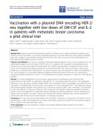
0.0001). For those patients with both plasma and CSF sam-
ples, the relative plasma and C SF chitotriosidase a ctivity for
each individual patient is s hown (Figure 2 ). The c orrelation
of the C SF and plasma activity l evels is < 0.0001.
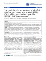
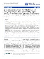
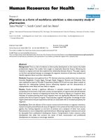
Correlations of Chitotriosidase Activity with Loes Score
We investigated whether plasma and spinal fluid chito-
triosidase activity correlated with extent of disease
based on Loes MRI scores. When CSF (Figure 3A) and
plasma (Figure 4A) chitotriosidase activity is analyzed
in relationship to the pre-transpla nt (baseline) Loes
BA
P = 0.0001P = 0.0001
Figure 1 Chitotriosidas e Activity is Elevated in Patients with ALD: Chitotriosidase activity was evaluated in the spinal fluid (Figure 1A) an d
plasma (Figure 2B) of patients with cerebral ALD or controls. There were 38 ALD patient samples and 16 controls represented in each group
Orchard et al. Journal of Neuroinflammation 2011, 8:144
/>Page 4 of 9
score, there was a statistically significant correlation (p
= 0.004 and 0.009, respectively). We also evaluated the
correlation between the pre-transplant chitotriosidase
activity and Loes score at one year, and also in the
change in Loes score (Delta score) before and one year
after transplant to determine whether chitotriosidase
activity pre- transplant is predi ctive of a change i n
Loes score. We found that t he spinal fluid chitotriosi-
dase significantly correlated with one-year post trans-
plant Loes score (Figure 3B; p = 0.0004), but not with
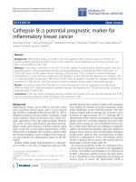
change in Loes score (Figure 3C). The plasma chito-
triosidase activity failed to correlate with either the
Loes score one year post transplant or the change in
Loes score (Figure 4B and 4C).
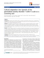
Correlations of Chitotriosidase Activity with Functional
Score
The functional scores of the patients prior to and one
year post-transplant were subsequently analyzed. The
change in functional score was determined by subtract-
ing the score at 1 year from the baseline score as a
measure o f clinical disease progression. The correlation
of CSF chitotriosidase activity to t he baseline func-
tional score is provided in Figure 3D; this correlation
is significant (p = 0.01). Importantly, the correlation
between chitotriosidase activ ity in the spinal fluid prior
to transplantation proved even more significant in the
linear regression analysis of the neurologic functional
score 1 year following transplantation (p < 0.0001; Fig-
ure 3E) and the change in the neurologic functional
score from baseline to 1 year post transplantation (p <
0.0001; Figure 3F). When this same analysis is per-
formed investigating the plasma chitotriosidase activity,
the correlation was high in regard to the baseline func-
tional score (p < 0.0001; Figure 4D) and the one-year
post transplantation functional score (p < 0.0001; Fig-
ure 4E) but less highly correlated with the change in
functional score (p = 0.0013; Figure 4F).
P < 0.0001
Paired Patients Samples: CSF and Plasma
Figure 2 Chitotriosidase Activity Correlates in C-ALD Plasma and Spinal Fluid: . For the 37 patients with cerebral ALD for which both
plasma and spinal fluid were available, the relative activity for both are depicted. For each patient, Statistical significance related to correlations
of the 2 groups is shown (Pearson two-tailed analysis).
Orchard et al. Journal of Neuroinflammation 2011, 8:144
/>Page 5 of 9
R
2
= 0.3063
P = 0.0004
R
2
= 0.3492
P = 0.0004
R
2
= 0.0959
P = 0.08
R
2
= 0.1742
P = 0.01
R
2
= 0.6514
P < 0.0001
R
2
= 0.5821
P < 0.0001
C
D
E
F
A
B
Figure 3 Spinal Fluid Chitotriosidase Determinations Are Associated with MRI and Functional Scores For ALD patients with cerebral
disease, the correlation of CSF chitotriosidase activity prior to transplantation and the baseline Loes MRI severity score (Fig 3A), the Loes score 1
year post transplantation (3B) and the relative increases in the Loes score from baseline to 1 year after transplantation (Loes Score; Delta; Fig 3C)
are presented. The correlation of CSF chitotriosidase activity to the Moser/Raymond functional score (Table 1) prior to transplantation (Fig 3D), at
1 year after transplantation (Fig 3E) and in regards to the change in the functional score from baseline to 1 year after transplant (Functional
Score; Delta; Fig 3F) are shown.
Orchard et al. Journal of Neuroinflammation 2011, 8:144
/>Page 6 of 9
R
2
= 0.4025
P < 0.0001
R
2
= 0.3053
P = 0.0013
R
2
= 0.1785
P = 0.009
R
2
= 0.1081
P = 0.08
R
2
= 0.0373
P = 0.3
C
D
E
F
A
B
R
2
= 0.4666
P < 0.0001
Figure 4 Plasma Chitotriosidase Determinations Are Associated w ith MRI and Functional Scores: For ALD patients with cerebral disease,
the correlation of plasma chitotriosidase activity prior to transplantation and the baseline Loes MRI severity score (Fig 4A), the Loes score 1 year
post transplantation (4B) and the relative increases in the Loes score from baseline to 1 year after transplantation (Loes Score; Delta; Fig 4C) are
presented. The correlation of plasma chitotriosidase activity to the Moser/Raymond functional score prior to transplantation (Fig 4D), at 1 year
after transplantation (Fig 4E) and in regards to the change in the functional score from baseline to 1 year after transplant (Functional Score;
Delta; Fig 4F) are shown.
Orchard et al. Journal of Neuroinflammation 2011, 8:144
/>Page 7 of 9
Discussion
We report for the first time highly significant elevations
of chitotriosida se activity in patients with cerebral ALD.
We reasoned that the chitotriosidase activity would be
elevated because of the previously documented presence
of monocytes and macrophages in the central nervous
system of individuals with cerebral A LD [10,20]. We
demonstrate that CHIT activity is elevated in both
plasma and spinal fluid, although levels are in general
much higher in CSF. Patients with higher CSF activity
also tend to have higher activity in the plasma (Figur e
2). We next asked whether CHIT activity in the CSF
and plasma correlated to the extent of disease as defined
by the MRI severity score described by Loes [16,21]. In
these analyses, both the CSF (Figure 3A) and p lasma
(Figure 4A) activity were significantly correlated t o the
“baseline” MRI scores, which would be closest in time
to when the samples were obtained (p = 0.0004 and
0.012, respectively). The correlation of chitotriosidase
activity was also a nalyzed in relationship to the MRI
severity scores at 1 year following transplant. In the case
of plasma activity (Figure 4B), t his correlation was not
significant (p = 0.08), while the CSF activity was highly
correlated to the Loes score at one year post transplant
(Figure 3 B, p = 0.0004). When the correlation of CHIT
activity to disease progression by MRI (Loes score;
Delta) is analyzed, neither plasma nor CSF activity
values were significantly correlated to the change in
Loes score (Figures 3C and 4C).
The majority of C-ALD patients transplanted early in
the course of their disease have minimal or no subse-
quent clinical manifestations. In contrast, patients with
more advanc ed disease often exhibit substantial disease
progression post transplant [22]. To better assess these
functional parameters, we used the Moser-Raym ond
scale (Table 1). The function al status of the patients was
determined prior to t ransplantation and at 1 year after
the transplant. Evidence of clinical disease progression
may be defined as the difference in these scores. Chito-
triosidase activity was shown to be highly correlated
with the pre-transplant functional score, but more
importantly, also to the clinical status of the patients
post transplantation. This is apparent when chitotriosi-
dase activity is assessed in relation to the 1-year scores
(CSF and plasma; p < 0.0001) and in relationship to the
change in functional status (p < 0.0001 and < 0.0013 in
CSF and plasma, respectively).
The ability to better establish prognosis in patients
being considered for allogeneic transplantation is of
great importance. Based on our experi ence and t hose of
others, patients early in the course of cerebral disease
are very likely to achieve disease stabilization without
significant clinical deterioration. In contrast, for patients
with more advanced disease there is great variation in
outcomes after transplantat ion, with relatively mild pro-
gression observed in some patients and dramatic dete-
rioration in others. Standard means of assessing these
patients include MRI, neurologic examination, neuropsy-
chological testing and potentially functional assessments.
The data presented in this study suggests that chitotrio-
sidase determinations can provide important prognostic
information, and may allow physicians and families to
make a much more informed decision on whether trans-
plantation is the best course of action.
Elevated chitotriosidase activity has been described i n
other neurologic disorders, including stroke and multi-
ple sclerosis (MS) [12,23-25]. While material that
appears similar to chitin was identified in Alzheimer’s
disease, it w as not shown to be present in multiple
sclerosis [26]. In the case of ALD the etiology cannot
be directly assessed, but it seems likely that the
increases in chitotriosidase activity are likely related to
inflammation, particularly since the elevations are also
apparent in the plasma of patients with ALD. Interest-
ingly, while chitotriosidase is elevated in the CSF in
both relapsing-remitting and primary progressive MS,
it is not elevated in the plasma [25]. This is in contrast
to our findings in ALD. This may suggest that the
inflammation in ALD is more systemic in nature than
that observed with MS.
These findings suggest other important questions
that cannot be addressed in this study. Is chitotriosi-
dase activity related directly to damage within the
CNS, or is it merely a biomarker of disease? Is there
any difference in the distribution of the chitotriosidase
24 base insert in exon 10 in ALD and the general
population? From our studies it would appear not, but
this could only be addressed with a larger population
of patients. Would determinations of plasma o r spinal
fluid chitotriosidase activity improve our ability to pre-
dict which patients diagnosed with ALD are likely to
progress to C-ALD? In addition, is chitotriosidase
activity increased in patients with adrenomyeloneuro-
pathy, or in female heterozygote “carriers"? Would it
be useful clinically in these conditions? Even more
intriguing is the possibility that chitotriosidase could
prove to be a biomarker for other neurodegenerative
diseases that have an inflammatory component, allow-
ing more rational therapeutic decisions. Additional
investigations will prove important in further establish-
ing the role of chitotriosidase i n ALD and other simi-
lar conditions.
Lists of abbreviations
ALD: Adrenoleukodystrophy; C-ALD: cerebral ALD; CHIT: chitotriosidase; CNS:
central nervous system; HSCT: hematopoietic stem cell transplantation; IRB:
institutional review board; LP: lumbar puncture; VLCFA: very long chain fatty
acids.
Orchard et al. Journal of Neuroinflammation 2011, 8:144
/>Page 8 of 9
Acknowledgements
We thank Teresa Kivisto for her integral work in patient care and data
monitoring, and Dr. Larry Charnas for his interest and thoughtful discussions
regarding this work. Also our appreciation to Todd Defor for his biostatistical
expertise and advice.
Support
These studies were supported by the Children’s Cancer Research Fund
(CCRF), as well as by an anonymous private foundation
Author details
1
Department of Pediatrics, Program in Blood & Marrow Transplantation,
University of Minnesota, Minneapolis, USA.
2
Department of Pediatrics,
Program in Neurology, University of Minnesota, Minneapolis, USA.
3
Department of Neurology, Kennedy Krieger Institute, Baltimore MD, USA.
4
Department of Diagnostic Radiology, University of Minnesota, Minneapolis,
USA.
5
Department of Experimental and Clinical Pharmacology, Center for
Orphan Drug Research, University of Minnesota, Minneapolis, USA.
Authors’ contributions
PJO was the Principal Investigator and primary author of the manuscript,
and his laboratory was used to perform the laboratory studies. TL
collaborated in the design of the laboratory studies, and discussions as to
the role of biomarkers in inherited disease with neuroinflammation. WM
reviewed clinical information regarding patient outcomes, including the
functional scoring system for the patients on this study. SMR reviewed
clinical information regarding patient outcomes, including the functional
scoring system for the patients on this study (this task was split between
WM and SMR). GR, an internationally established expert in peroxisomal
disease, established the scoring system used in these investigations and
provided assistance with the design and interpretation of the study. DN is a
neuroradiologist who read and scored the MRIs used in this analysis. LB is a
technician who performed the majority of the studies in the manuscript and
wrote the majority of the methods section. JC is a pharmacologist and
collaborator in clinical and laboratory studies on adrenoleukodystrophy, and
approaches associated with inflammation. JT is a laboratory collaborator
who assisted with PCR and chitotriosidase assay development and
interpretation. All authors critically reviewed, read, and approved the final
manuscript.
Competing interests
The authors declare that they have no competing interests.
Received: 16 May 2011 Accepted: 20 October 2011
Published: 20 October 2011
References
1. Mahmood A, Dubey P, Moser HW, Moser A: X-linked
adrenoleukodystrophy: therapeutic approaches to distinct phenotypes.
Pediatr Transplant 2005, 9(Suppl 7):55-62.
2. Moser HW, Raymond GV, Dubey P: Adrenoleukodystrophy: new
approaches to a neurodegenerative disease. JAMA 2005, 294:3131-3134.
3. Kemp S, Pujol A, Waterham HR, van Geel BM, Boehm CD, Raymond GV,
Cutting GR, Wanders RJ, Moser HW: ABCD1 mutations and the X-linked
adrenoleukodystrophy mutation database: role in diagnosis and clinical
correlations. Hum Mutat 2001, 18:499-515.
4. Powers JM, Pei Z, Heinzer AK, Deering R, Moser AB, Moser HW, Watkins PA,
Smith KD: Adreno-leukodystrophy: oxidative stress of mice and men. J
Neuropathol Exp Neurol 2005, 64:1067-1079.
5. Cartier N, Hacein-Bey-Abina S, Bartholomae CC, Veres G, Schmidt M,
Kutschera I, Vidaud M, Abel U, Dal-Cortivo L, Caccavelli L, et al:
Hematopoietic stem cell gene therapy with a lentiviral vector in X-linked
adrenoleukodystrophy. Science 2009, 326:818-823.
6. Loes DJ, Stillman AE, Hite S, Shapiro E, Lockman L, Latchaw RE, Moser H,
Krivit W: Childhood cerebral form of adrenoleukodystrophy: short-term
effect of bone marrow transplantation on brain MR observations. AJNR
Am J Neuroradiol 1994, 15:1767-1771.
7. Hollak CE, van Weely S, van Oers MH, Aerts JM: Marked elevation of
plasma chitotriosidase activity. A novel hallmark of Gaucher disease. J
Clin Invest 1994, 93:1288-1292.
8. Casal JA, Lacerda L, Perez LF, Pinto RA, Clara Sa Miranda M, Carlos Tutor J:
Relationships between serum markers of monocyte/macrophage
activation in type 1 Gaucher’s disease. Clin Chem Lab Med 2002, 40:52-55.
9. Aerts JM, Hollak CE, van Breemen M, Maas M, Groener JE, Boot RG:
Identification and use of biomarkers in Gaucher disease and other
lysosomal storage diseases. Acta Paediatr Suppl 2005, 94:43-46; discussion
37-48.
10. Powers JM, Liu Y, Moser AB, Moser HW: The inflammatory myelinopathy
of adreno-leukodystrophy: cells, effector molecules, and pathogenetic
implications. J Neuropathol Exp Neurol 1992, 51:630-643.
11. Boot RG, Renkema GH, Verhoek M, Strijland A, Bliek J, de Meulemeester TM,
Mannens MM, Aerts JM: The human chitotriosidase gene. Nature of
inherited enzyme deficiency. J Biol Chem 1998, 273:25680-25685.
12. Sotgiu S, Barone R, Arru G, Fois ML, Pugliatti M, Sanna A, Rosati G,
Musumeci S: Intrathecal chitotriosidase and the outcome of multiple
sclerosis. Mult Scler 2006, 12:551-557.
13. Ries M, Schaefer E, Luhrs T, Mani L, Kuhn J, Vanier MT, Krummenauer F,
Gal A, Beck M, Mengel E: Critical assessment of chitotriosidase analysis in
the rational laboratory diagnosis of children with Gaucher disease and
Niemann-Pick disease type A/B and C. J Inherit Metab Dis 2006,
29:647-652.
14. Schoonhoven A, Rudensky B, Elstein D, Zimran A, Hollak CE, Groener JE,
Aerts JM: Monitoring of Gaucher patients with a novel chitotriosidase
assay. Clin Chim Acta 2007, 381:136-139.
15. Vedder AC, Cox-Brinkman J, Hollak CE, Linthorst GE, Groener JE,
Helmond MT, Scheij S, Aerts JM: Plasma chitotriosidase in male Fabry
patients: a marker for monitoring lipid-laden macrophages and their
correction by enzyme replacement therapy. Mol Genet Metab 2006,
89:239-244.
16. Loes DJ, Hite S, Moser H, Stillman AE, Shapiro E, Lockman L, Latchaw RE,
Krivit W: Adrenoleukodystrophy: a scoring method for brain MR
observations. AJNR Am J Neuroradiol 1994, 15:1761-1766.
17. Moser HW, Raymond GV, Koehler W, Sokolowski P, Hanefeld F, Korenke GC,
Green A, Loes DJ, Hunneman DH, Jones RO, et al: Evaluation of the
preventive effect of glyceryl trioleate-trierucate ("Lorenzo’s oil”) therapy
in X-linked adrenoleukodystrophy: results of two concurrent trials. Adv
Exp Med Biol 2003, 544:369-387.
18. Malaguarnera L, Simpore J, Prodi DA, Angius A, Sassu A, Persico I, Barone R,
Musumeci S: A 24-bp duplication in exon 10 of human chitotriosidase
gene from the sub-Saharan to the Mediterranean area: role of parasitic
diseases and environmental conditions. Genes Immun 2003, 4:570-574.
19. Rodrigues MR, Sa Miranda MC, Amaral O: Allelic frequency determination
of the 24-bp chitotriosidase duplication in the Portuguese population by
real-time PCR. Blood Cells Mol Dis 2004, 33:362-364.
20. Takeda S, Ohama E, Ikuta F: Adrenoleukodystrophy–early ultrastructural
changes in the brain. Acta Neuropathol 1989, 78:124-130.
21. Loes DJ, Fatemi A, Melhem ER, Gupte N, Bezman L, Moser HW,
Raymond GV: Analysis of MRI patterns aids prediction of progression in
X-linked adrenoleukodystrophy. Neurology 2003, 61:369-374.
22. Peters C, Charnas LR, Tan Y, Ziegler RS, Shapiro EG, DeFor T, Grewal SS,
Orchard PJ, Abel SL, Goldman AI, et al: Cerebral X-linked
adrenoleukodystrophy: the international hematopoietic cell
transplantation experience from 1982 to 1999. Blood 2004, 104:881-888.
23. Palasik W, Fiszer U, Lechowicz W, Czartoryska B, Krzesiewicz M, Lugowska A:
Assessment of relations between clinical outcome of ischemic stroke
and activity of inflammatory processes in the acute phase based on
examination of selected parameters. Eur Neurol 2005, 53:188-193.
24. Sotgiu S, Barone R, Zanda B, Arru G, Fois ML, Arru A, Rosati G, Marchetti B,
Musumeci S: Chitotriosidase in patients with acute ischemic stroke. Eur
Neurol 2005,
54:149-153.
25. Verbeek MM, Notting EA, Faas B, Claessens-Linskens R, Jongen PJ: Increased
cerebrospinal fluid chitotriosidase index in patients with multiple
sclerosis. Acta Neurol Scand 2010, 121:309-314.
26. Sotgiu S, Musumeci S, Marconi S, Gini B, Bonetti B: Different content of
chitin-like polysaccharides in multiple sclerosis and Alzheimer’s disease
brains. J Neuroimmunol 2008, 197:70-73.
doi:10.1186/1742-2094-8-144
Cite this article as: Orchard et al.: Chitotriosidase as a biomarker of
cerebral adrenoleukodystrophy. Journal of Neuroinflammation 2011 8:144.
Orchard et al. Journal of Neuroinflammation 2011, 8:144
/>Page 9 of 9