báo cáo hóa học: " Post-traumatic hypoxia exacerbates neurological deficit, neuroinflammation and cerebral metabolism in rats with diffuse traumatic brain injury" pptx
Bạn đang xem bản rút gọn của tài liệu. Xem và tải ngay bản đầy đủ của tài liệu tại đây (1.23 MB, 16 trang )
Yan et al. Journal of Neuroinflammation 2011, 8:147
/>
JOURNAL OF
NEUROINFLAMMATION
RESEARCH
Open Access
Post-traumatic hypoxia exacerbates neurological
deficit, neuroinflammation and cerebral metabolism
in rats with diffuse traumatic brain injury
Edwin B Yan1,2†, Sarah C Hellewell1,3†, Bo-Michael Bellander4, Doreen A Agyapomaa1,3 and
M Cristina Morganti-Kossmann1,2*
Abstract
Background: The combination of diffuse brain injury with a hypoxic insult is associated with poor outcomes in
patients with traumatic brain injury. In this study, we investigated the impact of post-traumatic hypoxia in
amplifying secondary brain damage using a rat model of diffuse traumatic axonal injury (TAI). Rats were examined
for behavioral and sensorimotor deficits, increased brain production of inflammatory cytokines, formation of
cerebral edema, changes in brain metabolism and enlargement of the lateral ventricles.
Methods: Adult male Sprague-Dawley rats were subjected to diffuse TAI using the Marmarou impact-acceleration
model. Subsequently, rats underwent a 30-minute period of hypoxic (12% O2/88% N2) or normoxic (22% O2/78% N2)
ventilation. Hypoxia-only and sham surgery groups (without TAI) received 30 minutes of hypoxic or normoxic
ventilation, respectively. The parameters examined included: 1) behavioural and sensorimotor deficit using the Rotarod,
beam walk and adhesive tape removal tests, and voluntary open field exploration behavior; 2) formation of cerebral
edema by the wet-dry tissue weight ratio method; 3) enlargement of the lateral ventricles; 4) production of
inflammatory cytokines; and 5) real-time brain metabolite changes as assessed by microdialysis technique.
Results: TAI rats showed significant deficits in sensorimotor function, and developed substantial edema and
ventricular enlargement when compared to shams. The additional hypoxic insult significantly exacerbated
behavioural deficits and the cortical production of the pro-inflammatory cytokines IL-6, IL-1b and TNF but did not
further enhance edema. TAI and particularly TAI+Hx rats experienced a substantial metabolic depression with
respect to glucose, lactate, and glutamate levels.
Conclusion: Altogether, aggravated behavioural deficits observed in rats with diffuse TAI combined with hypoxia
may be induced by enhanced neuroinflammation, and a prolonged period of metabolic dysfunction.
Keywords: Traumatic brain injury, traumatic axonal injury, hypoxia, neurological deficit, cytokine, brain edema, ventricle, metabolism
Background
Traumatic brain injury (TBI) remains a major health
burden in both developed and developing countries. TBI
consists of two temporal pathological phases spanning
the initial traumatic impact and a multitude of secondary cascades, resulting in progressive tissue degeneration
* Correspondence:
† Contributed equally
1
National Trauma Research Institute, The Alfred Hospital, 89 Commercial
Road, Melbourne 3004, Australia
Full list of author information is available at the end of the article
and neurological impairment [1-3]. The pathological
consequences of TBI can be variable and largely depend
on the presentation of injury as either focal or diffuse,
or a combination of both. Diffuse brain injury may
result from rotational forces and/or acceleration/deceleration of the head during a traumatic impact, often
leading to diffuse axonal injury. Although difficult to
diagnose due to the absence of lesions or overt pathology [4,5], diffuse axonal injury is a common presentation, accounting for up to 70% of all TBI cases [6]. The
pathology of diffuse axonal injury develops over a
© 2011 Yan et al; licensee BioMed Central Ltd. This is an Open Access article distributed under the terms of the Creative Commons
Attribution License ( which permits unrestricted use, distribution, and reproduction in
any medium, provided the original work is properly cited.
Yan et al. Journal of Neuroinflammation 2011, 8:147
/>
delayed time course, and is frequently aggravated by the
occurrence of subsequent insults, which are known to
worsen morbidity and mortality in TBI patients [7]. Epidemiological studies have revealed that up to 44% of
severe head trauma patients experience brain hypoxia,
which has been associated with adverse neurological
outcomes [8-13]. Hypoxia can be initiated by TBIinduced cerebral hypoperfusion, apnoea and hypoventilation mostly related to brainstem injury [14-16]. In
addition, systemic hypoxia can be caused by extracranial
injuries often co-existing with head trauma such as
obstructed airways, lung puncture and excessive blood
loss [9,17]. Despite these clinical observations, the exact
mechanisms leading to the exacerbation of brain
damage concomitant to posttraumatic hypoxia remain
to be elucidated.
One putative sequel of TBI in contributing to secondary tissue damage is the activation of cellular and
humoral neuroinflammation. This response is characterised by the accumulation of inflammatory cells in the
injured area, as well as the release of pro- and antiinflammatory cytokines, which may either promote the
repair of injured tissue, or cause additional damage [18].
The activation of inflammatory cascades in human and
rodent TBI have previously been reported [19-21]. In
severe TBI patients, ourselves and others have demonstrated a robust longitudinal increase of multiple cytokines and chemokines in cerebrospinal fluid (CSF)
[22-27]. More recently, these findings have been corroborated with the upregulation of TNF, IL-1b, IL-6, IFNg protein and gene expression in post-mortem human
brain tissue after acute TBI [28]. Animal models of
brain hypoxia or trauma can independently activate
acute expression of cytokines IL-1b, IL-6 and TNF
[29-31]. Furthermore, in models of focal TBI, additional
post-traumatic hypoxia was shown to worsen brain tissue damage [32-34], cerebral edema [35], and exacerbate
sensorimotor, behavioural and cognitive impairment
[32,34,36-38]. The detrimental role of neuroinflammation can be elicited by its ability to induce the production of excitotoxic substances including reactive oxygen
and nitrogen radicals [39-41] contributing to the development of brain edema [42,43], blood brain barrier
(BBB) disruption [44,45], and apoptotic cell death
[43,46-49]. However, almost all the studies on post-TBI
hypoxia used focal brain injury models, while epidemiological data on large patient populations reported that
the majority of TBI patients present with diffuse brain
injury leading to worse neurological outcome especially
if associated with hypoxia [6]. The few studies by us and
others examining the effect of post-traumatic hypoxia
after diffuse traumatic axonal injury (TAI; the experimental counterpart of human diffuse axonal injury) have
demonstrated enhanced neurological deficits [34,38],
Page 2 of 16
exacerbated edema and cerebral blood flow, and diminished vascular reactivity [50-54]. In a recent study using
the Marmarou rat model of diffuse TAI with additional
post-trauma systemic hypoxia, we demonstrated a
greater axonal damage in the corpus callosum and
brainstem co-localising with a robust macrophage infiltration and enhanced astrogliosis, when compared with
TAI animals without hypoxia [54-56]. Therefore, using
this model of TAI, we aimed to further investigate
whether post-traumatic hypoxia also aggravates behavioural and sensorimotor function, cerebral edema,
enlargement of lateral ventricles, production of inflammatory cytokines in the brain, and impairment in cerebral energy metabolism.
Methods
Induction of trauma
Animal experiments were conducted in accordance with
the Code of Practice for the Care and Use of Animals
for Scientific Purposes (National Health and Medical
Research Council, Australia), and received approval
from the institutional Animal Ethics Committee. Adult
male Sprague-Dawley rats were housed under a 12-hour
light/dark cycle with food and water ad libitum. Rats
aged 12-16 weeks and weighing 350-375 g on the day of
surgery were subjected to TAI (n = 27), TAI followed
by a 30-min systemic hypoxia (TAI+Hx; n = 27),
hypoxia only (n = 27) or sham surgery (n = 27). Briefly,
rats were anaesthetized in a mixture of 5% isoflurane in
22% O2/78% N2, intubated, and mechanically ventilated
with a maintenance dose of 2-3% isoflurane in 22% O2/
78% N 2 . A steel disc (10 mm in diameter and 3 mm
thickness) was adhered to the skull between bregma and
lambda suture lines using dental acrylic. Animals were
briefly disconnected from the ventilator and moved onto
a foam mattress (Type E polyurethane foam, Foam2Size,
VA, USA) underneath a trauma device where a weight
of 450 g was allowed to fall freely though a vertical tube
from 2 m. Following the impact, animals were reconnected to the ventilator, and ventilated continuously for
a further 30 min using an appropriate concentration of
isoflurane (0.5-1%) in either hypoxic (12% O2/88% N2)
or normoxic (22% O2/78% N2) gas mixture. Consistent
with the literature [32,36] we have previously demonstrated that such systemic hypoxic conditions result in
an sO2 of 47 ± 4.3% and pO2 of 48.5 ± 3.8 mmHg, and
cause a significant hypotensive episode, with mean arterial blood pressure (MABP) dropping to 69.5 ± 29.5 midway through the insult (i.e. 15 min). The reduction of
sO 2 , pO 2 , and MABP returned to sham values by 15
min following the conclusion of the hypoxic period [55].
Consistent with the original description of this model by
Foda et al. (1994) [40], the intubation and ventilation of
rats after injury resulted in a mortality rate of ~10%
Yan et al. Journal of Neuroinflammation 2011, 8:147
/>
which was confirmed in our study. When the two
insults were combined, there was no significant increase
in mortality. Hypoxia-only and sham operated animals
were surgically prepared as described for TAI rats with
the exception of the traumatic impact, and ventilated
with hypoxic or normoxic gas, respectively. Rats were
housed in individual cages after surgery and placed on
heat pads (37°C) for 24 h to maintain normal body temperature during the recovery period.
Microdialysis probe implantation
Following trauma, 5 rats from each of TAI, TAI+Hx,
hypoxia-only and sham groups were inserted with
microdialysis probes into the brain for measuring realtime metabolite changes. If the microdialysis probe was
implanted soon after the completion of TAI, high severity of the injury together with the ongoing anesthesia
would result in a higher mortality rate. Therefore, we
allowed the animals to recover for a period of 4 h before
implantation of the microdialysis probe. Rats were then
anesthetized by isoflurane, intubated and mechanically
ventilated as described above. The head of the animal
was immobilized on a stereotactic frame with nose and
ear bars (David Kopf Instruments, California, USA). The
scalp was opened at the existing suture line, and a 1mm burr hole was drilled into the skull using a small
handheld drill at the coordinates of -4.52 mm to bregma
and -2 mm lateral to the midline on left hemisphere.
Care was taken not to damage the dura mater. Two
shallow holes were drilled posterior and anterior to the
burr hole, and screws were inserted to provide anchor
points for the microdialysis probe implantation. A guide
cannula for CMA12 microdialysis probe was adjusted to
3 mm in length, inserted into the brain and secured in
place by using dental cement (Dentsply, PA, USA) to
cover both the guide cannula and the anchor screws.
Once the dental cement solidified, the microdialysis
probe (CMA12, 100 kDa cutoff, CMA Microdialysis,
Solna, Sweden) was inserted into the guide tube to a
suitable length allowing the semi-permeable membrane
exposure outside of the guide tube for direct contact
with the brain tissue. The microdialysis probe was
immobilized by applying additional dental cement over
the probe and guide cannula. At surgery completion,
animals were allowed to recover in a microdialysis
experimental system (CAM 120, CMA Microdialysis)
which consists of a balanced arm with dual channel swivel allowing free movement of the animal and continuous collection of microdialysis samples. The
microdialysis probe was perfused at 1 μl/min using artificial cerebrospinal fluid (aCSF, CMA Microdialysis).
The effluent was collected as accumulative sample over
3 h (i.e. 180 μl/sample) using an automated refrigerated
microdialysis fraction collector (Harvard Apparatus,
Page 3 of 16
MA, USA). Samples were transferred to -80°C freezer
every 12 h and stored until analysis. At the end of the
experimental period, animals were killed and brains
were perfusion fixed to identify the location of the
microdialysis probe in the cortex. Only the animals with
the probe tip in the designated location were included
for analysis.
Assessment of sensorimotor functions
Rats were treated in each group as described above and
used for assessment of sensorimotor deficit by the
Rotarod test, beam balancing and walking test, and
adhesive tape removal from forepaws test (n = 10 per
group). Animals were trained for these tasks every second day starting 1 week before surgery. These sensorimotor tests were performed daily after TAI for a week,
then on every second day until 14 days. The Rotarod
allows assessment of movement coordination and function including motor, sensory and balancing skills. Rats
were placed on a rotating cylinder made of 18 rods (1
mm diameter) (Ratek, VIC, Australia). The rotational
speed of the device was increased in increments of 3
rpm/5 sec, from 0 to 30 revolutions per minute (rpm).
The maximal speed at which the rat was unable to
match and failed to stay on the device was recorded.
Body balancing and walking was assessed using a beamwalking test, in which rats were placed in the middle of
a 2-meter long, 2-cm wide beam suspended 60 cm
above the ground between 2 platforms. Rats were scored
as: [1] normal walking for at least 1 meter on the beam;
[2] crawling on the beam for at least 1 m with abdomen
touching the beam; [3] ability to stay on the beam but
failure to move; and [4] inability to balance on the
beam. Sensory and fine motor function was assessed by
the ability to remove adhesive tapes (5 × 10 mm; masking tape, Norton Tapes, NSW, Australia) placed on the
back of each forepaw. The number of tapes removed (0,
1 or 2) and the latency for each tape removal were
recorded within a 2-minute period.
Open field test
This test evaluates the animal’s normal exploratory
behavior. Rats were placed in an empty arena (70 × 70
× 60 cm, W×L×H) within an enclosed environment and
low lighting. The movement of the rats was recorded
for 5 min by a camera, and the distance walked was calculated using a custom made automated movementtracking program (Dr Alan Zhang, Department of Electrical Engineering, The University of Melbourne).
Brain edema measurement
Rats with TAI, TAI+Hx, hypoxia or sham surgery were
generated for assessment of brain edema. The wet-dry
weight method was used for determining the water
Yan et al. Journal of Neuroinflammation 2011, 8:147
/>
content of the brain at 2, 24, 48, 72, and 96 h after
treatment (n = 6 per timepoint per group). Briefly, the
left hemisphere was separated from the rest of brain tissue, weighed on a precision microbalance (Ohaus
Adventurer Analytical Balance Bradford, MA, USA), and
dried in an oven at 100°C for 24 h. The dry tissue was
weighed again, and cortical water content was calculated
as ([wet tissue weight - dry tissue weight]/wet tissue
weight) × 100.
Measurement of ventricle size
A cohort of rats for each experimental group was treated as described above and killed at 1 or 7 days after
injury (n = 6 per group per timepoint). Brains were perfusion fixed using 4% paraformaldehyde and embedded
in paraffin wax. Brain tissue blocks were cut into 10 μm
sections at the level of +1 mm relative to the bregma
and collected onto glass slides. Sections were dewaxed,
rehydrated, stained using hemotoxylin and eosin, and
visualized under a light microscope (Olympus BX50).
Multiple photographs were taken under 200× magnification to cover the entire sections. Image analysis software
(ImageJ, NIH, USA) was used to align images taken
from the same brain section to reconstruct a full section
view. The whole brain area and the area of the ventricle
were measured using ImageJ, with the area of the ventricle expressed as the percentage of total brain area.
Page 4 of 16
curve. Total protein concentration was determined in
each sample using the Bradford Assay (Bio-Rad
Laboratories).
Analysis of microdialysis samples
The microdialysis samples (180 μl/sample, n = 5 per
group) were freeze dried and suspended in small volume
of ddH2O to increase the concentration of solutes. The
samples were then analysed for glucose, lactate and glutamate using conventional enzymatic techniques performed in the ISCUS Analyser (CMA Microdialysis).
Due to a substantial time delay between sample collection and analysis, pyruvate was not measured as it is
known to be unstable after storage time of more than 3
months (CMA Microdialysis). The concentrations of
glucose, lactate and glutamate in each sample were calculated to the original concentration according to the
sample volume before and after the freeze-drying
procedure.
Data analysis
Sensorimotor function assessment, cytokine concentration, brain metabolites and brain edema results were
analysed using two-way repeated measures ANOVA.
The open field test and ventricular size measurement
were analysed by 1-way ANOVA. Data were presented
as mean ± standard error of the mean. Data were considered as significant where p < 0.05.
Cytokine measurements
The right hemisphere from each animal of edema study
was dissected, the cortex isolated, and stored at -80°C
until use. The cortex was homogenised in an extraction
solution containing Tris-HCl (50 mmol/L, pH 7.2),
NaCl (150 mmol/L), 1% Triton X-100, and 1 μg/mL
protease inhibitor cocktail (Complete tablet; Roche
Diagnostics, Basel, Switzerland) and agitated for 90 min
at 4°C. Tissue homogenates were centrifuged at 2000
rpm for 10 min, and the supernatants stored at -80°C
until use. The concentration of 6 cytokines (IL-1b, IL-2,
IL-4, IL-6, IL-10, TNF) in the brain cortex homogenates
was determined by multiplex assay as previously used in
our group [57] (Bio-Rad Laboratories, Hercules, CA,
USA). Briefly, colored beads conjugated with cytokine
antibodies were loaded into wells of 96-well filter plate.
Following washing, the standards, quality controls and
samples were added into the wells and incubated overnight at 4°C on a shaking platform. The wells were
washed by filtration, and subsequently a solution with a
mixture of biotinylated antibodies against each cytokine
was added and incubated for 1 h at room temperature.
Following the removal of excessive detection antibodies,
streptavidin-phycoerythrin was added. Cytokine concentration was measured using multiplex assay reader (BioRad Laboratories) and calculated against the standard
Results
Neurological outcome
The impact of post-TAI hypoxia on neurological dysfunction was explored using a number of sensorimotor
tests over a period of 2 weeks in TAI, TAI+Hx, hypoxia
alone and sham operated animal groups.
TAI+Hx rats show greater deficits on the Rotarod
compared to TAI
The Rotarod test involves examining complex body
movement and coordination, which showed severe
impairment in rats following TAI and TAI+Hx when
compared with shams. The maximal speed TAI rats
were able to maintain on the Rotarod was significantly
decreased at day 1 post-TAI (9.5 ± 1.6 rpm) as compared with shams (24.9 ± 1.3 rpm) (p < 0.05). Over time
TAI rats showed a gradual improvement in motor function, however the maximal speed recorded on the
Rotarod between day 2 and 6 post-injury (13.9 ± 1.8
and 19.3 ± 1.4 rpm, respectively) remained significantly
lower than sham control rats (average 25.83 ± 0.59 rpm)
(Figure 1A). Although the motor function in TAI rats
improved steadily, from 6 days onwards they failed to
recover further, showing a plateau speed on Rotarod
until 14 days. When compared to TAI-only rats, the
Yan et al. Journal of Neuroinflammation 2011, 8:147
/>
30
A
4
$
$ #
#
Deficit severity score
Revolution per minute
40
Page 5 of 16
$ $ $ $
# # # # #
20
10
.03 .01
.05
0
.07
$
#
3
$
#
$
# #
# #
1
-7 -5 -3 -1 1 2 3 4 5 6 8 10 12 14
Time Post Injury (days)
Time Post Injury (days)
C
2.5
Latency (min)
$
#
2
0
-7 -5 -3 -1 1 2 3 4 5 6 8 10 12 14
B
$
#
2.0
$
#
$
# $
Sham
TAI
TAI+Hx
$
#
#
1.5
#
1.0
0.5
0.0
-7 -5 -3 -1 1 2 3 4 5 6 8 10 12 14
Time Post Injury (days)
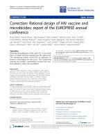
Figure 1 Sensorimotor function is aggravated following traumatic axonal injury combined with 30 min hypoxia. Graphics show changes
observed over 14 days for the 3 tests employed: (A) Rotarod, (B) beam walking and (C) adhesive tape removal from the front paws. Animals
were trained for these tasks for 7 days before trauma, and then tested daily for 6 days after surgery and on every second day until 14 days. $
indicates significant decrease in motor function on the Rotarod, and increase in beam walking deficit score and latency of adhesive tape
removal between TAI and sham animals, while # indicates significant difference in these tests between TAI+Hx and sham animals. Numbers in
(A) represent the p-values indicating significant differences between TAI and TAI+Hx at days 2, 5 and 6; and close to significant at day 1. The
results indicate that TAI+Hx rats require a longer period for neurological recovery towards sham levels, with significant differences between TAI
and TAI+Hx rats in the Rotarod test during the first 6 days post-injury. Although a similar deficit on the tape removal test was observed in TAI
and TAI+Hx groups versus sham in the first 5 days, TAI+Hx rats exhibited prolonged impairment over sham controls at 6 and 12 days. Data
shown as mean ± SEM, n = 10 per group per time point. Data was analysed by 2-way ANOVA repeated measures with Bonferroni post hoc test,
with a p-value of < 0.05 considered significant.
TAI+Hx group had substantially greater motor deficits
on the Rotarod, as indicated by a significant lower maximal walking speed at day 2 (9.2 ± 1.5 vs 13.9 ± 1.8
rpm), day 5 (12.1 ± 1.8 vs 17.5 ± 1.5 rpm) and day 6
(13.2 ± 1.8 vs 19.3 ± 1.4 rpm) after injury (p < 0.05)
(Figure 1A). These TAI+Hx rats also performed significantly worse on the Rotarod as compared to sham at 8
days (17.13 ± 1.81 vs 25 ± 1.55 rpm), demonstrating
that this deficit was prolonged as well as enhanced in
rats subjected to the combination of TAI and Hx.
Ability to balance and walk on a narrow beam is
impaired after TAI and TAI+Hx
The beam walk is a sensitive test to determine the
ability of injured rats to balance and walk on a narrow
beam. TAI and TAI+Hx induced severe impairment on
the beam walking test, whereby rats of both groups
were unable to balance or stay on the beam at 1 day
post-injury (Figure 1B). The deficit scores of beam
walking were significantly elevated in both TAI and
TAI+Hx groups, particularly during the first 5 days.
When compared to sham, TAI only rats displayed a
motor impairment which resolved after 5 days. On the
contrary, TAI+Hx rats had a significantly greater deficit in walking and balancing compared to sham controls which persisted up to 8 days after injury. Overall,
there was no significant difference in beam walking
test between TAI and TAI+Hx groups, with both
groups returning to sham function by 10 days post
TAI or TAI+Hx.
Yan et al. Journal of Neuroinflammation 2011, 8:147
/>
Page 6 of 16
20.8 ± 3.4 m either before sham operation or at days 3,
6 and 14 days post-surgery (Figure 2A). Hypoxia alone
did not alter the distance traveled, which was maintained at sham levels with no differences before or after
the insult (data not shown). In comparison to the above
sensorimotor function testing, TAI alone did not reduce
the voluntary walking distance at 3, 6 or 14 days postTAI over the pre-TAI levels (Figure 2B). However, an
additional hypoxic insult after TAI significantly
decreased the mobility of rats to 55.2% of the pre-TAI
+Hx level at day 3 post-injury (8.4 ± 2.6 m vs 15.1 ± 1.3
m, respectively; p < 0.05) (Figure 2C). By day 6, the distance of voluntary movement in TAI+Hx rats was
slightly increased (13.8 ± 2.2 m; p = 0.06) and was fully
restored to pre-TAI+Hx level at day 14 (17.7 ± 2.8 m)
after injury.
TAI+Hx rats have prolonged deficits in the adhesive tape
removal task
Both TAI and TAI+Hx rats took significantly longer to
sense, and subsequently remove the adhesive tapes
adhered on the back of forepaws (Figure 1C). In TAI
rats significant differences to sham function were
detected until day 5. The additional hypoxic insult postTAI caused further significant differences in latency of
adhesive tape removal on days 6 and 12 as compared
with TAI-only rats (latency 1.12 ± 0.27 vs 0.88 ± 0.21
min (day 6), 1.23 ± 0.26 vs 0.74 ± 0.22 min (day 12)).
Sham and hypoxia alone (not shown) rats did not
change their performance on the Rotarod, beam walking
and adhesive tape removal tests over the duration of
testing period.
Voluntary walking in an open field is compromised after
TAI+Hx
Brain water content is elevated after TAI and TAI+Hx
The ability of voluntary movement was determined by
calculating the distance traveled during the first 5 min
after the rats were placed in a testing chamber. In the
sham group, rats traveled between 12.3 ± 2.8 m and
Cerebral edema is a common pathophysiological consequence in this model of TAI [35,58,59]. Using the wetdry ratio method, we showed that brain water contents
in hypoxia-only and sham animals were within the
A
B
20
Distance (m)
25
20
Distance (m)
25
15
10
10
5
5
0
15
Pre
3
6
Injury Time (days)
Pre
3
6
Injury Time (days)
14
*
C
P = 0.06
25
*
20
Distance (m)
0
14
15
10
5
0
Pre
3
6
Injury Time (days)
14
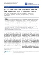
Figure 2 Spontaneous movement is only reduced after traumatic axonal injury with additional hypoxia. Distance travelled (metres) was
measured for 5 min as indicative of voluntary mobility in a novel open space. Diagrams depict: (A) Sham, (B) TAI, and (C) TAI+Hx. * indicates
significant differences between testing at the pre-injury (Pre) or post-injury at days 3, 6 and 14. Distance travelled is shown as mean ± SEM, n =
10 per group per time point. Note the significant reduction in walking distance in TAI+Hx rats at 3 and 6 days as compared to TAI and sham
rats. Data was analysed by 1-way ANOVA with Bonferroni post hoc test, with a p-value of < 0.05 considered significant.
Yan et al. Journal of Neuroinflammation 2011, 8:147
/>
Page 7 of 16
normal ranges reported in the literature [60] and
remained unchanged over time (not shown). In contrast,
whilst the brain water content of TAI and TAI+Hx rats
was similar to shams at 2 h post injury, by 24 h, it
increased significantly in TAI rats when compared with
sham (79.27 ± 0.14% vs 78.81 ± 0.14%, respectively; p <
0.05; Figure 3) and increased to near significance
between TAI+Hx and sham (79.27 ± 0.22% vs 78.81 ±
0.14%, respectively; p = 0.1147). The brain water content
remained elevated in both trauma groups for 48 h after
injury, and then decreased to sham levels by 72 h. Overall, brain water content was similar in TAI and TAI+Hx
groups at all time points examined.
The lateral ventricles are enlarged after TAI and TAI+Hx
Brain Water Content (%)
We measured the changes in lateral ventricle at +1.0
mm to bregma in concurrence with Paxinos and Watson rat brain atlas [61]. Ventricular size was unchanged
at all timepoints in animals that underwent sham surgery or hypoxia alone (data not shown). The ventricles
of TAI animals were significantly enlarged 1 day postinjury when compared to sham (2.55 ± 0.49% vs 0.65 ±
0.23%, p < 0.01; Figure 4A, B, C). Post-TAI hypoxia
resulted in a further, non significant increase in the size
of the ventricles at 1 day (3.50 ± 0.57%; Figure 4D)
when compared with TAI only rats (2.55 ± 0.49%). This
size was 5.4-fold larger than sham (3.50 ± 0.57% vs 0.65
± 0.23%; p < 0.001) (Figure 4A). By 7 days, although the
Sham
TAI
TAI+Hx
*
79.5
ventricular size was reduced as compared to day 1, they
were still larger than sham control rats being 2.43 ±
0.54% in TAI and 2.04 ± 0.45% in TAI+Hx animals.
The production of cytokines is enhanced following TAI
+Hx
The neuroinflammatory response was determined by measuring changes in cytokine production in the homogenised
cortex over 4 days (Figure 5). In these experiments six cytokines were measured: IL-6, IL-1b, TNF, IL-2, IL-4 and IL10. However, relevant differences were only detected in
three of them, IL-6, IL-1b and TNF. For the other cytokines including the pro-inflammatory IL-2 and anti-inflammatory IL-4 and IL-10, no changes were detected in either
the TAI or TAI+Hx groups, with values remaining comparable to those of sham animals over time (Figure 5D-F).
Hypoxia alone did not induce any changes in brain cytokine concentration at any time points (data not shown).
IL-6
In comparison to the cytokines measured in these experiments, IL-6 presented the highest concentration in the
injured cortex. By 2-way ANOVA, the overall increase of
IL-6 (all time points within the group analysed together)
was significantly more elevated in TAI+Hx brains when
compared to either the sham or TAI groups (p < 0.05, Figure 5A), while no changes were observed between sham
and TAI animals. Using post hoc analysis, we demonstrated
that hypoxia following TAI significantly increased the concentration of IL-6 in the brain at 24 h (12.67 ± 1.95 pg/mg
protein) and 48 h (11.30 ± 1.86 pg/mg protein) when compared with sham animals (6.71 ± 1.17 pg/mg protein, p <
0.05). In addition, TAI+Hx rats had significantly higher IL6 levels than TAI rats at 24 h post-injury (12.67 ± 1.95 pg/
mg protein vs 8.26 ± 0.65 pg/mg protein; p < 0.05).
79.0
IL-1b
78.5
78.0
S
2
24
48
72
96
Time Post-Injury (hours)
Figure 3 Increase in brain edema does not differ in traumatic
axonal injury rats with or without hypoxia. Brain water content
was determined at 2, 24, 48, 72 and 96 h post-injury, and calculated
as percentage of dry and wet ratio in the brain of sham (S), TAI
alone, and TAI with hypoxia (TAI+Hx) animals. * indicates significant
difference between groups. Both TAI and TAI+Hx showed similar
increases in brain water content, and no differences were found
between these groups. Data shown as mean ± SEM, n = 6 per
group per time point. Data was analysed by 1-way ANOVA with
Bonferroni post hoc test, with a p-value < 0.05 considered
significant.
In contrast to IL-6, the elevation of IL-1b occurred earlier and transiently after TAI (Figure 5B). In the TAI
group, a significant increase was observed 2 h post
injury (2.40 ± 0.15 pg/mg protein) as compared with
sham (1.76 ± 0.68 pg/mg protein; p < 0.05). In the TAI
+Hx group, a more striking significant increase was
observed at both 2 h (3.10 ± 0.56 pg/mg protein) and
24 h (2.44 ± 0.21 pg/mg protein) as compared with
sham (p < 0.05). A significant difference was also found
between TAI and TAI+Hx at 24 h post injury (1.81 ±
0.15 pg/mg protein vs 2.44 ± 0.21 pg/mg protein; p <
0.05). The concentration of IL-1b in both injury groups
returned to sham levels at 48 h post-injury.
TNF
No increase in TNF was detected at any timepoint
examined in the TAI group. Instead, similarly to IL-1b,
Yan et al. Journal of Neuroinflammation 2011, 8:147
/>
Page 8 of 16
Figure 4 Ventricular enlargement. Enlargement of the lateral ventricles following TAI and TAI with hypoxia (TAI+Hx) was quantified by
expressing the ventricle size as percentage of the entire brain section (A), at coronal plane of +1.0 mm to bregma in accordance with rat atlas
by Paxinos and Watson [61]. Coronal sections of (B) sham, (C) TAI alone and (D) TAI+Hx taken at +1 mm to bregma at 1 day after injury. *
indicates significant differences to sham group. Data shown as mean ± SEM, n = 6 per group per time point. Data was analysed by 1-way
ANOVA with Bonferroni post hoc test, with a p-value of < 0.05 considered significant.
the concentration of TNF in the brain of TAI+Hx rats
was significantly increased at 2 h when compared with
sham controls (2.67 ± 0.26 pg/mg protein vs 1.29 ± 0.26
pg/mg protein; p < 0.05). In TAI+Hx group TNF rapidly
returned close to the sham level at 24 h (Figure 5C).
Changes in metabolism after TAI and TAI+Hx
TBI is known to result in a reduction of oxidative metabolism [62]. We expected post-TAI hypoxia to aggravate
the metabolic disarray caused by diffuse axonal injury
and employed the microdialysis technique to monitor
changes of various metabolites over 4 days. Due to the
detection of significant alterations in brain metabolites
following the implantation of microdialysis probe in
uninjured sham animals as reported by others [63], we
chose to discard samples over the first 20 h following
probe implantation to reduce the artifact from the needle injury. In this study we were only present data of
glucose, lactate and glutamate from the microdialysates,
since pyruvate is known to become unstable after prolonged storage time (CMA Microdialysis).
Depression of glucose metabolism is prolonged after TAI
+Hx
Overall a significant hypoglycemia was observed in both
TAI and TAI+Hx groups when compared with sham (p
< 0.0001, Figure 6A). At 21 h post injury the concentration of glucose in TAI rats was similar to sham (0.09 ±
0.06 mmol/L vs 0.09 ± 0.04 mmol/L) and remained
similar until 33 h, after which time a substantial
decrease was observed, with glucose levels dropping to
30% of sham values (0.03 ± 0.02 mmol/L vs 0.09 ± 0.04
mmol/L) (Figure 6A &6B). Glucose levels remained low
until 51 h post-injury, when values gradually increased
toward to sham levels before they dropped again below
sham levels from 69 h until the end of experiment. In
TAI+Hx rats, glucose levels in the microdialysate were
approximately 50% lower than the levels of sham or
Yan et al. Journal of Neuroinflammation 2011, 8:147
/>
18
16
14
12
10
8
6
4
2
0
*
S
2
*
24
48
72
96
4
*
3
2
1
0
S
2
24
48
72
0.1
2
24
48
72
3
2
1
0
S
2
96
Time Post-Injury (hours)
24
48
72
96
Time Post-Injury (hours)
0.6
0.4
0.2
0.0
Brain IL-10 (pg/mg protein)
0.2
S
*
S
2
24
48
72
96
Time Post-Injury (hours)
F
0.3
0.0
*
*
Sham
TAI
TAI+Hx
0.8
96
Time Post-Injury (hours)
E
4
*
D
Brain IL-2 (pg/mg protein)
Brain TNF (pg/mg protein)
B
Time Post-Injury (hours)
C
Brain IL-4 (pg/mg protein)
*
Brain IL-1 (pg/mg protein)
Brain IL-6 (pg/mg protein)
A
Page 9 of 16
6
4
2
0
S
2
24
48
72
96
Time Post-Injury (hours)
Figure 5 Cytokines IL-6, IL-1b and TNF are increased in rats after traumatic axonal injury with additional hypoxia. The concentration
(pg/mg protein) of cytokines (A) IL-6, (B) IL-1b, (C) TNF, (D) IL-2, (E) IL-4 and (F) IL-10 was measured in cortical homogenates of sham (S), TAI
alone, and TAI with hypoxia (TAI+Hx) animals by multiplex assay over 4 days. * indicates significant differences between groups. Note the
significant increases of IL-6 and IL-1b in TAI+Hx vs TAI rats. TNF did not increase after TAI alone, and was only evident at 2 h in TAI+Hx rats.
Data shown as mean ± SEM, n = 6 per group per time point. Data was analysed by 1-way ANOVA with Bonferroni post hoc test, with a p-value
of < 0.05 considered significant.
TAI rats at 21 h (0.04 ± 0.02 mmol/L vs 0.09 ± 0.06
mmol/L and 0.09 ± 0.04 mmol/L, respectively) (Figure
6A &6B), with these low values subsisting until 51 h.
While the TAI rats showed some elevation in glucose
levels after 51 h, TAI+Hx rats had the opposite pattern,
with values further decreasing to less than 10% of those
observed in sham, (0.005 ± 0.002 mmol/L), and remaining under 10% of sham values for the study period.
Yan et al. Journal of Neuroinflammation 2011, 8:147
/>
Glucose
Sham
TAI
TAI+Hx
Glucose (mmol/L)
0.4
0.3
0.2
0.1
0.0
400
% change
from sham values
A
Page 10 of 16
TAI
TAI+Hx
200
100
21 33 39 45 51 57 63 69 75 81 87 93 99
Time Post Trauma (h)
C
Glucose
300
0
21 33 39 45 51 57 63 69 75 81 87 93 99
B
Time Post Trauma (h)
D
Lactate
Lactate
0.6
% change
from sham values
Lactate (mmol/L)
3000
0.4
2000
1000
0.2
0.0
100
0
21 33 39 45 51 57 63 69 75 81 87 93 99
21 33 39 45 51 57 63 69 75 81 87 93 99
Time Post Trauma (h)
E
Glutamate
20
Glutamate
1000
15
10
5
0
F
2000
% change
from sham values
Glutamate ( mol/L)
25
Time Post Trauma (h)
21 33 39 45 51 57 63 69 75 81 87 93 99
Time Post Trauma (h)
100
50
0
21 33 39 45 51 57 63 69 75 81 87 93 99
Time Post Trauma (h)
Figure 6 Metabolic alterations are exacerbated in rats exposed to traumatic axonal injury with additional hypoxia. Cerebral
microdialysis samples were analysed between 21 h and 99 h after sham surgery, TAI and TAI with 30 min hypoxia (TAI+Hx). Data are expressed
as both raw values and percentage changes from sham values for glucose (A, raw values; B, % change from sham levels), lactate (C, raw values;
D, % change from sham levels) and glutamate (E, raw values; F, % change from sham). Shaded area in (C) and (D) represents the peak period of
edema, which correlated with maximal lactate production. Overall a significant hypoglycaemic response was observed in both the TAI and TAI
+Hx groups compared to shams (2-way repeated measures ANOVA, p < 0.05). Data shown as mean ± SEM, n = 5 per group per time point.
Data was analysed by 2-way ANOVA repeated measures, with a p-value of < 0.05 considered significant.
Yan et al. Journal of Neuroinflammation 2011, 8:147
/>
Lactate is elevated after TAI+Hx and coincides with the
peak period of edema
Lactate levels in the microdialysates of sham-treated animals remained low for the duration of the study. Lactate
measurements for TAI animals were similar to sham
levels from 21 h until 51 h, when a substantial decrease
was observed to values less than 60% of sham (Figure
6C &6D). Values in TAI rats remained lower than sham
until 87 h, when lactate levels recovered to sham-level
were observations. Although not statistically significant,
TAI+Hx rats had lactate levels which were 400% higher
at 21 h when compared to sham or TAI (0.31 ± 0.2
mmol/L vs 0.08 ± 0.03 mmol/L and 0.09 ± 0.05 mmol/
L, respectively; Figure 6C &6D). The lactate levels in
TAI+Hx rats increased until 51 h correlating with the
peak period of edema observed in this study (shaded
area, Figure 6C &6D), then rapidly decreased to 10% of
sham levels at 75 h before recovering to sham- and
TAI-levels by 87 h (0.05 ± 0.01 mmol/L vs 0.05 ± 0.02
mmol/L and 0.01 ± 0.01 mmol/L, respectively).
Glutamate level is depressed after TAI and TAI+Hx
Glutamate levels in TAI animals at 21 h were approximately 40% less than those observed in sham animals
(4.58 ± 2.21 μmol/L vs 8.02 ± 2.74 μmol/L; Figure 6E
&6F), and remained low until 45 h, at which point the
microdialysate levels returned to sham levels until 87 h,
when another decrease was observed. TAI+Hx rats had
glutamate levels of approximately 50% of sham at 21 h,
and though not significant, a peak was observed at 39 h
to more than 200% of sham values (11.84 ± 8.54 μmol/L
vs 5.71 ± 1.64 μmol/L; Figure 6E &6F). From this time
onwards, glutamate levels in TAI+Hx rats decreased
again to 50% of sham values, and remained at between
30-60% of shams for the remainder of the experimental
period.
Discussion
Cerebral hypoxia, along with hypotension, is one of the
most critical factors worsening secondary brain damage
after TBI, and particularly following diffuse TBI [6,13].
Despite this clinical relevance, the underlying mechanisms by which hypoxia aggravates neurological outcome
following TBI have not been studied adequately.
Using focal or mixed focal-diffuse models, systemic
hypoxia following TBI in rats exacerbates neurological
deficit [32,37] and increases the lesion size, neuronal
death [33,34,37,64] and brain edema, while reducing cerebral blood flow [35,51]. However, the role of post-traumatic hypoxia elicited after diffuse brain injury has
rarely been addressed. Therefore, we explored the
impact of hypoxia using a model of diffuse TAI
[40,65,66] followed by a 30-min hypoxic ventilation.
Using this combinatorial insult model, we previously
Page 11 of 16
reported enhanced axonal damage and macrophage
infiltration within the corpus callosum and the brain
stem [55]. Thus, in this follow-up study we further
investigated changes in neurological outcome, brain
edema, ventricle enlargement, cerebral cytokines, and
energy metabolism.
We found that in comparison to TAI alone, an additional hypoxic insult enhanced sensorimotor deficits on
the Rotarod, beam walk and tape removal tests, reduced
spontaneous exploratory behavior, and delayed recovery.
These data closely relate to clinical studies on TBI
patients showing that post-traumatic hypoxia worsens
neurological outcome and prolongs the recovery period
[7,8,67]. The behavioural data in this model of TAI are
consistent with similar deficits shown at day 1 in previous studies using diffuse or focal TBI models in combination with hypoxia [32,34,36-38,68]. However, in
extension of this early work, our results show that an
additional hypoxic insult has a detrimental effect on
behaviour, inflammatory and metabolic outcomes for an
extended period of time.
Brain swelling is a major contributor for the development of secondary ischemia causing raised ICP and
decreased cerebral perfusion pressure [69]. Enlargement
of the brain due to edema [70] and/or obstruction of
CSF flow [71] is a common event in severe TBI patients
and a frequent cause of death. Cytotoxic edema results
from excessive accumulation of ion and water within
the cell, while vasogenic edema is caused by increased
vascular permeability and subsequent fluid extravasation
into the parenchyma. Here, we demonstrated that at 2 h
after TAI, brain water content was similar to sham animals, but it increased to a peak between 24 and 48 h,
and remained elevated until 72 h. Although hypoxia following TAI exacerbated sensorimotor deficit, it did not
further increase cerebral edema when compared with
TAI only animals, corroborating previous observations
using diffuse-weighted imaging [35]. Interestingly, using
MRI, others demonstrated that acute brain swelling
after TAI (both with and without hypoxia), as early as
60 min post-injury, was associated with increased extracellular fluid and BBB dysfunction, indicative of vasogenic edema [72-75]. This early brain swelling was
transient, with values quickly returning to sham levels
[53,58,75]. Since the earliest timepoint examined in our
study was 2 h, it is likely that we missed this initial peak
in edema, as no differences were detected between TAI,
TAI+Hx and sham rats later on. However, other studies
have also demonstrated that a modest, widespread second edematous response occurs at 24 h after TAI
despite the intact BBB, which suggests ongoing cytotoxic
edema [58,75]. Our results are consistent with this modest yet significant increase of edema at 24 h, which was
maintained until 48 h. It is possible that the peak in
Yan et al. Journal of Neuroinflammation 2011, 8:147
/>
brain water content observed at 24 h in both the TAI
and TAI+Hx rats (approximately 79.3%) reflects a sort
of saturation level, with the brain unable to tolerate any
further water accumulation. Other studies also demonstrated peak edema of similar degree after TBI
[59,76,77].
An interesting observation was the enlargement of the
lateral ventricles after TAI, and even greater following
TAI+Hx. Recent clinical neuroimaging studies have
shown correlations between ventricular enlargement and
long-term neurological impairment [78-80]. The prognostic value of ventricular dilatation had high sensitivity
and specificity for the prediction of cognitive outcome
[80-83]. In this study, we showed that the lateral ventricles are markedly enlarged at 1 day post-injury after
TAI and even larger in TAI+Hx animals, when compared to sham or rats with isolated hypoxia. Although
we did not examine the mechanism leading to ventricular enlargement after TAI, imaging studies on TBI
patients suggested that white matter degeneration
around the lateral ventricle may be a contributing factor
[84]. However, since ventricular enlargement in TAI rats
was an early and transient effect, it could be most likely
attributed to the onset of post-traumatic hydrocephalus,
caused by impaired CSF circulation due to edema compressing the aqueduct of sylvius.
Neuroinflammation has been extensively investigated
in hypoxia-ischemia and TBI in both humans and animal models [85] and all these studies have reported a
robust elevation of cytokines in the central nervous system [19,28,86-89]. More relevant for this study, our preliminary data on severe TBI patients with additional
hypoxic insult have shown enhanced and prolonged production of cytokines in the CSF (Yan et al: Neuroinflammation and brain injury markers in TBI patients:
Differences in focal and diffuse brain damage, and normoxic or hypoxic status on neurological outcome;
manuscript in preparation). Consistently, here we
demonstrated exacerbated production of IL-6, IL-1b,
and TNF in the brains after TAI with additional
hypoxia.
IL-1b is a key mediator of the inflammatory response,
which exacerbates neuronal injury and induces BBB dysfunction by stimulating matrix metalloproteinases [90].
IL-1b mRNA is upregulated within minutes after TBI,
and increased protein levels are detectable within an
hour after TBI [21,91-93]. In this study, IL-1b increased
early after TAI alone, peaking at 2 h. Post-TAI hypoxia
significantly enhanced IL-1b concentration at 2 h compared to TAI-only rats. In addition, whilst the elevation
of IL-1b in TAI-only rats appeared to be transient, in
TAI+Hx rats IL-1b was still significantly elevated at 24
h, suggesting that the addition of hypoxia prolongs
neuroinflammation.
Page 12 of 16
The neurotoxic effects of IL-1b are synergistically
enhanced in the presence of TNF [94], as both share
many of the same physiologic effects. However, the role
of TNF following TBI is controversial, neuronal toxicity
of TNF has been demonstrated with local TNF administration inducing breakdown down of the BBB and
increased leukocyte recruitment [95-98]. Clinically, high
levels of TNF in the CSF of brain-injured patients correlated with BBB dysfunction [99]. TNF inhibition also
reduced cerebral ischemia/reperfusion injury [100],
decreased TBI induced neuronal damage [101], and
ameliorated BBB dysfunction after closed head injury
[102]. However, studies on TNF deficient mice demonstrated an early functional improvement between 24-48
h after TBI, but failed to produce further amelioration
at 4 weeks [103]. Taken together, these studies suggest
that TNF may be deleterious in the acute phase postinjury, but beneficial for long-term recovery. In accordance with Kamm et al. [93], no changes in TNF levels
were detected in rats subjected to TAI alone, whereas
the combination of TAI and hypoxia elicited a significant early increase in TNF at 2 h post-injury, lasting up
to 72 h post-injury. These early enhancement in the
TAI+Hx rats possibly reflects a more severe brain
damage in this combined insult model.
Similar to IL-1b and TNF, at 24 h IL-6 was significantly higher in TAI+Hx rats compared to TAI alone.
IL-1b is an early mediator inducing the production of
IL-6 at both mRNA and protein levels [21]. IL-6 displays
pleiotropic functions with both deleterious and beneficial effects in the injured brain [104-106]. Using the
mild severity (250 g/2 m) of the Marmarou model, we
showed that IL-6 increased in rat CSF within 24 h and
IL-6 protein and mRNA was found expressed on neurons [95]. Studies of IL-6 gene-deficient mice have provided more information in regards to the protective
function of IL-6, by having a compromised immune
response, increased oxidative stress and neurodegeneration [107]. In this study, we demonstrate significantly
heightened IL-6 levels in the TAI+Hx rats at 24 h,
which remained elevated above TAI levels until 96 h.
Altogether, the increased acute production of IL-1b and
TNF may be associated with disruption of BBB integrity
and consequently formation of cerebral edema, while
late elevation of IL-6 may trigger repair mechanisms
[24,99].
We also investigated changes in energy metabolism in
this combinatorial insult model. Due to the nature of
the impact acceleration injury, it is impractical to
implant a microdialysis probe prior to injury without
compromising the integrity of the trauma. It is also difficult to implant the probe directly after trauma as it
resulted in higher mortality rate. Carré and colleagues
implanted the probe 2 weeks prior to injury, but without
Yan et al. Journal of Neuroinflammation 2011, 8:147
/>
success [108]. We therefore allowed the rats to recover
for 4 hours after TAI before implanting the microdialysis probe. In accordance with others [63], our study has
shown that in sham rats energy metabolism is altered
during the first 24 h following microdialysis probe
implantation, therefore we chose to examine only the
data from 20 h onwards to reduce the “probe effect”.
At 21 h, the glucose values for TAI+Hx rats were
substantially lower compared to TAI or sham rats, and
dropped to extremely low levels from 57 h onwards.
These low levels of cerebral glucose could be the result
of low glucose availability and/or hyperglycolysis in the
acute post-injury phase. Hyperglycolysis has previously
been shown as common early event following neurotrauma both experimentally and in the clinic [109,110].
It is often followed by a prolonged period of metabolic
depression beginning as early as 6 h post-injury,
remaining for as long as 5 days [111,112], a phenomenon which has also been demonstrated in the present
study. Interestingly, rats subjected to TAI experienced
only a brief period of glucose depletion between 39 h
and 57 h, at which time glucose levels returned to
sham values for the remaining duration of monitoring.
It is possible that the additional hypoxic insult
depleted available glucose stores in the TAI+Hx animals, and thus a prolonged compensatory period of
anaerobic respiration occurred to provide essential
ATP and generate lactate as by-product. Our experiments have demonstrated that this is a protracted process, lasting for 51 h after TAI. Lactate may be utilized
by the brain during periods of increased brain energy
requirements in which ATP and glucose stores are
exhausted, such as following TBI [113,114]. In a situation of prolonged glucose depletion, high concentrations of lactate and high-level energy usage for
neuronal repair or alternative metabolic pathways may
further reduce the ATP reserves, with a subsequent
mismatch between glucose transport, uptake and ATP
production [115,116]. This may explain the further
drop in glucose concentrations at 57 h post TAI, in
that the restoration of aerobic metabolism decreases
lactate concentration but further reduces glucose.
Post-traumatic impairment in energy metabolism is a
major contributor to cytotoxic edema, and interestingly, the period of elevated lactate in the TAI+Hx rats
between 21 h and 57 h overlaps with the peak of
increased brain water content. As edema begins to
reside, lactate levels in these rats return to sham
values. This prolonged period of metabolic crisis also
extends to glutamate production, which was depressed
below sham levels for TAI, and particularly TAI+Hx
rats, for the duration of the monitoring by
microdialysis.
Page 13 of 16
Conclusion
In this study, we reproduced a frequent debilitating condition contributing to poor neurological outcome in
humans by using a rat a model of diffuse TAI combined
with an hypoxic insult. Consistent with our hypothesis,
we demonstrated exacerbation of sensorimotor deficits
and delayed neurological recovery in TAI+Hx rats, as
well as a significant enlargement of the lateral ventricles
after TAI and TAI+Hx. However, no differences were
detected in brain edema, which was similarly increased
in both TAI and TAI+Hx injury groups. Enhanced neuroinflammation via amplified cerebral production of IL1b, TNF and IL-6 corroborates our previous findings of
exacerbated macrophage/microglial accumulation in
regions of axonal pathology in the corpus callosum and
brainstem of TAI+Hx animals [55]. Interestingly, while
TAI rats had a gradual recovery in glucose levels, metabolic depression was sustained in TAI+Hx rats, showing
elevated lactate in microdialysates coinciding with the
period of increased brain edema. Overall, the morphological and behavioural changes of this combined model
of diffuse TBI and hypoxia has similar characteristic of
the reported severe brain damage and poor outcomes in
patients with diffuse brain injury and hypoxia.
List of abbreviations
ATP: adenosine triphosphate; BBB: blood brain barrier; CSF: cerebrospinal
fluid; Hx: hypoxia; IFN: interferon; IL: interleukin; MABP: mean arterial blood
pressure; MRI: magnetic resonance imaging; pO2: partial pressure of oxygen;
rpm: revolutions per minute; sham: sham-operated animals; sO2: oxygen
saturation; TAI: traumatic axonal injury; TAI+Hx: traumatic axonal injury with
hypoxia; TBI: traumatic brain injury: TNF: tumor necrosis factor.
Acknowledgements
This study was supported by the National Health and Medical Research
Council Australia and the Victorian Neurotrauma Initiative.
Author details
1
National Trauma Research Institute, The Alfred Hospital, 89 Commercial
Road, Melbourne 3004, Australia. 2Department of Surgery, Monash University,
89 Commercial Road, Melbourne 3004, Australia. 3Department of Medicine,
Monash University, 89 Commercial Road, Melbourne 3004, Australia.
4
Department of Clinical Neuroscience, Section for Neurosurgery, Karolinska
University Hospital, Karolinskavägen, Solna, Stockholm 171 76, Sweden.
Authors’ contributions
EBY designed the study, performed all animal work and microdialysis probe
implantation, performed cytokine measurements, drafted the manuscript,
and performed statistical analysis. SCH assisted with animal work, performed
sensorimotor experiments, carried out the histology and ventricle
measurements, performed statistical analysis, and drafted the manuscript.
BMB carried out microdialysis sample measurements and assisted with
manuscript preparation. DAA assisted with animal work, carried out
sensorimotor and open field exploration experiments, and performed edema
experiments. CMK conceived of the study and oversaw its design and
coordination, and drafted the manuscript. All authors have read and
approved the final manuscript.
Competing interests
The authors declare that they have no competing interests.
Yan et al. Journal of Neuroinflammation 2011, 8:147
/>
Page 14 of 16
Received: 7 September 2011 Accepted: 28 October 2011
Published: 28 October 2011
24.
References
1. Maas AIR, Stocchetti N, Bullock MR: Moderate and severe traumatic brain
injury in adults. Lancet Neurol 2008, 7:728-741.
2. Gaetz M: The neurophysiology of brain injury. Clin Neurophysiol 2004,
115:4-18.
3. Masel BE, DeWitt DS: Traumatic brain injury: A disease process, not an
event. J Neurotrauma 2010, 27:1529-1540.
4. Myburgh JA, Cooper JC, Finfer SR, Venkatesh B, Jones D, Higgins A,
Bishop N, Higlett T: Epidemiology and 12-month outcomes from
traumatic brain injury in Australia and New Zealand. J Trauma 2008,
64:854-862.
5. Ding K, Marquez de la Plata C, Wang JY, Mumphrey M, Moore C, Harper C,
Madden C, McColl R, Whittemore A, Devous MD, Diaz-Arrastia RR: Cerebral
atrophy after traumatic white matter injury: correlation with acute
neuroimaging and outcome. J Neurotrauma 2008, 25:1433-1440.
6. McHugh GS, Engel DC, Butcher I, Steyerberg EW, Lu J, Mushkudiani N,
Hernández AV, Marmarou A, Maas AIR, Murray GD: Prognostic value of
secondary insults in traumatic brain injury: results from the IMPACT
study. J Neurotrauma 2007, 24:287-293.
7. Chesnut RM, Marshall LF, Klauber MR, Blunt BA, Baldwin N, Eisenberg HA,
Jane JA, Marmarou A, Faulkes MA: The role of secondary brain injury in
determining outcome from severe head injury. J Trauma 1993,
34:216-222.
8. Jeremitsky E, Omert L, Dunham CM, Protetch J, Rodriguez A: Harbingers of
poor outcome the day after severe brain injury: hypothermia, hypoxia,
and hypotension. J Trauma 2003, 54:312-319.
9. Silverston P: Pulse oximetry at the roadside: a study of pulse oximetry in
immediate care. Br Med J 1989, 298:711-713.
10. McHugh GS, Engel DC, Butcher I, Steyerberg EW, Lu J, Mushkudiani N,
Hernández AV, Marmarou A, Maas AIR, Murray GD: Prognostic value of
secondary insults in traumatic brain injury: results from the IMPACT
study. J Neurotrauma 2007, 24:287-293.
11. Gentleman D, Jennett B: Audit of transfer of unconscious head-injured
patients to a neurosurgical unit. The Lancet 1990, 335:330-334.
12. Jones PA: Measuring the burden of secondary insults in head-injured
patients during intensive care. J Neurosurg Anasthesiol 1994, 6:4-14.
13. Miller JD: Head Injury. J Neurol Neurosurg Pschiatry 1993, 56:440-447.
14. Adams JH, Graham DI, Murray L, Scott G: Diffuse axonal injury due to
nonmissile head injury in humans: an analysis of 45 cases. Ann Neurol
1982, 12:557-563.
15. Newcombe VFJ, Williams GB, Scoffings D, Cross J, Carpenter TA, Pickard JD,
Menon DK: Aetiological differences in neuroanatomy of the vegetative
state: insights from diffusion tensor imaging and functional implications.
J Neurol Neurosurg Psychiatry 2010, 81:552-561.
16. Chesnut RM: Guidelines for the management of severe traumatic brain
injury. Emerg Med Clin North Am 1997, 15:581-604.
17. Siegel JH: The effect of associated injuries, blood loss, and oxygen debt
on death and disability in blunt traumatic brain injury: the need for
early physiologic predictors of severity. J Neurotrauma 1995, 12:579-590.
18. Lenzlinger PM, Morganti-Kossmann MC, Laurer HL, McIntosh T: The duality
of the inflammatory response to traumatic brain injury. Mol Neurobiol
2001, 24:169-181.
19. Ziebell JM, Morganti-Kossmann MC: Involvement of pro- and antiinflammatory cytokines and chemokines in the pathophysiology of
traumatic brain injury. Neurotherapeutics 2010, 7:22-30.
20. Helmy A, Carpenter KL, Menon DK, Pickard JD, Hutchinson PJ: The cytokine
response to human traumatic brain injury: temporal profiles and
evidence for cerebral parenchymal production. J Cereb Blood Flow Metab
2011, 31:658-670.
21. Yan HQ, Banos MA, Herregodts P, Hooghe R, Hooghepeters EL: Expression
of interleukin (IL)-1-beta, IL-6 and their respective receptors in the
normal rat brain after injury. Eur J Immunol 1992, 22:2963-2971.
22. Kossmann T, Stahel PF, Lenzlinger PM, Redl H, Dubs RW, Trentz O, Schlag G,
Morganti-Kossmann MC: Interleukin-8 released into the cerebrospinal
fluid after brain injury is associated with nerve growth factor
production. J Cereb Blood Flow Metab 1997, 17:280-289.
23. Kossmann T, Hans VH, Imhof HG, Stocker R, Grob P, Trentz O, MorgantiKossmann MC: Intrathecal and serum interleukin-6 and the acute-phase
25.
26.
27.
28.
29.
30.
31.
32.
33.
34.
35.
36.
37.
38.
39.
40.
41.
42.
response in patients with severe traumatic brain injuries. Shock 1995,
4:311-317.
Kossmann T, Hans VH, Imhof HG, Trentz O, Morganti-Kossmann MC:
Interleukin-6 released in human cerebrospinal fluid following traumatic
brain injury may trigger nerve growth factor production in astrocytes.
Brain Res 1996, 713:143-152.
Hans VH, Kossmann T, Joller H, Otto V, Morganti-Kossmann MC: Interleukin6 and its soluble receptor in serum and cerebrospinal fluid after
cerebral trauma. Neuroreport 1999, 5:409-412.
Bell MJ, Kochanek PM, Doughty LA, Carcillo JA, Adelson PD, Clark RSB,
Wisniewski SR, Whalen MJ, DeKosky ST: Interleukin-6 and interleukin-10 in
cerebrospinal fluid after severe traumatic brain injury in children. J
Neurotrauma 1997, 14:451-457.
Whalen MJ, Carlos TM, Kochanek PM, Wisniewski SR, Bell MJ, Clark RSB,
DeKosky ST, Marion DW, Adelson PD: Interleukin-8 is increased in
cerebrospinal fluid of children with severe head injury. Crit Care Med
2000, 28:929-934.
Frugier T, Morganti-Kossmann MC, O’Reilly D, McLean CA: In situ detection
of inflammatory mediators in post mortem human brain tissue after
traumatic injury. J Neurotrauma 2010, 27:497-507.
Hagberg H, Gilland E, Bona E, Hanson LÅ, Hahn-Zoric M, Holst M, McRae A,
Söder O: Enhanced expression of interleukin (IL)-6 and IL-6 messenger
RNA and bioactive protein after hypoxia-ischemia in neonatal rats.
Pediatr Res 1996, 40:603-609.
Williams AJ, Wei HH, Dave JR, Tortella FC: Acute and delayed
neuroinflammatory response following experimental penetrating ballistic
brain injury in the rat. J Neuroinflammation 2007, 4.
Shreeniwas R, Koga M, Pinsky D, Kaiser E, Brett J, Wolitzky BA, Norton C,
Plocinski J, Benjamin W, Burns DK, et al: Hypoxia-mediated induction of
endothelial-cell interleukin-1-alpha- an autocrine mechanism promoting
expression of leukocyte adhesion molecules on the vessel surface. J Clin
Invest 1992, 90.
Clark RSB, Kochanek PM, Dixon CE, Chen M, Marion DW, Heineman S,
DeKosky ST, Graham SH: Early neuropathologic effects of mild or
moderate hypoxemia after controlled cortical impact injury in rats. J
Neurotrauma 1997, 14:179-189.
Matsushita Y, Bramlett HM, Alonso O, Dietrich WD: Posttraumatic
hypothermia is neuroprotective in a model of traumatic brain injury
complicated by a secondary hypoxic insult. Crit Care Med 2001,
29:2060-2066.
Ishige N, Pitts L, Hashimotos T, Nishimura M, Bartkowski H: The effects of
hypoxia on traumatic brain injury in rats: Part 1: alterations in
neurologic function, electroencephalograms, and histopathology.
Neurosurgery 1987, 20:848-853.
Van Putten HP, Bouwhuis MG, Muizelaar JP, Lyeth BG, Berman RF:
Diffusion-weighted imaging of edema following traumatic brain injury
in rats: effects of secondary hypoxia. J Neurotrauma 2005, 22:857-872.
Bramlett HM, Dietrich WD, Green EJ: Secondary hypoxia following fluid
percussion brain injury in rats exacerbates sensorimotor and cognitive
deficits. J Neurotrauma 1999, 16:1035-1047.
Robertson CL, Clark RSB, Dixon CE, Alexander BS, Graham SH, Wisniewski SR,
Marion DW, Safar PJ, Kochanek PM: No long-term benefit from
hypothermia after severe traumatic brain injury with secondary insult in
rats. Crit Care Med 2000, 28:3218-3223.
Beaumont A, Marmarou A, Czigner A, Yamamoto M, Demetriadou K,
Shirotani T, Marmarou C, Dunbar J: The impact-acceleration model of
head injury: injury severity predicts motor and cognitive performance
after trauma. Neurol Res 1999, 21:742-754.
Lewén A, Matz P, Chan PH: Free radical pathways in CNS injury. J
Neurotrauma 2000, 17.
Foda MA, Marmarou A: A new model of diffuse brain injury in rats. Part
II: Morphological characterization. J Neurosurg 1994, 80:301-313.
Tavazzi B, Signoretti S, Lazzarino G, Amorini G, Delfini R, Cimatti M,
Marmarou A, Vagnozzi R: Cerebral oxidative stress and depression of
energy metabolism correlate with severity of diffuse brain injury in rats.
Neurosurgery 2005, 56:582-589.
Ando Y, Inoue M, Hirota M, Morino Y, Araki S: Effect of a superoxide
dismutase derivative on cold-induced brain edema. Brain Res 1989,
477:286-291.
Yan et al. Journal of Neuroinflammation 2011, 8:147
/>
43. Holmin S, Mathiesen T: Intracerebral administration of interleukin-1 beta
and induction of inflammation, apoptosis, and vasogenic edema. J
Neurosurg 2000, 92:108-120.
44. Habgood MD, Bye N, Dziegielewska KM, Ek CJ, Lane MA, Potter A,
Morganti-Kossmann MC, Saunders NR: Changes in blood-brain barrier
permeablility to large and small molecules following traumatic brain
injury in mice. Eur J Neurosci 2007, 25:231-238.
45. Banks WA, Erickson MA: The blood-brain barrier and immune function
and dysfunction. Neurobiol Dis 2010, 37:26-32.
46. Pleines UE, Morganti-Kossmann MC, Rancan M, Trentz O, Kossmann T: S100 beta reflects the extent of injury and outcome, whereas neuronal
specific enolase is a better indicator of neuroinflammation in patients
with severe traumatic brain injury. J Neurotrauma 2001, 18:491-498.
47. Bye N, Habgood MD, Callaway JK, Malakooti N, Potter A, Kossmann T,
Morganti-Kossmann MC: Transient neuroprotection by minocycline
following traumatic brain injury is associated with attenuated microglial
activation by no changes in cell apoptosis or neutrophil infiltration. Exp
Neurol 2007, 204:220-233.
48. Shohami E, Beit-Yannai E, Horowitz M, Kohen R: Oxidative stress in closedhead injury: Brain antioxidant capacity as an indicator of functional
outcome. J Cereb Blood Flow Metab 1997, 17:1007-1019.
49. Stahel PF, Shohami E, Younis FM, Kariya K, Otto V, Lenzlinger PM,
Grosjean MB, Trentz O, Kossmann T, Morganti-Kossmann MC: Experimental
closed head injury: analysis of neurological outcome, blood-brain barrier
dysfunction, intracranial neutrophil infiltration, and neuronal cell death
in mice deficient in genes for pro-inflammatory cytokines. J Cereb Blood
Flow Metab 2000, 20:369-380.
50. Gabrielian L, Willshire LW, Helps SC, van den Heuvel C, Mathias J, Vink R:
Intracranial pressure changes following traumatic brain injury in rats:
lack of significant change in the absence of mass lesions or hypoxia. J
Neurotrauma 2011.
51. Ishige N, Pitts LH, Berry I, Carlson SG, Nishmura MC, Moseley ME,
Weinstein PR: The effect of hypoxia on traumatic head-injury in ratsalterations in neurologic function, brain edema, and cerebral blood-flow.
J Cereb Blood Flow Metab 1987, 7:759-767.
52. Ito J, Marmarou A, Barzo P, Fatouros P, Corwin F: Charaterization of edema
by diffusion-weighted imaging in experimental traumatic brain injury. J
Neurosurg 1996, 84:97-103.
53. Ghabriel M, Zhu C, Imran A, Blumbergs P, Reilly P: Blood-brain barrier
ultrastructural changes in impact acceleration head trauma. 7th
International Neurotrauma Symposium Adelaide, Australia: Medimond; 2004,
89-92.
54. Gao G, Oda Y, Wei EP, Povlishock JT: The adverse pial arteriolar and
axonal consequences of traumatic brain injury complicated by hypoxia
and their therapeutic modulation with hypothermia in rat. J Cereb Blood
Flow Metab 2010, 30:628-637.
55. Hellewell SC, Yan EB, Bye N, Agyapomaa D, Morganti-Kossmann MC: Posttraumatic hypoxia exacerbates brain tissue damage: Analysis of axonal
injury and glial responses. J Neurotrauma 2010, 27:1997-2010.
56. Hallam TM, Floyd CL, Folkerts MM, Lee LL, Gong Q-Z, Lyeth BG,
Muizelaar JP, Berman RF: Comparison of behavioural deficits and acute
neuronal degeneration in rat lateral fluid percussion and weight-drop
brain injury models. J Neurotrauma 2004, 21:521-539.
57. Semple BD, Bye N, Rancan M, Ziebell JM, Morganti-Kossmann MC: Role of
CCL2 (MCP-1) in traumatic brain injury (TBI): evidence from severe TBI
patients and CCL2-/- mice. J Cereb Blood Flow Metab 2010, 30:769-782.
58. Barzo P, Marmarou A, Fatouros PP, Hayasaki K, Corwin F: Contribution of
vasogenic and cellular edema to traumatic brain swelling measured by
diffusion-weighted imaging. J Neurosurg 1997, 87:900-907.
59. Bouzat P, Francony G, Thomas S, Valable S, Mauconduit F, Fevre M-C,
Barbier EL, Bernaudin M, Lahrech H, Payen J-F: Reduced brain edema and
functional deficits after treatment of diffuse traumatic brain injury by
carbamylated erythropoietin derivative. Crit Care Med 2011, 39.
60. Vaz R, Sarmento A, Borges N, Cruz C, Azevedo T: Experimental traumatic
cerebral contusion: morphological study of brain microvessels and
characterization of the oedema. Acta Neurochir (Wien) 1998, 140:76-81.
61. Paxinos G, Watson C, (Eds.): The rat brain in stereotaxic coordinates. 2
edition. London: Academic Press; 1986.
62. Hovda DA, Yoshino A, Kawamata T, Katayama Y, Becker DP: Diffuse
prolonged depression of cerebral oxidative metabolism following
Page 15 of 16
63.
64.
65.
66.
67.
68.
69.
70.
71.
72.
73.
74.
75.
76.
77.
78.
79.
80.
81.
82.
83.
84.
85.
concussive brain injury in the rat: a cytochrome oxidase histochemistry
study. Brain Res 1991, 3:1-10.
Sumbria RK, Klein J, Bickel U: Acute depression of energy metabolism
after microdialysis probe implantation is distinct from ischemia-induced
changes in mouse brain. Neurochem Res 2011, 36:109-116.
Nawashiro H, Shima K, Chigasaki H: Selective vulnerability of hippocampal
CA3 neurons to hypoxia after mild concussion in the rat. Neurol Res 1995,
17:455-460.
Csuka E, Hans VH, Ammann E, Trentz O, Kossmann T, MorgantiKossmann MC: Cell activation and inflammatory response following
traumatic axonal injury in the rat. Clin Neurosci 2000, 11:2587-2590.
Marmarou A, Foda MA, Van Den Brink W, Campbell J, Kita H,
Demetriadou K: A new model of diffuse brain injury in rats. Part I:
Pathophysiology and biomechanics. J Neurosurg 1994, 80:291-300.
Kim Y-J: A systematic review of factors contributing to outcomes in
patients with traumatic brain injury. J Clin Nurs 2011, 20:1518-1532.
Bauman RA, Widholm JJ, Petras JM, McBride K, Long JB: Secondary
hypoxemia exacerbates the reduction of visual discrimination and
neuronal cell density in the dorsal lateral geniculate nucleus resulting
from fluid percussion injury. J Neurotrauma 2000, 17:679-693.
Marmarou A, Fatouros P, Barzó P, Portella G, Yoshihara M, Tsuji O,
Yamamoto T, Laine F, Signoretti S, Ward JD, et al: Contribution of edema
and cerebral blood volume to traumatic brain swelling in head-injured
patients. J Neurosurg 2000, 93:183-193.
Unterberg AW, Stover J, Kress B, Kiening KL: Edema and brain trauma.
Neuroscience 2004, 129:1021-1029.
Poca MA, Sahuquillo J, Mataro M, Benejam B, Arikan F, Baguena M:
Ventricular enlargement after moderate or severe head injury: A
frequent and neglected problem. J Neurotrauma 2005, 22:1303-1310.
Barzo P, Marmarou A, Fatouros P, Corwin F, Dunbar J: Magnetic resonance
imaging monitored acute blood-brain barrier changes in experimental
traumatic brain injury. J Neurosurg 1996, 85:1113-1121.
Marmarou A: Pathophysiology of traumatic brain edema: Current
concepts. Acta Neurochir Suppl 2003, 86:7-10.
Shohami E, Novikov M, Bass R: Long-term effect of HU-211, a novel noncompetitive NMDA antagonist, on motor and memory functions after
closed head injury in the rat. Brain Res 1995, 674:55-62.
Beaumont A, Marmarou A, Hayasaki K, Barzo P, Fatouros P, Corwin F,
Marmarou C, Dunbar J: The permissive nature of blood brain barrier
(BBB) opening in edema formation following traumatic brain injury. Acta
Neurochir Suppl 2000, 76:125-129.
Clausen F, Hånell A, Israelsson C, Hedin J, Ebendal T, Mir AK, Gram H,
Marklund N: Neutralization of interleukin-1β reduces cerebral edema and
tissue loss and improves late cognitive outcome following traumatic
brain injury in mice. Eur J Neurosci 2011, 34:110-123.
Taya K, Marmarou C, Okuno K, Prieto R, Marmarou A: Effect of secondary
insults upon aquaporin-4 water channels following experimental cortical
contusion in rats. J Neurotrauma 2010, 27:229-239.
Levin HS, Meyers CA, Grossman RG, Sarwar M: Ventricular enlargement
after closed head injury. Arch Neurol 1981, 38:623-629.
Meyers CA, Levin HS, Eisenberg HM, Guinto FC: Early versus late lateral
ventricular enlargment following closed head injury. J Neurol Neurosurg
Psychiatry 1983, 46:1092-1097.
Adams JH, Jennett B, Murray LS, Teasdale GM, Gennarelli TA, Graham DI:
Neuropathological findings in disabled survivors of a head injury. J
Neurotrauma 2011, 28:701-709.
Reider-Groswasser I, Cohen M, Costeff H, Groswasser Z: Late CT findings in
brain trauma- relationship to cognitive and behavioral sequelae and to
vocational outcome. American Journal Roentgenology 1993, 160:147-152.
Levin HS, Amparo EG, Eisenberg HM, Miner ME, High WM, Ewingcobbs L,
Fletcher JM, Guinto FC: Magnetic-resonance imaging after closed head
injury in children. Neurosurgery 1989, 24:223-227.
Uzzell BP, Dolinskas CA, Wiser RF, Langfitt TW: Influence of lesions
detected by computer tomography on outcome and neuropsychological
recovery after severe head injury. Neurosurgery 1987, 20:396-402.
Ariza M, Mataro M, Poca MA, Junque C, Garnacho A, Amoros S,
Sahuquillo J: Influence of extraneurological insults on ventricular
enlargement and neuropsychological functioning after moderate and
severe traumatic brain injury. J Neurotrauma 2004, 21:864-876.
Morganti-Kossmann MC, Satgunaseelan L, Bye N, Kossmann T: Modulation
of the immune response by head injury. Injury 2007, 38:1392-1400.
Yan et al. Journal of Neuroinflammation 2011, 8:147
/>
86. del Zoppo G, Hallenbeck JM, Iadecola C, Wang XK, Feuerstein GZ:
Inflammation and stroke: Putative role for cytokines, adhesion molecules
and iNOS in brain response to ischemia. Brain Pathol 2000, 10:95-112.
87. Macrez R, Ali C, Toutirais O, Le Mauff B, Defer G, Dirnagl U, Vivien D: Stroke
and the immune system: from pathophysiology to new therapeutic
strategies. Lancet Neurol 2011, 10:471-480.
88. Xia W, Han J, Huang G, Ying W: Inflammation in ischaemic brain injury:
Current advances and future perspectives. Clin Exp Pharmacol Physiol
2010, 37:253-258.
89. Stover JF, Beyer TF, Woiciechowsky C, Unterberg AW: Temporal profile of
cerebrospinal fluid glutamate, interleukin-6, and tumor necrosis factoralpha in relation to brain edema and contusion following controlled
cortical impact in rats. Neurosci Lett 2000, 288:25-28.
90. Vecil GG, Larsen PH, Corley SM, Herx LM, Besson A, Goodyer CG, Yong VW:
Interleukin-1 is a key regulator of matrix metalloproteinase-9 expression
in human neurons in culture and following mouse brain trauma in vivo.
J Neurosci Res 2000, 61:212-224.
91. Fassbender K, Schneider S, Bertsch T, Schlueter D, Fatar M, Ragoschke A,
Kuhl S, Kischka U, Hennerici M: Temporal profile of release of interleukin-1
beta in neurotrauma. Neurosci Lett 2000, 284:135-138.
92. Gourin CG, Shackford SR: Production of tumor necrosis factor-alpha and
interleukin-1 beta by human cerebral microvascular endothelium after
percussive trauma. J Trauma 1997, 42:1101-1107.
93. Kamm K, VanderKolk W, Lawrence C, Jonker M, Davis AT: The effect of
traumatic brain injury upon the concentration and expression of
interleukin-1β and interleukin-10 in the rat. J Trauma 2006, 60:152-157.
94. Chao CC, Hu SX, Ehrlich L, Peterson PK: Interleukin-1 and tumor necrosis
factor-alpha synergistically mediate neurotoxicity: Involvement of nitric
oxide and of N-methyl-D-aspartate receptors. Brain Behavior and
Immunity 1995, 9:355-365.
95. Hans VH, Kossmann T, Lenzlinger PM, Probstmeier R, Imhof H-G, Trentz O,
Morganti-Kossmann MC: Experimental axonal injury triggers interleukin-6
mRNA, protein syntheisis and release into cerebrospinal fluid. J Cerebral
Blood Flow and Metabolism 1999, 19:184-194.
96. deVries HE, BlomRoosemalen MCM, vanOosten M, deBoer AG,
vanBerkel TJC, Breimer DD, Kuiper J: The influence of cytokines on the
integrity of the blood-brain barrier in vitro. J Neuroimmunol 1996,
64:37-43.
97. Kim KS, Wass CA, Cross AS, Opal SM: Modulation of blood-brain barrier
permeability by tumor necrosis factor and antibody to tumor necrosis
factor in the rat. Lymphokine Cytokine Res 1992, 11:293-298.
98. Megyeri P, Abraham CS, Temesvari P, Kovacs J, Vas T, Speer CP:
Recombinant human tumor necrosis factor alpha constricts pial
arterioles and increases blood-brain barrier permeability in newborn
piglets. Neurosci Lett 1992, 148:137-140.
99. Csuka E, Morganti-Kossmann MC, Lenzlinger PM, Joller H, Trentz O,
Kossmann T: IL-10 levels in cerebrospinal fluid and serum patients with
severe traumatic brain injury: relationship to IL-6, TNF-α, TGF-β and
blood-brain barrier function. J Neuroimmunol 1999, 101:211-221.
100. Lavine SD, Hofman FM, Zlokovic BV: Circulating antibody against tumor
necrosis factor-alpha protects rat brain from reperfusion injury. J Cereb
Blood Flow Metab 1998, 18:52-58.
101. Shohami E, Ginis I, Hallenbeck JM: Dual role of tumor necrosis factor
alpha in brain injury. Cytokine Growth Factor Rev 1999, 10:119-130.
102. Shohami E, Gallily R, Mechoulam R, Bass R, BenHur T: Cytokine production
in the brain following closed head injury: Dexanabinol (HU-211) is a
novel TNF-alpha inhibitor and an effective neuroprotectant. J
Neuroimmunol 1997, 72:169-177.
103. Scherbel U, Raghupathi R, Nakamura M, Saatman KE, Trojanowski JQ,
Neugebauer E, Marino MW, McIntosh T: Differential acute and chronic
responses of tumor necrosis factor-deficient mice to experimental brain
injury. Proc Natl Acad Sci USA 1999, 96:8721-8726.
104. Morganti-Kossmann MC, Lenzlinger PM, Hans VH, Stahel PF, Csuka E,
Ammann E, Stocker R, Trentz O, Kossmann T: Production of cytokines
following brain injury: Beneficial and deleterious for the damaged tissue.
Mol Psychiatry 1997, 2:133-136.
105. Morganti-Kossmann MC, Rancan M, Stahel PF, Kossmann T: Inflammatory
response in acute traumatic brain injury: A double-edged sword. Curr
Opin Crit Care 2002, 8:101-105.
106. Gadient RA, Otten UH: Interleukin-6 (IL-6)- a molecule with both
beneficial and destructive potentials. Prog Neurobiol 1997, 52:379-390.
Page 16 of 16
107. Penkowa M, Giralt M, Carrasco J, Hadberg H, Hidalgo J: Impaired
inflammatory response and increased oxidative stress and
neurodegeneration after brain injury in interleukin-6-deficient mice. Glia
2000, 32:271-285.
108. Carré E, Cantais E, Darbin O, Terrier J-P, Lonjon M, Palmier B, Risso J-J:
Technical aspects of an impact acceleration traumatic brain injury rat
model with potential suitability for both microdialysis and PtiO2
monitoring. J Neurosci Methods 2004, 140:23-28.
109. Bergsneider MA, Hovda DA, Shalmon E, Kelly DF, Vespa PM, Martin NA,
Phelps ME, McArthur DL, Caron MJ, Kraus JF, Becker DP: Cerebral
hyperglycolysis following severe human traumatic brain injury: a
positron emission tomography study. J Neurosurg 1997, 86:241-251.
110. Vespa PM, McArthur D, O’Phelan K, Glenn T, Etchepare M, Kelly D,
Bergsneider M, Martin NA, Hovda DA: Persistently low extracellular
glucose correlates with poor outcome 6 months after human traumatic
brain injury despite a lack of increased lactate: a microdialysis study. J
Cereb Blood Flow Metab 2003, 23.
111. Yoshino A, Hovda DA, Kawamata T, Katayama Y, Becker DP: Dynamic
changes in local cerebral glucose utilization following cerebral
concussion in rats: evidence of a hyper- and subsequent hypometabolic
state. Brain Res 1991, 561:106-119.
112. Hovda DA, Becker DP, Katayama Y: Secondary injury and acidosis. J
Neurotrauma 1992, 9:S47-60.
113. Prieto R, Tavazzi B, Taya K, Barrios L, Amorini AM, Di Pietro V, Pascual JM,
Marmarou A, Marmarou CR: Brain energy depletion in a rodent model of
diffuse traumatic brain injury is not prevented with administration of
sodium lactate. Brain Res 2011, 1404:39-49.
114. Ros J, Pecinska N, Alessandri B, Landolt H, Fillenz M: Lactate reduces
glutamate-induced neurotoxicity in rat cortex. J Neurosci Res 2001,
66:790-794.
115. Bartnik BL, Lee SM, Hovda DA, Sutton RL: The fate of glucose during the
period of decreased metabolism after fluid percussion injury: a 13C NMR
study. J Neurotrauma 2007, 24:1079-1092.
116. Moro N, Ghavim SS, Hovda DA, Sutton RL: Delayed sodium pyruvate
treatment improves working memory following experimental traumatic
brain injury. Neurosci Lett 2011, 491:158-162.
doi:10.1186/1742-2094-8-147
Cite this article as: Yan et al.: Post-traumatic hypoxia exacerbates
neurological deficit, neuroinflammation and cerebral metabolism in rats
with diffuse traumatic brain injury. Journal of Neuroinflammation 2011 8:147.
Submit your next manuscript to BioMed Central
and take full advantage of:
• Convenient online submission
• Thorough peer review
• No space constraints or color figure charges
• Immediate publication on acceptance
• Inclusion in PubMed, CAS, Scopus and Google Scholar
• Research which is freely available for redistribution
Submit your manuscript at
www.biomedcentral.com/submit