báo cáo hóa học: " Origin and consequences of brain Toll-like receptor 4 pathway stimulation in an experimental model of depression" docx
Bạn đang xem bản rút gọn của tài liệu. Xem và tải ngay bản đầy đủ của tài liệu tại đây (483.22 KB, 14 trang )
RESEARCH Open Access
Origin and consequences of brain Toll-like
receptor 4 pathway stimulation in an
experimental model of depression
Iciar Gárate
1,4,5
, Borja García-Bueno
1,4,5
, José LM Madrigal
1,4,5
, Lidia Bravo
3,4
, Esther Berrocoso
3,4
, Javier R Caso
2,4,5
,
Juan A Micó
3,4
and Juan C Leza
1,4,5*
Abstract
Background: There is a pressing need to identify novel pathophysiological pathways relevant to depression that
can help to reveal targets for the development of new medications. Toll-like receptor 4 (TLR-4) has a regulatory
role in the brain’s response to stress. Psychological stress may compromise the intestinal barrier, and increased
gastrointestinal permeability with translocation of lipopolysaccharide (LPS) from Gram-negative bacteria may play a
role in the pathophysiology of major depression.
Methods: Adult male Sprague-Dawley rats were subjected to chronic mild stress (CMS) or CMS+intestinal antibiotic
decontamination (CMS+ATB) protocols. Levels of components of the TLR-4 signaling pathway, of LPS and of
different inflammatory, oxidative/nitrosative and anti-inflammatory mediators were measured by RT-PCR, western
blot and/or ELISA in brain prefrontal cortex. Behavioral despair was studied using Porsolt ’ s test.
Results: CMS increased levels of TLR-4 and its co-receptor MD-2 in brain as well as LPS and LPS-binding protein in
plasma. In addition, CMS also increased interleukin (IL)-1b, COX-2, PGE
2
and lipid peroxidation levels and reduced
levels of the anti-inflammatory prostaglandin 15d-PGJ
2
in brain tissue. Intestinal decontamination reduced brain
levels of the pro-inflammatory parameters and increased 15d-PGJ
2
, however this did not affect depressive-like
behavior induced by CMS.
Conclusions: Our results suggest that LPS from bacterial translocation is responsible, at least in part, for the TLR-4
activation found in brain after CMS, which leads to release of inflammatory mediators in the CNS. The use of Gram-
negative antibiotics offers a potential therapeutic approach for the adjuvant treatment of depression.
Keywords: neuroinflammation, chronic mild stress, depression, innate immunity, TLR-4, LPS
Background
The complete remission of symptoms, while not the
cure, is the goal of treatment of any disease, but in neu-
ropsychiatric disorders (such as depression) patients fre-
quently fail to m aintain a long-term symptom-free
status [1,2]. When depression does not respond ade-
quately to treatment with an antidepressant, clinicians
should be able to choose different strategies including
adding another compound to the pharmacological treat-
ment or other non-pharmacol ogical strategies. However,
despite advances in our understanding of depression,
resistance is still a significant challenge for clinicians
and their patients, with non-response in at least one-
third of cases [3]. Exposure to external stressors is
widely acknowledged as a predisposing and precipitating
factor of depression, and an increasing body of ev idence
presented in recent years has shown that exposure to
certain psychological experiences, including stress-
induced diseases, is associated with variations in
immune parameters. In some cases both depression and
chronic stressors have been associated with decreased
adaptative/adquired immunity and inflammation but it
has been only recently demonstrated that af ter stress
exposure or during certain episodes of depression an
* Correspondence:
1
Department of Pharmacology, Faculty of Medicine, Universidad
Complutense, Madrid 28040, Spain
Full list of author information is available at the end of the article
Gárate et al. Journal of Neuroinflammation 2011, 8:151
/>JOURNAL OF
NEUROINFLAMMATION
© 2011 Gárate et al; licensee BioMed Central Ltd. This is an Open Access article distributed under the terms of the Creative Commons
Attribution License ( ), which permits unrestricted use, distribution, and reproduction in
any medium, provided the original work is properly cited.
innate inflammatory/immune response is strongly act i-
vated [4-7]. A matter of special relevance is that,
although the brain has long been considered to be an
“immune-privileged” organ, this immune status is far
from absolute, especially when blood-brain barrier
(BBB) structure or function may be affected, as is the
case after stress exposure in animal models of depres-
sion or in humans with depression [8-12].
The brain monitors peripheral immune responses by
several means acting in parallel [6]: some involve locally
produced cytokines or pro-inflammatory cytokine trans-
porters at the BBB and cells surrounding the perivascu-
lar space; in a nother humoral pathway, Toll-like
receptors (TLRs) on macrophage-like cells residing in
the CNS respond to circulating pathogen components
by producing pro-inflammatory cytokines and other
pro-inflammatory mediators.
Recently, several studies have focused on TLRs and
their potential roles in neuropathology [13]. The discov-
ery that not on ly immune cells, but also neurons, astro-
cytes and resident microglia express a large majority of
the already d iscovered 10 TLRs has challenged the way
neuroscience explai ns the role of the immune system in
the brain and, as a result, the view of the brain as an
immune privileged organ has been re-evaluated.
TLRs are pattern recognition receptors. Their expres-
sion is not static, being rapidly modulated in response
to pathogens, a variety of cytokines, and environmental
stresses [14]. One of these, TLR-4, has been reported to
have a regulatory role in the adrenal response to stress-
ful inflammatory stimuli as well as in the brain’ s
response to stress [15,16]. TLR-4 res ponds predomi-
nantly to lipopolysaccharide (LPS) from Gram-negative
bacteria. To achieve specificity of sig naling, TLRs recruit
some co-receptors such as, in the case o f TLR-4, the
myeloid differ entiation factor MD-2. After vario us steps
in the transduction pathway (i.e. specific kinases), the
signal leads to activation of the prototypic i nflammatory
nuclear transcription factor NF-Bandotherssuchas
AP-1 [14]. Activation of NF- B culminates in produc-
tion of NF-B-dependent pro-inflammatory mediators,
such as the products of the inducible isoforms of the
enzymes nitric oxide synthase (iNOS) and cyclooxygen-
ase ( COX-2). This cellular pathway has been described
in brain cells (neurons and glia) where inflammatory
and oxidative-ni trosative damage takes place after stress
exposure and in humans with depression [5,17-19].
Two major mechanisms have been proposed to acti-
vate TLR-4 after immune/inflammatory stimuli (stress
exposure included): the first is related to endogenous
molecules or DAMPs (damage-associated molecular pat-
terns) released from disrupted cells and extracellular
matrix degradation products that may contribute to
immune activation and inflammation after tissue injury
[20]. The second comes from models of stress that show
increased intestinal permeability and resultant bacterial
translocation to the systemic circulation [21,22]. These
circulating Gram-negative enterobacteria are a major
source of LPS, the main activator of TLR-4 expression
in the CNS, inducing a neuroinflammatory response.
This proposed mechanism, known as “leaky gut“,also
takes place in depressed patients and has been related
to the inflammatory pathophysiology of the disease [23].
Thus, the aims of the prese nt study were to evaluate
(1) activation of the TLR-4 pathway in brain after
chronic stress exposure, (2) the possible role of LPS,
resulting from intestinal bacterial traslocation after
stress, in this activation, and (3) the potential role of
new pharmacological approaches to control stress-
induced neuroinflammation. To accomplish these aims,
we used a chronic mild stress model in rats widely
accepted as an experimental model of depression.
Methods
Animals
Male Sprague-Dawley rats, initially weighing 200-220 g,
were used. All ani mals were housed under standard
conditions of temperature and humidity in a 12-hour-
light/dark cycle (lights on at 08:00 h), with free access
to food and water, and were maintained under constant
conditions for 15 days prior to induct ion of stress. All
experimental prot ocols adhered to the guidelines of the
Animal Welfare Committee of the University of Cadiz
following European legislation (2003/65/EC).
Experimental groups
Four groups (n = 8-10 in each group) were used: (1) a
control group (Control); (2) a chronic mild stress group
(CMS); (3) a control group treated with antibiotics
(Control+ATB) and (4) a chronic mild stress group trea-
ted with antibiotics (CMS+ATB). The antibiotic-treated
groups were designed to test the possibility of Gram-
negative LPS induction of TLR-4 caused by intestinal
bacterial translocation after stress.
Intestinal antibiotic decontamination
We followed a previously described protocol for rats
[24]. Briefly, animals were given drinking water ad libi-
tum containing streptomycin sulphate (2 mg/ml) and
penicill in G (1,500 U/ml), from the first day of stress (at
08:00 h) until the moment of sacrifice, to reduce indi-
genous gastrointestinal microflora.
Chronic mild stress and tissue samples
The CMS protocol used was a modification of the one
proposed by Willner [25]. The protocol consists of a
series of different stressor s that were changed daily for a
period of 21 days. The stressors included: (a) food
Gárate et al. Journal of Neuroinflammation 2011, 8:151
/>Page 2 of 14
deprivation, (b) water deprivation, (c) cage tilting, (d)
soiled cage, (e) grouped housing after a period of water
deprivation (f), strob oscopic illumination (150 flashes/
min) and (g) intermittent illumination every 2 hours.
To avoid variations in corticosterone levels caused by
circadian rhythms, all animals were s acrificed at the
same time of day (15:00 h) and, specifically, CMS-
exposed animals were killed immediately after the 21
days of stress, using chloral hydrate (400 mg/kg i.p.).
Blood for plasma determinations was collected by car-
diac puncture and anti-coagulated in the presence of tri-
sodium citrate (3.15% w:v, 1 vol citrate per 9 vol blood).
After decapitation, brains were removed from the skull
and both cortical areas were excised from the brain an d
frozen at -80°C until assayed. Rat brain prefrontal cortex
was chosen because of its high levels of pro-inflamma-
tory (NF-B, COX-2) mediators, its susceptibility to the
neuroinflammatory process elicited b y stress [5] and,
finally, because this brain area is an important neural
substrate for regulation of the hypothalamic/pituitary/
adrenal (HPA) axis response to stress [26]. TLR-4
expression has been found after different immune/
inflammatory challenges in murine primary cortical neu-
rons, astrocytes, microglia and endothelial cells [27-30].
Plasma corticosterone levels
Plasma was obtained from blood samples by centrifu-
ging samples at 1500 g for 10 min immediately after
stress. All plasma samples were stored at -40°C until
assayed by means of a commercially available RIA
(Coat-a-Count
®
, Siemens). The value s obtained in basal
conditions (182.9 ± 20.20 ng/mL) were in accordance
with the values o btained in previous stu dies for adu lt
rats at the time of blood extraction (15:00 h) [31].
Behavioral studies
In order to verify depressive-like behavior, one set of
animals (including control, CMS, control+ATB and
CMS+ATB) was tested after 21 days of CMS exposure
by the modified forced swimming test (mFST) based in
the method described by Porsolt [32]. The mFST is by
itself an important stressor; thus, we decided to use a
different set of animals for behavioral studies after CMS.
Briefly, the rats were placed individually into plexiglas
cylinders (height 40 cm, diameter 18 cm) filled with
water (25 ± 1°C). Two different sessions were performed
with a 15 min pre-test followed by a test of 5 min per-
formed 24 hours later. The two sessions were assessed
using a camera connected to a video tr acking system.
The time of climbing was measured when the rats made
upward-directed movements of the forepaws along the
side of the swim chamber. The time of swimming was
measured when the rats showed active swimming move-
ment throughout the swim chamber that also included
crossing into another quadrant. Immobility was consid-
ered when the rats did not show additional activity
other than movements necessary to keep their heads
above water. Depressive-like behavior (behavioral des-
pair) was defined as an increase in time of immobility.
Some other physiological measures were taken: weight
loss during the entire 21-day protocol and number of
faecal boli during the test session.
Plasma LPS (lipopolysaccharide) and LBP
(lipopolysaccharide binding protein) levels
Plasma LPS and LBP levels were deter mined using com-
mercially available kits following the manufacturer’s
instructions (Hycult Biotech, The Netherlands). Plasma
LPS was measured using a chromogenic endpoint assay.
The principle of the test is based on the fact that bac-
teria cause intravascular coagulation in the American
horseshoe crab, Limulus polyphemus. Endotoxin causes
an opacity and gelation in Limulus amebocyte lysate
(LAL), which is based on an enzymatic reaction that
cause a yellow color. LPS was measured at 450 nm in a
spectrophotometer (Molecular Devices
®
). Results are
expressed as endotoxin units (EU) per mL (EU/mL).
LPS binding protein (LBP) is a type 1 acute phase pro-
tein that is constitutively produced by the liver and
rapidly up-regulated durin g the acute phase response.
LBP plays a central role in the response to LPS by cata-
lysing its monomerization and its transfer to receptors
and lipoproteins. LBP was measured at 450 nm in a
spectrophotometer (Molecular Devices
®
). The results
are expressed as ng/mL of plasma.
Western blot analysis
To determine expression levels of TLR-4, the TLR-4 co-
receptor MD-2 (myeloid differentiation factor 2) and the
inflammatory transcription factor NFB subunit p65,
brain prefrontal cortex was homogenized by sonication
in 400 μl of PBS (pH = 7) mixed with a protease inhibi-
tor cocktail (Complete, Roche
®
)followedbycentrifuga-
tion at 12.000 g for 10 minutes at 4°C. After adjusting
protein levels in the resultant supernatants, homoge-
nates were mixed with Laemmli sample buffer (Bio Rad,
Hercules, CA, USA) (SDS 10%, distilled H
2
O, glycerol
50%, Tris HCl 1 M pH 6,8, dithiotreit ol and blue bro-
mophenol). Then, 10 μl (1 mg/ml) were loaded and the
proteins size-separated by 10% SDS-polyacrylamide gel
electrophoresis (90 V). In the case of the NF-kB subunit
p65, analyses were carried out o n nuclear extracts (see
next point).
Afterward the membranes were blocked in 30 ml Tris-
buffered saline containing 0.1% Tween 20 and 5% skim
milk/BSA; then the membranes were incubated with
specific primary antibodies against p65, MD-2 and TLR-
4 (Santa Cruz Biotechnology, 1:1000) and, after washing
Gárate et al. Journal of Neuroinflammation 2011, 8:151
/>Page 3 of 14
with a TBS-Tween solution, the membranes were incu-
bated with the respective horseradish peroxidase-conju-
gated secondary antibodies for 90 min at room
temperature and revealed by ECL™-kit following manu-
facturer’s instructions (Amersham Ibérica, Spain). Auto-
radiographs were quant ified by densitometry using
ImageJ
®
software and expressed as optical density (O.
D.). Several exposition times were analyzed to ensure
linearity of the band intensities, and the housekeeping
proteins b-actin and sp-1 were used as loading controls
for cytosolic and nuclear protein fractions, respectively
(blots shown in the respective figures). Antibodies were
from Santa Cruz, CA, USA, except for b-actin (from
Sigma Spain).
Preparation of cytosolic and nuclear extracts
In order to quantify the transcription f actor NF-B
components, we used cytosolic or nuclear extracts. Acti-
vation of NF-B occurs by enzymatic degradation of the
bound inhibitory protein, predominantly IBa, allowing
movement of the p50/65 subunits from the cytoplasm
to the nucleus where they bind to consensus B
sequences in DNA.
Tissues (brain frontal cortex) were homogenized in
300 μL of buffer [10 mmol/L N-2-hydroxyethylpipera-
zine-N-2-ethanesulfo nic acid (pH 7.9); 1 mmol/L EDTA,
1 mmol/L EGTA, 10 mmol/L KCl, 1 mmol/L dithio-
threitol, 0.5 mmol/L phenylmethylsulfonyl fluoride, 0.1
mg/ml aprotinin, 1 mg/mL leupeptin, 1 mg/mL Na-p-
tosyll-lysine-chloromethyl ketone, 5 mmol/L NaF, 1
mmol/L NaVO
4
, 0.5 mol/L sucrose, and 10 mmol/L
Na
2
MoO
4
]. After 15 minutes, Nonidet P-40 (Roche
®
,
Mannheim, Germany) was added to reach a 0.5% con-
centration. The tubes were gently vortexed for 15 sec-
onds, and nuclei were collected by centrifugation at
8000 g for 5 min. Supernatants were considered to be
the cytosolic fraction. The pellets were resuspended in
100 ml buffer supplemented with 20% glycerol and 0.4
mol/liter KCl and gently shaken for 30 min at 4°C.
Nuclear protein extracts were obtained by centrifugatio n
at 13,000 g for 5 min, and aliquots of the supernatant
were stored at -80°C. All steps of the fractionation were
carried out at 4°C. As an analysis of purity, extracts
were assayed against IBa, sp-1 or b-actin (in cytosol:
83 ± 4 ; 19 ± 5; 98 ± 1 [% of total OD signal] respec-
tively; in nuclei: 16 ± 9; 81 ± 7; 99 ± 1 [% of total OD
signal] respectively).
Nuclear factor kappa B (NF-B) activity
The activity of nuclear factor B was measured in
nuclear extracts (obtained as described above) through a
commercially available NF-B (p65) Transcription Fac-
tor Assay (Cayman Chemicals, MI, USA) following the
manufacturer’s instructions. Briefly, a specific double-
stranded DNA (dsDNA) sequence containing the NF-B
response element was immobilized to wells of a 96-well
plate and nuclear extract was added. NF-B (p65) was
detected by additio n of a specific primary antibody
directed against it and a secondary antibody conjugated
to HRP was added to provide a sensitive colorimet ric
readout at 450 nm. The plate was read in a spectrophot-
ometer (BioTek
®
, S ynergy 2). Th e optical density (O.D.)
was normalized using the amount of protein p resent in
the nuclear fraction - (O.D.)/mg of protein - and the
results are presented as percentage of control.
PCR analysis
Total cytoplasmic RNA was prepared from cells using
Trizol
®
reagent (Invitrogen, Carlsbad, CA, USA); ali-
quots were converted to cDNA using random hexamer
primers. Quantitative changes in mRNA levels were esti-
matedbyrealtimePCR(Q-PCR)usingthefollowing
cycling conditions: 35 cycles of de naturation at 95°C for
10 s, annealing at 58-61°C for 15 s depending on the
specific set of primers, and extension at 72°C for 20 s.
Reactions were carried out in the presence of SYBR
green (1:10000 dilution of stock solution f rom Molecu-
lar Probes, Eugene, OR, USA), carried out in a 20-L
reaction in a Rotor-Gene (Corbett Research, Mortlake,
NSW, Australia).
The primers used were: for iNOS, forward: 5’ -GGA
CCA CCT CTA TCA GGA A-3’ , and reverse: 5 ’-CCT
CAT GAT AAC GTT TCT GGC-3’ ,forCOX-2for-
ward: 5’ -CTT CGG GAG CAC AAC AGA G-3’ ,and
reverse: 5’-GCG GAT GCC AGT GAT AGA G-3’,for
TLR4,forward:5’ -AGT TGG CTC TGC CAA GTC
TCA GAT- 3’,reverse:5’ -TGG CAC TCA TCA GGA
TGA CAC CAT-3’ ,forMD-2forward:5’ -CAT AGA
ATT GCC GAA GCG CAA GGA-3’,reverse:5’-ACA
CAT CTG TGA TGG CCC TTA GGA-3’ ,forNFB
subunit p65, forward: 5’ -CAT GCG TTT CCG TTA
CAA GTG CGA-3’, reverse: 5’-TGG GTG CGT CTT
AGT GGT ATC TGT-3’ ,forIBa forward: 5’-TGG
CCT TCC TCA ACT TCC AGA ACA-3’, reverse: 5’-
TCA GGA TCA CAG CCA GCT TTC AGA-3’ ,for
tubulin, forward: 5’-CCC TCG CCA TGG TAA ATA
CAT-3’ , reverse: 5’ -ACT GGA TGG TAC GCT TGG
TCT-3’ ,forIL-1b,forward:5’ -ACC TGC TAG TGT
GTG ATG TTC CCA-3’ , a nd reverse: 5’ -AGG TGG
AGA GCT TTC AGC TCA CAT-3’.
Relative mRNA concentrations were calculated from
the t ake-off point of reactions using included software,
and tubulin levels were used to normalize data.
Lipid peroxidation
As a marker of reactive oxygen species attack to the lipi-
dic components of a particular tissue, lipid peroxid ation
rates were measured in brain cortex homogenates using
Gárate et al. Journal of Neuroinflammation 2011, 8:151
/>Page 4 of 14
the thiobarbituric acid test for malonildialdehyde (MDA)
following the method described by Das and Ratty with
some modifications [33]. Briefly, cortical fragments were
sonicated in 10 vol 50 mM phosphate buffer and depro-
teinised with 40% trichloroacetic acid and 5 M HCl, fol-
lowe d by the addition of 2% (w/v) thiobarbituric acid in
0.5 M NaOH. The reaction mixture was heated in a
water bath at 90°C for 15 min and centrifuged at 12,000
g for 10 min. The pink chromogen was measured at 532
nm (BioTek
®
, Synergy 2). The results are expressed as
nanomols per milligram (nmol/mg) of protein.
Brain PGE
2
levels
Prostaglandin E
2
(PGE
2
) prefrontal cortex levels were
determined using an enzyme immun oassay kit (Cayman
Chemicals, MI, USA). PGE
2
is known as one of the
main inflammatory and oxido-nitrosative mediators in
brain after multiple stimuli [34]. Samples were purified
using polypropylene minicolumns C-18 (Waters Corp.
MA, USA). Tissues were homogenized by sonication in
ice-cold phosphate buffer (pH 7.4) containing EDTA (1
mM) and indomethacin (10 μM). Enzyme immunoassay
isolation and prostaglandin quantification were carried
out following manufacturer’s instructions.
Brain 15-deoxy-Δ
12,14
-PGJ
2
levels
Prefrontal cortex levels of 15-deoxy-Δ
12,14
-prostaglandin
J
2
(15d-PGJ
2
) were determined using an enzyme immu-
noassay kit (DRG Diagnostics, Marburg, Germany). 15d-
PGJ
2
is the main component of the anti-inflammatory
counterbalanc e mechanism in COX-containing cell s
[35]. Homogenization, purification of samples and quan-
tification procedures were the same as for the PGE
2
determination.
Protein assay
Protein levels were measured using the Bradford
method, based on the principle of protein-dye binding
[36].
Chemicals and statistical analyses
Unless otherwise stated, chemicals were from Sigma-
Aldrich (Spain). Data in text and figures are expressed
as mean ± SEM. For multip le comparisons, a one-way
ANOVA followed by the Newman-Keuls post hoc test to
compare all pairs of means between groups was made.
When comparing only two experimental groups a two-
tailed t-test was employed. Two-way analysis of variance
(ANOVA) followed by a Bonferroni post hoc test was
used for the statistical analysis of the forced swimming
test. A p value < 0.05 was considered statistically
significant.
Results
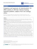
1 TLR-4 expression and signaling in brain cortex after
CMS exposure
To evaluate if the TLR-4 pathway is activated after
stress ex posure we studied the expression of TLR-4 and
its co-r eceptor, myeloid differentiation factor-2 (MD-2).
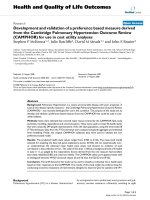
Stress exposure induced a significant increase in TLR-4
mRNA and protein levels in the brain cortex (Figure
1A&1B). Similarly, MD-2 was up-regulated after stress
(Figure 1C&1D).
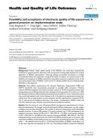
2 Possible regulatory mechanisms of TLR-4 activation in
brain cortex after CMS
Lipopolysaccharide (LPS) is a main ligand of TLR-4,
whose activation switches on intracellular inflammatory
pathways. In order to clarify the origin of the stress-
induced activation of t he TLR-4 pathway, we studied
plasma levels of LPS and LPS binding protein (LBP).
CMS exposure produced an increase in both LPS and
LBP plasma levels (Figure 2A&2B).
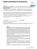
3 Inflammatory mediators in brain cortex after CMS
exposure
TLR-4 activation is followed by stimulation of the pro-
inflammatory transcription nuclear factor B(NF-B)
[37], whose p65 subunit can be determined in cell nuclei
to evaluate its activation (by cytoplasm-nuclear traffick-
ing) after stress or other im mune/inflammatory stimuli.
Under the conditions used in this study, a decreased
activity of NF-B after CMS exposure was detected (Fig-
ure 3A). Similarly, a decrease in mRNA levels and pro-
tein expression of p65 subunit (Figure 3B&3C) was
observed in nuclear fractions from brain cortex of
stressed i ndividuals as well. Stress also increased mRNA
expression of the NF-Binhibitoryprotein,IBa in the
cytoplasm (Figure 3D).
The pro-inflammatory enzymatic source inducible
cyclooxygenase (COX-2) was also assessed in co ntrol
and after stress-exposure conditions. An increase in
COX-2 mRNA and in levels of its main product in
brain, PGE
2
was observed after 21 days of chronic stress
(Figures 4A&4B). Taking into account that inflammation
is a regulated process, we decide to study the main com-
ponent of the anti- inflamma tory mechanism: levels of
15-deoxy-Δ
12,14
-prostaglandin J
2
(15d-PGJ
2
), an anti-
inflamma tory product of COX-2, were decreased in pre-
frontal cortex after CMS exposure (Figure 4C).
Another well known inflammatory agent in brain that
is activated after TLR-4 activation is the pro-inflamma-
tory cytokine IL-1b [6]. In this particular stress model,
an increase in IL-1b mRNA levels was also detected
(Figure 4D).
Gárate et al. Journal of Neuroinflammation 2011, 8:151
/>Page 5 of 14
4 Oxidative/nitrosative damage in brain cortex after
CMS exposure
Although neither inducible nitric oxide synthase (iNOS)
expression nor stable metabolites of nitric oxide
(nitrites) levels were modified in brain cortex after 21
days of CMS (data not shown), we decided to study pos-
sible (COX-2- and c ytokine-induced) oxidative/nitrosa-
tive damage after stress. As a final index of this type of
damagethatcouldbeaffectedbyCMS,wemeasured
the accumulation of the lipid peroxidation marker mal-
ondialdehyde (MDA) in brain prefrontal cortex of the
different groups of rats. MDA increased after CMS
exposure (Figure 5).
5 Effects of intestinal decontamination on CMS-induced
inflammatory and oxidative/nitrosative damage
In order to evaluate whether the source of LPS (and sub-
sequent TLR-4 activation) were bacteria translocated
from the digestive tract, the e ffects of intestinal
decontamination was assessed in our experimental set-
ting. Antibiotic (ATB) decontamination decreased both
stress-induced LPS and LBP increases in plasma (Table
1).
The effects of decontamination on stressed animals
extended to stress-induced TLR-4 a nd MD-2 up-regula-
tion at protein and mRNA levels, and to all of the other
inflammatory and oxidative parameters previously deter-
mined in brain tissue (Table 1). Interestingly, ATB decon-
tamination prevented the CMS-induced decrease in anti-
inflammatory 15d-PGJ
2
levels in the brain (Table 1).
6 Effects of CMS and intestinal decontamination on
plasma corticosterone levels
Chronic mild stress exposure increased plasma corticos-
terone levels when compared to the control group and
to the group of rats subjected to CMS plus intestinal
decontamination (CMS+ATB group). Antibiotic (ATB)
treatment decreased corticosterone levels of chronically
TLR-4
CONTROL CMS
0
20
40
60
80
100
120
*
mRNA relativ e
expression levels
A
B
C
D
MD-2
CONTROL CMS
0
20
40
60
80
100
120
*
mRNA relativ e
expression levels
TLR-4
CO
NTR
O
L
C
M
S
0
25
50
75
100
125
**
TLR-4/
E
actin (O.D.)
MD-2
CONTROL CMS
0
20
40
60
80
100
120
*
MD-2/
E
actin (O.D.)
CONTROL
CMS
TLR-4
ȕ actin
CONTROL
CMS
CONTROL
CMS
TLR-4
ȕ actin
- 95kDa
- 42kDa
CONTROL
CMS
MD-2
ȕ actin
CONTROL
CMS
CONTROL
CMS
MD-2
ȕ actin
-20kD
a
- 42kDa
Figure 1 TLR-4 pathway activation in brain cortex after stress exposure in rats. mRNA expression levels for TLR-4 (A) and MD-2 (C) in brain
in control and after CMS. Protein expression of TLR-4 (B) and MD-2 (D) in brain in control and after CMS. Data are mean ± SEM of 8-10 rats per
group. * p < 0.05, ** p < 0.01 vs. Control group (two-tailed t-test).
Gárate et al. Journal of Neuroinflammation 2011, 8:151
/>Page 6 of 14
stressed rats (CMS+ATB group) and these C MS+ATB
animals did not show differences in plasma corticoster-
one levels when compared to the control (non stressed)
group, show ing that intestinal decontamination inhibits
the increase o f corti costerone induced by the CMS pro-
tocol (Figure 6).
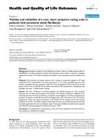
7 Effects of CMS and intestinal decontamination on
depressive-like behavior
After 21 days of the CMS protocol, separate groups of
animals (n = 10) were exposed to t he modified forced
swimming test (mFST). Data show that after CMS
exposure rats elicit a pro-depressive behavior (Figure
7A): immobility time is s ignificantly increased in CMS,
as shown by significant decreases in swimming time
compared to the control group. Analysis of time climb-
ing did not reveal significant differences between groups.
Furthermore, weight loss and number o f fecal boli were
increased in CMS (Figure 7B&7C). However, in spite of
the anti-inflammatory effects demo nstr ated in the brain
by the antibiotic intestinal decontamination protocol
used, ATB did not modify immobility or swimming
behaviors after mFST in stressed animals (Figure 7A).
Discussion
The present work points to a role for bacterial translo-
cation and subsequent TLR-4 pathway stimulation in
the neuroinflammation induced by an experimental
model of depression. To our knowledge, our results
demonstrate for the first time that the TLR-4 signaling
pathway becomes activated in brain cortex of rats
exposed to an animal model of depression. This activa-
tion occurs with increased levels of the pro-inflamma-
tory cytokine IL-1b and of one of the main enzymatic
sources of inflammatory and oxidative mediato rs, COX-
2 and its product PGE
2
. Interestingly, after 21 days of
CMS, the COX-derived anti-inflammatory mediator
15d-PGJ
2
appears decreased. As a consequence of this
misbalance and the resulting enhancement of inflamma-
tion and oxidation in brain cortex after CMS exposure,
an increment in lipid peroxidation takes place.
In the search for a mechanistic explanation for the
observed TLR-4 activation, exper iments using antibiotic
intestinal decontamination suggest a pivotal role for
anaerobic Gram-negative bacteria translocation on TLR-
4-signaling pathway activation after stress exposure in
brain cortex of rats.
In accordance with other studies carried out in differ-
ent models of stress exposure, including CMS, our data
show that there is inflammatory and oxidative/nitrosa-
tive damage in the brain after CMS [5,38-40]. The
increase of IL-1b mRNA levels detected in brain cortex
also correlates with results obtained in previous studies
[41-43]. This can be considered particularly signific ant,
bearing in mind that this cytokine plays a central role in
the sickness behavior detected in animals after LPS
injection (LPS induces its release) and has been pro-
posed as a possible actor involved in the pathophysiol-
ogy of depression [ 6,44]. Moreover, the ac tions of IL-1b
in the CNS include increases in the production of other
pro-inflammatory cytokines which can stimulate en zy-
matic sources of oxidative and nitrosative mediators
[45].
Apart from cytokines, other mediators such as bacter-
ial endotoxin (i.e. LPS, which we are showing here also
increased after CMS) rapi dly induce COX-2 and PGE
2
LPS
CONTROL CMS
0.0
0.1
0.2
0.3
0.4
*
EU/mL plasma
LBP
CO
NTR
O
L
C
M
S
0
200
400
600
800
1000
*
ng/mL plasma
A
B
Figure 2 LPS (A) and LBP (B) levels in plasma in control and
after CMS. Data are mean ± SEM of 8-10 rats per group. * p < 0.05
vs. Control group (two-tailed t-test).
Gárate et al. Journal of Neuroinflammation 2011, 8:151
/>Page 7 of 14
production [46,47]. The induction of COX-2 in the CNS
by stress and the increase in the PGE
2
levels in the
brain cortex are well documented phenomena [48,49] of
significant importance in experimental models of
depression and in depressive disorders [50], bearing in
mind that PGE
2
, in turn, stimulates production of pro-
inflammatory cytokines, expression of COX-2 and, as a
co-factor, activity of indoleamine 2,3-dioxygenase (IDO),
which reduces levels of 5-HT, a hallmark of depression.
On the other hand, it has been previously shown that,
during the production of prostaglandins, reactive oxygen
species (ROS) are generated, which are a main cause of
oxidative/ni trosative damage as has been shown to
occur after CMS, leading t o an increase in lipid peroxi-
dation markers (increase in the amount of MDA) [51].
Although previous studies have revealed an increase in
inducible nitric oxide synthase (iNOS) levels in the
brain after acute and subacute stress protocols [5], after
chronicexposuretoaseriesof stressors of mild inten-
sity (as occurs in CMS) the main isoform implicated is
the constitutive, neuronal NOS (nNOS) isoform [52].
Thus, the increase in lipid peroxidation observed in the
specific experimental setting used in the prese nt study
should be attributed mainly to cyclooxygenase-derived
products.
Activation of the transcription factor nuclear factor
kappa B(NF-B) controls the transcription of many
acute-phase proteins a nd inflammatory genes both in
humans and rodents, and is one of the earliest events in
the stress-inflammation response in the brain [53,54].
This transcription factor resides silent in the cytoplasm
bound by an inhibitory pro tein, I kappa B alpha (IBa).
When a specific cellular pathway is stimulated, it pro-
duces phosphorylation and s ubsequent degradation of
IBa, activat ing NF-B which translocates to cell
nucleus where it recognizes specific DN A sequences in
NF-
N
B p65
CONTROL CMS
0
20
40
60
80
100
120
*
NF-
N
B p65/sp-1 (O.D.)
NF-
N
B p65
CONTROL CM
S
0
20
40
60
80
100
120
*
mRNA relative
expression levels
I
N
B
D
CONTROL CMS
0
20
40
60
80
100
120
**
mRNA relative
expression levels
A
B
C
D
NF-
N
B p65 Activity
CONTROL CMS
0
20
40
60
80
100
120
**
p65 Activity/ mg prot.
% Control
CONTROL
C
M
S
NF-
N
Bp65
sp-1
- 65kDa
- 95-105kD
a
Figure 3 NF-B signaling in brain cortex after CMS exposure: p65 activity (A), p65 mRNA levels (B) and p65 protein expression (C) in
nuclear fractions of brain cortex in control and CMS.IBa mRNA levels in cytoplasmic fractions of cortex in control and CMS (D). Data are
mean ± SEM of 8-10 rats per group. * p < 0.05, ** p < 0.01 vs. Control group (two-tailed t-test).
Gárate et al. Journal of Neuroinflammation 2011, 8:151
/>Page 8 of 14
the promoter of target genes, among which are those
that code for proteins involved in inflammation. Inter-
estingly, no clear stimulation of NF-B occurs in the
brain cortex after CMS when its p65 subunit is ana-
lyzed. Ho wever, our results show that I Ba mRNA
levels are increased after CMS. As it has b een described
to occur in other experimental settings, the increase in
IBa mRNA is an autoregulatory pathway switched on
by NF-B after prolonged stimulation as may be the
case in CMS, thus restricting NF- Bactionwhen
chronically stimulated [55,56].
Having described some components of the inflamma-
tory response in the brain cortex to CMS exposure, we
focused on a search for possible external stressors sti-
mulating this response, as recently reviewed by Kubera
et al. [39]. All of the inflammatory parameters described
up to this point can be induced by the Toll-like recep-
tors (TLRs) pathway stimulation. TLRs, being the first
line of defense against invading microorganisms, consti-
tute the main agents of the innate immune response.
Stimulation of TLRs causes an immediate defensive
COX-2
CONTROL CMS
0
25
50
75
100
125
150
**
mRNA relative
expression levels
15d-P
G
J
2
CONTROL CMS
0
20
40
60
80
100
*
pg/mg prot.
PGE
2
CO
NTR
O
L
C
M
S
0
20
40
60
80
100
*
pg/mg prot.
A
B
C
D
IL-1
E
CO
NTR
O
L
C
M
S
0
20
40
60
80
100
120
**
mRN A r elativ e
expression levels
Figure 4 Inflammatory parameters in brain cort ex after CMS. Protein expressi on of COX-2 in control and after CMS in the brain (A). Brain
levels of the pro-inflammatory prostaglandin PGE
2
(B), the anti-inflammatory one 15d-PGJ
2
(C), and interleukin-1b (IL-1b) mRNA levels in control
and after CMS in the brain (D). Data are mean ± SEM of 8-10 rats per group. * p < 0.05, ** p < 0.01 vs. Control group (two-tailed t-test).
MDA
CO
NTR
O
L
C
M
S
0.000
0.001
0.002
0.003
0.004
0.005
*
nmol
/
mg prot.
Figure 5 Lipid peroxidati on in brain after CMS: l evels of
malondialdehyde (MDA; a marker of reactive oxygen species
attack and resultant lipid peroxidation) in control rats and
after CMS exposure in brain cortex. Data are mean ± SEM of 8-
10 rats per group. * p < 0.05 (two-tailed t-test).
Gárate et al. Journal of Neuroinflammation 2011, 8:151
/>Page 9 of 14
response, including the production of an array of anti-
microbial peptides and inflammatory/oxidative media-
tors [ 37]. During the last several years numerous studies
have appeared rega rding the role of TLRs in the patho-
physiology of diverse CNS diseases such as multiple
sclerosis, Alzheimer’ s disease and brain i schemia
[16,57,58]. Now, our results show for the first time
increases in expression of and mRNA levels for Toll-like
receptor 4 (TLR-4 ) in the brain cortex in an experimen-
tal model of depression in rodents . Additionally, we
have also found that CMS induces protein expression
and synthesis of MD-2, which is the molecule that con-
fers lipopolysaccharide responsiveness to TLR-4 [59].
Taken as a whole, the results presented here suggest
that TLR-4 could be an important regulatory factor in
the consequences of chronic stress in the brain, and also
support a possibility for pharmacological or genetic
manipulations of this pathway - although to date the
selective inhibition o f TLR-4 has proved to be a difficult
challenge [60] - in order to minimize oxidative and
inflammatory damage in the CNS after stress and in
stress-related psycho- and neuro-pathologies such as
depression.
There are several studies exploring endogenous
ligands that activate TLR-4 after brain damage (e .g. pro-
tein S100 or nuclear protein high-mobility g roup box 1
after cerebral ischemia, pro-inflammatory cytokines after
brain trauma) [60]. However, knowledge about mechan-
isms that regulate TLR-4 activation in the brain in mod-
els of neuro psychiatric pathologies comes from pre vious
studies based on stress exposure, which have shown
increased intestinal permeability and a resultant bacter-
ial translocation to the systemic circulation after stress
Table 1 Antibiotic intestinal decontamination (ATB) effect on stress-induced inflammatory, anti-inflammatory and
oxidative/nitrosative parameters in control and CMS-exposed rats.
Control CMS Control+ATB CMS+ATB
Plasma determinations
LPS (EU/mL) 0.2856 ± 0.027 0.3546 ± 0.006** 0.248 ± 0.022 0.3008 ± 0.016
#
LBP (ng/mL) 799.8 ± 39.75 955.6 ± 35.57* 840.0 ± 19.52 804.2 ± 32.97
#
Brain determinations
TLR-4 (mRNA) 96.84 ± 2.618 109.8 ± 3.285** 102.5 ± 2.703 101.0 ± 1.278
TLR-4 (OD) (protein) 99.26 ± 4.455 116.9 ± 3.093** 88.09 ± 4.142 97.01 ± 3.162
##
MD-2 (mRNA) 98.01 ± 2.575 108.4 ± 2.178** 91.74 ± 2.432 96.86 ± 3.912
#
MD-2 (OD) (protein) 94.94 ± 2.977 108.6 ± 2.578* 102.6 ± 2.842 104.9 ± 4.381
NF-B p65 Activity
(% Control)
100.0 ± 4.571 85.48 ± 3.277* 96.73 ± 15,33 71.66 ± 3.1**
NF-B p65 (mRNA) 101.8 ± 2.546 94.21 ± 2.193* 90.28 ± 2.052 88.16 ± 2.879
NF-B p65 (OD) (protein) 100.6 ± 3.363 87.23 ± 3.554* 103.2 ± 4.530 99.43 ± 3.442
#
IBa (mRNA) 100.0 ± 4.286 118.7 ± 6.436* 95.55 ± 3.265 99.42 ± 5.101
#
COX-2 (mRNA) 99.89 ± 5.056 137.2 ± 8.159** 124.6 ± 7.084 107.1 ± 6.181
#
PGE
2
(pg/mg prot.)
45.14 ± 6.485 78.69 ± 12.24* 48.58 ± 8.973 36.75 ± 7.877
#
15d-PGJ
2
(pg/mg prot.)
83.45 ± 13.99 42.00 ± 6.775* 83.28 ± 13.78 107.8 ± 21.68
#
IL-1b (mRNA) 94.59 ± 4.000 114.0 ± 2.318** 95.91 ± 9.424 91.35 ± 3.886
##
MDA
(nmol/mg prot.)
0.00279 ± 0.000256 0.00372 ± 0.000285* 0.00187 ± 0.000142 0.00242 ± 0.000344
##
Data are means ± SEM of 8-10 rats per group; * p < 0.05; ** p < 0.01 vs. Control;
#
p < 0.05;
##
p < 0.01 vs. CMS. One-way ANO VA followed by the Newman-Keuls
post hoc test.
Corticosterone
CO
NTR
O
L
C
M
S CO
NTR
O
L+ATB
C
M
S
+ATB
0
100
200
300
400
**
#
ng
/
mL plasma
Figure 6 Plasma corticosterone levels of control (non-stressed),
CMS-exposed, control+intestinal antibiotic-decontamination
(CONTROL+ATB) and CMS+ATB animals. Data are mean ± SEM of
8-10 rats per group. ** p < 0.01 vs. Control group; #p < 0.05 vs.
CMS group. One-way analysis of variance (ANOVA) followed by the
Newman-Keuls post hoc test.
Gárate et al. Journal of Neuroinflammation 2011, 8:151
/>Page 10 of 14
exposure [21,22]. As a result, there are circulating
Gram-negative enterobacteri a, which are a major source
of LPS and can activate brain TLR-4 inducing a neu-
roinflammatory response. In order to clarify the origin
of stress-induced activation of the TLR-4 pathway in
CMS, we studie d LPS and its binding protein (LBP;
which serves as a lipid transfer protein that facil itates
the transportation of LPS to the recognition protein
CD14 and to TLR-4) levels in plasma. O ur results sho w
tha t CMS exposure produ ces increases in b oth LPS and
LBP plasma levels. Thus, it is possible that CMS is caus-
ing an intestinal dysfunction followed by bacterial trans-
location, as occurs in different stress models in rodents
[22], with LPS (from those Gram-negative bacteria)
being the reason for the TLR-4 activation. This pro-
posed mechanism, known as “leaky gut“, also takes place
in depressed patients, and has been related to the
inflammatory pathophysiology of major depressive disor-
der [23].
To assess, in our experimental setting, whether the
source of LPS and the consequent TLR-4 pathway
stimulation, are bacte ria translocated from the gut, we
examined the eff ects of intestinal decontamination on
the stress-induced inflammatory and oxidative/nitrosa-
tive changes rev ealed above. We used a standard strin-
gent protocol (strepto mycin and penicillin G) for only
intes tinal decontamination. This protoco l has been used
because it has demonstrated to lack any neuroprotective
or an ti-inflammatory effects on the CNS when used in
other related protocols [24,61]. By using this protocol,
we can separate possible effects on the brain of the anti-
biotic used (i.e. the anti-neuroinflammatory effect of
minocycline) from the effects caused by intestinal
decontamination.
Our data show that animals subjected to CMS plus
intestinal decontamination present a return to basal
levels (control group values) for pro-inflammatory and
oxidative/nitrosative parameters previously analyzed,
including LPS and LBP plasma concentrations and TLR-
4 and MD-2 expression and mRNA levels.
In this vein, of special relevance i s the finding that
antibiotic intestinal decontamination promotes decreases
A
C
B
modified Forced Swimming Test (mFST)
Immobility Swimming Climbing
0
5
10
15
20
25
30
35
40
45
CONTROL
CMS
CONTROL+ATB
CMS+ATB
*
*
Mean co unts
Body weight
CONTROL CMS CONTROL+ATB CMS+ATB
0
50
100
150
200
***
***
Weight after 21 days
(% basal)
Fecal boli after FST
CO
NTR
O
L
C
M
S CO
NTR
O
L+ATB
C
M
S
+ATB
0
2
4
6
8
***
***
***
number
Figure 7 Behavioral parameters (time in immobility, swimming and cli mbing, in seconds) during the modified forced swimming test
(mFST) (A), weight change after 21 days of CMS exposure (B) and fecal boli (number) (C). Data are means ± SEM of 9-10 rats per group;
* p < 0.05; ** p < 0.01 vs. Control. Two-way analysis of variance (ANOVA) followed by Bonferroni post hoc test.
Gárate et al. Journal of Neuroinflammation 2011, 8:151
/>Page 11 of 14
in IL-1b and COX-2/PGE
2
in brain cortex. This result
supports the notion that LPS from translocated bacteria
stimulates TLR-4, and in that way produces the
increases in IL-1b and COX-2/PGE
2
levels in the CNS
previously detected. More interestingly, intestinal decon-
tamination is able to restore the disbalance between
COX-derived inflammatory (PGE
2
) and anti-inflamma-
tory (15d-PGJ
2
) components in the brain.
Our results also indica te that plasma c orticosterone
levels are increased after 21 days of CMS when com-
pared with the control group, showing that even after
this chronic stress exposure the hypothalam ic-pituitary-
adrenal (HPA) axis of these animals remains function-
ing. Additionally, it has been previously demonstrated
that LPS stimulates the HPA a xis [62] and thus, it is
conceivable that the increase in the corticosterone levels
after CMS could be caused, at least in part, by the
increase in LPS levels detected here and not only by the
stressors themselves. Supporting this idea, the intestinal
decontamination that decreases LPS after CMS, also
decreases plasma corticosterone level s, again supporting
the role of intestinal bacteria as a source for the LPS
detected in our study.
The effects of intestinal decontamination on depres-
sive-like behavior were analyzed using a modified forc ed
swimming test based o n the method described by Por-
solt [32], measuring behavioral despair. In spite of its
anti-inflammatory effects after decreasing LPS levels,
antibiotic decontamination failed to reverse the depres-
sive-like behavior induced by CMS, which indicates a
role for LPS-induced neuroinflammation after CMS
without (at this level) behavioral consequences. None-
theless, the fact that CMS-induced neuroinflammation is
reversed by antibiotic intest inal decontamination is par-
ticularly relevant because neuroinflammation is consid-
ered an important biological even t that might increase
the risk o f major depressive episodes much like more
traditional psychosocial factors [6]. Further studies using
mixed protocols of experimental depression plus infec-
tive or inflammat ory agents would aid in explaining the
role of comorbid depression in inflammatory or
immune-related pathologies.
The results presented here are in line with a hypoth-
esis recently presented [38] according to which, external
stressors to the brain, such as LPS, may up-regulate
immune receptors such as TLR-4 that, in turn, may
aggravate neuroinflammation due to locally produced
internal stressors (prostanoids, some cytokines, tran-
scription factors) thus causing a superinduction of
(neuro)inflammatory responses.
Conclusions
In conclusion, our results suggest that LPS from bacter -
ial translocation is responsible, at least in part, for the
TLR-4 activation found in the brain after chronic mild
stress exposure which leads to the release of inflamma-
tory mediators in the CNS (including IL-1b and COX-2)
(Figure 8). In addition, antibiotic intestinal decontamina-
tion decreases LPS systemic levels and neuroinflamma-
tion showing a possible protective role of antib iotic
decontamination in stress-related conditions and offer-
ing a potential therapeutic target for the adjuvant treat-
ment of depression.
Acknowledgements
This work was supported by Spanish Ministry of Science and Innovation
(SAF07-63138), the Instituto de Salud Carlos III (FIS PI10/00123, PI07/0687,
PI10/01221)”, “Junta de Andalucía; Consejería de Innovación , Ciencia y
Empresa (CTS - 4303), Centro de Investigación Biomédica en Red de Salud
Mental, CIBERSAM, and Foundation Santander-UCM (GR 58/08). IG is a FPI
fellow (MICINN). JRC is a Juan de la Cierva fellow (MICINN).
Author details
1
Department of Pharmacology, Faculty of Medicine, Universidad
Complutense, Madrid 28040, Spain.
2
Department of Psychiatry, Faculty of
Medicine, Universidad Complutense, Madrid 28040, Spain.
3
Department of
Neurosciences, Faculty of Medicine, Universidad de Cádiz, Cádiz 11003,
Spain.
4
Centro de Investigación Biomédica en Red de Salud Mental
(CIBERSAM), Spain.
5
Instituto de Investigación Sanitaria Hospital 12 de
Octubre, Madrid 28026, Spain.
Authors’ contributions
IG contributed to acquisition, analysis and interpretation of data; BGB
contributed to acquisition, analysis and interpretation of data, drafting the
manuscript and revising it critically; JLMM contributed to analysis and
interpretation of data and revising the manuscript critically; LB and EB
contributed to acquisition, analysis and interpretation of CMS model and
behavioural data; JAM and JRC revised the manuscript critically; and JCL
contributed to conception and design, drafting the manuscript and revising
it critically for important intellectual content. All authors have given final
approval of the version to be published.
CMS
BLOODPREFRONTAL CORTEX
BEHAVIOR
LPS
LBP
+
TLR-4/MD-2
+
ATB
-
-
-
PROINFLAMMATORY MEDIATORS
COX-2, PGE
2
, IL-1
E
CELL DAMAGE
Lipid peroxidation
DEPRESSIVE LIKE BEHAVIOR
+
+
?
-
+
+
Figure 8 Schematic representati on of the results obtained
from and the effects of antibiotic intestinal decontamination
(ATB: intestinal antibiotic decontamination). See text for
abbreviations.
Gárate et al. Journal of Neuroinflammation 2011, 8:151
/>Page 12 of 14
Competing interests
The authors declare that they have no competing interests.
Received: 24 August 2011 Accepted: 3 November 2011
Published: 3 November 2011
References
1. Machado M, Iskedjian M, Ruiz I, Einarson TR: Remission, dropouts, and
adverse drug reaction rates in major depressive disorder: a meta-
analysis of head-to-head trials. Curr Med Res Opin 2006, 22:1825-1837.
2. Shelton RC, Osuntokun O, Heinloth AN, Corya SA: Therapeutic options for
treatment-resistant depression. CNS Drugs 2010, 24:131-161.
3. Rush AJ, Trivedi MH, Wisniewski SR, Nierenberg AA, Stewart JW, Warden D,
Niederehe G, Thase ME, Lavori PW, Lebowitz BD, McGrath PJ,
Rosenbaum JF, Sackeim HA, Kupfer DJ, Luther J, Fava M: Acute and longer-
term outcomes in depressed outpatients requiring one or several
treatment steps: a STAR*D report. Am J Psychiatry 2006, 163:1905-1917.
4. Herbert TB, Cohen S: Depression and immunity: a meta-analytic review.
Psychol Bull 1993, 113:472-486.
5. Garcia-Bueno B, Caso JR, Leza JC: Stress as a neuroinflammatory condition
in brain: damaging and protective mechanisms. Neurosci Biobehav Rev
2008, 32:1136-1151.
6. Dantzer R, O’Connor JC, Freund GG, Johnson RW, Kelley KW: From
inflammation to sickness and depression: when the immune system
subjugates the brain. Nat Rev Neurosci 2008, 9:46-56.
7. Miller AH, Maletic V, Raison CL: Inflammation and its discontents: the role
of cytokines in the pathophysiology of major depression. Biol Psychiatry
2009, 65:732-741.
8. Madrigal JL, Moro MA, Lizasoain I, Lorenzo P, Leza JC: Stress-induced
increase in extracellular sucrose space in rats is mediated by nitric
oxide. Brain Res 2002, 938:87-91.
9. Esposito P, Chandler N, Kandere K, Basu S, Jacobson S, Connolly R, Tutor D,
Theoarides TC: Corticotropin-releasing hormone and brain mast cells
regulate blood-brain-barrier permeability induced by acute stress. J
Pharmacol Exp Ther 2002, 303:1061-1066.
10. de Klerk OL, Bosker FJ, Willemsen AT, Van WA, Visser AK, de Jager T,
Dagyte G, den Boer JA, Dierckx RA, Meerlo P: Chronic stress and
antidepressant treatment have opposite effects on P-glycoprotein at the
blood-brain barrier: an experimental PET study in rats. J Psychopharmacol
2010, 24:1237-1242.
11. Hampel H, Muller-Spahn F, Berger C, Haberl A, Ackenheil M, Hock C:
Evidence of blood-cerebrospinal fluid-barrier impairment in a subgroup
of patients with dementia of the Alzheimer type and major depression:
a possible indicator for immunoactivation. Dementia 1995, 6:348-354.
12. Hampel H, Kotter HU, Moller HJ: Blood-cerebrospinal fluid barrier
dysfunction for high molecular weight proteins in Alzheimer disease
and major depression: indication for disease subsets. Alzheimer Dis Assoc
Disord 1997, 11:78-87.
13. Crack PJ, Bray PJ: Toll-like receptors in the brain and their potential roles
in neuropathology. Immunol Cell Biol 2007, 85:476-480.
14. Akira S, Uematsu S, Takeuchi O: Pathogen recognition and innate
immunity. Cell 2006, 124:783-801.
15.
Zacharowski K, Zacharowski PA, Koch A, Baban A, Tran N, Berkels R,
Papewalis C, Schulze-Osthoff K, Knuefermann P, Zähringer U, Schumann RR,
Rettori V, McCann SM, Bornstein SR: Toll-like receptor 4 plays a crucial
role in the immune-adrenal response to systemic inflammatory response
syndrome. Proc Natl Acad Sci USA 2006, 103:6392-6397.
16. Caso JR, Pradillo JM, Hurtado O, Leza JC, Moro MA, Lizasoain I: Toll-like
receptor 4 is involved in subacute stress-induced neuroinflammation
and in the worsening of experimental stroke. Stroke 2008, 39:1314-1320.
17. Monje FJ, Cabatic M, Divisch I, Kim EJ, Herkner KR, Binder BR, Pollak DD:
Constant Darkness Induces IL-6-Dependent Depression-Like Behavior
through the NF-κB Signaling Pathway. J Neurosci 2011, 31:9075-9083.
18. Pace TW, Mletzko TC, Alagbe O, Musselman DL, Nemeroff CB, Miller AH,
Heim CM: Increased stress-induced inflammatory responses in male
patients with major depression and increased early life stress. Am J
Psychiatry 2006, 163:1630-1633.
19. Koo JW, Russo SJ, Ferguson D, Nestler EJ, Duman RS: Nuclear factor-
kappaB is a critical mediator of stress-impaired neurogenesis and
depressive behavior. Proc Natl Acad Sci USA 2010, 107:2669-2674.
20. Koedel U, Merbt UM, Schmidt C, Angele B, Popp B, Wagner H, Pfister HW,
Kirschning CJ: Acute brain injury triggers MyD88-dependent, TLR2/4-
independent inflammatory responses. Am J Pathol 2007, 171:200-213.
21. Collins SM: Stress and the Gastrointestinal Tract IV. Modulation of
intestinal inflammation by stress: basic mechanisms and clinical
relevance. Am J Physiol Gastrointest Liver Physiol 2001, 280:G315-G318.
22. Ponferrada A, Caso JR, Alou L, Colon A, Sevillano D, Moro MA, Lizasoain I,
Menchén P, Gómez-Lus ML, Lorenzo P, Cos E, Leza JC, Menchén L: The role
of PPARgamma on restoration of colonic homeostasis after experimental
stress-induced inflammation and dysfunction. Gastroenterology 2007,
132:1791-1803.
23. Maes M, Kubera M, Leunis JC: The gut-brain barrier in major depression:
intestinal mucosal dysfunction with an increased translocation of LPS
from gram negative enterobacteria (leaky gut) plays a role in the
inflammatory pathophysiology of depression. Neuro Endocrinol Lett 2008,
29:117-124.
24. Ando T, Brown RF, Berg RD, Dunn AJ: Bacterial translocation can increase
plasma corticosterone and brain catecholamine and indoleamine
metabolism. Am J Physiol Regul Integr Comp Physiol 2000, 279:R2164-R2172.
25. Willner P: Chronic mild stress (CMS) revisited: consistency and
behavioral-neurobiological concordance in the effects of CMS.
Neuropsychobiology 2005, 52:90-110.
26. Radley JJ, Arias CM, Sawchenko PE: Regional differentiation of the medial
prefrontal cortex in regulating adaptive responses to acute emotional
stress. J Neurosci 2006, 26:12967-12976.
27. Tang SC, Arumugam TV, Xu X, Cheng A, Mughal MR, Jo DG, Lathia JD,
Siler DA, Chigurupati S, Ouyang X, Magnus T, Camandola S, Mattson MP:
Pivotal role for neuronal Toll-like receptors in ischemic brain injury and
functional
deficits. Proc Natl Acad Sci USA 2007, 104:13798-13803.
28. Caso JR, Pradillo JM, Hurtado O, Lorenzo P, Moro MA, Lizasoain I: Toll-like
receptor 4 is involved in brain damage and inflammation after
experimental stroke. Circulation 2007, 115:1599-1608.
29. Olson JK, Miller SD: Microglia initiate central nervous system innate and
adaptive immune responses through multiple TLRs. J Immunol 2004,
173:3916-3924.
30. Singh AK, Jiang Y: How does peripheral lipopolysaccharide induce gene
expression in the brain of rats? Toxicology 2004, 201:197-207.
31. Garcia-Bueno B, Serrats J, Sawchenko PE: Cerebrovascular cyclooxygenase-
1 expression, regulation, and role in hypothalamic-pituitary-adrenal axis
activation by inflammatory stimuli. J Neurosci 2009, 29:12970-12981.
32. Porsolt RD, Le PM, Jalfre M: Depression: a new animal model sensitive to
antidepressant treatments. Nature 1977, 266:730-732.
33. Das NP, Ratty AK: Studies on the effects of the narcotic alkaloids,
cocaine, morphine, and codeine on nonenzymatic lipid peroxidation in
rat brain mitochondria. Biochem Med Metab Biol 1987, 37:258-264.
34. Turrin NP, Rivest S: Unraveling the molecular details involved in the
intimate link between the immune and neuroendocrine systems. Exp
Biol Med (Maywood) 2004, 229:996-1006.
35. Kapadia R, Yi JH, Vemuganti R: Mechanisms of anti-inflammatory and
neuroprotective actions of PPAR-gamma agonists. Front Biosci 2008,
13:1813-1826.
36. Bradford MM: A rapid and sensitive method for the quantitation of
microgram quantities of protein utilizing the principle of protein-dye
binding. Anal Biochem 1976, 72:248-254.
37. Akira S: Toll-like receptor signaling. J Biol Chem 2003, 278:38105-38108.
38. Mallo T, Matrov D, Koiv K, Harro J: Effect of chronic stress on behavior
and cerebral oxidative metabolism in rats with high or low positive
affect. Neuroscience 2009, 164:963-974.
39. Kubera M, Obuchowicz E, Goehler L, Brzeszcz J, Maes M: In animal models,
psychosocial stress-induced (neuro)inflammation, apoptosis and reduced
neurogenesis are associated to the onset of depression. Prog
Neuropsychopharmacol Biol Psychiatry 2011, 35:744-759.
40. Tagliari B, Tagliari AP, Schmitz F, da Cunha AA, Dalmaz C, Wyse AT: Chronic
variable stress alters inflammatory and cholinergic parameters in
hippocampus of rats. Neurochem Res 2011, 36:487-493.
41. Minami M, Kuraishi Y, Yamaguchi T, Nakai S, Hirai Y, Satoh M:
Immobilization
stress induces interleukin-1 beta mRNA in the rat
hypothalamus. Neurosci Lett 1991, 123:254-256.
42. Johnson JD, Campisi J, Sharkey CM, Kennedy SL, Nickerson M,
Greenwood BN, Fleshner M: Catecholamines mediate stress-induced
Gárate et al. Journal of Neuroinflammation 2011, 8:151
/>Page 13 of 14
increases in peripheral and central inflammatory cytokines. Neuroscience
2005, 135:1295-1307.
43. Deak T, Bordner KA, McElderry NK, Barnum CJ, Blandino P Jr, Deak MM,
Tammariello SP: Stress-induced increases in hypothalamic IL-1: a
systematic analysis of multiple stressor paradigms. Brain Res Bull 2005,
64:541-556.
44. Goshen I, Kreisel T, Ben-Menachem-Zidon O, Licht T, Weidenfeld J, Ben-
Hur T, Yirmiya R: Brain interleukin-1 mediates chronic stress-induced
depression in mice via adrenocortical activation and hippocampal
neurogenesis suppression. Mol Psychiatry 2008, 13:717-728.
45. Lucas SM, Rothwell NJ, Gibson RM: The role of inflammation in CNS injury
and disease. Br J Pharmacol 2006, 147(Suppl 1):S232-S240.
46. Hoffmann C: COX-2 in brain and spinal cord implications for therapeutic
use. Curr Med Chem 2000, 7:1113-1120.
47. Norris PC, Reichart D, Dumlao DS, Glass CK, Dennis EA: Specificity of
eicosanoid production depends on the TLR-4-stimulated macrophage
phenotype. J Leukoc Biol 2011.
48. Yamagata K, Andreasson KI, Kaufmann WE, Barnes CA, Worley PF:
Expression of a mitogen-inducible cyclooxygenase in brain neurons:
regulation by synaptic activity and glucocorticoids. Neuron 1993,
11:371-386.
49. Madrigal JL, Moro MA, Lizasoain I, Lorenzo P, Fernandez AP, Rodrigo J,
Boscá L, Leza JC: Induction of cyclooxygenase-2 accounts for restraint
stress-induced oxidative status in rat brain. Neuropsychopharmacology
2003, 28:1579-1588.
50. Muller N, Schwarz MJ: The immune-mediated alteration of serotonin and
glutamate: towards an integrated view of depression. Mol Psychiatry
2007, 12:988-1000.
51. Phillis JW, Horrocks LA, Farooqui AA: Cyclooxygenases, lipoxygenases, and
epoxygenases in CNS: their role and involvement in neurological
disorders. Brain Res Rev 2006, 52:201-243.
52. Zhou QG, Hu Y, Hua Y, Hu M, Luo CX, Han X, Zhu XJ, Wang B, Xu JS,
Zhu DY: Neuronal nitric oxide synthase contributes to chronic stress-
induced depression by suppressing hippocampal neurogenesis. J
Neurochem 2007, 103:1843-1854.
53. Black PH: The inflammatory consequences of psychologic stress:
relationship to insulin resistance, obesity, atherosclerosis and diabetes
mellitus, type II. Med Hypotheses 2006, 67:879-891.
54. Madrigal JL, Moro MA, Lizasoain I, Lorenzo P, Castrillo A, Boscá L, Leza JC:
Inducible nitric oxide synthase expression in brain cortex after acute
restraint stress is regulated by nuclear factor kappaB-mediated
mechanisms. J Neurochem 2001, 76:532-538.
55. Sun SC, Ganchi PA, Ballard DW, Greene WC: NF-kappa B controls
expression of inhibitor I kappa B alpha: evidence for an inducible
autoregulatory pathway. Science 1993, 259:1912-1915.
56. Sun SC, Ganchi PA, Beraud C, Ballard DW, Greene WC:
Autoregulation of
the NF-kappa B transactivator RelA (p65) by multiple cytoplasmic
inhibitors containing ankyrin motifs. Proc Natl Acad Sci USA 1994,
91:1346-1350.
57. Jin JJ, Kim HD, Maxwell JA, Li L, Fukuchi K: Toll-like receptor 4-dependent
upregulation of cytokines in a transgenic mouse model of Alzheimer’s
disease. J Neuroinflamm 2008, 5:23.
58. Wang YC, Lin S, Yang QW: Toll-like receptors in cerebral ischemic
inflammatory injury. J Neuroinflamm 2011, 8:134.
59. Shimazu R, Akashi S, Ogata H, Nagai Y, Fukudome K, Miyake K, Kimoto Ml:
MD-2, a molecule that confers lipopolysaccharide responsiveness on
Toll-like receptor 4. J Exp Med 1999, 189:1777-1782.
60. Kong Y, Le Y: Toll-like receptors in inflammation of the central nervous
system. Int Immunopharmacol 2011, 11:1407-1414.
61. Caso JR, Hurtado O, Pereira MP, Garcia-Bueno B, Menchen L, Alou L,
Gómez-Lus ML, Moro MA, Lizasoain I, Leza JC: Colonic bacterial
translocation as a possible factor in stress-worsening experimental
stroke outcome. Am J Physiol Regul Integr Comp Physiol 2009, 296:
R979-R985.
62. Dunn AJ, Ando T, Brown RF, Berg RD: HPA axis activation and
neurochemical responses to bacterial translocation from the
gastrointestinal tract. Ann N Y Acad Sci 2003, 992:21-29.
doi:10.1186/1742-2094-8-151
Cite this article as: Gárate et al.: Origin and consequences of brain Toll-
like receptor 4 pathway stimulation in an experimental model of
depression. Journal of Neuroinflammation 2011 8:151.
Submit your next manuscript to BioMed Central
and take full advantage of:
• Convenient online submission
• Thorough peer review
• No space constraints or color figure charges
• Immediate publication on acceptance
• Inclusion in PubMed, CAS, Scopus and Google Scholar
• Research which is freely available for redistribution
Submit your manuscript at
www.biomedcentral.com/submit
Gárate et al. Journal of Neuroinflammation 2011, 8:151
/>Page 14 of 14