báo cáo hóa học: " Reduced inflammation accompanies diminished myelin damage and repair in the NG2 null mouse spinal cord" potx
Bạn đang xem bản rút gọn của tài liệu. Xem và tải ngay bản đầy đủ của tài liệu tại đây (8.16 MB, 13 trang )
RESEARCH Open Access
Reduced inflammation accompanies diminished
myelin damage and repair in the NG2 null mouse
spinal cord
Karolina Kucharova
1*
, Yunchao Chang
1,2
, Andrej Boor
3
, Voon Wee Yong
4
and William B Stallcup
1
Abstract
Background: Multiple sclerosis (MS) is a demyelinating disease in which blood-derived immune cells and activated
microglia damage myelin in the central nervous system. While oligodendrocyte progenitor cells (OPCs) are
essential for generating oligodendrocytes for myelin repair, other cell types also participate in the damage and
repair processes. The NG2 proteoglycan is expressed by OPCs, pericytes, and macrophages/microglia. In this report
we investigate the effects of NG2 on these cell types during spina l cord demyelination/remyelination.
Methods: Demyelinated lesions were created by microinjecting 1% lysolecithin into the lumbar spinal cord.
Following demyelination, NG2 expression patterns in wild type mice were studied via immunostaining.
Immunolabeling was also used in wild type and NG2 null mice to compa re the extent of myelin damage, the
kinetics of myelin repair, and the respective responses of OPCs, pericytes, and macrophages/microglia. Cell
proliferation was quantified by studies of BrdU incorporation, and cytokine expression levels were evaluated using
qRT-PCR.
Results: The initial volume of spinal cord demyelination in wild type mice is twice as large as in NG2 null mice.
However, over the ensuing 5 weeks there is a 6-fold improvement in myelination in wild type mice, versus only a
2-fold improvement in NG2 null mice. NG2 ablation also results in reduced numbers of each of the three affected
cell types. BrdU incorporation studies reveal that reduced cell proliferation is an important factor underlying NG2-
dependent decreases in each of the three key cell populations. In addition, NG2 ablation reduces macrophage/
microglial cell migration and shifts cytokine expression from a pro-inflammatory to anti-inflammatory phenotype.
Conclusions: Loss of NG2 expression leads to decreased proliferation of OPCs, pericytes, and macrophages/
microglia, reducing the abundance of all three cell types in demyelinated spinal cord lesions. As a result of these
NG2-dependent changes, the course of demyelination and remyelination in NG2 null mice differs from that seen in
wild type mice, with both myelin damage and repair being reduced in the NG2 null mouse. These studies identify
NG2 as an important factor in regulating myelin processing, suggesting that therapeutic targeting of the
proteoglycan might offer a means of manipulating cell behavior in demyelinating diseases.
Keywords: Inflammation, myelin repair, NG2 ablation, oligodendrocyte progenitors, pericytes, macrophages
Background
During the acute phase of multiple sclerosis (MS),
damage to the blood-brain barrier allows infiltration of
blood-derived cells that cause disruption of the myelin
sheath [1-5]. The capability of the CNS for myelin repair
is mediated by the action of oligodendrocyte progenitor
cells (OPCs), which not only generate oligodendrocytes
during CNS development, but also persist as the largest
cycling population in the mature CNS [6-9]. These
“adult ” OPCs serve as a source of cells fo r myelin repair
[8,10-12], but also exhibit oth er functions of mature glia
[13], including contributions to nodes of Ranvier [14-16]
and reception of synaptic input [17,18]. OPC function
and remyelination of axons nevertheless often fail i n
both relapsing-remitting and progressive MS [19-21].
* Correspondence:
1
Sanford-Burnham Medical Research Institute, La Jolla, CA 92037, USA
Full list of author information is available at the end of the article
Kucharova et al. Journal of Neuroinflammation 2011, 8:158
/>JOURNAL OF
NEUROINFLAMMATION
© 2011 Kucharova et al; licensee BioMed Central Ltd. This is an Open Access a rticle distributed under the terms of the Creative
Commons Attribution License ( which permits unrestricted use, distribution, and
reproduction in any me dium, pr ovided the original work is properly cited.
The inability of O PCs to produce adequate numbers of
myelinating oligodendrocytes has been attributed to sev-
eral fa ctors, including failu re of OPC proliferation, fail-
ure of OPC recruitment to the lesion, failure of OPC
differentiation, and failure of OPCs or oligodendrocytes
to interact with neurons. Compounding this complexity,
MS is a multifactorial disease, involving participation of
multiple factors in both myelin damage and myelin
repair. A better understanding of the molecular mechan-
isms of myelin degradation and regeneration is clearly
required for improved treatment of this primary demye-
linating disease.
Here we show that the NG2 proteoglycan is expressed
by three cell types that invade demyelinated CNS
lesions: OPCs , macrophages/microglia, and microvascu-
lar pericytes. In addition to serving as a marker for
these cell types [22,23], NG2 also promotes cell prolif-
eration and motility. In the neonatal NG2 null mouse,
decreased OPC proliferationreducesthepoolofpro-
genitors available for generating myelinating oligoden-
drocytes, resulting in reduced developmental
myelination in the cerebellum [24]. Ablation of NG2
also causes deficits in pericyte function. Decreased peri-
cyte recruitment and interaction with endothelial cells
lead to diminished vascularization in both ocular and
tumor models in the NG2 null mouse [25,26]. We
therefore have the ability to investigat e the role of NG2
in multiple cell types during the processes of demyelina-
tion and remyelination.
Following microin jection of L-a-lysolecithin into the
spinal cord white matter, we have investigated the acti-
vation, proliferation, recruitment, and maturation of
cells that are normally NG2-positive in the wild type
mouse. The importance of the NG2 molecule and NG2-
positive cells in demyelination and remyelination has
been evaluated via comparisons of wild type and NG2
knockout animals. The absence of NG2 causes signifi-
cant deficits in the behavior of OPCs, macrophages/
microglia, and pericytes, accompanied by quantitative
changes in the phenomena associated with axon demye-
lination and remyelination.
Methods
Animals
Animal work was performed according to guidelines
issued by the National Institutes of Health, following pro-
cedures approved by the Office of Laboratory Animal
Welfare. All experimental protocols were approved by
the Sanford-Burnham Institutional Animal Care and Use
Committee. The current experiments ut ilized male wild
type (NG2+/+) and NG2 null (NG2-/-) mice between the
ages of 3 -5 months. NG2 null mice were generated by a
homologous recombination strategy and backcrossed for
10 generations onto the C57Bl/6 background [27].
Lysolecithin-induced demyelination in the spinal cord of
mice
For spinal cord surgery, male mice (28-38 g) were
anesthetized with Ketamine/Xylazine (100/10 mg/kg)
administered intraperitoneally. Depth of anesthesia was
assured by monitoring lack of response to a noxious
foot pinch prior to commencing surgery. A skin incision
was m ade above the lower thoracic vertebrae. Paraver-
tebral muscles on both sides of the Th
11
-L
1
vertebrae
were cut, and t he vertebral column was stabilized with
transverse process clamps (Stoelting). The spinal cord
was exposed between the Th
12
-Th
13
vertebrae, and a
small incision was made in the dura just l ateral to the
posterior spinal vein. A 1.5 μl solution of 1% L-a-lysole-
cithin (Lysophosphatidylcholine; Sigma, St. Louis, MO)
in 0.1 M phosphate buffer wa s injected 0.5 mm deep
into the dorsal column at a rate of 0.75 μl/minute. This
was accomplished using a micromanipulator (Stoelting,
Wood Dale, IL), 32 G needle, 5 μl syringe (7762-05,
87930; Hamil ton), and d igital injector (Harvard Appara-
tus, Holliston, MA). As a sham control, injections were
done with 0.1 M PBS. The needle was left in place for
an additional 2 min to avoid backflow of the lysolecithin
or PBS. The muscle and skin incisions were sutured
with gut and nylon, respectively (Harvard apparatus). In
order to reduce postoperative pain after recovery from
anesthes ia, animals received a subcutaneous injection of
buprenorphine (1.0 mg/kg).
Tissue preparation and immunocytochemistry
Some animals received intraperitoneal doses of 5-
bromo-2-deoxyuridine (BrdU, 80 mg/kg) on post-sur-
gery day 4, three days prior to euthanasia at day 7. At 1,
2, and 6 weeks after lysolecithin injection, animals were
deeply anesthetized with Ketamine/Xylazine (100/10
mg/kg) and transcardially perfused with 0.1 M PBS, fol-
lowed by 4% paraformaldehyde (pH 7.4). Spinal cords
were removed and post-fixed for 24 hours at 4°C in the
same fixative used for transcardial perfusion. Spinal
cords were cryoprotected for 24 h ours at 4°C in 0.1 M
phosphate buffer containing 20% sucrose. Transverse
sections (30 μm) were cut at -16°C on a cryostat micro-
tome (Cryocut, 1800), and collected free-floating in 0.1
M PBS containing 0.02% sodium azide.
For immunostaining, free-floa ting sections we re first
incubated for 60 min at room temperature in 0.1 M
PBS containing 5% normal goat serum and 0.5% Triton
X-100. Sections were then incubated overnight at 4°C
with primary antibodies diluted in PBS containing 0.8%
Triton X-100, 0.02% sodium azide, and 5% normal goat
serum. The following primary antibodies were used: 1)
guinea pig anti-NG2 (1:25; [28]); 2) rabbit anti-PDGFRa
(1:100; [29]); 3) rat anti-BrdU (OBT0030G, Serotec,
1:50); 4) mouse anti-Pan-Axonal Neurofilament (smi-
Kucharova et al. Journal of Neuroinflammation 2011, 8:158
/>Page 2 of 13
312R, Sternberger, 1:500); 5) mouse or rabbit anti-mye -
lin ba sic protein (MBP, Stern berger MSMI 94, 1:500 or
Chemicon, AB980 1:100); 6) rabbit anti-PDGFRb (1:100;
[28]); 7) rat anti-mouse CD11b (550282, BD Pharmin-
gen); 8) rabbit anti-IBA-1 (019-197 41, Wako). After
three 1 0-min washes with PBS, the sections were incu-
bated with appropriate combinations of secondary anti-
bodies: goat anti-mouse (Alexa 488; A11029,
Invitrogen), anti-rabbit (Alexa 568; A11036 or Alexa
647; A21245, Invitrogen), donkey anti-guinea pig (Cy2
or Cy3; 706-225-148 or 706-165-148, Jackson Immu-
noResearch), and/or goat anti-rat (Alexa 488; A11006,
Invitrogen). Secondary antibodies were diluted 1:250 in
the same solution as the primary antisera. In the case o f
BrdU, sections were incubated in 2N HCl for 30 min at
37°C, followed by boric acid neutralization (pH 8.5) for
10 min, and then processed via the immunostaining
protocol described above. 4’-6-diamidino-2-phenylindole
(DAPI, 4 μg/mL, D 3571, Invitrogen) was used for gen-
era l nuclear staining of all sections. After washing three
times for 10 min with PBS, the sections were mounted
on slides, air-dried, and then cover-slipped with Vecta-
shield (H-1000, Vector lab).
In some cases myelin was also visualized histochemi-
cally in 5 μm thick paraffin sections using K iernan’s
Eriochrome Cyanin technique [30], coupled with coun-
terstaining by Nuclear Fast Red (H-3403, Vector lab).
Quantitative RT-PCR analysis
For quantitative RT-PCR analysis, 6 mice of each gen o-
type at 7 days postsurgery were deeply anesthetized with
Ketam ine/Xyl azine and rapidly decapitated. Spinal cords
removed by hydroextrusion were immersed in RNA sta-
bilization reagent (76104, Qiagen), and 6 mm segments
were dissected, spanning from 3 mm above to 3 mm
below the lysolecithin injection site. Dissected spinal
cord segments were immersed for 30 seconds in isopen-
tane on dry ice and then stored at -80°C. For RNA isola-
tion, the frozen spinal cords were homogenized in liquid
nitrogen, and total RNA was isolated using an RNeasy
®
Lipid Tissue Mini Kit (# 74804, Qiagen) following the
manufacturer’s instructions. Co mplementary DNA was
prepared from 1-2.5 μg of total RNA from each sample
using the Superscript
®
First-Strand RT-PCR kit (#
11904018, Invitrogen). Diluted cDNA aliquots were then
used for 20 μl PCR reactions with Brilliant
®
II SYBR
®
Green qPCR Master Mix (Stratagene) and appropriate
primers at concentrations of 200 nM each. PCR reac-
tions were run in duplicate for each primer pair, and
transcripts were quantified in the MXP 3000 qPCR Sys-
tem (Stratagene). Transcript levels were normalized to
expression of mRNA for t he housekeeping gene gly cer-
aldehyde-3-phosphate dehydrogenase (GAPDH), a nd
normalized expression levels for each test gene in the
NG2 null mouse were compared to levels found in wild
type mice, which were defined as being equal to 1. Fol-
lowing qRT-PCR, the identity of RT-PCR products was
confirmed by agarose gel electrophoresis. Sequences of
oligonucleotide primers us ed in this study are shown in
the Table 1.
Image processing and quantification
At least 4 wild type and 4 NG2 null male mice w ere
exami ned at each time point for quantitative analyses of
various aspects of demyelination and rem yelination. For
calculation of demyelination volume, every 10
th
section
from a 6 mm segment of spinal cord (i.e., a total of
twenty 30 μm sections spanning f rom 3 mm above to 3
mm below the injection site) was immunostai ned for
MBP. A Nikon fluorescence microscope was used to
acquire imag es of each section, allowing determination
of individual areas of demyelination (mm
2
) via image
analysis (Image Pro P lus 5.1; Media Cybernetics). Each
individual value was multiplied by 10 to obtain the
demyelinated volume for that particular segment of 10
sections, and all 20 values were then summed to obtain
the total volume of demyelination. For animals of the
same genotype and s urvival period, an average volume
of demyelination was obtained and expressed as a mean
value ± SD.
The location and abundan ce of PDGFRa,PDGFRb,
and CD11b immunoreactive cells in the dorsal column
were analyzed in 7 sections spanning 1 mm of the cen-
tral part of the demyelinated lesion. Immunostained sec-
tions were scanned via confocal microscopy (FV 1000
and FV10-ASW Ver. 2.0, Olympus). From each scan, we
assembled a z-stack of 11 optical sections, each sepa-
rated by 1 μm. Data from each of the z-stacks were
averaged to yield values for the density of immunoreac-
tive cells.
Colocalization of PDGFRa, PDGFRb, CD11b, or IBA-
1 immunoreactivity with immunostaining for either
NG2 or BrdU was analyzed in a single optical section
obtained from each of 7 sections. For t hese double
Table 1 Primer sequences used in qRT-PCR
Gene Primer Sequence
GAPDH forward 5’-CCA GTA TGA CTC CAC TCA CG-3’
GAPDH reverse 5’-GAC TCC ACG ACA TAC TCA GC-3’
IFNg forward 5’-TGC TGA TGG GAG GAG ATG TCT-3’;
IFNg reverse 5’-TTT CTT TCA GGG ACA GCC TGT T-3’;
IL-4 forward 5’-AGG TCA CAG GAG AAG GGA CGC C-3’
IL-4 reverse 5’-TGC GAA GCA CCT TGG AAG CCC-3’
IL-10 forward 5’-CTG GAC AAC ATA CTG CTA ACC G-3’
IL-10 reverse 5’-GGG CAT CAC TTC TAC CAG GTA A-3’
IL-1b forward 5’-GCC CAT CCT CTG TGA CTC AT-3’
IL-1b reverse 5’-AGG CCA CAG GTA TTT TGT CG-3’
Kucharova et al. Journal of Neuroinflammation 2011, 8:158
/>Page 3 of 13
labeling studies, the th reshold for image capture was set
high enough to avoid low levels of diffuse staining due
to the presence of p roteolytically shed NG2. This
allowed us to focus on localization of cell surface NG2.
Mitotic indices for PDGFRa, PDGFRb and IBA-1
immunoreactive cells were calcula ted as the percentage
of BrdU-positive cells in each of the three cellular
populations.
Throughout the various analyses, images were pro-
cessed with Adobe Photoshop CS3 Ver. 10.0 (Adobe
Systems) to standardize brightness and contrast. All data
were analyzed stati stically using ANOVA and un-paired
t-tests. P-values less than 0.05 were considered statisti-
cally significant.
Results
NG2 expression in wild type animals following
lysolecithin injection
Compared to sham-operated animals injected with 1.5
μL of PBS (Figure 1A), wild type mice injected with
lysolecithin exhibited increased NG2 expression in the
damaged region of the spinal cord (Figure 1B, C). The
greatest increase in NG2 expression was detected 1
week after lysolecithin injection (Figure 1B and Table 2).
At the injury site one week after lysolecithin injection,
we also detected more than a 3-fold increase in cell den-
sity compared to the dorsal columns of sham-operated
mice. In Figures 1D-G, invading cells are present at sites
of axonal demyelination, vi sualized by antibodie s against
neurofilament protein (NF) and myelin basic protein
(MBP). NG2-positive cells are seen in close proximity to
completely or partially (arrow) demyelinated axons (Fi g-
ure 1F) and in association with vessel-like structures
(arrowheads). MBP was observed within some NG2-
immuno reactive cells (Figure 1F, asterisk ), possibly indi-
cative of phagocytosis by macrophages. Double immu-
nolabeling shows that NG2 is expressed by platelet-
derived growth factor receptor alpha-positive OPCs (Fig-
ure 1H, arrow), CD11b-positive macrophages/microglial
cells (Figure 1I, asterisk), and PDGFRb-positive pericytes
(Figure 1J, arro whead) in the inflammator y region. For
these studies, CD11b was chosen over other macro-
phage/microglial markers because of its expression on a
relatively high percentage of NG2-positive cells.
Lysolecithin-induced demyelination in wild type and NG2
null mice
Use of MBP staining (green) to compare demyelinated
regions in the white matter of wild type and NG2 null
mouse spinal cords one week after lysolecithin injection
reveals a 43.67 ± 11.54% decrease in injury volume in
the absence of NG2 (Figures 2A, D and 2G). However,
over the ensuing 5 weeks, only a small degree of damage
repair is seen in NG2 null mice (Figures 2E and 2F),
whileamarkedimprovementisobservedinwildtype
mice (Figures 2B and 2C). At 6 weeks post-injection, a
6-fold repair of myelin is found in the wild type mice,
comparedtoonlya2-foldrecoveryinNG2nullmice
(Figure 2G). Qualitatively similar results were obtained
using eriochrome cya nin staining to quantify the extent
of myelin damage (data not shown) . Thus, des pite the
initially larger extent of inflammation and loss of myelin
in wild type mice, myelin repair is superior in these
mice to the recovery observed in NG2 null mice. We
used double immunostaining for MBP (green) a nd NF
(red) to evaluate the extent to which axons were remye-
linated in the two sets of mice at 6 weeks post-injectio n
(Figures 2C and 2F). Quantification of NF-positive
axons tightly associated with MBP reve aled that more
dorsal column axons remained unmyelina ted in the
absence of NG2 (Figure 2H).
Effects of NG2 ablation on abundance of specific cell
types during demyelination and remyelination
Along with comparisons of demyelination and remyeli-
nation in wild type and NG2 null mice, we evaluated
the recruitment and abundance of specific cell types
during the injury and repair processes. We focused on
OPCs, macrophages/microglial cells, and pericytes; i.e.
the cells in wild type mice that express NG2 under phy-
siological or pathological conditions.
The abundan ce of OPCs, mac rophages/microglial
cells, and pericytes in demyelinated lesions was deter-
mined by immunostaining for PDGFRa, CD11b or IBA-
1, and PDGFR b , respectively, and positive areas of
immunoreactivity in the dorsal column of the spinal
cord were quantified by image analysis. The dorsal col-
umns of uninjured wild type and NG2 null mice did not
exhibit statistically significant differences in the numbers
of PDGFRa-positive OPCs o r PDGFRb-positive peri-
cytes, although there was a trend toward lower numbers
in the NG2 null mouse in both cases (Table 2). How-
ever, one week after lysolecithin injection, lesion sites in
wild type and NG2 null mice contained significantly dif-
ferent numbers of these cell types. Compared to wild
type animals, lesions in NG2 null mice contained almost
30% fewer OPCs than lesions in wild type mice (Figures
3A, B and Table 2). This same trend was also observed
in the cases of macrophages/microglial cells (Figures 3E
and 3F) and pericytes (Figures 3I and 3J). The most
remarkable difference was found in the case of macro-
phages/microglial cells, where approximately 3-fold
fewer CD11b-positive macrophages/microglial cells wer e
found in NG2 null mice than in wild type animals
(Table 2).
Six weeks after lysolecithin injection, CD11b-positive
macrophages/microglial cells are still seen less fre-
quently in NG2 null lesions than in wild type lesions
Kucharova et al. Journal of Neuroinflammation 2011, 8:158
/>Page 4 of 13
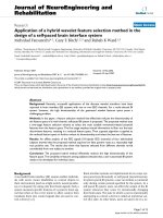
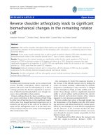
Figure 1 NG2 expression in the dorsal column of wild type animals following lysolecthin injection. Compared to sham-operated animals
(A), an increased number of dapi (blue) positive nuclei, as well as increased NG2 expression (red), are observed within the injury site at 1 week
(B and D-J) and 6 weeks (C) after lysolecithin injection. Panels D-G depict labeling in the same tissue section with multiple markers. More intense
expression of pan-neurofilament (NF, cyan) is seen in axons in the inflammatory region (D-G). NG2 (red) positive cells are seen (1) closely
apposed to vessel-like structures (arrowheads), (2) in close proximity to completely or partially demyelinated axons (arrow), as judged by NF
(cyan) and MBP (green) staining (F and G), and (3) in association with MBP labeling (asterisk, yellow, F). Each of these patterns of NG2 expression
is magnified in the inset panels in F. Double labeling for NG2 along with PDGFR alpha (arrow, Pa, H), CD11b (asterisk, I), or PDGFR beta
(arrowhead, Pb, J) reveals NG2 co-expression by oligodendrocyte progenitors (H), macrophages/microglial cells (I), or pericytes/mesenchymal
stem cells (J). Scale bar = 100 μm (A-C), 50 μm (D-G), and 30 μm (H-J).
Kucharova et al. Journal of Neuroinflammation 2011, 8:158
/>Page 5 of 13
(Figures 3G and 3H). However, PDGFa-positive OPCs
(Figures 3C and 3D) and PDGFRb-positive pericytes
(Figures 3K and 3L) now appear to be more abundant
in NG2 null lesions than in wild type lesions. We
believe this is due to delayed recruitmen t of immature
OPCs and pericytes in the absence of NG2. In wild type
animals, maturing cells recruited at earlier time points
may have already down-regulated expression of the
PDGFRa and PDGFRb markers.
Effect of NG2 ablation on cytokine expression
In addition to reduced influx of CD11b-immunoreact ive
macrophages/microglial cells into the damaged white
matter one week after lysolecithin injection into NG2
null mice , we also observed changes in cytokine levels
indicative of a shift from a pro-inflammatory to anti-
inflammatory phenotype [31]. Analysis of transcript
levels by qRT-PCR revealed that transcripts for the pro-
inflammatory cytokines interferon gamma (IFNg)and
interleukin 1-beta (IL-1b) were reduced in NG2 null
mice. In co ntrast, t he expression of cytokines character-
istic of an anti-inflammatory phenotype (IL-4 and IL-10)
was increased by ablation of NG2 (Figure 4).
Effects of NG2 ablation on cell proliferation and motility
Proliferation of OPCs, pericytes, and macrophages/
microglial cells in demyelinated lesions in wild type and
NG2 null mice was evaluated by BrdU incorpora tion.
BrdU was injected 4 days afte r surgery and animals
were euthanized after an additional 3 days (i.e. at day 7).
We found that the mitotic indices of OPCs, pericytes,
and macrophages/microglia were all reduced in the
absence of NG2 (Table 3). While OPCs proliferated in
proximity t o demyelinated axons inside the lesion site,
some BrdU-positive macrophages/microglial cells were
also seen outside the lesion (Figure 5). For these studies
we used the IBA-1 marker because of its expression on
both resident microglia and infiltrating m acrophages/
microglial cells, thus al lowing us to assess proliferation
in both populations. The presence of extra-lesional
BrdU-labeled IBA-1-posit ive cells suggested the possibi-
lity that microgl ial cells generated outside the demyeli-
nated region might invade the lesion, contributing to
the poo l of inflammatory cells present in this area. To
examine this possibility, we examined BrdU incorpora-
tion after a one-day incubation period. BrdU was admi-
nistered at 4 days after lysolecithin injection, and
animals were euthanized on the following day. Table 4
shows that in both wi ld type and NG2 null mice, about
10% of IBA-1, BrdU-double positive cells were located
outside the demyelinated area on this first day. However
by day 3, only 1% of these cells were still outside the
lesion in wild type mice, whereas at least 7% of the cells
in NG2 null mice were still located external to the
lesion. This result indicates a possible role for NG2 in
the motility of macrophages/microglia.
Discussion
In the CNS, myelination is accomplished by mature oli-
godendrocytes that arise from OPCs. During CNS devel-
opment, a substantia l pool of OPCs must be generated
for production of mature oligodendrocytes in sufficient
numbers for adequate myelination of axons. The adult
CNS still contains large numbers of OPCs that differ
somewhat from perinatal progenitors in their capability
for motility and proliferation, yet respond to most of the
same stimuli and express a similar set of phenotypic
markers as their perinatal counterparts. Adult OPCs
account for a large percen tage of the proliferating cells
in the mature CNS [7,9] and are responsible for produc-
tion of new oligodendrocytes to replace damaged cells.
Newly-differentiated oligodendrocytes derived from
adult OPCs, rather than pre-existing oligodendrocytes,
are responsible for remyelination of axons that occurs
Table 2 Abundance of NG2, PDGFR alpha, CD11b, and PDGFR beta expressing cells in wild type and NG2 null mice 1,
2, and 6 weeks after lysolecithin injection.
NG2 (%) PDGFRa (%) CD11b (%) PDGFRb (%)
Sham WT 6.12 ± 1.7
c
8.32 ± 2.1
c
- 6.62 ± 0.9
c
KO - 6.77 ± 2.6
c
- 4.82 ± 1.4
c
1W WT 100 ± 8.2 100 ± 11.5 100 ± 9.1 100 ± 17.7
KO - 72.69 ± 16.8* 33.23 ± 10.3*** 60.59 ± 15*
2W WT 75.45 ± 6
b
111.4 ± 1.9 174.83 ± 18.6
c
121.71 ± 18.7
KO - 89.7 ± 6.6*** 103.13 ± 16.6
c
** 87.71 ± 12.4
a
*
6W WT 23.99 ± 11.7
c
33.01 ± 4.9
c
6.41 ± 0.8
c
33.83 ± 2.6
c
KO - 40.8 ± 6.4
a
4.21 ± 1.3
c
* 44.69 ± 7*
The abundance of various cell types (NG2+, PDGFRa+, CD11b+, PDGFRb+) in demyelinated lesions from 1-6 weeks post-lysolecithin injection is illustrated by
normalizing cell density values to cell densities found in wild type mice at 1 week post-surgery (these 1 week values are designated as 100%). Values represent
means ± S.D. Statistically significant differences are indicated by * < 0.05; ** < 0.01; *** < 0.001 when values were compared between WT and NG2 null miceat
the same post-injection week.
a
< 0.05;
b
< 0.01;
c
< 0.001 represent statistically significant differences between values obtained for mice of the same genotype
when compared to the 1
st
week post-injection.
Kucharova et al. Journal of Neuroinflammation 2011, 8:158
/>Page 6 of 13
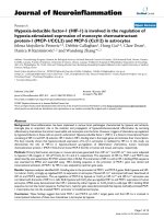
Figure 2 Demyelination and remyelination in dorsal columns of wild type (WT) and NG2 null (KO) mice. Immunolabeling for MBP (green)
and neurofilament (NF, red) reveals greater initial demyelination in wild type (A) compared to NG2 null spinal cord (D) during the first post-
surgery week. However, better repair is seen in wild type (B and C) than in knockout (E and F) spinal cord at 6 weeks after surgery. The higher
resolution images in C and F allow identification of NF-positive axons (red) associated with (arrowheads) or lacking association with (arrows)
MBP-positive myelin (green) at 6 weeks post-injury. Quantification of white matter lesion volumes, defined as MBP-negative regions (see panels
A, B, D and E), in wild type and NG2 null mice reveals larger lesions in wild type mice one week after lysolecithin injection, but diminished repair
of lesions in NG2 null mice six weeks post-injury. Lesion volumes are expressed as mean values ± SD. (G). An increased number of demyelinated
axons (H), determined by MBP and NF double labeling (see panels C and F), were present in the dorsal column of NG2 null mice 6 weeks after
lysolecithin injection. Statistically significant differences are indicated by * < 0.05; ** < 0.01 when values for WT and KO mice are compared at
the same time point;
b
< 0.01;
c
< 0.001 indicate statistically significant differences within the same genotype at 1 and 6 weeks after lysolecithin
injection. Scale bar = 100 μm (A, B, D and E) and 8 μm (C and F).
Kucharova et al. Journal of Neuroinflammation 2011, 8:158
/>Page 7 of 13
following various types of demyelinating events
[8,10,32-34]. Factors that influence O PC proliferation
and differentiation are therefore of great importance for
our understanding of both developmental myelination
and myelin repair.
The NG2 proteoglycan contributes to the proliferation
of OPCs during CNS development. In the NG2 null
mouse, decreased OPC proliferation red uces the size o f
the OPC pool, leading to a delay in production of nor-
mal numbers of mature oligodendrocytes and to a cor-
respondingdelayinaxonmyelination[24].Wehave
used lysolecithin-induced demyelination of the spinal
cord to examine the possibility that ablation of NG2
also impedes repair o f myelin damage in the adult CNS.
Following microinjectio n into CNS white matter, lysole-
cithin replaces phospholipids and forms micelles in the
membrane bilayer [35], rapi dly inducing lo cal myelin
destruction [36], blood-brain barrier damage, and
recruitment of macrophages and local microglial cells
into the lesion site [4]. This commonly-used demyelina-
tion model [4,19,35-37] has the advantage that the site
and extent of the injury are well-defined and
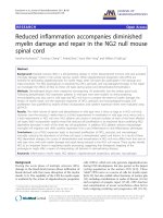
Figure 3 Distribution of PDGFR alpha, CD11b, and PDGFR beta immunoreactive cells in injured spinal cord white matter.PanelsA-D
show the distribution of PDGFR alpha positive OPCs (green) at 1 (A, B) and 6 (C, D) weeks after demyelination insult. Panels E-H present CD11b
immunoreactive myeloid cells (green) at 1 (E, F) and 6 weeks (G, H) post-injury, while panels I-L show PDGFR beta positive cells (green) at 1 (I, J)
and 6 weeks (K, L) post-injury. The first and third columns show sections from wild type mice at 1 and 6 weeks, respectively, after demyelination
insult. The second and fourth columns show sections from NG2 null mice at 1 and 6 weeks, respectively, after demyelination insult. Blue: DAPI.
Scale bar = 100 μm.
Kucharova et al. Journal of Neuroinflammation 2011, 8:158
/>Page 8 of 13
reproducible, facilitating data acquisition. In addition,
lysolecithin-induced demyelination occurs as an acute
event, such that all subsequent phenomena are asso-
ciated with the regenerative response. This provides a
useful means of separating events and mechanisms asso-
ciated with the respective processes of demyelination
and remyelination [21].
The regenerati on of myelin following demyelination is
a multifactorial process, due in part to the involvement
of multiple cell types in the damage and repair me chan-
isms. In additi on to neurons and OPCs, microglia,
macrophages, and pericytes also contribute to these pro-
cesses [38-41]. Our work shows that the NG2 proteogly-
can is expressed by three cell types that invade
demyelinated lesions: OPCs, pericytes, and macro-
phages/microglia. The differential contributions of these
three cell types to the damage and repair processes,
combined with differences in NG2 function in the
respective cell types, are probably responsible for the
complex patterns of demyelination and remyelination
that we see in the global NG2 null mouse. Figure 2
shows that although the extent of initial demyelination
is reduced in the NG2 null mouse, repair of this lesion
nevertheless proceeds more slo wly than repair of the
larger lesion found in the wild type mouse. The impact
of NG2 ab lation on OPCs is likely confined to deficien-
cies seen during the repair process, since OPCs generate
oligodendrocytes that carry out remyelination. Conver-
sely, diminished involvement of macrophages/microglia
probably pro vides the best explanation for the reduced
extent of initial demyelination seen in the NG2 null
mouse. However, macrophages/microglial cells also con-
tribute to myelin repair by clearing myelin debris and by
producing cytokines and growth factors that promote
recruitment of O PCs and prime interactions between
OPCs and axons. Thus, NG2-dependent deficits in
macrophage/microglia function may also contribute to
the reduced myelin repair seen in the NG2 null mouse.
Similarly, it is possible that pericytes affect both myelin
damage and repair. The recruitment of pericytes for
revascularization of the lesion and repair of t he blood
brain barrier likely plays an important rol e in the heal-
ing process. How ever, vascularization also provides
increased access to inflammatory cells a nd cytokines
that contrib ut e to myelin damage [40,42-45]. Since
many of the pericytes in lysolecithin-ind uced lesions are
not associated with vascular endothelial cells, another
consideration is the ability of pericytes to serve as
mesenchymal stem cells [46,47] with immunomodula-
tory properties that can promote myelin repair via their
effects on the activities of inflammatory cells [48].
Our evidence suggests that promoting cell prolifera-
tion is a key functional role for NG2 in OPCs, pericytes,
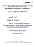
Figure 4 Relative expression levels of IFNg,IL-1b, IL-4, and IL-10 transcripts 7 days after lysole cithin injecti on.CytokinelevelsinNG2
null mice were normalized to those seen in wild type mice, defined as being equal to 1. Relative cytokine expression levels are expressed as
mean values ± SD. Statistically significant differences between WT and KO values are indicated by * < 0.05, ** < 0.01, and *** < 0.001.
Table 3 Proliferation of PDGFRa, PDGFRb, and IBA-1
expressing cells in wild type and NG2 null mice.
WT KO
Total Pa positive cells 178.23 ± 20.3 127.94 ± 18.4*
Pa/BrdU positive cells 26.4 ± 2.2 10.76 ± 2.9
Mitotic indices 14.81 ± 3.1% 8.41 ± 0.6%**
Total Pb positive cells 29.8 ± 5.7 21.91 ± 3.4*
Pb/BrdU positive cells 4.53 ± 0.8 2.31 ± 0.9
Mitotic indices 15.2 ± 0.6% 10.52 ± 0.6%***
Total IBA-1 positive cells 217.94 ± 14.8 74.45 ± 12.3***
IBA1/BrdU positive cells 19.66 ± 3.6 4.71 ± 3.5
Mitotic indices 9.02 ± 1.2% 6.32 ± 1.8%*
Total numbers of PDGFRa (Pa), PDGFRb (Pb), and IBA-1 positive cells, along
with BrdU incorporation, were determined in 0.1 mm
2
areas of the dorsal
column at 7 days postsurgery. Mitotic labeling indices for OPCs, pericytes, and
macrophages/microglial cells are expressed as the percentage of each cell
type that is BrdU positive. Data represent the mean ± S.D. Statistically
significant differences between wild type and NG2 null mice are indicated by
* < 0.05; ** < 0.01; *** < 0.001.
Kucharova et al. Journal of Neuroinflammation 2011, 8:158
/>Page 9 of 13
and macrophages/microglia. BrdU incorporation reve als
significant reductions in mitotic index for all three cell
types in demyelinated lesions in the NG2 null mouse. In
the case of OPCs, this confirms a similar result obtained
in our studies of developmental myelination: namely,
that ablation of NG2 reduces the OPC mitotic index,
with a corresponding decrease in the number of
myelinating oligode ndrocytes [24]. Thus, NG2 is impor-
tant for promoting the proliferation of both perinatal
OPCs and adult OPCs. The BrdU results also confirm
our report that ablation of NG2 diminishes pericyte pro-
liferation during pat hological retinal neovascularization,
leading to decreased blood vessel formation in the reti-
nas of NG2 null mice [25]. This negative effect of NG2
ablation on cell proliferation may be a fairly general
one, since we also observe diminished keratinocyte pro-
liferation in the skin of newborn NG2 null mice [49].
Our in vitro studies also support a rol e for NG2 in pro-
moting cell proliferation. NG2 is able to enhance prolif-
eration via two mechanisms: promotion of signaling by
b1 integrins [50] and promotion of signaling by recep-
tors for the growth factors PDGF and FGF [27,51].
In vitro studies also indicate that NG2-dependent sig-
naling by b1 integrins and growth factor receptors can
promote cell motility as well as cell proliferation
Figure 5 Proliferation of I BA-1 immunoreactive macrophages/microglial cells in the injured spinal cord. Sections of injured spinal cord
were evaluated for BrdU incorporation (green) and IBA-1 labeling (red) 7 days after injury (3 days after BrdU injection). Compared to wild type
animals (A-C), fewer proliferating IBA-1-positive cells are seen in NG2 null mice (D-F). Boundaries of demyelinated lesions are indicated by white
dotted lines in C and F. In NG2 null mice, IBA-1/BrdU double-labeled cells (arrows) remain outside the lesion to a greater extent than in wild
type mice. Scale bar = 100 μm.
Table 4 Percentage of IBA-1/BrdU-positive cells outside
the demyelinated region.
WT KO
5
th
postsurgery day 7.74 ± 0.44% 10.29 ± 0.31%
7
th
postsurgery day 1.12 ± 0.01%
a
7.22 ± 0.12% ***
The percentage of BrdU-labeled cells IBA-1-positive cells outside the area of
demyelination was evaluated in wild type (WT) and NG2 null (KO) mice at 5
and 7 days following lysolecithin injection. Data represent the mean ± S.D. A
statistically significant difference between wild type and NG2 null mice at day
7 is indicated by *** < 0.001.
a
< 0.05 indicates the statistically significant
difference in wild type mice between the 5
th
and 7
th
post-injection days.
Kucharova et al. Journal of Neuroinflammation 2011, 8:158
/>Page 10 of 13
[27,50,52,53]. In vivo, one indication of this effect is
seen in our current studies on macrophage invasion into
demyelinated lesions. BrdU tracking studies at day 5,
one day after lysolecithin injection, show that 8 to 10%
of the macrophages/microglia in dorsal column white
matter are located outside demyelinated lesions. By 7
days post-injection in wild type mice , 90% of these per-
ipherally-located cells have migrated into the les ion. By
contrast, only 20% of extra-lesional ce lls have migrated
into the lesion in NG2 null mice, indicat ive of the NG2
dependence of macrophage motility. Similar measure-
ments were no t possible in the case of OPCs or peri-
cytes due to the rare occurrence of BrdU-labeled cells
outside of demyelinated lesions.
Our finding of changes in cytokine expression fol-
lowing NG2 abl ation may also be important in under-
standing changes in demyelination and remyelination
in the NG2 null mouse. Although it remains to be
determined whether changes in cytokine expression in
the NG2 null mouse are associated with changes in
macrophages as opposed to other inflammatory cell
types, decreased levels of IFNg and IL -1b coupled with
increased levels of IL-4 and IL-10 suggest that NG2
ablation shifts a pro-inflammatory phenotype to an
anti-inflammatory one. IFNg provokes acute re-occur-
rence of demyelination in MS patients [54], and IL-1b
is present in CNS-infiltrating myeloid cells in MS
models [55]. It therefore seems possible that decreased
levels of IFNg and IL-1b in spinal cord lesions i n the
NG2 null mouse (or altered activities of cells expres-
sing these cytokines) are responsible for the reduced
white matter dam age seen in these mice . Moreover,
decreased IL-4 production in the CNS exacerbates
experimental autoimmune encephalitis, and is asso-
ciated with increased infiltration of inflammatory cells
[56], while increased IL-10 expression is associated
with reduced inflammation [57]. The possibility that
NG2 null macrophages/microglia may exhibit less
inflammatory properties than wild type cells is in line
with the in vitro finding of a reduced inflammatory
phenotype upon knockdown of NG2 in microglia [58].
We speculate that diminished occurrence of myeloid
cells in NG2 null spinal cord lesions, coupled with
alterations in the intrinsic properties/functions of
NG2-negative macrophages/microglial cells, can affect
the progression of demyelination and remyelination in
NG2 null mice.
Conclusions
In summary, our results demonstrate that the func-
tions of all three of the NG2-positive cell type s (OPCs,
pericytes, and macrophages/microglia) associated with
demyelinated lesions are compromised by the ablation
of NG2. As a result of changes in multiple cell types,
the respective processes of myelin damage and myelin
repair are both altered in NG2 null mice. The com-
plexity of the demyelination/remyelination phenotype
in the global NG2 null mouse suggests that cell type-
specific ablation of the proteoglycan will be a useful
strategy for elucidating the respective contributions of
NG2-positive cell types to the myelin damage and
repair processes. The use of NG2 floxed mice in con-
junction with appropriate Cre drivers will allow us to
perform t he desired NG2 ablations.
List of abbreviations
BrdU: 5-bromo-2-deoxyuridine; CNS: central nervous system; DAPI: 4’ -6-
diamidino-2-phenylindole; GAPDH: glyceraldehyde-3-phosphate
dehydrogenase; IFN: interferon; IL: interleukin; MS: Multiple sclerosis; MBP:
myelin basic protein; NF: Pan-Axonal Neurofilament; NG2-/-: NG2 null mice;
NG2+/+: wild type mice; OPCs: oligodendrocyte progenitor cells; PDGFR:
Plate derived growth factor receptor; qRT-PCR: quantitative reverse
transcription-polymerase chain reaction.
Acknowledgements
This work was supported by Postdoctoral Fellowship 82922 from the Craig
H. Neilsen Foundation (KK) and by NIH grants PO1 HD25938 and RO1
CA95287 (WBS). We thank Dr. Michael Hefferan and Dr. Viktor Skihar for help
with spinal cord surgery, and Francisco Beltran and Adriana Charbono for
assistance with portions of the animal work.
Author details
1
Sanford-Burnham Medical Research Institute, La Jolla, CA 92037, USA.
2
St
Jude Children’s Research Hospital, Memphis, TN 38105, USA.
3
Department of
Pathology, P.J. Safárik University, Faculty of Medicine, Kosice 04001, Slovak
Republic.
4
Departments of Oncology and Clinical Neurosciences, University
of Calgary, Calgary, Alberta, T2N 4N1, Canada.
Authors’ contributions
WBS and KK designed and performed research, and prepared the
manuscript. KK also evaluated the data. YC and AB performed research. VWY
designed research. All authors have read and approved the final version of
the manuscript.
Competing interests
The authors declare that they have no competing interests.
Received: 5 October 2011 Accepted: 13 November 2011
Published: 13 November 2011
References
1. Grigoriadis N, Grigoriadis S, Polyzoidou E, Milonas I, Karussis D:
Neuroinflammation in multiple sclerosis: evidence for autoimmune
dysregulation, not simple autoimmune reaction. Clin Neurol Neurosurg
2006, 108:241-244.
2. Mikita J, Dubourdieu-Cassagno N, Deloire MS, Vekris A, Biran M, Raffard G,
Brochet B, Canron MH, Franconi JM, Boiziau C, Petry KG: Altered M1/M2
activation patterns of monocytes in severe relapsing experimental rat
model of multiple sclerosis. Amelioration of clinical status by M2
activated monocyte administration. Mult Scler 2011, 17:2-15.
3. Petry KG, Brochet B, Dousset V, Vignes JR, Boiziau C: Inflammation induced
neurological handicap processes in multiple sclerosis: new insights from
preclinical studies. J Neural Transm 2010, 117:907-917.
4. Ousman SS, David S: Lysophosphatidylcholine induces rapid recruitment
and activation of macrophages in the adult mouse spinal cord. Glia
2000, 30:92-104.
5. Rosenberg GA: Matrix metalloproteinases and neuroinflammation in
multiple sclerosis. Neuroscientist 2002, 8:586-595.
6. Arnett HA, Fancy SP, Alberta JA, Zhao C, Plant SR, Kaing S, Raine CS,
Rowitch DH, Franklin RJ, Stiles CD: bHLH transcription factor Olig1 is
Kucharova et al. Journal of Neuroinflammation 2011, 8:158
/>Page 11 of 13
required to repair demyelinated lesions in the CNS. Science 2004,
306:2111-2115.
7. Dawson MR, Levine JM, Reynolds R: NG2-expressing cells in the central
nervous system: are they oligodendroglial progenitors? J Neurosci Res
2000, 61:471-479.
8. Gensert JM, Goldman JE: Endogenous progenitors remyelinate
demyelinated axons in the adult CNS. Neuron 1997, 19:197-203.
9. Horner PJ, Power AE, Kempermann G, Kuhn HG, Palmer TD, Winkler J,
Thal LJ, Gage FH: Proliferation and differentiation of progenitor cells
throughout the intact adult rat spinal cord. J Neurosci 2000, 20:2218-2228.
10. Keirstead HS, Blakemore WF: Identification of post-mitotic
oligodendrocytes incapable of remyelination within the demyelinated
adult spinal cord. J Neuropathol Exp Neurol 1997, 56:1191-1201.
11. Nguyen L, Borgs L, Vandenbosch R, Mangin JM, Beukelaers P, Moonen G,
Gallo V, Malgrange B, Belachew S: The Yin and Yang of cell cycle
progression and differentiation in the oligodendroglial lineage. Ment
Retard Dev Disabil Res Rev 2006, 12:85-96.
12. Patel JR, McCandless EE, Dorsey D, Klein RS: CXCR4 promotes
differentiation of oligodendrocyte progenitors and remyelination. Proc
Natl Acad Sci USA 2010, 107:11062-11067.
13. Nishiyama A, Watanabe M, Yang Z, Bu J: Identity, distribution, and
development of polydendrocytes: NG2-expressing glial cells. J Neurocytol
2002, 31:437-455.
14. Butt AM, Duncan A, Hornby MF, Kirvell SL, Hunter A, Levine JM, Berry M:
Cells expressing the NG2 antigen contact nodes of Ranvier in adult CNS
white matter. Glia 1999, 26:84-91.
15. Butt AM, Kiff J, Hubbard P, Berry M: Synantocytes: new functions for novel
NG2 expressing glia. J Neurocytol 2002, 31:551-565.
16. Ong WY, Levine JM: A light and electron microscopic study of NG2
chondroitin sulfate proteoglycan-positive oligodendrocyte precursor
cells in the normal and kainate-lesioned rat hippocampus. Neuroscience
1999, 92:83-95.
17. Lin SC, Bergles DE: Synaptic signaling between GABAergic interneurons
and oligodendrocyte precursor cells in the hippocampus. Nat Neurosci
2004, 7:24-32.
18. Paukert M, Bergles DE: Synaptic communication between neurons and
NG2+ cells. Curr Opin Neurobiol 2006, 16:515-521.
19. Blakemore WF, Franklin RJ: Remyelination in experimental models of
toxin-induced demyelination. Curr Top Microbiol Immunol 2008,
318
:193-212.
20.
Bramow S, Frischer JM, Lassmann H, Koch-Henriksen N, Lucchinetti CF,
Sorensen PS, Laursen H: Demyelination versus remyelination in
progressive multiple sclerosis. Brain 2010, 133:2983-2998.
21. Franklin RJ: Why does remyelination fail in multiple sclerosis? Nat Rev
Neurosci 2002, 3:705-714.
22. Stallcup WB: The NG2 proteoglycan: past insights and future prospects. J
Neurocytol 2002, 31:423-435.
23. Stallcup WB, Huang FJ: A role for the NG2 proteoglycan in glioma
progression. Cell Adh Migr 2008, 2:192-201.
24. Kucharova K, Stallcup WB: The NG2 proteoglycan promotes
oligodendrocyte progenitor proliferation and developmental
myelination. Neuroscience 2010, 166:185-194.
25. Ozerdem U, Stallcup WB: Pathological angiogenesis is reduced by
targeting pericytes via the NG2 proteoglycan. Angiogenesis 2004,
7:269-276.
26. Huang FJ, You WK, Bonaldo P, Seyfried TN, Pasquale EB, Stallcup WB:
Pericyte deficiencies lead to aberrant tumor vascularizaton in the brain
of the NG2 null mouse. Dev Biol 2010, 344:1035-1046.
27. Grako KA, Ochiya T, Barritt D, Nishiyama A, Stallcup WB: PDGF (alpha)-
receptor is unresponsive to PDGF-AA in aortic smooth muscle cells from
the NG2 knockout mouse. J Cell Sci 1999, 112(Pt 6):905-915.
28. Ozerdem U, Grako KA, Dahlin-Huppe K, Monosov E, Stallcup WB: NG2
proteoglycan is expressed exclusively by mural cells during vascular
morphogenesis. Dev Dyn 2001, 222:218-227.
29. de Castro R, Tajrishi R, Claros J, Stallcup WB: Differential responses of
spinal axons to transection: influence of the NG2 proteoglycan. Exp
Neurol 2005, 192:299-309.
30. Kiernan JA: Histological and Histochemical Methods: Theory and Practice. 3
edition. Butterworth-Heinemann, Oxford; 1999.
31. Gordon S: Alternative activation of macrophages. Nat Rev Immunol 2003,
3:23-35.
32. Watanabe M, Toyama Y, Nishiyama A: Differentiation of proliferated NG2-
positive glial progenitor cells in a remyelinating lesion. J Neurosci Res
2002, 69:826-836.
33. Piaton G, Williams A, Seilhean D, Lubetzki C: Remyelination in multiple
sclerosis. Prog Brain Res 2009, 175:453-464.
34. Miron VE, Kuhlmann T, Antel JP: Cells of the oligodendroglial lineage,
myelination, and remyelination. Biochim
Biophys Acta 2011, 1812:184-193.
35. Gregson NA: Lysolipids and membrane damage: lysolecithin and its
interaction with myelin. Biochem Soc Trans 1989, 17:280-283.
36. Hall SM: The effect of injections of lysophosphatidyl choline into white
matter of the adult mouse spinal cord. J Cell Sci 1972, 10:535-546.
37. Blakemore WF, Eames RA, Smith KJ, McDonald WI: Remyelination in the
spinal cord of the cat following intraspinal injections of lysolecithin. J
Neurol Sci 1977, 33:31-43.
38. Cristofanilli M, Harris VK, Zigelbaum A, Goossens AM, Lu A, Rosenthal H,
Sadiq SA: Mesenchymal stem cells enhance the engraftment and
myelinating ability of allogeneic oligodendrocyte progenitors in
dysmyelinated mice. Stem Cells Dev .
39. Pohl HB, Porcheri C, Mueggler T, Bachmann LC, Martino G, Riethmacher D,
Franklin RJ, Rudin M, Suter U: Genetically induced adult oligodendrocyte
cell death is associated with poor myelin clearance, reduced
remyelination, and axonal damage. J Neurosci 2011, 31:1069-1080.
40. Yong VW: Inflammation in neurological disorders: a help or a hindrance?
Neuroscientist 2010, 16:408-420.
41. Yaguchi M, Ohta S, Toyama Y, Kawakami Y, Toda M: Functional recovery
after spinal cord injury in mice through activation of microglia and
dendritic cells after IL-12 administration. J Neurosci Res 2008,
86:1972-1980.
42. Watzlawik J, Warrington AE, Rodriguez M: Importance of oligodendrocyte
protection, BBB breakdown and inflammation for remyelination. Expert
Rev Neurother 2010, 10:441-457.
43. Holley JE, Newcombe J, Whatmore JL, Gutowski NJ: Increased blood vessel
density and endothelial cell proliferation in multiple sclerosis cerebral
white matter. Neurosci Lett 2009, 470:65-70.
44. Kirk S, Frank JA, Karlik S: Angiogenesis in multiple sclerosis: is it good, bad
or an epiphenomenon? J Neurol Sci 2004, 217:125-130.
45. Roscoe WA, Welsh ME, Carter DE, Karlik SJ: VEGF and angiogenesis in
acute and chronic MOG((35-55)) peptide induced EAE. J Neuroimmunol
2009, 209:6-15.
46. Caplan AI: All MSCs are pericytes? Cell Stem Cell 2008, 3:229-230.
47. Crisan M, Yap S, Casteilla L, Chen CW, Corselli M, Park TS, Andriolo G, Sun B,
Zheng B, Zhang L, et al: A perivascular origin for mesenchymal stem cells
in multiple human organs. Cell Stem Cell 2008, 3:301-313.
48. Siatskas C, Payne NL, Short MA, Bernard CC:
A consensus statement
addressing
mesenchymal stem cell transplantation for multiple sclerosis:
it’s time! Stem Cell Rev 2010, 6:500-506.
49. Kadoya K, Fukushi J, Matsumoto Y, Yamaguchi Y, Stallcup WB: NG2
proteoglycan expression in mouse skin: altered postnatal skin
development in the NG2 null mouse. J Histochem Cytochem 2008,
56:295-303.
50. Makagiansar IT, Williams S, Mustelin T, Stallcup WB: Differential
phosphorylation of NG2 proteoglycan by ERK and PKCalpha helps
balance cell proliferation and migration. J Cell Biol 2007, 178:155-165.
51. Goretzki L, Burg MA, Grako KA, Stallcup WB: High-affinity binding of basic
fibroblast growth factor and platelet-derived growth factor-AA to the
core protein of the NG2 proteoglycan. J Biol Chem 1999, 274:16831-16837.
52. Makagiansar IT, Williams S, Dahlin-Huppe K, Fukushi J, Mustelin T,
Stallcup WB: Phosphorylation of NG2 proteoglycan by protein kinase C-
alpha regulates polarized membrane distribution and cell motility. J Biol
Chem 2004, 279:55262-55270.
53. Fukushi J, Makagiansar IT, Stallcup WB: NG2 proteoglycan promotes
endothelial cell motility and angiogenesis via engagement of galectin-3
and alpha3beta1 integrin. Mol Biol Cell 2004, 15:3580-3590.
54. Satoh J, Kuroda Y: Differing effects of IFN beta vs IFN gamma in MS:
gene expression in cultured astrocytes. Neurology 2001, 57:681-685.
55. Burger D, Molnarfi N, Weber MS, Brandt KJ, Benkhoucha M, Gruaz L,
Chofflon M, Zamvil SS, Lalive PH: Glatiramer acetate increases IL-1
receptor antagonist but decreases T cell-induced IL-1beta in human
monocytes and multiple sclerosis. Proc Natl Acad Sci USA 2009,
106:4355-4359.
Kucharova et al. Journal of Neuroinflammation 2011, 8:158
/>Page 12 of 13
56. Ponomarev ED, Maresz K, Tan Y, Dittel BN: CNS-derived interleukin-4 is
essential for the regulation of autoimmune inflammation and induces a
state of alternative activation in microglial cells. J Neurosci 2007,
27:10714-10721.
57. Reder AT, Genc K, Byskosh PV, Porrini AM: Monocyte activation in multiple
sclerosis. Mult Scler 1998, 4:162-168.
58. Gao Q, Lu J, Huo Y, Baby N, Ling EA, Dheen ST: NG2, a member of
chondroitin sulfate proteoglycans family mediates the inflammatory
response of activated microglia. Neuroscience 2010, 165:386-394.
doi:10.1186/1742-2094-8-158
Cite this article as: Kucharova et al.: Reduced inflammation accompanies
diminished myelin damage and repair in the NG2 null mouse spinal
cord. Journal of Neuroinflammation 2011 8:158.
Submit your next manuscript to BioMed Central
and take full advantage of:
• Convenient online submission
• Thorough peer review
• No space constraints or color figure charges
• Immediate publication on acceptance
• Inclusion in PubMed, CAS, Scopus and Google Scholar
• Research which is freely available for redistribution
Submit your manuscript at
www.biomedcentral.com/submit
Kucharova et al. Journal of Neuroinflammation 2011, 8:158
/>Page 13 of 13