báo cáo hóa học: " The Dapsone Hypersensitivity Syndrome revisited: a potentially fatal multisystem disorder with prominent hepatopulmonary manifestations" pdf
Bạn đang xem bản rút gọn của tài liệu. Xem và tải ngay bản đầy đủ của tài liệu tại đây (360.23 KB, 9 trang )
BioMed Central
Page 1 of 9
(page number not for citation purposes)
Journal of Occupational Medicine
and Toxicology
Open Access
Case report
The Dapsone Hypersensitivity Syndrome revisited: a potentially
fatal multisystem disorder with prominent hepatopulmonary
manifestations
Semaan G Kosseifi
1
, Bhuvana Guha
1
, Dima N Nassour
1
, David S Chi
1
and
Guha Krishnaswamy*
1,2
Address:
1
Department of Internal Medicine, Quillen College of Medicine, East Tennessee State University, Johnson City, TN, USA and
2
Department
of Internal Medicine, Division of Allergy and Clinical Immunology, Quillen College of Medicine, East Tennessee State University, Johnson City,
TN, USA
Email: Semaan G Kosseifi - ; Bhuvana Guha - ; Dima N Nassour - ;
David S Chi - ; Guha Krishnaswamy* -
* Corresponding author
Abstract
4,4'-Diaminodiphenylsulphone (Dapsone) is widely used for a variety of infectious, immune and
hypersensitivity disorders, with indications ranging from Hansen's disease, inflammatory disease
and insect bites, all of which may be seen as manifestations in certain occupational diseases.
However, the use of dapsone may be associated with a plethora of adverse effects, some of which
may involve the pulmonary parenchyma. Methemoglobinemia with resultant cyanosis, bone
marrow aplasia and/or hemolytic anemia, peripheral neuropathy and the potentially fatal dapsone
hypersensitivity syndrome (DHS), the focus of this review, may all occur individually or in
combination. DHS typically presents with a triad of fever, skin eruption, and internal organ (lung,
liver, neurological and other systems) involvement, occurring several weeks to as late as 6 months
after the initial administration of the drug. In this sense, it may resemble a DRESS syndrome
(Drug Rash with Eosinophilia and Systemic Symptoms). DHS must be promptly identified, as
untreated, the disorder could be fatal. Moreover, the pulmonary/systemic manifestations may be
mistaken for other disorders. Eosinophilic infiltrates, pneumonitis, pleural effusions and interstitial
lung disease may be seen. This syndrome is best approached with the immediate discontinuation
of the offending drug and prompt administration of oral or intravenous glucocorticoids. An
immunological-inflammatory basis of the syndrome can be envisaged, based on the pathological
picture and excellent response to antiinflammatory therapy. Since dapsone is used for various
indications, physicians from all specialties may encounter DHS and need to familiarize themselves
with the salient features about the syndrome and its management.
Background
Dapsone has been used for many indications because of
its antibiotic and anti-inflammatory effects [1]. Not only
has it been the drug of choice for the treatment of leprosy
(Hansen's disease) since the middle of the 20
th
century,
but it has also been used for the treatment of brown rec-
luse spider bite [1,2], dermatitis herpetiformis, vesicobul-
lous dermatoses, cutaneous vasculitis, polyarteritis
Published: 06 June 2006
Journal of Occupational Medicine and Toxicology 2006, 1:9 doi:10.1186/1745-6673-1-9
Received: 06 January 2006
Accepted: 06 June 2006
This article is available from: />© 2006 Kosseifi et al; licensee BioMed Central Ltd.
This is an Open Access article distributed under the terms of the Creative Commons Attribution License ( />),
which permits unrestricted use, distribution, and reproduction in any medium, provided the original work is properly cited.
Journal of Occupational Medicine and Toxicology 2006, 1:9 />Page 2 of 9
(page number not for citation purposes)
nodosa, nodulocystic acne, cutaneous mycetoma and
multiples other dermatologic indications all of which
may be seen as manifestations in certain occupational dis-
eases [1]. Since the advent of the era of the Acquired
Immunodeficiency Syndrome (AIDS), dapsone has been
increasingly utilized in the chemoprophylaxis of Pneumo-
cystis carinii infection in combination with Trimethoprim/
Sulfamethoxazole. This has led to increasing incidence of
dapsone-related complications. Table 1 lists the multiple
adverse effects of Dapsone, including the dapsone hyper-
sensitivity syndrome (DHS) and DRESS syndrome.
Hemolysis (more likely to occur with deficiency of glu-
cose 6-phosphate dehydrogenase or G6-PD), bone mar-
row aplasia, renal disease, peripheral neuropathy,
methemoglobinemia, nausea, dizziness, fatigue and other
systemic manifestations may occur singly or in combina-
tion in patients on dapsone therapy. Of these, DHS is
characterized by the onset of fever, skin eruption and
internal organ involvement several weeks to as late as 6
months after patients are given this drug. Untreated the
syndrome can lead to severe organ dysfunction and even
death. The definitive mechanism for DHS is unclear, but
it is hypothesized that it is mediated by immune activa-
tion and elaboration of inflammatory cytokines. This case
report emphasizes the pulmonary effects associated with
DHS. The following sections provide an overview of the
presentations, pathogenesis, diagnosis and management
of DHS.
Case report
A twenty-one-year old previously healthy nursing student,
with no significant past medical or surgical history, pre-
sented with an apparent reaction to an insect bite on her
left shin. She was evaluated and based on the presumed
diagnosis that this represented a hypersensitivity reaction
to an insect bite, and she was treated with dapsone (100
mg twice a day for 7 days) as an anti-inflammatory agent.
This was based on some data supporting the use of this
drug for insect bites [2], although this now appears to be
a controversial approach. After seven days of therapy, the
patient developed progression of her symptoms, develop-
ing fever with chills, myalgia and nausea associated with
diffuse abdominal pain, dark-colored urine and jaundice.
She also noticed a new onset non-pruritic, ascending,
maculo-papular skin eruption, with progressive shortness
of breath. She denied cough, hemoptysis, or chest pain at
this time. On admission, the patient was jaundiced, tach-
ypenic with a respiratory rate of 28 breath/minute, heart
rate of 103 beats/minute, blood pressure of 130/92 mm
of Hg, and a temperature of 99°F. The patient's oxygen
saturation was 86 % on room air, and subsequently she
was given higher amount of oxygen, to the point that she
was requiring 100 % non-rebreather mask in order to
maintain her oxygen saturation above 92 %. Oral mucosa
was normal, with no visible lesions. Neck examination
was supple, with no palpable lymphadenopathy or evi-
dence of thyroid enlargement. Lung examination revealed
bilateral diffuse crackles, decreased breath sounds in both
bases and dullness to percussion. Cardiac examination
showed tachycardia without audible murmur while the
abdominal examination revealed hypoactive bowel
sounds with diffuse mild tenderness on deep palpation,
without palpable organomegaly or evidence of rebound
tenderness. The patient demonstrated a diffuse maculopa-
pular eruption with sparing of the hands, feet, and
mucosa. There was no blanching on pressure. There was a
healing ulcer on her left shin with a dried-up scar (site of
presumed insect bite), without surrounding erythema or
other signs of inflammation or infection. The absence of
significant necrosis or surrounding inflammation sug-
gested that the symptoms the patient was experiencing
were independent of the insect bite.
The admitting laboratory data is provided in Table 2. Liver
function abnormalities consistent with transaminitis were
seen. The peripheral smear review showed a normocytic,
Table 1: Adverse reactions to Dapsone
System Manifestations Mechanisms
Systemic
• *DHS Dermatitis, hepatitis Idisosyncratic
• **DRESS syndrome Dermatitis, eosinophilia Idiosyncratic
• Nonspecific Nausea, headache, dizzy Weakness/fatigue Predictable
Hematological Hemolytic anemia Predictable (G6PDD)
Methemoglobinemia Predictable
Neurological Peripheral neuropathy Predictable
Dermatological Stevens-Johnson Syndrome Idiosyncratic
Toxic epidermal necrolysis Idiosyncratic
Hepatic Colestasis, hepatitis Idiosyncratic
Renal Nephritis Idiosyncratic
Pulmonary Pneumonitis, ***PIE Idiosyncratic
Thyroid Hypothyroidism Idiosyncratic
DHS = dapsone hypersensitivity syndrome, DRESS = drug rash, eosinophilia and systemic symptoms, PIE = pulmonary infiltration with eosinophilia
Journal of Occupational Medicine and Toxicology 2006, 1:9 />Page 3 of 9
(page number not for citation purposes)
normochromic anemia with mild neutrophilia and toxic
granulations. The patient also demonstrated mild aniso-
cytosis, poikilocytosis and reticulocytosis. The urine sam-
ple tested positive for blood but the patient had negative
urine, blood and stool cultures. A chest roentgenogram
was done on admission and showed a diffuse bilateral air
space disease involving the majority of the lung with spar-
ing of the lung apices with bilateral pleural effusions.
Computed tomography of the abdomen and pelvis with
contrast showed gallbladder wall-thickening and mild dil-
atation, with small amount of abdominal fluid (ascites).
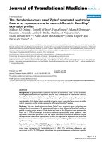
Computed tomography of the chest showed bilateral
infiltrates mainly on the left, along with bilateral pleural
effusion (Figure 1). Evaluation for other infectious and/or
immunological etiologies was negative, and included a
hepatitis panel, ELISA for HIV 1–2 and testing for
Legionella, Rocky Mountain Spotted Fever, cold aggluti-
nins, Group B streptococcus, Influenza A and B,
Monotest, direct Coombs, Leptospirosis, Ehlerichiosis,
Ebstein-Barr virus (EBV), Cytomegalovirus (CMV) and
Parvovirus.
Based on the clinical picture of multisystem involve-
ment, lack of a defining microbiological cause and the
progression of disease, a diagnosis of DHS was made.
The patient was immediately initiated on intravenous glu-
cocorticoids (methylprednisolone) with dramatic
improvement in her symptoms and clinical disease. This
improvement is shown in Table 2 and includes dramatic
reversal of anemia, improvement in transaminitis and in
inflammatory parameters. This was also evidenced by sta-
bilization of hypoxemia with less need for oxygen supple-
ment.
Dapsone Hypersensitivity Syndrome
Background
DHS is characterized by a hypersensitivity response to the
drug, dapsone which is a sulfone. Dapsone (4,4'-Diamin-
odiphenylsulphone) is used mainly as an anti-inflamma-
tory and anti-bacterial agent for the treatment of skin
diseases, bacteria, and fungi [1]. The anti-inflammatory
effects of dapsone are mainly related to its interference
with neutrophil chemotactic migration and adherence
[1]. Other multiple mechanisms are listed in the litera-
ture. These describe suppression of neutrophil recruit-
ment, inhibition of local production of toxic respiratory
and secretory products, as well as formation of oxidants.
These not only kill bacteria but also damage bystander tis-
sues. In addition, dapsone can inhibit the release of pros-
taglandins and leukotrienes by blocking their
inflammatory effects [1,3-5]. Overall, the side effects of
dapsone are very low if plasma concentration is below 5
mg/l [1,4].
A list of adverse effects of dapsone (some predictable,
some idiosyncratic or allergic) is provided in Table 1.
A rare syndrome, DHS which was first described by Allday,
Lowe, and Barnes [4,5] as a hypersensitivity vasculitis syn-
drome [1,3-8]. The incidence of DHS ranges from 0.5% to
3 % [1,3,6,8], while the median latency before symptoms
onset can be as early as 2 to 6 hours in previously sensi-
tized patients, to as late as 6 months. This variability of the
latency time was explained by several factors. Some of
those were related not only to the variability of the
acetylators but also to the dose and the modality of treat-
ment [1,4,6,8]. This wide variability of the latency period
Table 2: Representing the laboratory data.
Measurement admission day 1 day 2 day 3 day 4 day 5 Normal
Values
WBC8 8.310.5*6.77.38.34.0 – 9.2 × 10
3
/
mm3
Hb 6.9* 8.6* 11.5* 11.9* 13.3 15 12.1 – 15.2 g/dL
Platelet 217 246 318 380 468* 425 150 – 450 ×
10
3
/mm
3
ALT 103* 100* 86* 100* 130* 77* 10 – 60 IU/L
AST 179* 111* 91* 75* 68* 30 10 – 42 IU/L
ESR 21* 11 0 – 20 mm/hour
T.Bil. 8* 5* 1.8* 2.3* 1.5* 1.2* 0.2 – 1 mg/dL
LDH 778* 1182* 589* 90 – 180 IU/L
CPK 112 30 – 165 IU/L
*means abnormal values.
Black arrow indicating the timing when intravenous glucocorticoids were given to the patient.
WBC = White blood count, MCV = Mean corpuscular volume, Hb = Hemoglobin, ALT = Alanine aminotransferase, AST = Aspartate
aminotransferase, T.Bil = Total bilirubin, LDH = Lactate dehydrogenase, INR = International ratio, CPK = Creatinine phosphokinase.
Journal of Occupational Medicine and Toxicology 2006, 1:9 />Page 4 of 9
(page number not for citation purposes)
suggests a multi-organ hypersensitivity reaction, but the
exact mechanisms are unclear and reviewed later [4-7].
Clinical features
The classic triad of DHS consists of fever, eruption, and
internal organ involvement (Table 3). Fever, hepatitis,
exfoliative dermatitis, adenopathy and hemolytic anemia
might be seen in varying combinations and sequences [9].
While traditionally a hepatitis or transaminitis is seen,
cholangitis has also been described as a component of the
DHS [9]. Studies have shown that this syndrome may
begin as early as 7–10 days after administration of the
drug or as late as 6 months into therapy with dapsone.
Cutaneous lesions can range from erythematous papules
as in our patient, to plaques, pustules, and eczematous
lesions [1,3-8]. The severity of the cutaneous changes does
not correlate with the severity or extent of internal organ
involvement which may remain asymptomatic of even
life-threatening [10]. Cutaneous lesions usually begin to
resolve 2 weeks after stopping therapy. Some patients may
also develop severe dermatitis and complications, such as
the Steven Johnson Syndrome or toxic epidermal necroly-
sis (Table 1). These patients may experience prolonged
morbidity and even mortality with the complications.
Especially in severe cases, malnutrition, protein loss and
secondary infection may complicate the illness, and these
patients need to be monitored for complications more
aggressively. In traditional DHS, antibiotics have little or
no role unless obvious infection, cellulitis or sepsis is
present.
Pulmonary manifestations
Infiltrative lung disease is the most common pattern of
drug-induced injury [10]. This pattern can cause intersti-
Computed chest tomography of the patient described in this report, showing bilateral interstitial infiltrates (yellow arrow) and pleural effusions (red arrow)Figure 1
Computed chest tomography of the patient described in this report, showing bilateral interstitial infiltrates (yellow arrow) and
pleural effusions (red arrow). Image taken at a mid-thoracic level.
Journal of Occupational Medicine and Toxicology 2006, 1:9 />Page 5 of 9
(page number not for citation purposes)
tial lung damage, alveolar damage or vasculitic involve-
ment (e.g., vasculitis) [10]. As listed in Table 3, DHS can
also involve the lung, with 10 cases reported in the litera-
ture. In the patient described by us, pulmonary infiltrates
and pleural effusions with severe hypoxemia were present
suggesting interference with alveolocapillary oxygen
transfer. Other manifestations have also been described in
the literature.
Table 4 summarizes the different pulmonary manifesta-
tions of dapsone. Pulmonary manifestations of the DHS
are sometimes dominant features, as occurred in our
patient. Manifestations have included the development of
eosinophilic pneumonia [11-15], hypersensitivity pneu-
monia [16], pleural effusion [17]. Many reported cases of
eosinophilic pneumonia have an associated systemic
blood eosinophilia, which however, was not present in
our case. The clinical/radiological presentation of our
patient, along with the dramatic clinical response to glu-
cocorticoids, strongly suggest a drug-induced hypersensi-
tivity pneumonitis. The rapid clinical deterioration seen
with DHS can lead to respiratory failure as seen in our
patient and sometimes death, if untreated or unrecog-
nized.
Other manifestations
Other manifestations of this syndrome include hepato-
biliary dysfunction (such as jaundice[1], hepatomegaly
[1]and cholangitis [9], splenomegaly[1], eosi-
nophilia[1,13], photosensitivity[1], elevated sedimenta-
tion rate [1], and a mononucleosis picture that can mimic
EBV and CMV infection [1,7]. In addition, DHS can
include peripheral neuropathy [18], psychosis [19], renal
involvement (such as nephrotic syndrome [1] and renal
papillary necrosis [20] and pancreatitis [17] as found in
our patient. As listed in Table 3, a maculopapular erup-
tion, bullous disease, photosensitivity and oral erosions
can occur.
Differential diagnosis
The differential diagnosis of multisystem illness present-
ing in a patient on dapsone is shown in Table 5. These
include diseases such as: DRESS syndrome and its vari-
ants, vasculitis (Churg Strauss syndrome), Hypereosi-
nophilic syndrome, TENS (Toxic epidermal necrolysis
syndrome), Steven Johnson Syndrome, Still's disease,
Hematological disorders (leukemia, lymphoma), parane-
oplastic disorders and certain connective tissue disorders.
We will discuss a few of these important conditions in this
section.
Systemic organ involvement is often a feature of both
DHS and the DRESS syndrome. In the case of the latter,
drug rash, eosinophilia and systemic symptoms are often
present. DRESS syndrome can be seen with a variety of
medications, including anticonvulsants, sulfonamides,
allopurinol, calcium channel blockers, NSAIDS (Non-
steroidal anti-inflammatory drugs) and dapsone [21].
Fever, skin eruption, adenopathy, eosinophilia and inter-
nal organ involvement might also be seen [21]. Slow
acetylators may be associated with an increased risk for
development of DRESS syndrome. DHS can be consid-
ered a variant of the DRESS syndrome.
TENS and Stevens-Johnson syndrome represent a derma-
tologic emergency. They are characterized by diffuse ery-
Table 3: DHS clinical manifestations.
Systemic
1. Fever*
2. Pneumonitis*
3. Lymphadenopathy
4. Hepatitis*
5. Hemolytic anemia*
6. Carditis
Dermatological
1. Exfoliative dermatitis
2. Eczematous/maculopapular eruption*
3. Oral erosions
4. Vesicles and bullae
5. Photosensitivity
Laboratory
1. Hemolysis*
2. Anemia*
3. Eosinophilia
4. Atypical lymphocytosis
5. Transaminitis/elevated bilirubin/alkaline phosphatase*
6. Hypogammaglobulinemia
*-manifestations present in the patient described.
Journal of Occupational Medicine and Toxicology 2006, 1:9 />Page 6 of 9
(page number not for citation purposes)
thematous or purpuric macules with involvement of more
than 30 % of body surface area with epidermal necrosis
and mucosal membranes involvement. It is important to
note, that both disorders may overlap. A prodromal
phase, similar to a flu-like illness, lasting up to14 days,
may precede the skin eruption. The acute phase of TENS
consists of persistent fevers, generalized epidermal
sloughing and mucous membrane involvement [22].
Cutaneous vasculitis which can be part of a systemic dis-
ease needs also to be excluded. The evaluation of such
patients needs a complete work-up for the presence of sys-
temic disease. Skin biopsy and immunofluorescence stud-
ies may also helpful in the diagnosis of cutaneous
vasculitis in which immunoglobulin and complement
deposition are found [23].
If dermatitis and pulmonary disease is the dominant fea-
ture, necrotizing vasculitides and Churg Strauss syndrome
needs to be excluded. If dermatological manifestations
dominate the presentation, with or without mucosal
involvement, Steven Johnson Syndrome and TENS need
to be excluded. In some patients with a dominant eosi-
nophilia and pulmonary infiltration, PIE syndrome [10]
needs to be excluded.
Evaluation
Laboratory tests can include a complete blood count and
differential, comprehensive chemistry profile, sedimenta-
tion rate, urine analysis, arterial blood gases and a chest
roentgenogram. In selected cases, chest computed tomog-
raphy, hepatic ultrasound and/or liver or skin biopsy may
be required. Skin biopsy may not be specific but will assist
in excluding vasculitis or hematological malignancies. It
might be important to obtain a thyroid stimulating hor-
mone level in patients 3–4 months after the diagnosis of
DHS as detailed below.
Molecular and immunopathogenesis of DHS
The exact immune mechanism behind DHS is unclear. A
few mechanisms have been proposed. For one, DHS
might be a combination of type I, type IV, and perhaps
type III Gel and Coombs hypersensitivity reactions [5].
Alternately, DHS could be a modified graft versus host
disease mediated by activated T-lymphocytes [5]. It is
worth noting that DHS is not a dose-related effect [3-6,8],
whereas dapsone hepatotoxicity is a dose-dependent
effect [3].
According to Prussick and Shear, there is some evidence
suggesting that the metabolic differences in the produc-
tion and detoxification of reactive metabolites are an
important factor in sulfonamide hypersensitivity reac-
Table 5: Differential Diagnosis of DHS
DRESS syndrome and it's variants
Vasculitis (Churg Strauss syndrome)
Hypereosinophilic syndrome
Toxic epidermal necrolysis
Steven Johnson Syndrome
Still's disease
Hematological disorders (leukemia, lymphoma)
Paraneoplastic disorders
Certain connective tissue disorders
Table 4: Reported cases of Dapsone Hypersensitivity Syndrome with pulmonary manifestations.
Age/Sex Pulmonary manifestations Treatment Reference
22/M' Crepitations DW*, Steroid (6)
47/F Pulmonary eosinophilia DW, Steroid (11)
65/F Pulmonary eosinophilia DW (12)
45/M Pulmonary eosinophilia DW, Steroid (13)
60/F Eosinophilic pneumonia DW (14)
23/F Eosinophilic pneumonia DW, Steroid (15)
31/M Eosinophilic pneumonia with
pulmonary infarction
DW, Steroid (15)
37/F Eosinophilic pneumonia DW (15)
40/F" Hypersensitivity pneumonitis DW, Steroid (16)
15/M Right sided pleural effusion DW, Steroid (17)
*DW means drug withdrawal with supportive therapy only.
' M = male, " F = female.
Journal of Occupational Medicine and Toxicology 2006, 1:9 />Page 7 of 9
(page number not for citation purposes)
tions [24]. After absorption from the gastrointestinal tract,
dapsone is transported through the portal circulation to
the liver where it is metabolized primarily via two path-
ways: N-acetylation and N-hydroxylation [1,24]. N-
acetylation which has a bimodal pattern of activity (slow
and fast acetylation), has been shown not to determine
total clearance of dapsone [24]. However, the N-hydroxy-
lation pathway which is mediated primarily by human
liver microsomal enzymes P4503A4, 2C6, and 2C11
[1,24], is shown to be the initial step in the formation of
toxic intermediate metabolites, such as nitrosamines and
possibly other compounds, which can induce hemolytic
anemia and methemoglobinemia [24]. It is presumed that
these molecules are also important in the pathogenesis of
DHS. While N-hydroxylation yields a potentially toxic
metabolite known as the hydroxylamine, produced by
cytochrome P-450, acetylation by N-acetyltransferase
yields the nontoxic metabolites monoacetyl dapsone and
diacetyl dapsone [1]. Moreover, it has been shown that a
reduction in either quantity or activity of N-hydroxylation
enzyme systems resulted in decreased total clearance of
dapsone. Furthermore, this information is supported by
studies showing an extensive population and individual
variation in this ability involving both genetic (increased
or decreased P450 activity, decreased reduced glutathione
[GSH]) and environmental (drugs or chemicals such as
smoking inducing P450, cirrhosis and drugs inhibiting
P450, decreased GSH such as in AIDS, deficiency of anti-
oxidants such as Vitamin E, C, selenium) [24]. Fortu-
nately, other factors such as increased age and preexisting
liver disease (e.g., cirrhosis) offer relative protection
against adverse events because of decreased enzyme activ-
ity and, therefore, decreased production of toxic metabo-
lites [24]. However, with the lack of clinical studies, those
aforementioned protective factors remain speculations.
The mechanisms behind the pulmonary manifestations
may be similar, and due to the occurrence of eosinophilia
and pulmonary infiltrations, suggest the role for cytokines
such as tumor necrosis factor alpha (TNF-α), interleukin-
5 (IL-5) and chemokines [25,26]. These have not been
routinely studied. The rapid response to glucocorticoids
[27] also suggests that activation and nuclear transloca-
tion of nuclear factor kappaB (NF-kappa B) may occur,
resulting in a massive inflammatory response [25,26,28].
Glucocorticoids have also been shown to have both
genomic and nongenomic mechanisms that influence
inflammatory diseases [27]. How such events may relate
to the ultimate pathogenesis and evolution of this syn-
drome are unclear but need further examination.
Treatment
Treatment options for DHS are listed in Table 6. The main
treatment for DHS is immediate discontinuation of the
drug with initiation of oral or parenteral glucocorticoids,
depending on severity [1,5]. Glucocorticoids (such as
prednisone, prednisolone or methylprednisolone) have
profound anti-inflammatory actions, as summarized ear-
lier. The usual starting doses are mentioned in Table 6, but
are approximations only. Glucocorticoids should be
tapered gradually over a period of more than one month
because dapsone is found to persist in the body for up to
35 days [5]. It should be remembered that glucocorticoids
are medications with multiple side effects, including
hyperglycemia, hypokalemia, osteoporosis, glaucoma
and cataracts. Patients with risk factors for any of these
complications need close monitoring. Measurements of
blood sugar, bone mineral density, frequent measure-
ments of electrolytes and a thorough ophthalmologic
evaluation all constitute appropriate monitoring strate-
gies.
According to Reeve et al., [8] patients can be desensitized
to dapsone following DHS, by a very gradual re-introduc-
tion of the drug at low doses [8], but this hypothesis was
not supported by Pavithran K. et al. [4] who did not advise
re-challenge with dapsone. According to the literature,
patients with viral hepatitis (B, E) are at increased risk for
the development of DHS, suggesting the need to perform
a screening test for hepatitis B before starting dapsone
[5,29].
Table 6: Treatment approaches to DHS
Intervention Comments
1. Withdrawal of offending medication (dapsone) Drug discontinuation
2. Supportive therapy
• Volume replacement Intravenous fluid replacement
• Nutritional support Enteral or parenteral nutrition
• Antibiotics Early antibiotic institution in case of concomitant sepsis
• Skin care Preventing skin superinfection
3. Specific therapy
• Glucocorticoids Recommended dose 1 mg/kg/day
• Thyroid hormone replacement Associated late hypothyroidism
• Family counseling Genetic factors involvement
Journal of Occupational Medicine and Toxicology 2006, 1:9 />Page 8 of 9
(page number not for citation purposes)
Another important issue to remember in the management
of patients with DHS is that those patients might be at
higher risk for the development of hypothyroidism after
three months, suggesting the need to repeat thyroid func-
tion tests at three-month intervals and considering thy-
roid replacement therapy if the patient develops clinical
hypothyroidism as a delayed complication (Table 6). The
etiology attributed to the development of hypothy-
roidism, seems linked to the presence of autoantibodies,
including antimicrosomal antibodies [30]. In fact,
patients who are unable to detoxify reactive metabolites
produced by thyroid peroxidase will be more in favor of
developing a hypersensitivity reaction leading to hypothy-
roidism [30].
It is important to consider supportive therapies in patients
with more severe and protracted illness as listed in Table
6. Nutritional support (either total parenteral nutrition,
nutrition supplementation or enteral feeding), meticu-
lous fluid and electrolyte balance, control and prevention
of infectious complications (cellulitis, sepsis) and skin
care if necrotizing disease (TENS or Steven Johnson Syn-
drome were to ensue). For patients with dapsone-induced
hemolysis, Vitamin E supplementation might be benefi-
cial while in patients with methemoglobinemia coadmin-
istration of cimetidine can have an ameliorative effect.
In selected cases where glucocorticoids cannot be used or
are associated with severe complications (glaucoma,
severe osteoporosis, hyperglycemia or severe psychosis),
alternative drug therapies might be required. Unfortu-
nately, this being a rare disorder, other therapeutic
options such as methotrexate, azathioprine, cyclosporine
or hydroxychloroquin, have not been vigorously studied.
These drugs may provide benefit in the occasional patient.
Severe internal organ involvement such as carditis, hepa-
titis, pneumonitis and colitis, can cause death. These can
occur at any time and hence vigilance is required in these
sick patients. Also to be remembered is that in some
patients, in spite of drug withdrawal and high steroid ther-
apy, a relapsing and chronic course might ensue [22].
Since genetic factors are involved in the pathogenesis of
DHS, relatives should be instructed about DHS and their
enhanced risk of developing similar adverse reactions
should they take dapsone [10].
Conclusion
A high index of suspicion is needed for early diagnosis of
DHS. Patients initiated on dapsone for various indica-
tions need to be observed carefully for the development of
the DHS. If and when this occurs, DHS can be mistaken
for progression of the primary disease. If the drug is not
withdrawn, it could have deleterious and potentially fatal
effects due to major organ dysfunction. In our patient, the
association of hypoxemic pulmonary disease, pleural
effusions and anemia led to serious problems with oxy-
genation. The prompt withdrawal of the offending drug,
administration of glucocorticoids and supportive man-
agement led to rapid recovery in our patient
References
1. Zhu YI, Stiller MJ: Dapsone and sulfones in dermatology: over-
view and update. J Am Acad Dermatol 2001, 45:420-434.
2. Singletary EM, Rochman AS, Bodmer JC, Holstege CP: Envenoma-
tions 1. Med Clin North Am 2005, 89:1195-1224.
3. Leslie KS, Gaffney K, Ross CN, Ridley S, Barker TH, Garioch JJ: A
near fatal case of the dapsone hypersensitivity syndrome in
a patient with urticarial vasculitis. Clin Exp Dermatol 2003,
28:496-498.
4. Pavithran K, Bindu V: Dapsone syndrome: hepatitis-B infection
a risk factor for its development? Int J Lepr Other Mycobact Dis
1999, 67:171-172.
5. Knowles SR, Shapiro LE, Shear NH: Reactive metabolites and
adverse drug reactions: clinical considerations. Clin Rev Allergy
Immunol 2003, 24:229-238.
6. Rao PN, Lakshmi TS: Increase in the incidence of dapsone
hypersensitivity syndrome an appraisal. Lepr Rev 2001,
72:57-62.
7. Christiansen J, Tegner E, Irestedt M: Dapsone hypersensitivity
syndrome in a patient with cutaneous lupus erythematosus.
Acta Derm Venereol 1999, 79:482.
8. Reeve PA, Ala J, Hall JJ: Dapsone syndrome in Vanuatu: a high
incidence during multidrug treatment (MDT) of leprosy. J
Trop Med Hyg 1992, 95:266-270.
9. Itha S, Kumar A, Dhingra S, Choudhuri G: Dapsone induced
cholangitis as a part of dapsone syndrome: a case report.
BMC Gastroenterol 2003, 3:21.
10. Camus P, Bonniaud P, Fanton A, Camus C, Baudaun N, Foucher P:
Drug-induced and iatrogenic infiltrative lung disease 8. Clin
Chest Med 2004, 25:479-519, vi.
11. Begbie S, Burgess KR: Maloprim-induced pulmonary eosi-
nophilia. Chest 1993, 103:305-306.
12. Janier M, Guillevin L, Badillet G: Pulmonary eosinophilia associ-
ated with dapsone. Lancet 1994,
343:860-861.
13. Arunthathi S, Raju S: Dapsone induced pulmonary eosinophilia
without cutaneous allergic manifestations an unusual
encounter a case report. Acta Leprol 1998, 11:3-5.
14. Jaffuel D, Lebel B, Hillaire-Buys D, Pene J, Godard P, Michel FB, Blayac
JP, Bousquet J, Demolyi P: Eosinophilic pneumonia induced by
dapsone. BMJ 1998, 317:181.
15. Davidson AC, Bateman C, Shovlin C, Marrinan M, Burton GH, Cam-
eron IR: Pulmonary toxicity of malaria prophylaxis. BMJ 1988,
297:1240-1241.
16. Tobin-D'Angelo MJ, Hoteit MA, Brown KV, Ray SM, King MD: Dap-
sone-induced hypersensitivity pneumonitis mimicking Pneu-
mocystis carinii pneumonia in a patient with AIDS. Am J Med
Sci 2004, 327:163-165.
17. Corp CC, Ghishan FK: The sulfone syndrome complicated by
pancreatitis and pleural effusion in an adolescent receiving
dapsone for treatment of acne vulgaris. J Pediatr Gastroenterol
Nutr 1998, 26:103-105.
18. Mery L, Dega H, Prost C, Dubertret L: [Dapsone-induced sensory
peripheral neuropathy]. Ann Dermatol Venereol 2003,
130:447-449.
19. Fine JD, Katz SI, Donahue MJ, Hendricks AA: Psychiatric reaction
to dapsone and sulfapyridine 1. J Am Acad Dermatol 1983,
9:274-275.
20. Hoffbrand BI: Dapsone and renal papillary necrosis 1. Br Med J
1978, 1:78.
21. Volcheck GW: Clinical evaluation and management of drug
hypersensitivity 5. Immunol Allergy Clin North Am 2004, 24:357-71,
v.
22. McKenna JK, Leiferman KM: Dermatologic drug reactions 2.
Immunol Allergy Clin North Am 2004, 24:399-423, vi.
Publish with BioMed Central and every
scientist can read your work free of charge
"BioMed Central will be the most significant development for
disseminating the results of biomedical research in our lifetime."
Sir Paul Nurse, Cancer Research UK
Your research papers will be:
available free of charge to the entire biomedical community
peer reviewed and published immediately upon acceptance
cited in PubMed and archived on PubMed Central
yours — you keep the copyright
Submit your manuscript here:
/>BioMedcentral
Journal of Occupational Medicine and Toxicology 2006, 1:9 />Page 9 of 9
(page number not for citation purposes)
23. McKenna KE, Robinson J: The dapsone hypersensitivity syn-
drome occurring in a patient with dermatitis herpetiformis.
Br J Dermatol 1997, 137:657-658.
24. Prussick R, Shear NH: Dapsone hypersensitivity syndrome. J Am
Acad Dermatol 1996, 35:346-349.
25. Krishnaswamy G: Treatment strategies for bronchial asthma:
an update 33. Hosp Pract (Off Ed) 2001, 36:25-35.
26. Shakoory B, Fitzgerald SM, Lee SA, Chi DS, Krishnaswamy G: The
role of human mast cell-derived cytokines in eosinophil biol-
ogy 14. J Interferon Cytokine Res 2004, 24:271-281.
27. Rhen T, Cidlowski JA: Antiinflammatory action of glucocorti-
coids - New mechanisms for old drugs 1. New England Journal of
Medicine 2005, 353:1711-1723.
28. Fitzgerald SM, Chi DS, Hall HK, Reynolds SA, Aramide O, Lee SA,
Krishnaswamy G: GM-CSF induction in human lung fibroblasts
by IL-1beta, TNF-alpha, and macrophage contact 24. J Inter-
feron Cytokine Res 2003, 23:57-65.
29. Chogle A, Nagral A, Soni A, Agale S, Jamadar Z: Dapsone hypersen-
sitivity syndrome with coexisting acute hepatitis E. Indian J
Gastroenterol 2000, 19:85-86.
30. Gupta A, Eggo MC, Uetrecht JP, Cribb AE, Daneman D, Rieder MJ,
Shear NH, Cannon M, Spielberg SP: Drug-induced hypothy-
roidism: the thyroid as a target organ in hypersensitivity
reactions to anticonvulsants and sulfonamides 1. Clin Pharma-
col Ther 1992, 51:56-67.