Báo cáo hóa học: "Trimellitic anhydride-conjugated serum albumin activates rat alveolar macrophages in vitro" ppt
Bạn đang xem bản rút gọn của tài liệu. Xem và tải ngay bản đầy đủ của tài liệu tại đây (287.98 KB, 8 trang )
BioMed Central
Page 1 of 8
(page number not for citation purposes)
Journal of Occupational Medicine
and Toxicology
Open Access
Research
Trimellitic anhydride-conjugated serum albumin activates rat
alveolar macrophages in vitro
Dingena L Valstar
1
, Marcel A Schijf
1
, Erietta Stelekati
1
, Frans P Nijkamp
1
,
Nanne Bloksma
1,2
and Paul AJ Henricks*
1
Address:
1
Pharmacology and Pathophysiology, Utrecht Institute for Pharmaceutical Sciences, Faculty of Science, Utrecht University, Utrecht, The
Netherlands and
2
Deparment of Biology, Faculty of Science, Utrecht University, Utrecht, The Netherlands
Email: Dingena L Valstar - ; Marcel A Schijf - ; Erietta Stelekati - ;
Frans P Nijkamp - ; Nanne Bloksma - ; Paul AJ Henricks* -
* Corresponding author
Abstract
Background: Occupational exposure to airborne low molecular weight chemicals, like trimellitic
anhydride (TMA), can result in occupational asthma. Alveolar macrophages (AMs) are among the
first cells to encounter these inhaled compounds and were previously shown to influence TMA-
induced asthma-like symptoms in the Brown Norway rat. TMA is a hapten that will bind to
endogenous proteins upon entrance of the body. Therefore, in the present study we determined
if TMA and TMA conjugated to serum albumin induced the production of the macrophage
mediators nitric oxide (NO), tumour necrosis factor (TNF), and interleukin 6 (IL-6) in vitro using
the rat AM cell line NR8383 and primary AMs derived from TMA-sensitized and naïve Brown
Norway rats.
Methods: Cells were incubated with different concentrations of TMA, TMA conjugated to bovine
serum albumin (BSA), and BSA as a control for 24 h and the culture supernatant was analyzed for
mediator content.
Results: TMA alone was not able to induce the production of mediators by NR8383 cells and
primary AMs from sensitized and sham-treated rats. TMA-BSA, on the contrary, dose-dependently
stimulated the production of NO, TNF, and IL-6 by NR8383 cells and of NO and TNF, but not IL-
6, by primary AMs independent of sensitization.
Conclusion: Results suggest that although TMA is a highly reactive compound, conjugation to a
suitable protein is necessary to induce mediator production by AMs. Furthermore, the observation
that effects of TMA-BSA were independent of sensitization suggests involvement of an
immunologically non-specific receptor. In the discussion it is argued that a macrophage scavenger
receptor is a likely candidate.
Background
Trimellitic anhydride (TMA) is a reactive low molecular
weight (LMW) chemical used in the manufacture of
paints, epoxy curing agents, printing inks and vinyl plasti-
cizers and is known to cause occupational asthma charac-
terized by airflow obstruction, airway inflammation and
Published: 23 June 2006
Journal of Occupational Medicine and Toxicology 2006, 1:13 doi:10.1186/1745-6673-1-13
Received: 21 February 2006
Accepted: 23 June 2006
This article is available from: />© 2006 Valstar et al; licensee BioMed Central Ltd.
This is an Open Access article distributed under the terms of the Creative Commons Attribution License ( />),
which permits unrestricted use, distribution, and reproduction in any medium, provided the original work is properly cited.
Journal of Occupational Medicine and Toxicology 2006, 1:13 />Page 2 of 8
(page number not for citation purposes)
non-specific bronchial hyperreactivity [1-3]. The develop-
ment of this allergic disease requires sensitization trig-
gered by dermal or respiratory exposure to TMA followed
by its binding to proteins [4]. These so formed TMA-pro-
tein conjugates will then be taken up by antigen-present-
ing cells, transported to the regional lymph node, and
presented to TMA-specific T cells resulting in T cell mem-
ory and the production of TMA-specific IgE antibodies.
These antibodies will then bind to the high-affinity IgE
receptor on mast cells [5,6] and upon renewed contact
with TMA mediate cross-linking of the IgE-receptors with
subsequent release of mediators, that in their turn cause
bronchoconstriction and attraction of inflammatory cells
[7-9].
Alveolar macrophages (AMs) are among the first cells that
encounter inhaled small particles and chemicals in the
airways, since these cells are located at the interface
between air and lung tissue. AMs are long-lived cells
belonging to the family of mononuclear phagocytes. They
represent a non-specific cellular host defence mechanism
and can do so by binding to and ingestion of micro-organ-
isms and macromolecules via pattern recognition recep-
tors, and the secretion of a broad repertoire of mediators
that regulate inflammatory and immune reactions in the
lung [10-14]. Previously, it has been shown that depletion
of AMs in TMA-sensitized Brown Norway (BN) rats prior
to inhalation challenge with TMA, but not TMA-BSA,
resulted in ameliorated lung function during the chal-
lenge [15,16]. Furthermore, an increased influx of inflam-
matory cells into the lung lumen was observed 24 h after
challenge with TMA as well as TMA-BSA in AM-depleted
rats compared to non-depleted control rats [15,16].
Therefore, we investigated the direct effects of TMA and
TMA-BSA on the production of nitric oxide (NO), tumour
necrosis factor alpha (TNF), and interleukin 6 (IL-6) by
AMs using the rat AM cell line NR8383 and AMs derived
from TMA-sensitized and naïve BN rats.
Methods
Materials
TMA (97% purity) was obtained from Aldrich (Brussels,
Belgium) and acetone (HPLC grade) from Merck (Darm-
stadt, Germany). Highly refined olive oil, 2,4,6-trini-
trobenzene sulphonic acid, bovine serum albumin (BSA;
cell culture tested), sulphanilamide, and naphthyl-ethyl-
enediamide were purchased from Sigma (St. Louis, MO).
Sodium pentobarbitone was obtained from Cevasante
Animale B.V. (Maassluis, the Netherlands). K-medium
contained Dulbecco's Modified Eagle Medium (DMEM;
Cambrex BioScience, Verviers, Belgium) supplemented
with 10% fetal calf serum (FCS; Invitrogen BV, Breda, the
Netherlands), 10 mM HEPES (Merck), 4 mM L-glutamine,
1 mM sodium pyruvate, 100 U/ml of penicillin, 100 mg/
ml of streptomycin, 0.05 mM β-mercapto-ethanol (all
from Sigma) and 100 mg/ml gentamycin (Invitrogen).
Ham's F12 medium was obtained from Invitrogen,
lipopolysaccharide (LPS; E. coli O111:B4) from Sigma,
and IFN-γ from Genentech Inc. (San Francisco, CA). The
TNF and IL-6 ELISA kits were purchased from R&D Sys-
tems Inc. (Minneapolis, MN).
Animals
Female, inbred Brown Norway/CrlBR rats (BN; 7–8 weeks
of age) were purchased from Charles River (Maastricht,
the Netherlands). The animals were acclimatized at least 5
days before the start of the study. They were kept under
conventional laboratory conditions and received food
(Tecnilab BMI, Helmond, the Netherlands) and tap water
ad libitum. All animal procedures were conducted in
accordance with the Animal Ethics Committee of Utrecht
University (Utrecht, the Netherlands).
Sensitization procedure
TMA was applied at a concentration of 50% (w/v) in a
vehicle solution of 4:1 (v/v) acetone and olive oil. Ani-
mals received 150 μl on each flank (approximately 12 cm
2
each), which had been shaved with an electrical razor 2–
3 days earlier. Seven days after the first sensitization the
animals received 75 μl of a 25% TMA solution on the dor-
sum of both ears. Control animals received vehicle solu-
tion. Increased TMA-specific IgE serum levels verified the
sensitization status of the TMA-sensitized rats [15,16].
Preparation of TMA-BSA conjugate
The TMA-BSA conjugate was prepared under aseptic con-
ditions by dissolving 10 mg/ml of BSA in 0.1 M sodium
borate buffer (pH 9.4), adding approximately 1.5 mg
TMA per ml BSA-solution and stirring at room tempera-
ture. After 1 h the same amount of TMA was added and
the mixture was stirred for 2 h at room temperature. After
centrifugation at 390 × g for 5 min, the supernatant was
dialyzed successively against phosphate-buffered saline
(PBS) and distilled water for 24 h at 4°C. The conjugate
was lyophilized and stored at 4°C until use. The degree of
substitution of the TMA-BSA was assessed by determina-
tion of remaining free amino groups by reaction with
2,4,6-trinitrobenzene sulphonic acid as described previ-
ously [17]. The conjugate substitution ratio was approxi-
mately 40 mol TMA to 1 mol of BSA.
Cell culture and stimulation
AMs from TMA-sensitized and naïve rats were obtained by
lung lavage at day 20 after treatment. Rats were killed with
an overdose of sodium pentobarbitone (0.6 g/kg, i.p.). A
cannula was inserted into the trachea, the lungs were lav-
aged 4 times with 8 ml aliquots of PBS warmed to 37°C
and the lavage fluid was immediately thereafter put on ice.
The cells were collected by centrifugation for 10 min at
390 × g (4°C). After washing 3 time swith PBS, the cells
Journal of Occupational Medicine and Toxicology 2006, 1:13 />Page 3 of 8
(page number not for citation purposes)
were resuspended in K-medium and incubated at 37°C
for 2 h in 100 ml culture flasks (Greiner Bio-One, Alphen
a/d Rijn, the Netherlands). After washing away non-
adherent cells, the adherent cells (AMs) were scraped off
in fresh K-medium supplemented with 1% FCS, adjusted
to 1 × 10
6
cells/ml, and seeded in a total volume of 100 μl
into sterile flat-bottom 96-wells plates (Costar, Cam-
bridge, MA). After 1 h, 25 μl medium, stimulants (2 μg
LPS admixed with 10 U IFN-γ per ml, 0.1–3 mg/ml of
TMA-BSA, 0.1–3 mg/ml of BSA) in medium, or 10 and
100 μM TMA in 0.01 and 0.1% ethanol were added. All
stimulants were filtered through a 0.22 μm syringe-filter
(TPP, Trasadingen, Switzerland) before use. After 24 h the
supernatants were collected and kept at -20°C until use.
Control stimulation of cells with 0.01 and 0.1% ethanol
in medium did not affect cells (data not shown).
The AM cell line, NR8383, derived from Sprague Dawley
rats, was purchased from the ATCC (Manassas, VA, USA)
and maintained in Ham's F12 medium supplemented
with 15% FCS, 100 U/ml of penicillin and 100 μg/ml of
streptomycin. Cells were subcultured once per week. For
that purpose, floating and scraped-off adherent cells were
collected by centrifugation, resuspended in fresh medium,
and seeded into new culture flasks. For stimulation exper-
iments, both adherent and floating cells were harvested,
resuspended in Ham's F12 medium supplemented with
1% FCS, penicillin, and streptomycin, and seeded into
flat-bottom 96-wells plates at a density of 1 × 10
6
cells/ml.
After 1 h, test compounds were added and the cells were
further incubated for 24 h as described above. All cells
were cultured at 37°C in a humidified atmosphere con-
taining 5% CO
2
. Combinations of several concentrations
of LPS and IFN-γ were tested in preliminary experiments
to find optimal conditions for maximal production of
NO, IL-6 and TNF by the macrophages used. Thereafter,
the dose-response effects of TMA and TMA-BSA were
measured and compared with the maximal production of
these mediators by macrophages used.
NO measurements
The amount of NO secreted into the culture supernatants
was assessed by determination of the concentration of its
reaction product, nitrite, using the Griess reaction [18]. In
solution, nitric oxide reacts with oxygen to form nitrite
and with superoxide anion to form nitrate. Nitrite consti-
tutes for approximately 60% of the total macrophage
nitrite and nitrate production [19]. Griess reagent (100 μl
of 1% sulphanilamide and 0.1% naphthyl-ethylenedi-
amide in 5% phosphoric acid) was added to 100 μl of
sample medium. After incubation at room temperature
for 10 min the optical density was measured at 550 nm
using a microplate reader (Bio-Rad Laboratories, Her-
cules, CA). Calibration curves were made with NaNO
2
dis-
solved in the culture medium.
Cytokine assays
Levels of TNF and IL-6 in the culture supernatant of con-
trol and stimulated cell cultures were measured using
commercial ELISA kits according to the manufacturer
instructions. The IL-6 and TNF ELISA kits had detection
limits of 10 pg/ml and 15 pg/ml, respectively. A micro-
plate reader was used to measure the optical density at
450 nm.
Statistical analysis
All data are expressed as mean ± SEM. NO, TNF, and IL-6
levels were statistically analyzed using an unpaired t-test.
Differences were considered statistically significant if p <
0.05. Analyses were performed by the usage of Graphpad
Prism (version 3.0, San Diego, U.S.A.).
Results
Effects of different stimuli on the production of NO, TNF,
and IL-6 by NR8383 cells
NR8383 cells produced very low levels of NO and TNF
and undetectable amounts of IL-6 when cultured for 24 h
in medium (Fig. 1). Incubation with LPS/IFN-γ induced
the production of TNF and IL-6 already after 6 h (data not
shown). After 24 h of incubation, LPS/IFN-γ further
increased the production of NO, TNF and IL-6. TMA was
not able to stimulate significant production of any of the
mediators by NR8383 cells at any time, but TMA-BSA
induced TNF and IL-6 production in a concentration-
dependent manner after 6 h (data not shown). After 24 h
of incubation, TMA-BSA induced the production of NO
and further increased the production of TNF and IL-6 pro-
duction by NR8383 cells in a concentration-dependent
fashion. However, the lowest concentrations of TMA-BSA
to induce significant production of the separate media-
tors, and the amounts produced relative to those induced
by LPS/IFN-γ diverged. NO was already induced by the
lowest concentration of TMA-BSA (0.1 mg/ml) and the
NO levels induced by the two highest concentrations were
similar to the level induced by LPS/IFN-γ. TNF was
induced at TMA-BSA concentrations of 0.3 mg/ml or
higher and the maximum TNF level that was induced by 3
mg/ml was approximately 65% of that induced by LPS/
IFN-γ. IL-6 was induced at TMA-BSA concentrations of 1
mg/ml or higher and the maximum IL-6 level that was
induced by 3 mg/ml was approximately 25% of that
induced by LPS/IFN-γ.
NR8383 cells were pre-incubated with 5, 10, and 15%
serum derived from either TMA-sensitized or control rats
for 1 h to study the effects of passive sensitization with
TMA-specific IgE. These treatments did not affect the
capacity of LPS/IFN-γ, TMA, TMA-BSA, or BSA to induce
the production of NO, TNF, and IL-6 (data not shown)
Journal of Occupational Medicine and Toxicology 2006, 1:13 />Page 4 of 8
(page number not for citation purposes)
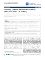
Effect of different stimuli on the production of NO, TNF, and IL-6 by NR8383 cellsFigure 1
Effect of different stimuli on the production of NO, TNF, and IL-6 by NR8383 cells. Cells were incubated with medium (white
bars), LPS/IFN-γ (black bars), TMA (striped bars) TMA-BSA (dark grey bars; 1 mg/ml corresponds with 13.5 μM TMA-BSA) and
BSA (light grey bars; 1 mg/ml corresponds with 15.1 μM BSA) for 24 h. The culture supernatants were analyzed for nitrite (a),
TNF (b) and IL-6 (c). Results are expressed as mean ± SEM. Significant differences are denoted by *: p < 0.05 compared to
medium incubation.
medium
L
P
S/I
F
Nγ
10
1
00
0
.1
0
.3
1
2
3
1
2
3
0
10
20
30
40
50
mg/ml TMA-BSA mg/ml BSA
μM TMA
*
*
*
*
*
nitrite (mM)
a
*
m
edium
LPS/I
FN
γ
10
100
0
.
1
0
.
3
1
2
3
1
2
3
0
1000
2000
3000
mg/ml TMA-BSA mg/ml BSA
μM TMA
*
*
*
*
b
*
TNF (pg/ml)
m
edi
um
LP
S/IFNγ
10
100
0.
1
0.
3
1
2
3
1
2
3
0
1000
2000
3000
4000
5000
mg/ml TMA-BSA mg/ml BSA
μM TMA
*
*
*
c
IL-6 (pg/ml)
*
Journal of Occupational Medicine and Toxicology 2006, 1:13 />Page 5 of 8
(page number not for citation purposes)
Effect of different stimuli on the production of NO, TNF,
and IL-6 by AMs from TMA-sensitized and naïve rats
AMs from naïve and TMA-sensitized rats produced very
low levels of NO, TNF and undetectable levels of IL-6
when cultured for 24 h in medium. Incubation with LPS/
IFN-γ induced equal production of mediators by AMs
from both naïve and TMA-sensitized rats (Fig. 2). The
LPS/IFN-γ induced NO production by AMs was compara-
ble to that by similarly stimulated NR8383 cells. The LPS/
IFN-γ-induced TNF and IL-6 production by AMs, however,
was lower than by NR8383 cells. TMA did not induce the
production of mediators by AMs after 24 h. TMA-BSA,
however, induced a concentration-dependent increase in
NO and TNF production by AMs, but did not induce the
production of IL-6. Significant levels of both NO and TNF
were induced by 0.3 mg/ml of TMA-BSA or more. The
maximum NO level that was induced by TMA-BSA was
approximately 60% of that induced by LPS/IFN-γ (Fig. 2a)
and the maximum TNF level induced by TMA-BSA was
approximately 15% of that induced by LPS/IFN-γ (Fig.
2b).
Discussion
The present study showed that TMA-BSA conjugates, but
not free TMA or BSA, were able to induce the production
of the mediators NO, TNF, and IL-6 by the cell line
NR8383, and, IL-6 excepted, by primary AMs in vitro. This
stimulation is probably not immunologically specific for
two reasons. Firstly, preincubation with serum containing
TMA-specific IgE did not affect the capacity of TMA-BSA to
stimulate mediator production by NR8383 cells. Sec-
ondly, primary AMs from TMA-sensitized and naïve rats
reacted similarly to TMA-BSA. The lack of effect of serum
from TMA-sensitized BN rats that contained high levels of
TMA-specific IgE was not expected, since AMs express the
IgE receptors, FcεRI and CD23. Moreover, their expression
is increased in the presence of IgE [20] and upon in vivo
sensitization [21,22]. Apparently, there is no cross-linking
of IgE at the surface of AMs by TMA-BSA or cross-linking
does not trigger production of the mediators inresponse
to TMA-BSA, although similarly prepared conjugates have
been reported to trigger degranulation of IgE-primed mast
cells [6].
Regarding the nature of the apparent immunological non-
specific AM stimulation, structural similarities between
TMA-conjugated BSA and maleylated-BSA [23] may point
at the involvement of a member of the family of scavenger
receptors. These receptors belong to the large family of
pattern recognition receptors that exhibit binding specifi-
city for structural patterns typically displayed by cell sur-
face molecules of many micro-organisms [24].
Macrophages are known to express multiple scavenger
receptors [25-27] and a variety of ligands, including
maleylated-BSA and LPS, have been shown to induce the
production of NO, TNF, and IL-6 via scavenger receptors
[23,28,29]. Since these mediators were also induced after
stimulation of AMs with TMA-BSA, but not free BSA, and
because of the structural similarities of TMA-BSA with
maleylated-BSA, it is likely that the observed effects were
mediated via scavenger receptors.
In the present study, stimulation with LPS/IFN-γ induced
the production of higher TNF and IL-6 levels by NR8383
cells than by primary AMs. Since both NR8383 cells and
primary cells were AMs, differences in genetic background
may explain the variation in mediator production, given
the fact that the NR8383 cells were derived from Sprague
Dawley rats [30], while the primary AMs were derived
from BN rats. Differences in amounts of mediators pro-
duced by AMs obtained from Sprague Dawley and from
BN rats have been reported [31,32]. Furthermore, differ-
ences between primary cells and immortalized cells may
be implicated, since Rao et al. [33] demonstrated that
stimulation of NR8383 cells with LPS activated three dif-
ferent mitogen-activated protein kinases, while only one
of them was activated in primary AMs derived from
Sprague Dawley rats.
The observation that TMA-BSA induced equal amounts of
NO in NR8383 cells as LPS/IFN-γ and only 40% less in
primary AMs indicates that TMA-BSA is a powerful macro-
phage activating agent. It is probably more potent than
LPS as such, since the amount of NO produced by AMs in
response to LPS was reported to be only 0–35 % of the
response after LPS/IFN-γ incubation of primary AMs
derived from Sprague Dawley and BN rats [31]. Despite
the potent in vitro AM-activating capacity of TMA-BSA and
the lack of effect of TMA in this respect, inhalation chal-
lenge of TMA-sensitized BN rats with either TMA or TMA-
BSA induced similar immediate reduction in minute ven-
tilation [15,16]. However, depletion of AMs prior to chal-
lenge of sensitized rats ameliorated the decrease in minute
ventilation in case of TMA challenge [15], but not in case
of TMA-BSA challenge [16], although both compounds
induced an influx of inflammatory cells in the airways of
these animals. The substantial differences between TMA
and TMA-BSA in their in vitro AM-activating capacity are
apparently not at play upon inhalation challenge with
these compounds. A possible explanation for this contro-
versy might be that inhalation of TMA leads to rapid con-
jugation to endogenous proteins in vivo while formation
of such conjugates is not feasible in vitro due to the static
culture conditions. The observation that TMA challenge of
BN rats caused immediate bronchoconstriction [15] is
indicative of rapid conjugation, since the immediate
bronchoconstriction is likely to be due to mast cell
degranulation triggered by IgE receptor cross-linking with
a multivalent TMA ligand as formed upon binding of mul-
tiple TMA molecules to self-proteins. Since formation of
Journal of Occupational Medicine and Toxicology 2006, 1:13 />Page 6 of 8
(page number not for citation purposes)
Effect of different stimuli on the production of NO, TNF, and IL-6 by AMs derived from either naïve or TMA-sensitized BN ratsFigure 2
Effect of different stimuli on the production of NO, TNF, and IL-6 by AMs derived from either naïve or TMA-sensitized BN
rats. Rats received 150 μl vehicle or 50% TMA in vehicle on each shaved flank on day 0 and 75 μl vehicle or 25% TMA on the
dorsum of both ears on day 7. On day 21 the animals were sacrificed and the lungs were lavaged. AMs obtained from the lung
lavage fluid were incubated with medium (white bar), LPS/IFN-γ (black bar), TMA (striped bars) TMA-BSA (dark grey bars; 1
mg/ml corresponds with 13.5 μM TMA-BSA) and BSA (light grey bars; 1 mg/ml corresponds with 15.1 μM BSA) for 24 h. The
culture supernatants were analyzed for nitrite (a), TNF (b) and IL-6 (c). Results are expressed as mean ± SEM. Significant differ-
ences are denoted by *: p < 0.05 compared to medium incubation.
medium
LP
S
/
I
F
Nγ
10
100
0.
1
0.3
1
2
3
1
3
medium
L
P
S/I
F
Nγ
10
100
0.
1
0.
3
1
2
3
1
3
0
50
100
150
200
250
800
1200
1600
mg/ml TMA-BSA
mg/ml
BSA
μM
TMA
mg/ml TMA-BSA
mg/ml
BSA
μM
TMA
naive TMA-sensitized
*
*
*
*
*
*
*
*
b
*
*
TNF (pg/ml)
medium
L
PS
/
IF
N
γ
1
0
1
00
0.
1
0
.
3
1
2
3
1
3
medium
LPS/IFNγ
1
0
1
00
0.1
0.3
1
2
3
1
3
0
1000
2000
3000
4000
mg/ml TMA-BSA
mg/ml
BSA
μM
TMA
mg/ml TMA-BSA
mg/ml
BSA
μM
TMA
TMA-sensitized
c
naive
*
*
IL-6 (pg/ml)
m
ed
i
um
LPS/IF
N
γ
10
100
0.1
0.3
1
2
3
1
3
med
i
um
LPS/IFNγ
10
100
0.1
0.3
1
2
3
1
3
0
10
20
30
40
50
*
*
*
*
*
*
*
*
mg/ml TMA-BSA
mg/ml
BSA
μM
TMA
mg/ml TMA-BSA
mg/ml
BSA
μM
TMA
TMA-sensitized
nitrite (mM)
a
naive
*
*
Journal of Occupational Medicine and Toxicology 2006, 1:13 />Page 7 of 8
(page number not for citation purposes)
conjugates of TMA with endogenous proteins is consid-
ered to be required for sensitization [34] and if such pro-
tein-conjugates, like TMA-BSA in vitro, induce the
production of NO and proinflammatory cytokines in vivo,
then TMA can be considered as an inducer of danger sig-
nals. Thus, TMA-protein-conjugates, like the danger sig-
nalling molecules of bacteria, can act as an adjuvant for
TMA sensitization. An interesting question in this respect
is, whether the most potent inducers of LMW chemical-
induced occupational respiratoryallergic disease share this
intrinsic adjuvant activity. If so, toxicological hazard iden-
tification may benefit from screening for macrophage-
activating activity of reactive LMW compounds conju-
gated with suitable carrier proteins.
Conclusion
In summary, the results of the present study demonstrate
that although TMA is a highly reactive chemical, it needs
to be conjugated to suitable protein to exert an effect on
mediator production by AMs, as observed for the TMA-
BSA conjugate. The effects of TMA-BSA on AMs were not
dependent on sensitization, indicating that the interac-
tion of TMA-BSA with AMs is probably mediated via an
immunologically non-specific scavenger receptor.
Competing interests
The author(s) declare that they have no competing inter-
ests.
Authors' contributions
DLV conducted part of the study and was involved in the
design of the study, the analysis of the data and the writ-
ing of the manuscript. MAS prepared the TMA-BSA and
assisted in the study. ES conducted part of the study and
was involved in the analysis of the data. FPN helped to
obtain the research support and reviewed the manuscript.
NB and PAJH obtained the research support and partici-
pated in the design of the study, the interpretation of the
data and the writing of the manuscript. All authors read
and approved the final manuscript.
Acknowledgements
The authors thank M.C. Bello and A.M. Raktoe for their contributions to
this study.
References
1. Lombardo LJ, Balmes JR: Occupational asthma: A review. Environ
Health Perspect 2000, 108 Suppl 4:697-704.
2. Kirchner DB: The spectrum of allergic disease in the chemical
industry. Int Arch Occup Environ Health 2002, 75 Suppl:S107-S112.
3. Mapp CE, Boschetto P, Maestrelli P, Fabbri LM: Occupational
asthma. Am J Respir Crit Care Med 2005, 172:280-305.
4. Karol MH: Bonding and transfer: do epithelial conjugates have
a role in chemical asthma? Clin Exp Allergy 2001, 31:357-360.
5. Holliday MR, Coleman JW, Dearman RJ, Kimber I: Induction of
mast cell sensitization by chemical allergens: a comparative
study. J Appl Toxicol 1993, 13:137-142.
6. Vento KL, Dearman RJ, Kimber I, Basketter DA, Coleman JW: Selec-
tivity of IgE responses, mast cell sensitization, and cytokine
expression in the immune response of Brown Norway rats to
chemical allergens. Cell Immunol 1996, 172:246-253.
7. Prussin C, Metcalfe DD: 4. IgE, mast cells, basophils, and eosi-
nophils. J Allergy Clin Immunol 2003, 111:S486-94.
8. Hart PH: Regulation of the inflammatory response in asthma
by mast cell products. Immunol Cell Biol 2001, 79:149-153.
9. Hamid Q, Tulic MK, Liu MC, Moqbel R: Inflammatory cells in
asthma: mechanisms and implications for therapy. J Allergy
Clin Immunol 2003, 111:S5-S17.
10. Laskin DL, Weinberger B, Laskin JD: Functional heterogeneity in
liver and lung macrophages. J Leukoc Biol 2001, 70:163-170.
11. Dorger M, Munzing S, Allmeling AM, Messmer K, Krombach F: Phe-
notypic and functional differences between rat alveolar,
pleural, and peritoneal macrophages. Exp Lung Res 2001,
27:
65-76.
12. Peiser L, Mukhopadhyay S, Gordon S: Scavenger receptors in
innate immunity. Curr Opin Immunol 2002, 14:123-128.
13. Careau E, Bissonnette EY: Adoptive transfer of alveolar macro-
phages abrogates bronchial hyperresponsiveness. Am J Respir
Cell Mol Biol 2004, 31:22-27.
14. Peters-Golden M: The alveolar macrophage. The forgotten
cell in asthma. Am J Respir Cell Mol Biol 2004, 31:3-7.
15. Valstar DL, Schijf MA, Nijkamp FP, Storm G, Arts JHE, Kuper CF,
Bloksma N, Henricks PAJ: Alveolar macrophages have a dual
role in a rat model for trimellitic anhydryde-induced occupa-
tional asthma. Toxicol Appl Pharmacol 2006, 211:20-29.
16. Valstar DL, Schijf MA, Arts JH, Kuper CF, Nijkamp FP, Storm G,
Bloksma N, Henricks PAJ: Alveolar macrophages suppress non-
specific inflammation caused by inhalation challenge with
trimellitic anhydride conjugated to albumin. Arch Toxicol
2006:in press (DOI: 10.1007/s00204-006-0081-5)
17. Sashidhar RB, Capoor AK, Ramana D: Quantitation of ε-amino
group using amino acids as reference standards by trini-
trobenzene sulfonic acid. A simple spectrophotometric
method for the estimation of hapten to carrier protein ratio.
J Immunol Methods 1994, 167:121-127.
18. Green LC, Wagner DA, Glogowski J, Skipper PL, Wishnok JS, Tan-
nenbaum SR: Analysis of nitrate, nitrite, and [15N]nitrate in
biological fluids. Anal Biochem 1982, 126:131-138.
19. Stuehr DJ, Marletta MA: Mammalian nitrate biosynthesis:
mouse macrophages produce nitrite and nitrate in response
to Escherichia coli lipopolysacharide. Proc Natl Acad Sci U S A
1985, 82:7738-7742.
20. Boltz-Nitulescu G, Plummer JM, Spiegelberg HL: Increased expres-
sion of the IgE Fc receptors on rat macrophages induced by
elevated serum IgE levels. Immunology 1984, 53:9-16.
21. Mencia-Huerta JM, Dugas B, Boichot E, Petit-Frere C, Paul-Eugene N,
Lagente V, Capron M, Liu FT, Braquet P: Pharmacological modu-
lation of the antigen-induced expression of the low-affinity
IgE receptor (Fc
εRII/CD23) on rat alveolar macrophages. Int
Arch Allergy Appl Immunol 1991, 94:295-298.
22. Humbert M, Grant JA, Taborda-Barata L, Durham SR, Pfister R, Menz
G, Barkans J, Ying S, Kay AB: High-affinity IgE receptor (FcεRI)-
bearing cells in bronchial biopsies from atopic and nonatopic
asthma. Am J Respir Crit Care Med 1996, 153:1931-1937.
23. Alford PB, Xue Y, Thai SF, Shackelford RE: Maleylated-BSA
enhances production of nitric oxide from macrophages. Bio-
chem Biophys Res Commun 1998, 245:185-189.
24. Gordon S: Pattern recognition receptors: doubling up for the
innate immune response. Cell 2002, 111:927-930.
25. Peiser L, Gordon S: The function of scavenger receptors
expressed by macrophages and their role in the regulation of
inflammation. Microbes Infect 2001, 3:149-159.
26. Gough PJ, Gordon S: The role of scavenger receptors in the
innate immune system. Microbes Infect 2000, 2:305-311.
27. Greaves DR, Gordon S: Recent insights into the biology of mac-
rophage scavenger receptors. J Lipid Res 2005, 46:11-20.
28. Coller SP, Paulnock DM: Signaling pathways initiated in macro-
phages after engagement of type A scavenger receptors. J
Leukoc Biol 2001, 70:142-148.
29. Paulnock DM, Demick KP, Coller SP: Analysis of interferon-γ-
dependent and -independent pathways of macrophage acti-
vation. J Leukoc Biol 2000, 67:677-682.
30. Helmke RJ, Boyd RL, German VF, Mangos JA: From growth factor
dependence to growth factor responsiveness: the genesis of
Publish with BioMed Central and every
scientist can read your work free of charge
"BioMed Central will be the most significant development for
disseminating the results of biomedical research in our lifetime."
Sir Paul Nurse, Cancer Research UK
Your research papers will be:
available free of charge to the entire biomedical community
peer reviewed and published immediately upon acceptance
cited in PubMed and archived on PubMed Central
yours — you keep the copyright
Submit your manuscript here:
/>BioMedcentral
Journal of Occupational Medicine and Toxicology 2006, 1:13 />Page 8 of 8
(page number not for citation purposes)
an alveolar macrophage cell line. In Vitro Cell Dev Biol 1987,
23:567-574.
31. Jesch NK, Dorger M, Messmer K, Krombach F: Formation of nitric
oxide by rat and hamster alveolar macrophages: an inter-
strain and interspecies comparison. Toxicol Lett 1998, 96-
97:47-51.
32. Careau E, Sirois J, Bissonnette EY: Characterization of lung
hyperresponsiveness, inflammation, and alveolar macro-
phage mediator production in allergy resistant and suscepti-
ble rats. Am J Respir Cell Mol Biol 2002, 26:579-586.
33. Rao KM, Meighan T, Bowman L: Role of mitogen-activated pro-
tein kinase activation in the production of inflammatory
mediators: differences between primary rat alveolar macro-
phages and macrophage cell lines. J Toxicol Environ Health A
2002, 65:757-768.
34. Karol MH, Macina OT, Cunningham A: Cell and molecular biology
of chemical allergy. Ann Allergy Asthma Immunol 2001, 87:28-32.