báo cáo hóa học: " Lipopolysaccharide induced inflammation in the perivascular space in lungs" docx
Bạn đang xem bản rút gọn của tài liệu. Xem và tải ngay bản đầy đủ của tài liệu tại đây (1.59 MB, 5 trang )
BioMed Central
Page 1 of 5
(page number not for citation purposes)
Journal of Occupational Medicine
and Toxicology
Open Access
Research
Lipopolysaccharide induced inflammation in the perivascular space
in lungs
Thomas Tschernig*
†1
, Kyathanahalli S Janardhan
†2,3
, Reinhard Pabst
1
and
Baljit Singh
2
Address:
1
Dept. of Functional and Applied Anatomy -4120-, Medical School of Hannover, Carl-Neuberg-Str. 1, 30625, Hannover, Germany,
2
Immunology Research Group, Departments of Veterinary Biomedical Sciences and Veterinary Microbiology, Western College of Veterinary
Medicine, University of Saskatchewan, 52 Campus Drive, Saskatoon, SK, S7N 5B4, Canada and
3
Diagnostic Medicine and Pathobiology, 1800
Denison Avenue, Kansas State University, Manhattan, Kansas 66506, USA
Email: Thomas Tschernig* - ; Kyathanahalli S Janardhan - ;
Reinhard Pabst - ; Baljit Singh -
* Corresponding author †Equal contributors
Abstract
Background: Lipopolysaccharide (LPS) contained in tobacco smoke and a variety of
environmental and occupational dusts is a toxic agent causing lung inflammation characterized by
migration of neutrophils and monocytes into alveoli. Although migration of inflammatory cells into
alveoli of LPS-treated rats is well characterized, the dynamics of their accumulation in the
perivascular space (PVS) leading to a perivascular inflammation (PVI) of pulmonary arteries is not
well described.
Methods: Therefore, we investigated migration of neutrophils and monocytes into PVS in lungs of
male Sprague-Dawley rats treated intratracheally with E. coli LPS and euthanized after 1, 6, 12, 24
and 36 hours. Control rats were treated with endotoxin-free saline. H&E stained slides were made
and immunohistochemistry was performed using a monocyte marker and the chemokine
Monocyte-Chemoattractant-Protein-1 (MCP-1). Computer-assisted microscopy was performed to
count infiltrating cells.
Results: Surprisingly, the periarterial infiltration was not a constant finding in each animal although
LPS-induced alveolitis was present. A clear tendency was observed that neutrophils were appearing
in the PVS first within 6 hours after LPS application and were decreasing at later time points. In
contrast, mononuclear cell infiltration was observed after 24 hours. In addition, MCP-1 expression
was present in perivascular capillaries, arteries and the epithelium.
Conclusion: PVI might be a certain lung reaction pattern in the defense to infectious attacks.
Background
Lipopolysaccharide (LPS) is a glycolipid of gram-negative
bacterial cell walls and is present in many different air-
borne particles, such as tobacco smoke and a variety of
environmental and occupational dusts [1,2]. Inhalation
of LPS in man and administration through various routes
in animal models result in inflammation [3,4]. LPS
induces inflammatory cell signalling through its binding
Published: 30 July 2008
Journal of Occupational Medicine and Toxicology 2008, 3:17 doi:10.1186/1745-6673-3-17
Received: 29 March 2007
Accepted: 30 July 2008
This article is available from: />© 2008 Tschernig et al; licensee BioMed Central Ltd.
This is an Open Access article distributed under the terms of the Creative Commons Attribution License ( />),
which permits unrestricted use, distribution, and reproduction in any medium, provided the original work is properly cited.
Journal of Occupational Medicine and Toxicology 2008, 3:17 />Page 2 of 5
(page number not for citation purposes)
to LPS binding protein and subsequent interaction with
Toll like receptor-4 (TLR-4) and other molecules such as
CD14 and MD2 [5-7]. Lungs from LPS-treated animals
show recruitment of neutrophils and monocytes into
alveolar and vascular compartments through a complex
interplay of cytokines, chemokines and adhesion mole-
cules [8].
Recently, we identified the perivascular space (PVS)
around pulmonary arteries as a unique morphological
compartment with possible impact on inflammatory
responses in the lung [9,10]. The PVS of pulmonary arter-
ies increases in size in inflammation due to influx of fluids
and inflammatory cells, which may come from perivascu-
lar capillaries that follow the arteries [11]. In addition,
lymph vessels are located in the PVS and run in the oppo-
site direction to the central draining lymph nodes. While
the PVS is increased in various models of lung inflamma-
tion, anti-inflammatory agents such as anti-IL-9 agent
reduce the amount of cellular infiltrates within this area
[10,12]. It appears that the PVS may be an important loca-
tion for the accumulation and actions of inflammatory
cells in acute and chronic lung inflammation. Theogaraj et
al. [13] found the PVS of the rat rich in white cells, includ-
ing T- and B-lymphocytes, and suggested a significant role
in host defence for this compartment.
Currently, there are no data on the temporary migration
of inflammatory cells into the PVS in LPS-induced lung
inflammation. Therefore, we conducted this study to
define the kinetics of neutrophils and monocytes/macro-
phages into PVS in lungs of LPS-treated rats. In addition,
we examined the immune-histological expression of
monocyte chemoattractant protein-1 (MCP-1) in PVS of
normal and LPS-treated rats.
Methods
Rats and treatment groups
The experimental protocols were approved by the Univer-
sity of Saskatchewan Committee on Animal Care Assur-
ance and experiments were conducted according to the
Canadian Council on Animal Care Guidelines. Specific
pathogen-free, ten-week-old, male Sprague-Dawley rats
were procured from Charles River laboratories, Canada.
Rats were maintained in the animal care unit and were
acclimatized for a period of one week and randomly
divided into six groups of five each (Table 1).
Acute lung inflammation
The procedure was performed as described before [5].
Briefly, rats were anesthetized by intraperitoneal adminis-
tration of xylazine (20 mg/Kg) and ketamine (100 mg/
Kg). The trachea was dissected surgically and endotoxin-
free saline (Sigma, St. Louis, MO, USA) or E. coli LPS
diluted in 80 μl of saline (250 μg; serotype 0128:B12;
Sigma, St. Louis, MO, USA) was injected in the trachea.
Animals were euthanized at 1, 6, 12, 24 and 36 hours (n
= 5 per group) post-treatment and were observed during
the post-LPS treatment period hourly during the day after
application. Although LPS-treated rats appeared to be
sluggish, none of them died prior to euthanasia. Control
animals (n = 5) were euthanized at 6 hours post saline
treatment (Table 1). Only this time point has been chosen
for the control treatment because the influence of saline
instillation had only very mild effects as compared to LPS.
Tissue collection and processing
After induction of deep anesthesia the animals were
exsanguinated, and the lungs were obtained. Rat lungs
were collected for light microscopy without instillation or
perfusion with fixatives to avoid any dislocation of leuko-
cytes within the air space or lung vessels. Lung pieces for
histology were fixed in 4% paraformaldehyde for 16
hours. Lungs were processed through ascending grades of
alcohol and embedded in paraffin. Five μm sections were
cut from 6 lung specimens of each rat.
Immunohistology
Tissue sections were prepared and stained as described
before [5]. Briefly, sections were deparaffinized in xylene
and rehydrated in descending grades of alcohol followed
by treatment with 5% hydrogen peroxide in methanol to
quench endogenous peroxidase. Sections were treated
with pepsin at room temperature (2 mg/ml in 0.01N
hydrochloric acid; Sigma, St. Louis MO, USA) for 45 min-
utes to unmask the antigens and with 1% bovine serum
albumin (Sigma) to block non-specific binding. Sections
were incubated with primary antibodies against rat mono-
cyte/macrophage (1:75; ED1, Serotec Inc. NC, USA) or rat
MCP-1 (1:300; Torrey Pines Biolabs, Inc. TX, USA), fol-
lowed by appropriate horseradish peroxidase(HRP)-con-
jugated secondary antibodies (1:100; Dako cytomation,
ON, Canada). The antigen-antibody complex was visual-
ized using a color development kit (Vector laboratories,
ON, Canada). Controls consisted of staining without pri-
mary antibody or with isotype matched immunoglobulin
instead of primary antibody. Proper quenching of endog-
enous peroxidase was confirmed by omitting both pri-
mary and secondary antibodies.
Tissue evaluation
The evaluation was performed by a person blinded to the
identity of groups with a microscope using a software
Table 1: Experimental design
Hours after
instillation of
16122436
LPS n = 5 n = 5 n = 5 n = 5 n = 5
Saline n = 5
Journal of Occupational Medicine and Toxicology 2008, 3:17 />Page 3 of 5
(page number not for citation purposes)
assisted determination of edematous area (PVS areas)
around pulmonary arteries (PA). We have not evaluated
PVS areas around pulmonary veins because the changes
there are weaker as compared to pulmonary arteries. Only
those PA were included in the analyses, which were fully
captured in a cross section and had inner diameter of
more than 100 μ. The PVS area was digitally displayed and
determined in square microns by the delineation with the
cursor. In most of the cases the PVS was clearly separated
from the adjacent alveolar tissue as well as the adventitia
of the adjacent bronchi and other vessels (Figure 1A). The
total number of infiltrating leukocytes was determined
using a 200× magnification and the number of neu-
trophils by using a 400× magnification. Three areas per
animal on different lung sections have been evaluated.
This semi-quantitive procedure seemed to be adequate
because many sections of the same lung revealed similar
results as has been checked in single lungs. The cells per
area were calculated and statistics performed (MS Office
2003). Mean values (MV) and standard errors (SE) were
calculated. Each time point after treatment was compared
with the saline treated control group using the non-para-
metric Mann-Whitney U-test for unmatched pairs and sig-
nificance was indicated for p < 0.05.
Results and discussion
Lungs from saline-treated rats showed normal histology
and no accumulation of inflammatory cells in alveoli or
the PVS. In contrast, lungs from LPS-treated rats displayed
a typical accumulation of cells (Fig. 1AB). Single lungs of
the LPS-treated rats did not develop edema and perivascu-
lar inflammation although they showed alveolitis (Fig.
1C). At time points later than 1 hour after LPS treatment,
occasional lymphatic vessels characterized by thin walls
and larger diameter were seen in the periphery of PVS and
filled with mononuclear and polymorphonuclear cells.
Interestingly, aggregates of granulocytes were found
within lymphatic vessels after 6 hours of the treatment.
The 12 hour groups showed foci of interstitial inflamma-
tion which became larger by 24 hours after the LPS treat-
ment and filled most of the section area of peripheral lung
tissue after 36 hours. At 12 hours and later, alveolitis and
hemorrhages were seen in most of the lungs. The strategy
to determine leukocyte kinetics within the PVS was to
count in a first step all round cells which are "all leuko-
cytes" in Table 2. These are a) monomorphonuclear cells
(MMN) such as monocytes/macrophages and lym-
phocytes and b) polymorphonuclear cells (PMN) repre-
senting the granulocytes. In this model only neutrophil
granulocytes could be observed. In a second step the
PMNs has been counted separately because only this cell
type was changing in numbers very early after the applica-
tion of LPS. The phenotype of the MMN has not been dif-
ferentiated in this study because that would be important
at later time points in type IV immune reactions. Exem-
plary the population of monocytes/macrophages in PVS
areas has been documentes using the monoclonal anti-
body ED-1 (Fig. 1D). Low numbers of leukocytes were
found in the PVS of the control group and 1 hour after LPS
exposition (Table 2). The total number of leukocytes
showed a gradual increase in the LPS groups beginning 6
hrs after the challenge reaching significance after 36
hours. In contrast, the neutrophil numbers in the PVS
showed an abrupt and significant rise at 6 hours after the
intratracheal instillation of LPS, declining to control val-
ues at 24 hours. Because MCP-1 is critical for the recruit-
ment of monocytes/macrophages, lung sections were
stained with an MCP-1 antibody. Intense expression of
MCP-1 (Fig. 1EF) was detected in lung sections from all of
the LPS-treated rats especially at time points later than 6
hours in bronchial epithelium, airway and vascular
smooth muscles and leukocytes. MCP-1 expression was
mild and only in some of the blood vessels including
those in the PVS. Lungs from the control rats showed weak
staining for MCP-1. To our knowledge, this is the first
study to characterize infiltration of neutrophils and
monocytes and expression of MCP-1 in PVS of LPS-treated
lungs. The study was conducted in a well characterized
model of acute lung inflammation induced following
intratracheal instillation of E. coli LPS. To minimize
changes to morphology and introduction of artifacts, the
lungs were neither lavaged nor perfused. The histological
signs of lung inflammation observed were similar to those
reported in various other studies using intratracheal instil-
lation of LPS [4,5,14].
Our study showed distinct patterns of recruitment of leu-
kocytes into the PVS. The total leukocyte numbers
increased slightly 6 hours post-LPS treatment. Signifi-
cance was calculated only after 36 hours which was due to
the moderate increase with high variations and to a small
number of animals used in this study. The neutrophils
were primarily absent and were increased rapidly after 1
and 6 hours and disappeared again after 24 hours. Com-
pared to the alveolar recruitment leukocytes came slow
but neutrophil migration into the PVS was as quick as into
the alveoli [5,14,15]. The slight increase of monocytes in
the PVS the LPS-challenge was in contrast to our recent
data showing clear increases of monocytes within the
alveoli already 3 hours after an LPS-challenge [14] indicat-
ing the PVS as a compartment which is functional distinct
from the alveolar space.
The mechanisms and route of recruitment of inflamma-
tory cells into PVS remain largely unknown. Recently, we
reported that there are strain-dependent differences in
inflammatory cell recruitment into PVS in acute and
chronic airway inflammation [16]. Although we showed
expression of Vascular Adhesion Protein (VAP-1) in pul-
monary arteries in a mouse model of airway inflamma-
Journal of Occupational Medicine and Toxicology 2008, 3:17 />Page 4 of 5
(page number not for citation purposes)
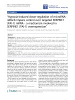
A-F: H&E histology demonstrating the PVI 12 h after instillation of 25 μg LPS (A, B)Figure 1
A-F: H&E histology demonstrating the PVI 12 h after instillation of 25 μg LPS (A, B). A massive alveolitis can be
seen (C). The leukocytes are mainly ED1 positive monocytes/macrophages (D). MCP-1 expression is demonstrated on the api-
cal epithelium and on the endothelium and most of the leukocytes as well (E control, F MCP-1).
Journal of Occupational Medicine and Toxicology 2008, 3:17 />Page 5 of 5
(page number not for citation purposes)
tion, we are aware of a significant structural barrier
afforded by their thick wall [17]. Therefore, we believe
that entry and existence of inflammatory cells into PVS
possibly occur through capillaries and lymph vessels
present in the PVS. However, other authors believe that
cells exit the blood stream and pass immediately into the
PVS [13]. This is supported through our direct observa-
tions of neutrophils in the lumen of microvessels in the
PVS. One of the critical requirements for inflammatory
cell recruitment is expression of chemoattractants [15]. A
classical chemoattractant for monocytes/macrophages is
MCP-1. Our data show expression of MCP-1 in recruited
cells in PVS along with airway epithelium and vascular
endothelium. Immuno-histological localization of chem-
okines such as MCP-1 is difficult and does not provide
direct information on their functions. The intense expres-
sion of MCP-1 observed in PVS in lungs of LPS-treated rats
may indicate its role in promoting monocyte/macrophage
entry into PVS.
Conclusion
We conclude that PVS may be a unique anatomical and
functional site for the migration of inflammatory cells in
acute lung inflammation. Therefore, PVS may contribute
to immune responses in lung inflammation provoked
through various stimuli. We still need to answer intrigu-
ing questions such as the route and mechanisms of migra-
tion of inflammatory cells into PVS.
Competing interests
The authors declare that they have no competing interests.
Authors' contributions
KSJ performed the animal experiments and was involved
in the morphological evaluation and helped to draft the
manuscript. TT performed the analysis of the lung sec-
tions and drafted the manuscript. RP was involved in
coordination of the study and helped to draft the manu-
script. BS conceived of the study, participated in its design
and helped to draft the manuscript. All authors read and
approved the final manuscript.
Acknowledgements
We thank Sheila Fryk for correction of the English. The study was sup-
ported through grants from the Natural Sciences and Engineering Research
Council of Canada to Baljit Singh, and Dr. Tschernig's visit to the Western
College of Veterinary Medicine was supported through a DLT Smith Visit-
ing Professorship. Dr. Janardhan is a recipient of an Interprovincial Gradu-
ate Fellowship from the Western College of Veterinary Medicine. Further
support came from the "Deutsche Forschungsgemeinschaft" (SFB 587, B1).
References
1. Larsson L, Szponar B, Pehrson C: Tobacco smoking increases
dramatically air concentrations of endotoxin. Indoor Air 2004,
14:421-24.
2. Charavaryamath C, Janardhan KS, Townsend HG, Willson P, Singh B:
Multiple exposures to swine barn air induce lung inflamma-
tion and airway hyper-responsiveness. Respir Res 2005, 6:50.
3. Wallin A, Pourazar J, Sandstrom T: LPS-induced bronchoalveolar
neutrophilia; effects of salmeterol treatment. Respir Med
2004, 98:1087-92.
4. Remick DG, Strieter RM, Eskandari MK, Nguyen DT, Genord MA,
Raiford CL, Kunkel SL: Role of tumor necrosis factor-alpha in
lipopolysaccharide-induced pathologic alterations. Am J Pathol
1990, 136:49-60.
5. Janardhan KS, McIsaac M, Fowlie J, Shrivastav A, Caldwell S, Sharma
RK, Singh B: Toll like receptor-4 expression in lipopolysaccha-
ride induced lung inflammation. Histol Histopathol 2006,
21:687-96.
6. Reaves TA, Chin AC, Parkos CA: Neutrophil transepithelial
migration: role of toll-like receptors in mucosal inflamma-
tion. Mem Inst Oswaldo Cruz 2005, 100:191-98.
7. Wassef A, Janardhan KS, Pearce JW, Singh B: Toll-like receptor 4
in normal and inflamed lungs and other organs of pig, dog
and cattle. Histol Histopathol 2004, 19:1201-8.
8. Vernooy JH, Dentener MA, van Suylen RJ, Buurman WA, Wouters EF:
Long-term intratracheal lipopolysaccharide exposure in
mice results in chronic lung inflammation and persistent
pathology. Am J Respir Cell Mol Biol 2002, 26:152-9.
9. Pabst R, Tschernig T: Perivascular capillaries in the lung: an
important but neglected vascular bed in immune reactions?
J Allergy Clin Immunol 2002, 110:209-14.
10. Pabst R: The periarterial space in the lung: its important role
in lung edema, transplantation, and microbial or allergic
inflammation. Pathobiology 2004, 71:287-94.
11. Guntheroth WG, Luchtel DL, Kawabori I: Pulmonary microcircu-
lation: tubules rather than sheet and post. J Appl Physiol 1982,
53:510-5.
12. Cheng G, Arima M, Honda K, Hirata H, Eda F, Yoshida N, Fukushima
F, Ishii Y, Fukuda T: Anti-interleukin-9 antibody treatment
inhibits airway inflammation and hyperreactivity in mouse
asthma model. Am J Respir Crit Care Med 2002, 166:409-16.
13. Theogaraj E, John CD, Dewar A, Buckingham JC, Smith SF: The long-
term effects of perinatal glucocorticoid exposure on the host
defence system of the respiratory tract. J Pathol 2006,
210:85-93.
14. Janardhan KS, Appleyard GD, Singh B: Expression of integrin sub-
units alpha-v and beta3 in acute lung inflammation. Histochem
Cell Biol 2004, 121:383-90.
15. Doerschuk CM: Mechanisms of leukocyte sequestration in
inflamed lungs. Microcirculation 2001, 8:71-88.
16. Singh B, Shinagawa K, Taube C, Gelfand EW, Pabst R: Strain-specific
differences in perivascular inflammation in lungs in two
murine models of allergic airway inflammation. Clin Exp Immu-
nol 2005, 141:223-9.
17. Singh B, Tschernig T, van Griensven M, Fieguth A, Pabst R: Expres-
sion of vascular adhesion protein-1 in normal and inflamed
mice lungs and normal human lungs. Virchows Arch 2003,
442:491-5.
Table 2: Density of leukocytes and neutrophils in PVS at
different time points after NaCl- or LPS-instillation (cells per
mm
2
, mean value ± SEM)
Group/time All leukocytes Neutrophils
Saline 6 hours 0.3 ± 0.05 0*
LPS 1 hour 0.3 ± 0.09 0.02 ± 0.02
LPS 6 hours 0.6 ± 0.32 0.2 ± 0.14
+
LPS 12 hours 0.5 ± 0.23 0.1 ± 0.09
LPS 24 hours 0.6 ± 0.23 0*
LPS 36 hours 0.6 ± 0.09
+
0*
+
significant p < 0.05, *single cells were found
PMN = polymorphonuclear cells/granulocytes (>99% neutrophils)