báo cáo hóa học: " Lead exposure study among workers in lead acid battery repair units of transport service enterprises, Addis Ababa, Ethiopia: a cross-sectional study" pot
Bạn đang xem bản rút gọn của tài liệu. Xem và tải ngay bản đầy đủ của tài liệu tại đây (601 KB, 8 trang )
BioMed Central
Page 1 of 8
(page number not for citation purposes)
Journal of Occupational Medicine
and Toxicology
Open Access
Research
Lead exposure study among workers in lead acid battery repair
units of transport service enterprises, Addis Ababa, Ethiopia: a
cross-sectional study
Kemal Ahmed
1
, Gonfa Ayana
2
and Ephrem Engidawork*
1
Address:
1
Department of Pharmacology, School of Pharmacy, Addis Ababa University, Addis Ababa, Ethiopia and
2
Department of Clinical
Chemistry, Ethiopian Health and Nutrition Research Institute, Addis Ababa, Ethiopia
Email: Kemal Ahmed - ; Gonfa Ayana - ; Ephrem Engidawork* -
* Corresponding author
Abstract
Background: Lead exposure is common in automobile battery manufacture and repair, radiator repair,
secondary smelters and welding units. Urinary Aminolevulinic acid has validity as a surrogate measure of
blood lead level among workers occupationally exposed to lead. This study had therefore assessed the
magnitude of lead exposure in battery repair workers of three transport service enterprises.
Methods: To this effect, a cross-sectional study was carried out on lead exposure among storage battery
repair workers between November 2004 and May 2005 from Anbasa, Comet and Walia transport service
enterprises, Addis Ababa, Ethiopia. Subjective information from the workers was obtained by making use
of structured questionnaire. Other information was obtained from walkthrough evaluation of the repair
units. Aminolevulinic acid levels in urine were used as an index of the exposure. This was coupled to
measurements of other relevant parameters in blood and urine collected from adult subjects working in
the repair units as well as age matched control subjects that were not occupationally exposed to lead.
Aminolevulinic acid was determined by spectrophotometry, while creatinine clearance, serum creatinine,
urea and uric acid levels were determined using AMS Autolab analyzer.
Results: Urinary aminolevulinic acid levels were found to be significantly higher in exposed group (16 μg/
ml ± 2.0) compared to the non-exposed ones (7 μg/ml ± 1.0) (p < 0.001). Alcohol taking exposed subjects
exhibited a significant increase in urinary aminolevulinic acid levels than non-alcohol taking ones (p < 0.05).
Moreover, urinary aminolevulinic acid levels of exposed subjects increased with age (p < 0.001) as well as
duration of employment (p < 0.001). Whereas serum uric acid levels of exposed subjects was significantly
higher than non-exposed ones (p < 0.05), no statistically significant difference had been found in renal
indices and other measured parameters between exposed and non-exposed subjects. From the
questionnaire responses and walkthrough observations, it was also known that all the repair units did not
implement effective preventive and control measures for workplace lead exposure.
Conclusion: Taken together, these findings indicated that workers in lead acid battery repair units of the
transport service enterprises are not protected from possibly high lead exposure. Thus, strict
enforcement of appropriate and cost effective preventive and control measures is required by all the
enterprises.
Published: 28 November 2008
Journal of Occupational Medicine and Toxicology 2008, 3:30 doi:10.1186/1745-6673-3-30
Received: 2 March 2006
Accepted: 28 November 2008
This article is available from: />© 2008 Ahmed et al; licensee BioMed Central Ltd.
This is an Open Access article distributed under the terms of the Creative Commons Attribution License ( />),
which permits unrestricted use, distribution, and reproduction in any medium, provided the original work is properly cited.
Journal of Occupational Medicine and Toxicology 2008, 3:30 />Page 2 of 8
(page number not for citation purposes)
Background
Lead (Pb) is a highly toxic metal with no known physio-
logical benefits and is a ubiquitous pollutant in the eco-
system as a result of its natural occurrence and its
industrial use. Mankind has used lead for over 6000 years
[1]. Lead's toxicity was recognized and recorded as early as
2000 BC and the widespread use of lead has been a cause
of endemic chronic plumbism in several societies
throughout history.
Significant occupational lead exposures are not limited to
traditional heavy industries. Automobile battery manu-
facture and repair, radiator repair, secondary smelters
(including scrap metal refiners) are found in most coun-
tries and are common sources of lead exposure. These
small domestic versions of secondary smelters are typi-
cally located within or in close proximity to homes and
lead fumes and dust generated in such operations also
poses health hazard to children and adults [1,2]. In devel-
oping countries the distinction between home and work-
place lead exposure is non-existent [3].
The prevention of occupational hazards is far more effec-
tive and less costly when considered during the early
stages. Lead poisoning amongst occupationally exposed
persons is known to pose serious health problems on the
nervous system, heme biosynthesis, kidneys, reproductive
system, hepatic, hearing, endocrinal, gastrointestinal,
blood pressure and cardiovascular system [4-6]. The effect
of lead on heme synthesis is attributed to inhibition of
enzymes involved in heme synthesis, resulting in abnor-
mal concentrations of heme precursors in blood and
urine. Essentially, lead interferes with the activity of three
enzymes: it indirectly stimulates the mitochondrial
enzyme aminolevulinic acid synthetase (ALAS); directly
inhibits the activity of the cytoplasmic enzyme aminole-
vulinic acid dehydratase (ALAD); and it interferes with the
normal functioning of intramitochondrial ferrochelatase
[2]. The functional changes on kidney are related to lead
effect on mitochondrial respiration and phosphorylation
in proximal tubules of nephron [5]. Typical measures of
renal failure, e.g. blood urea nitrogen (BUN) and creati-
nine are elevated as a consequence of lead induced renal
failure. Chronic occupational lead exposure is also related
to low urate excretion and a high incidence of gout in lead
workers [7].
Significant human suffering related to occupation is unac-
ceptable and often results in appreciable financial loss due
to the burden on health and social security systems, which
negatively impacts production [8]. There are a number of
occupational hazards in all workplaces worldwide due to
lack of adequate prevention and control measures [9].
Occupational exposure to lead still occurs in many coun-
tries of the world. Especially in many developing coun-
tries, occupational lead exposure is entirely unregulated
and no monitoring of exposures exists [10]. The present
study was therefore aimed at investigating lead exposure
among lead-acid battery repair workers and relating the
exposure to health effects.
Methods
Study population
A total of 51 subjects (45 male and 6 female) aged
between 23 and 57 years and who had worked for over six
months in lead acid battery repair units of transport serv-
ice enterprises in Addis Ababa (Anbasa, Comet and
Walia) had participated in this study (Table 1). Fifty
healthy non-exposed age matched subjects (48 male and
2 female) were taken as control for comparison with
exposed group.
All subjects were informed about the purpose, benefits
and risks of the study and their right to withdraw at any
time point. Following this, each of them had given their
consent of participation in the study. The study was
approved by the IRB of the School of Pharmacy, Addis
Ababa University. For each subject, information on per-
sonal particulars, work experience, health risks and other
relevant factors that might influence lead exposure were
collected using a pre-tested and standardized structured
questionnaire.
Sample collection
Urine samples were collected between 9:00 and 11:00
a.m. from study participants using light protected plastic
urine containers (wrapped with aluminum foil), which
contained 2 g barbituric acid as preservative. The measure-
ment of volumes was done using graduated cylinder. In
parallel, 3 ml blood samples were collected and centri-
fuged, and serum was separated for analyses. Both serum
and urine specimens were refrigerated at 4°C immediately
in the dark until time of analyses. The specimen container
was labeled with codes that represent each participant;
Table 1: Study sites and demographic data of lead exposed
workers (n = 51)
Exposed workers Number %
Enterprise
Anbasa 23 45.1
Comet 17 33.3
Walia 11 21.6
Age
20–35 47.9
36–45 22 43.1
46+ 25 49.0
Sex
Male 45 88.2
Female 6 11.8
Journal of Occupational Medicine and Toxicology 2008, 3:30 />Page 3 of 8
(page number not for citation purposes)
date and time of collection for identification purposes
[11,12].
Measurement of urinary delta-Aminolevulinic acid
Urinary delta-Aminolevulinic acid (δ-ALA) levels were
determined spectrophotometrically as described else-
where [13]. Briefly, urine samples were heated with buff-
ered ethyl acetoacetate to produce pyrrole derivatives. This
δ-ALA derivative was purified by extraction into ethyl ace-
tate. Ehrlich reagent was then added to produce a reddish
color and absorbance was measured at 553 nm. For the
analyses, four tubes were prepared as follows: Tube A:
Water blank (1 ml water +1 ml acetate buffer + 0.2 ml
ethyl acetoacetate + 3 ml ethylacetate + 2 ml Ehrlich's rea-
gent), Tube B: Subject specimen blank (1 ml urine + 1 ml
acetate buffer + 3 ml ethylacetate + 2 ml Ehrlich's reagent),
Tube C: Subject specimen (1 ml urine + 1 ml acetate buffer
+ 0.2 ml ethyl acetoacetate + 3 ml ethylacetate +2 ml Ehr-
lich's reagent), and Tube D: Subject specimen (1 ml urine
+ 1 ml acetate buffer + 0.2 ml ethyl acetoacetate + 3 ml
ethylacetate + 2 ml Ehrlich's reagent). Tube A served as a
blank for tube B while tube B was a blank for tubes C and
D. All tubes were heated in a boiling water bath for 10 min
and allowed to cool in cold water. The glass stoppers were
then removed and centrifuged (1000 g ×) for 1 min to sep-
arate the phases. 2 ml of the upper ethyl acetate phase was
removed using volumetric pipette. Then, Ehrlich's reagent
was added, mixed, left for 10 min and the absorbance at
553 nm was taken using water to zero the spectrophotom-
eter.
For calibration known concentrations of δ-ALA in μg/ml
were analyzed by regression analysis to establish the best
line that relates measured absorbance to concentration.
The analysis gave the following least squares equation,
which was used to calculate δ-ALA concentration in μg/
ml.
X = Y - 0.01953/0.06399, where X = concentration of δ-
ALA (μg/ml) and Y = absorbance.
The urinary levels of δ-ALA were then categorized into
four groups, i.e. normal range (<6 μg/ml), acceptable (6–
20 μg/ml), high (20–40 μg/ml) and dangerous (> 40 μg/
ml) [14].
Measurement of creatinine clearance, urea and uric acid
Creatinine clearance was used to assess glomerular filtra-
tion rate (GFR) function after determining the serum and
urinary creatinine concentrations and urine volume over
2 h. Fluitest kit (Biocon
®
Diagnostic Hecke 8, 34516 Vöhl/
Marienhagen, Germany) based on Jaffe Kinetic Colori-
metric Method was used for the determination of creati-
nine. Measurements were done using AMS Autolab
analyzer (Roche, Basel, Switzerland). Urea Kit (Biocon
®
Diagnostic Hecke 8, 34516 Vöhl/Marienhagen, Germany)
based on Berthelet method was used for the determina-
tion of urea, whilst uric Acid was analyzed using uric Acid
PAP Kit (Human Biological Diagnostic, Germany). These
tests were also run on the same AMS Autolab analyzer
described above.
Statistical analyses
Data were entered using Excel spread sheet and results
were analyzed using STAT ver 6. Student t-test was used to
compare urinary δ-ALA level per se and in relation to sex
and alcohol taking habits as well as blood uric acid, serum
creatinine, creatinine clearance and serum urea levels of
both exposed and non-exposed groups. F-ANOVA was
also done to relate levels of δ-ALA with duration of expo-
sure, age and enterprises. Values are expressed as means ±
SEM. A probability value of less than 5 percent was used
as the level of significance. The distribution was regarded
as normal distribution and reference values were also con-
sidered.
Results
The distribution of exposed subjects was Anbasa (45.1%),
Comet (33.3%) and Walia (21.6%) and about 88% of the
interviewed study participants were males (Table 1). The
duration of employment in the same position ranged
from 1 up to 32 years (Table 2).
Levels of urinary
δ
-ALA
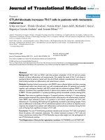
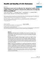
Biochemical analysis of urinary δ-ALA revealed a two-fold
increase (p < 0.001) in exposed than non-exposed sub-
jects (Fig. 1). The mean levels of urinary δ-ALA were 16 ±
2.0 μg/ml in exposed subjects and 7 ± 1.0 μg/ml in non-
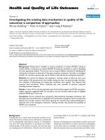
exposed ones. Exposed and non-exposed subjects were
categorized using classification proposed [14] to see the
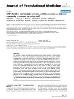
intra-group distribution of δ-ALA. Accordingly, whilst
84% of subjects from the non-exposed group were within
normal range, the percent for exposed ones was as low as
9.8% (Fig. 2). Furthermore, more than half of the exposed
subjects had acceptable levels and about a third had high
levels. By contrast, among non-exposed subjects about
16% displayed acceptable range and none of them had
high levels of urinary δ-ALA.
Inter-enterprise analysis of urinary δ-ALA was also done to
have an idea whether preventive measures were in place or
Table 2: Employment duration of lead exposed workers.
Employment duration (years) Number of workers %
≤ 10 9 17.7
11–20 17 33.3
21–25 9 17.7
25+ 16 31.3
Journal of Occupational Medicine and Toxicology 2008, 3:30 />Page 4 of 8
(page number not for citation purposes)
not. Although levels in Comet (12.6 ± 2.9 μg/ml) tended
to be lower than the other two (18.3 ± 3.9 μg/ml for
Anbessa and 18.7 ± 6.0 μg/ml for Walia), it failed to reach
statistical significance. Categorization of exposed subjects
using Lane et al's classification was also applied to enter-
prises and no dangerous levels had been found in any of
the Enterprises. However, the rank order of proportion for
high urinary δ-ALA levels was Anbessa ≥
Walia>>>>Comet, with the proportion being about 50%
for the former two and about 6% for Comet.
To examine whether urinary δ-ALA levels vary with age,
subjects were stratified into different age groups and sta-
tistical analysis was performed. The result indicated that
urinary δ-ALA levels increased with age in exposed group
(p < 0.001) but failed to show any significant difference in
non-exposed group (Table 3). Likewise, analysis made to
assess the impact of sex on urinary δ-ALA levels failed to
show any significant sex-related differences, although lev-
els in male (16.9 ± 2.6 μg/ml) tended to increase than
females (13.4 ± 4.5 μg/ml).
The impact of duration of employment on levels of uri-
nary δ-ALA was also analyzed and δ-ALA was found to be
a function of duration of employment (Table 4). Indeed,
δ-ALA was noted to significantly increase with duration of
employment (p < 0.001).
Serum creatinine, creatinine clearance and urea levels
In order to see the long-term effects of lead on kidney, dif-
ferent renal indices were measured in both exposed and
non-exposed groups and the results are presented in Table
5. No detectable differences were observed in serum creat-
inine, creatinine clearance and blood urea levels between
exposed and non-exposed groups. However, it is worth
noting that creatinine clearance decreased by about 11%
in exposed subjects, although it fell short of reaching sta-
tistical significance. In parallel, an attempt was made to
look whether there was deviation from reference values
given by the manufacturer and interestingly all were
found to lie within the normal range. The normal ranges
according to the manufacturer of the kit were: serum cre-
atinine (male, 7–13 μg/ml and female, 6–11 μg/ml); cre-
atinine clearance (male, 94–140 ml/min and female, 72–
110 ml/min); and blood urea (150–450 μg/ml for both
sex).
Uric acid levels
Lead is known to inhibit uric acid secretion thereby
increasing serum uric acid levels. Serum uric acid levels
were therefore measured to use it as an indirect measure
of lead exposure, along with urinary δ-ALA. Consistent
with the aforementioned notion, exposed subjects dis-
played increased uric acid levels than non-exposed sub-
Urinary ALA levels in exposed and unexposed subjectsFigure 1
Urinary ALA levels in exposed and unexposed sub-
jects. urine samples collected from 51 exposed and 50 non-
exposed persons were analyzed for levels of δ-ALA using
double beam spectrophotometer. Inter-group analysis was
performed using Student t-test. ***P < 0.001.
Distribution of subjects by group using urinary ALA levelsFigure 2
Distribution of subjects by group using urinary ALA
levels. urinary ALA levels determined as described in the
legend of Fig. 1 were used to classify subjects into different
groups using ranges given by the manufacturer of the kit i.e.
normal (<6 μg/ml), acceptable (6–20 μg/ml), high (20–40 μg/
ml) and dangerous (>40 μg/ml).
Table 3: Levels of urinary δ-ALA by age group
Age δ-ALA (μg/ml) ± SEM
Non-Exposed Exposed
20–35 7.3 ± 2.2 6.1 ± 2.2***
36–45 6.7 ± 1.2 12.4 ± 3.2***
46+ 6.8 ± 1.4 21.7 ± 2.5***
Urinary ALA levels determined as described in the legend of Fig. 1
were compared between similar age groups of exposed and non-
exposed subjects. Data were analyzed using F-ANOVA. ***P < 0.001.
Journal of Occupational Medicine and Toxicology 2008, 3:30 />Page 5 of 8
(page number not for citation purposes)
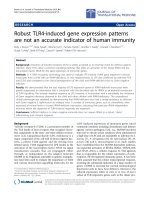
jects, which were significantly higher by about 8% in
exposed subjects than non-exposed ones (p < 0.05) (Fig.
3).
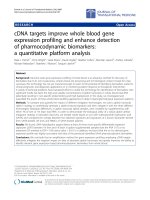
Intra-group sub-classification of uric acid levels using nor-
mal ranges supplied along with the kit revealed that about
69% exposed subjects had abnormal serum uric acid lev-
els, while this was 36% in non-exposed subjects (Fig. 4).
Uric acid normal range was 34–70 μg/ml in male and 26–
60 μg/ml in female.
Data mined from questionnaire
Reported illnesses that were compiled from the structured
questionnaire included illnesses linked with lead poison-
ing, while life style factors were alcohol intake, smoking,
meals at workplace and work related hobbies which could
result in additional exposure to lead. Twenty of the
exposed workers interviewed during this study reported
that they had suffered from illnesses, which are known to
be commonly linked with lead poisoning and include,
among others, visual problems, asthma, gastrointestinal
and kidney problems (in order of proportion of respond-
ents).
The effort to associate the habit of alcohol drinking and
lead exposure revealed that 60.8% of the respondents
were alcohol takers, consuming approximately 6 glass of
draught beer per week. Levels of δ-ALA were found to be
significantly higher (p < 0.05) in alcohol taking workers
(18.9 μg/ml ± 1.5, n = 31) than non-alcohol taking ones
(13.1 μg/ml ± 1.7, n = 20). Moreover, workers were also
unaware of the effects of alcohol consumption on blood
lead levels. Among other life style factors that possibly
contribute to additional lead exposure in and outside the
workplace, having meal at the work place was the prime
candidate. About 88% of the respondents confessed that
they had meal at the work place at least once in a day.
Interviews and walkthrough evaluation also revealed that
none of the enterprises implement clear policy regarding
the use of personal protective equipments (PPEs). All
exposed subjects of the repair units reported that the
enterprises had not provided training regarding lead tox-
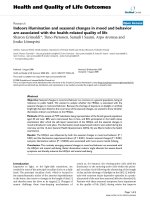
icity. It was also observed during the walkthrough evalua-
tion that all the enterprises workplace was dusty and did
not follow lead regulations (Fig 5). Moreover, the way
used batteries were disposed found to be inappropriate
and hazardous to the environment (Fig 6), particularly to
people living in the vicinity.
Discussion
The application of biomarkers has become a crucial and
widely used tool in understanding and assessment of
health effects [1]. At present, blood lead levels are fre-
quently measured to assess both lead exposure and effect
that will facilitate the risk assessment process. However, a
large body of evidence indicates that alternative biomark-
ers for lead that may be easily measured are also of major
importance, particularly in the heme biosynthetic path-
way [1,15]. Here we report for the first time the occupa-
tional hazard associated with lead exposure in Ethiopia.
Urinary
δ
-ALA levels
This study considered urinary excretion of δ-ALA as a sur-
rogate marker of blood lead in storage battery repair work-
ers; owing to lack of facilities to measure blood lead levels.
δ-ALA is excreted normally in small amounts in urine, but
levels increase with lead exposure. Previous studies
reported a five-fold increase in urinary excretion of δ-ALA
following lead intoxication [16]. This rise in concentra-
tion of δ-ALA during lead exposure is a function of prima-
rily decreased activity of enzymes involved in the heme
synthetic pathway. This inhibition would then result in
increased levels of δ-ALA in the blood and plasma, even-
tually leading to increased δ-ALA urinary excretion
[15,17].
Increased urinary δ-ALA levels found in exposed subjects
in the present study might be the impact of low-level long-
term lead exposure at the repair units and reinforces the
notion that δ-ALA can serve as a surrogate marker for lead
exposure. In addition, the high urinary δ-ALA levels
obtained from about 33.33% of exposed workers (Fig. 2)
is a clear indicator of cumulative lead exposure and
appears to be directly related to duration of employment
at the repair units (Table 2). Evidence for the contribution
Table 4: Urinary δ-ALA (μg/ml) mean levels of exposed workers
by employment duration.
Employment duration δ-ALA (μg/ml) ± SEM
≤ 10 5.4 ±1.6
11–20 14.5 ± 3.1
21–25 19.0 ± 3.7
25+ 23.4 ± 3.5
Table 5: Serum creatinine, creatinine clearance and urea levels of exposed and non-exposed groups.
Group Serum creatinine (μg/ml) ± SEM Creatinine clearance (ml/min.) ± SEM Urea (μg/ml) ± SEM
Non-Exposed 11.5 ± 0.3 115.33 ± 5.20 218.0 ± 7.9
Exposed 11.8 ± 0.2 102.91 ± 6.27 229.1 ± 7.9
Journal of Occupational Medicine and Toxicology 2008, 3:30 />Page 6 of 8
(page number not for citation purposes)
of lead exposure to elevated urinary δ-ALA levels comes
from the observation that 84% of non-exposed subjects
exhibited normal range and none of them had high levels.
This observation excludes the possibility that other factors
might have contributed to the observed high levels of δ-
ALA in exposed subjects. Chronic lead exposure as a cul-
prit for higher δ-ALA levels was also corroborated by the
observation that levels vary with duration of employment.
Urinary δ-ALA levels in workers who had served for 25
years was about fourfold to those served for ten years and
below. This finding is consistent with other reports that
show urinary δ-ALA of lead workers increases with an
increase in the duration of exposure [18].
Although findings published in the literature show that
both age and gender have influence on blood lead levels
[19], age but not sex was found to have effect on urinary
δ-ALA in the present study. Sex was found to have little or
no impact on urinary δ-ALA levels among the exposed
subjects, though females are expected to have higher
blood lead levels compared to males. This might have
something to do with small number of females available
for comparison. Whilst age was found not to be a neces-
sary or sufficient factor for levels of urinary δ-ALA in non-
exposed subjects, it had a significant correlation in
exposed subjects. Plasma lead levels are known to be
higher in children and decline with age, as bone density
increases and lead starts to redistribute to the skeletal
pool. However, in older people plasma lead again
increases due to decalcification of bones and eventual
release of lead into the plasma. Given this fact, the associ-
ation of urinary δ-ALA with age could probably be better
explained by duration of exposure rather than increase
with age per se, as the maximum age of an exposed subject
is an unlikely age where decalcification of bone starts.
Levels of blood uric acid levels in exposed and non-exposed subjectsFigure 3
Levels of blood uric acid levels in exposed and non-
exposed subjects. blood samples were taken from 51
exposed and 50 non-exposed persons and compared for lev-
els of uric Acid using AMS Autolab analyzer. Inter-group var-
iation was analyzed using Student t-test. *P < 0.05.
Distribution of subjects by group using blood uric acid levelsFigure 4
Distribution of subjects by group using blood uric
acid levels. uric acid levels determined as depicted in the
legend of Fig. 3 were used to stratify subjects into different
groups using ranges given by the manufacturer of the kit.
A partial view of storage battery repair unit in one of the enterprisesFigure 5
A partial view of storage battery repair unit in one of
the enterprises. workers are engaged in routine activities
without personal protective equipment, poor ventilation and
full of dust.
Disposal of used storage batteries in one of the enterprisesFigure 6
Disposal of used storage batteries in one of the
enterprises. used batteries are not properly disposed and
this has a long lasting environmental impact.
Journal of Occupational Medicine and Toxicology 2008, 3:30 />Page 7 of 8
(page number not for citation purposes)
Lifestyle factors other than the occupational settings can
have an effect on the exposure of a toxicant. Such factors
usually include smoking and alcohol taking. In this study,
the effect of alcohol, particularly draught beer, on urinary
δ-ALA levels of exposed subjects was analyzed and alco-
hol-taking subjects displayed increased levels than their
non-alcohol-taking peers and this is in line with other
reports [20]. The role of alcohol in blood lead levels is
unclear and is still a subject of controversy. Published
reports indicate that the draught dispensing equipment
rather than alcohol per se is responsible for the increased
lead concentration in alcohol-taking subjects [21]. They
argue that the equipment sometimes contains brass or
gunmetal that has low but significant amounts of lead.
Thus, it is plausible to assume that the same argument
might hold true for the observed increased urinary δ-ALA
in alcohol-taking exposed subjects.
Serum creatinine, creatinine clearance and urea levels
There is evidence to suggest that chronic low level lead
exposure may affect kidney function [22,23]. However,
the level of severity and duration of exposure leading to
renal damage is not clearly defined. Though urinary δ-ALA
increased in exposed subjects and appeared to be related
to duration of employment, none of the renal indices
were found to be different from the non-exposed subjects.
Surprisingly, levels of serum creatinine, creatinine clear-
ance and blood urea levels of both non-exposed and
exposed subjects were found to be within the normal
range (data not shown). Cross-sectional studies con-
ducted in lead-exposed workers showed that lead might
not cause adverse effects on renal glomerular and proxi-
mal tubular functions when there is long-term and less
severe exposure [24,25]. Lack of renal effects in this study
may point to the fact that exposure is not sufficient
enough to bring about appreciable damage to the kidney.
The notion that kidney damage is a function of degree/
intensity of exposure is supported by other studies [26].
These authors found that exposed workers at the smelter
had a greater serum creatinine levels and renal dysfunc-
tion, indicating that workers at the primary lead smelters
have a higher chance of kidney damage than those in
repair units.
Uric acid levels
A relationship between gout and lead nephropathy has
been recognized for centuries and gout occurs more fre-
quently in the presence of chronic lead nephropathy than
in any other type of chronic renal disease [22]. The fact
that large proportion of exposed subjects had high serum
uric acid levels than non-exposed ones is an indicator for
the possible contribution of lead exposure (Fig. 4). Con-
sistent with our finding, a growing body of evidence indi-
cates that chronic occupational lead exposure is associated
with low urate excretion [7,26]. Attempts were also made
to examine additional factors other than lead exposure
that might contribute for the rise in the levels of uric acid
in both exposed and non-exposed subjects. And it was
known that among exposed and non-exposed subjects
there was no one who had been taking medication(s) that
could contribute for the rise in the levels of uric acid, rul-
ing thus out this possibility.
Public health impact of the finding
In Ethiopia, there is no workplace regulation for lead
exposure. Therefore, workers at lead acid battery units of
the studied transport enterprises are clearly at high risk of
lead exposure (Fig 5), as 39% of exposed workers had
some of the common illnesses associated with lead poi-
soning. Not only the workers, but also people living
nearby the repair units are at high risk of exposure due to
failure to follow proper disposal method for used batter-
ies (Fig 6). It was also interesting to note that 44% of
exposed subjects reported that they had changed worksta-
tions through promotion but not because of the risks of
lead exposure, which would definitely affect productivity
of workers in the long run [10,27]. Health risks of lead
require due attention by the enterprises management and
periodic medical checkups should be put in place along
with promoting awareness about the risks associated with
lead exposure. It may not be feasible to quickly introduce
engineering controls so as to protect storage battery repair
workers. Biological monitoring from urine and/or blood
samples would, however, be useful in identifying and
lowering excess lead absorption. Furthermore, workers
should use PPEs very strictly. Enterprises need a clear pol-
icy regarding proper use of PPEs [28], besides training and
regular supervision of workers.
In another finding of this study, the structured question-
naire analysis showed that 88% of exposed subjects had
meals at workplaces on regular basis for at least once per
day and were assumed to have additional lead exposure.
All the enterprises should explore the possibility of estab-
lishing cloth changing facilities, decontamination serv-
ices, and dining rooms to ensure good performance and
well being of workers [29]. Improvements of hygienic
practices are more effective at lowering blood lead levels
than reducing ambient lead level [30]. Hygienic practices
might therefore be the preferred way to reduce lead expo-
sure at the workplace, especially in developing countries
like Ethiopia compared to the engineering controls.
Lead poisoning is a preventable disease provided an inte-
grated prevention program is organized and maintained.
Safety and health measures, such as general ventilation are
usually desirable to control exposure to airborne sub-
stances by diluting the airborne contaminants [9]. Ethio-
pian lead regulations need to be developed and regular
progress monitoring should be made in instituting new
Publish with BioMed Central and every
scientist can read your work free of charge
"BioMed Central will be the most significant development for
disseminating the results of biomedical research in our lifetime."
Sir Paul Nurse, Cancer Research UK
Your research papers will be:
available free of charge to the entire biomedical community
peer reviewed and published immediately upon acceptance
cited in PubMed and archived on PubMed Central
yours — you keep the copyright
Submit your manuscript here:
/>BioMedcentral
Journal of Occupational Medicine and Toxicology 2008, 3:30 />Page 8 of 8
(page number not for citation purposes)
workplace lead controls, implementing large scale health
screenings and lowering this all-pervasive and hidden epi-
demic, so that occupational lead exposure and its long-
term impacts on society are ultimately eliminated.
To sum up, raised levels of urinary δ-ALA and uric acid
obtained from the exposed subjects may indicate the pos-
sible parallel rise in blood lead levels. These measured val-
ues were mainly attributed from poor preventive and
control measures at the repair units. Improving the work
environment of the workers is quite important, as the next
workers who are assigned to work in the 'non-fit' environ-
ment would also be exposed to the same hazard that
entails an overall decrease in productivity of the enter-
prises. By and large occupational exposure to lead remains
a big problem in developing countries including Ethiopia.
Therefore, it is necessary that lead exposures at workplaces
be minimized by placement of appropriate and cost-effec-
tive integrated preventive and control measures.
Declaration of competing interests
The authors declare that they have no competing interests.
Authors' contributions
KA conception and design of the work, generation and
analysis of data, GA generation and data analysis and
commented the MS, EE conception and design of the
work, data analysis, drafted and developed the MS.
Acknowledgements
The authors are most grateful to Addis Ababa University for the financial
support and Ethiopian Health and Nutrition Research Institute for allowing
using the facilities.
References
1. International Program on Chemical Safety: Environmental Health
Criteria 165: Inorganic Lead. Geneva 1995.
2. Massaro EJ: Handbook of Human Toxicology. New York: CRC
Press; 1997.
3. Winder C: Toxicity of metals. In Occupational Toxicology Edited by:
Neill H Stacey. London: Taylor and Francis; 1993:169-175.
4. Wu TN, Shen CY, Ko KN, Guu CF, Gau HJ, Lai JS, Chen CJ, Chang
PY: Occupational Lead Exposure and Blood Pressure. Int J Epi-
demio 1996, 25:791-796.
5. Robert AG, Thomas WC: Toxic Effects of Metals. In Casarett and
Doull's Toxicology: The Basic Science of Poisons 6th edition. Edited by:
Curtis DK. New York: McGraw-Hill; 2001:827-834.
6. World Health Organization Regional Office for Europe: Air Quality
Guidelines. Copenhagen 2001.
7. Lin JL, Tan DT, Ho HH, Yu CC: Environmental lead exposure
and urate excretion in the general population. Am J Med 2002,
113:563-568.
8. Goelzer B: The harmonized development of occupational
hygiene: A need in developing countries. Am Ind Hygiene Asso-
ciation J 1996, 57:984.
9. World Health Organization: Hazard prevention and control in work envi-
ronment: Airborne Dust. Geneva 1999.
10. George AM, (Ed): Proceedings of the International Conference on Lead
Poisoning Prevention and Treatment: 8–10 February 1999; Bangalore The
George Foundation; 1999.
11. Henry JB: Clinical Diagnosis and Management by Laboratory
Methods. Philadelphia: W.B. Saunders Company; 1984.
12. Labbe RF, Lamon JM: Porphyrins and Disorders of Porphyrins
Metabolism. In Fundamentals of Clinical Chemistry 3rd edition. Edited
by: Tietz NW. Philadelphia: W.B.Saunders Co; 1987:825-841.
13. Tomokumi K, Ogata M: Simple method for determination of
urinary δ-ALA as an index of lead exposure. Clin Chem 1972,
18:1534-1536.
14. Lane RE, Hunter D, Malcolm D, Williams MK, Hudson AR, de Kretser
AJ, Zielhuis RL, Cramer K, Barry PSI, Beritic T, Vigliani EC, Truhaut
TR, Kehoe RA, King E: Diagnosis of inorganic lead poisoning: A
statement. Brit Med J 1968, 4:
501.
15. Sakai T: Biomarkers of Lead Exposure. Ind Health 2000,
38:127-142.
16. Bauer JD: Clinical Laboratory Methods. St. Louis Missouri: The
C.V. Mosby Company; 1982.
17. Higashikawa K, Furuki K, Takada S, Okamoto S, Ukai H, Yuasa T,
Ikeda M: Blood Lead Level to Induce Significant Increase in
Urinary δ-Aminolevulinic Acid Level among Lead-Exposed
Workers: A Statistical Approach. Industrial Health 2000,
38:181-188.
18. Lee BK: Occupational lead exposure of storage battery work-
ers in Korea. Br J Ind Med 1982, 39:283-289.
19. Hodgkings DG, Hinkamp DL, Robins TG, Schork MA, Krebs WH:
Influence of high past lead-in-air exposures on the lead – in –
blood levels of lead – acid – battery workers with continuing
exposure. J Occup Med 1991, 33:797-803.
20. Weyermann M, Brenner H: Alcohol consumption and smoking
habits as determinants of blood lead levels in a national pop-
ulation sample from Germany. Arch Environ Health 1997,
52:233-240.
21. Sherlock JC, Pickford CJ, White GF: Lead in alcoholic beverages.
Food Addit Contam 1986, 3:347-354.
22. Bernard BP, Becker CE: Environmental Lead Exposure and the
Kidney. J Toxicol Clin Toxicol 1988, 26(1-2):1-34.
23. Lim YC, Chia KS, Ong HY, Ng V, Chew YL: Renal dysfunction in
workers exposed to inorganic lead. Ann Acad Med Singapore
2001, 30(2):112-117.
24. Omae K, Sakurai H, Higashi T, Muto T, Ichikawa M, Sasaki N: No
adverse effects of lead on renal function in lead-exposed
workers. Ind Health 1990, 28:77-83.
25. Wang VS, Lee MT, Chiou JY, Guu CF, Wu CC, Wu TN, Lai JS: Rela-
tionship between blood lead levels and renal function in lead
battery workers. Int Arch Occup Environ Health 2002, 75:569-575.
26. Pinto de Almeida AR, Carvalho FM, Spinola AG, Rocha H:
Renal dys-
function in Brazilian lead workers. Am J Nephrol 1989,
7(6):455-458.
27. Smith JM: Ergonomics factors. In Occupational Health and safety
Encyclopedia Volume 34. 4th edition. Edited by: Stellman JM. London:
Taylor and Francis; 1997:22.
28. Stellman JM, Osinsky D: Encyclopaedia of Occupational Health
and Safety. Geneva: International Labor Organization; 1997.
29. International Labour Office: Ergonomics Checkpoints: Practical
and easy to implement solutions for improving safety, health
and working conditions. Geneva 1996.
30. Lai JS, Wu TN, Liou SH, Shen CY, Guu CF, Ko KN, Chi HY, Chang
PY: A study of the relationship between ambient lead and
blood lead among lead battery workers. Int Arch Occup Environ
Health 1997, 69:295-300.