báo cáo hóa học: " Inhibition of gap junctional Intercellular communication in WB-F344 rat liver epithelial cells by triphenyltin chloride through MAPK and PI3-kinase pathways" pdf
Bạn đang xem bản rút gọn của tài liệu. Xem và tải ngay bản đầy đủ của tài liệu tại đây (1.02 MB, 10 trang )
Lee et al. Journal of Occupational Medicine and Toxicology 2010, 5:17
/>Open Access
RESEARCH
© 2010 Lee et al; licensee BioMed Central Ltd. This is an Open Access article distributed under the terms of the Creative Commons At-
tribution License ( which permits unrestricted use, distribution, and reproduction in any
medium, provided the original work is properly cited.
Research
Inhibition of gap junctional Intercellular
communication in WB-F344 rat liver epithelial cells
by triphenyltin chloride through MAPK and
PI3-kinase pathways
Chung-Hsun Lee
1,2
, I-Hui Chen
2
, Chia-Rong Lee
2
, Chih-Hsien Chi
1
, Ming-Che Tsai
1
, Jin-Lian Tsai
2
and Hsiu-Fen Lin*
2,3,4
Abstract
Background: Organotin compounds (OTCs) have been widely used as stabilizers in the production of plastic,
agricultural pesticides, antifoulant plaints and wood preservation. The toxicity of triphenyltin (TPT) compounds was
known for their embryotoxic, neurotoxic, genotoxic and immunotoxic effects in mammals. The carcinogenicity of TPT
was not well understood and few studies had discussed the effects of OTCs on gap junctional intercellular
communication (GJIC) of cells.
Method: In the present study, the effects of triphenyltin chloride (TPTC) on GJIC in WB-F344 rat liver epithelial cells
were evaluated, using the scrape-loading dye transfer technique.
Results: TPTC inhibited GJIC after a 30-min exposure in a concentration- and time-dependent manner. Pre-incubation
of cells with the protein kinase C (PKC) inhibitor did not modify the response, but the specific MEK 1 inhibitor PD98059
and PI3K inhibitor LY294002 decreased substantially the inhibition of GJIC by TPTC. After WB-F344 cells were exposed
to TPTC, phosphorylation of Cx43 increased as seen in Western blot analysis.
Conclusions: These results show that TPTC inhibits GJIC in WB-F344 rat liver epithelial cells by altering the Cx43 protein
expression through both MAPK and PI3-kinase pathways.
Background
Organotin compounds have been widely used as agricul-
tural biocides, antifouling agents in boat paint, wood pre-
servatives, and stabilizers for polyvinylchloride polymers
(PVC) in industry [1,2]. Triphenyltin (TPT) is an organo-
tin compound which is widely used as fungicides on
major food and food-stock crops. It is also used in anti-
fouling paints to prevent growth of barnacles and other
fouling organisms on boats and ships [3]. Organotin com-
pounds are known to be endocrine disruptors in marine
species and may be mahuman beings [4,5]. Tissue con-
centrations of TPT were correlated with the degree of
imposex in rock shells [6,7]. TPT compounds have
embryotoxic, myotoxic, genotoxic and immunotoxic
effects in mammals [8-11]. The organotin compounds
might be incorporated in the most abundant phospho-
lipid of eukaryotic membrane and caused toxicity [12].
Some toxic effects have been observed in aquatic and ter-
restrial organisms exposed to TPT, such as increased
tumor incidence and immune suppression [13,14]. Some
studies have revealed that TPT might inhibit the cyto-
toxic function of human natural killer cells and triphenyl-
tin hydroxide produced tumors in rats and mice [14-16].
Connexins (Cxs) are a group of at least 20 highly con-
served proteins that provide the basis for communication
through the direct exchange of ions, nutrients, second
messengers, electrical coupling, and small metabolites
from one cell to its neighboring cells [17-20]. Cell prolif-
eration, differentiation, apoptosis and adaptive responses
of differentiated cells can occur as a consequence of the
up- or down-regulation of GJIC [21-23]. Disruption in
GJIC may cause loss of homeostatic and cell growth con-
trol [18,24-26]. Growing evidence suggests that connexin
* Correspondence:
2
Graduate Institute of Occupational Safety and Health, College of Health
Science, Kaohsiung Medical University, Kaohsiung 80708, Taiwan
Full list of author information is available at the end of the article
Lee et al. Journal of Occupational Medicine and Toxicology 2010, 5:17
/>Page 2 of 10
43 (Cx43), a major gap junction protein, functions as a
tumor suppressor gene. Expression of Cx43 is often
decreased in human tumor cells and tissues, including
those involved in human mammary carcinoma, prostate
cancer, human glioblastoma, skin squamous cell carci-
noma, lung cancer, esophagus cancer, adrenocortical
tumors, ovarian carcinoma, cervical cancer, endometrial
carcinoma, and human mesothelioma [27-37]. It has been
assumed that using pharmacological stimulation to effi-
ciently restore GJIC in tumor cells might represent a
strategy for anti-neoplastic therapies [38-42].
The carcinogenicity of TPT remained unclear. The
present work was undertaken to define the effects of
TPTC on GJIC in WB-F344 rat liver epithelial cells.
Materials and methods
Chemicals
Powder of TPTC was supplied by MERCK (Darmstadt,
Germany).
Lucifer yellow, DMSO (dimethylsulfoxide), formalde-
hyde, MTT (3-[4,5-dimethyl-2-thiazolyl]-2,5-diphenyl-
2H-tetrazolium bromide) were supplied by Sigma-
Aldrich (St. Louis, MO, USA). D medium and newborn
calf serum were from Gibco (Invitrogen cooperation, CA,
USA), Trizole was from Invitrogen Life Technologies
(Rockville, MD, USA) and 2 X SYBR green PCR master
mix was from Applied Biosystems (Foster, CA, USA). The
protein kinase C (PKC) inhibitor GF109203X, extracellu-
lar signal-regulated protein kinase (ERK) inhibitor
PD98059 and PI3 kinase inhibitor LY294002 were from
Sigma (St. Louis, MO, USA). Immobilon Western HRP
Substrate Peroxide Solution and luminal reagent were
supplied by Millipore Corporation (Billerica, MA). All
chemicals used in the study were of the highest available
purity.
Cell culture and treatment with chemicals
WB-F344 rat liver epithelial cells [43] were cultured in D
medium supplemented with 5% fetal bovine serum and
1% [v/v] penicillin/streptomycin antibiotic. The cells
were grown at 37°C in a 5% CO
2
incubator before being
used in the different experiments. Confluent cells, grown
in plates, were exposed to various concentrations of
TPTC. To prepare the TPTC stock solution, 0.01 g of
TPTC powder was dissolved in 10 ml DMSO and then
diluted to a final concentration of 1000 ppm.
Cell toxicity assay of TPTC
The effect of TPTC on the survival of WB F344 cells was
assessed using MTT toxicity assay as described previ-
ously [44]. In brief, the cells were plated in 100 μl media
in 48-well plates (1 × 10
4
/well). On the following day, the
experimental medium containing different TPTC con-
centrations (0, 0.25, 0.5, 1, 2, 3, 4, and 5 ppm) was added,
and then incubated for 30 and 60 minutes. Fifty μl of
MTT solution (2 mg/ml in PBS) was added to each well
and incubated for 6-8 hours. After careful removal of the
medium, 150 μl of DMSO was added to each well, and
then after careful shaking, the absorbance was read at 570
nm using an ELISA microplate reader (Zenyth 200rt with
ADAP software, Anthos Labtec Instruments, Autria).
Cell viability was expressed as a percentage of control
cells not treated with TPTC and was designated as 100%.
Colony forming-efficiency assay
Colony forming-efficiency experiments were performed
as previously described [45]. In brief, exponentially grow-
ing cells were plated at 500 cells/100 mm tissue culture
dish in 10 ml D medium, treated with different concen-
trations of TPTC. Following treatment, the plates were
washed two times with the medium. The medium was
not replaced, and colonies were fixed and stained after 14
days in culture by water: addition of methanol (1:1) con-
taining crystal violet (1 g/l). Colonies with cell clusters
containing more than 50 cells were counted under a dis-
secting microscope. Data indicate survival as a percent-
age relative to untreated cells.
GJIC inhibition assay
GJIC assay was carried out in 35 × 10 mm tissue culture
dishes with 100% confluent monolayer cells grown in 2
ml D-medium supplemented with 5% newborn calf
serum, 100 U/ml penicillin and streptomycin 100 μg/ml.
GJIC was detected using the scrape-loading and dye
transfer (SL/DT) technique developed by el-Flouly [46].
Assays for different treatments and vehicle control were
run in triplicate in cell culture dishes. Monolayer cells
with 100% confluence were incubated with target com-
pounds. For dose-dependent inhibition of GJIC, we
treated cells with 0.5, 1.0, 1.5 and, 2.0 ppm TPTC for 30
min. For time-dependent inhibition of GJIC, analysis was
performed with 1.5 ppm TPTC for 15, 30, 45, and 60 min.
After exposure to the target compounds, the cells were
rinsed three times with PBS and 1 ml of lucifer yellow
solution was then added to the cell cultures and scrape-
loaded with several scrapes using a steel surgical blade.
The dye solution was left on the cell cultures for 3 min,
and then discarded. The cell cultures were carefully
rinsed three times with PBS to remove detached cells and
background fluorescence. Several drops of 4% formalin in
PBS were added to fix the cell cultures. An inverted fluo-
rescence microscope equipped with a digital camera
(Nikon Eclipse TE 2000-U system, Nikon ACT-1 version
2.62, Nikon Corporation, Japan) was employed to record
the migration of the lucifer yellow dye from the edge cells
of the scrape. The migration was measured on the micro-
graph. An average value of 30 measurements for each
treatment (10 measurements per dish) was regarded as
Lee et al. Journal of Occupational Medicine and Toxicology 2010, 5:17
/>Page 3 of 10
the migration of dye in the cell cultures. The percentage
of migration of dye in cell cultures exposed to target com-
pounds to the migration of dye traveling in the vehicle
control was employed to evaluate the inhibition of GJIC.
For inhibition studies, cultures were pre-incubated for 30
min with various pathway inhibitors prior to treatment
with 1.5 ppm TPTC for 30 min.
Western blot analysis
WB F344 liver cells were treated with TPTC of 1.5 ppm
for 15 and 30 min. After treatment, the medium was
removed and cells were washed twice with PBS and lysed
with 0.5% SDS. Lysates were stored at -80°C. Cell lysates
were sonicated, and protein levels were determined using
a protein detection assay (BioRad). Sample blue buffer
(30% sucrose, 10% SDS, 0.1% bromophenol blue, and
0.2% dithiothreitol) was added and the samples were
heated for 10 min at 100°C and loaded onto gels (10%
SDS-PAGE). SDS-PAGE-separated proteins were blotted
onto a PVDF membrane (Immobilon-PSQ, Millipore,
Bedford, MA) using a semi-dry blotter (VWR), and the
membrane was blocked with 5% milk in PBS-T buffer
[1000 ml PBS with 1 ml Tween 20 (pH 7.4)] for more than
1 h at room temperature. The protein was probed with
antibodies (Mouse IgG, Zymed) against connexin 43 at
4°C overnight and this was followed by incubation with
horseradish peroxidase-conjugated secondary antibodies
(Mouse anti-Goat IgG-HRP, Sigma). Protein visualization
was carried out using an enhanced chemiluminescence
kit (Pierce) according to the manufacturer's protocol.
Immunofluorescence staining
Immunofluorescence staining experiment s were per-
formed as previously described[47]. In brief, WB F344
liver cells were plated in 100 μl media in 12 well-plates
treated with 1.5 ppm TPTC for 30 min. After treatment,
the medium was removed and sections were washed with
PBS. 4% paraformaldehyde was added and washed sec-
tions with PBS 20 min later. 0.5% triton X-100 (Sigma)
was added for 20 min and washed out with PBS. After
treatment, diluted primary antibodies mouse IgG against
connexin 43 (Santa Cruz Biotechnology, Inc.) with 4% tri-
ton X-100 was added and incubated sections for 1 h at
room temperature. The sections were washed with PBS,
and diluted mouse IgG secondary antibody (Alexa
Fluor555 &488) with 4% triton X-100 was added and
incubated sections for 1 h at room temperature. After
treatment, 4,6'-diamidino-2-phenylindole (DAPI)(Sigma)
was added and incubated sections for 10 min at room
temperature. An inverted fluorescence microscope
equipped with a digital camera (Nikon Eclipse TE 2000-U
system, Nikon ACT-1 version 2.62, Nikon Corporation,
Japan) was employed to record the fluorescent intensity
of the cells.
Statistical analysis
Means ± SEM were calculated and the data are presented
as a percentage of control. All data were analyzed by
Sigma Plot 8.0 software using repeated measures.
ANOVA (SPSS for window version 12.0.1; SPSS, Inc.,
Chicago, IL) was performed to examine the effect of inde-
pendent variables (treatment, day, incubation time, time
point). Tests for contrasts were carried out to compare
the different levels of the independent variables. P values
≤ 0.05 were considered statistically significant.
Results
TPTC dissolved easily in DMSO but not in water. To
exclude the toxic effects of DMSO on cell viability and
diffusion length of GJIC, tests involving exposure to
DMSO were carried out. Results revealed that after expo-
sure to 2% DMSO for 30 minutes, the diffusion length of
GJIC did not obviously decrease as compared with that of
the control group (p > 0.05).
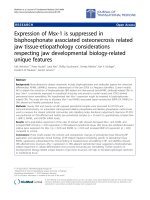
Cytotoxicity of TPTC
Cytotoxicity evoked by TPTC in WB-F 344 cells was
tested with 0, 0.25, 0.5, 1, 2, 3, 4, and 5 ppm of TPTC
using the MTT proliferation assay. After 30- and 60-min
exposure to TPTC, it was found that cell viability
decreased obviously with increasing concentration of
TPTC and the lethal concentration 50 (LC 50) in 60 min
calculated was 5 ppm (Fig. 1A.)
Colony-forming efficiency in WB-F 344 cells was evalu-
ated using TPTC of 0, 3, 9, 12, 15, 18 ppb. After 14 days of
exposure, the colony-forming efficiency decreased signif-
icantly when TPTC concentration exceeded 12 ppb (Fig.
1B.)
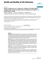
Dose- and time- dependent inhibition of GJIC by TPTC
Inhibition of GJIC has been suggested to be an important
activity of tumor promoters [36]. Therefore, the capacity
of TPTC to inhibit GJIC was measured in concentrations
with 0.5, 1.0, 1.5 and 2 ppm TPTC after 30 min of expo-
sure. As shown in Figure 2A, TPTC inhibited significantly
GJIC in WB-F344 liver cells. The migration of Lucifer yel-
low dye in scraped WB F344 liver cells treated with TPTC
was less than that in untreated cells, when the concentra-
tion was 1.0 ppm (*p < 0.05).
The effects of TPTC on GJIC were evaluated with cells
exposed to TPTC for 15 min, 30 min, 45 min, and 60 min.
After 15 min of exposure to 1.5 ppm of TPTC, the diffu-
sion length was significantly decreased as compared with
that of the control group (p < 0.05) (Fig. 2B). The diffu-
sion length reduced gradually with time and became
Lee et al. Journal of Occupational Medicine and Toxicology 2010, 5:17
/>Page 4 of 10
almost invisible after 60 min of exposure to 1.5 ppm of
TPTC.
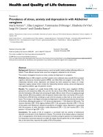
Effects of PKC, ERK and PI3 kinase on GJIC response
Organotin compounds showed that inhibition through
some kinase pathways is a possible mechanism involved
in the apoptotic effects [48]. The mitogen-activated pro-
tein kinase (MAPK) pathway has been shown to be
involved in the inhibition of GJIC by TPA [49-54]. Its role
in the TPTC-induced inhibition of GJIC was studied
next. No specific inhibitor of MAPK was available, but
PD98059, a MEK1 inhibitor that blocks ERK activation,
was used as an inhibitor of the pathway [55-57]. MEK 1 is
the direct upstream activator kinase of MAPKs. The cells
were pre-exposed to 50 μM PD98059 for 30 min prior to
co-exposure to TPTC (1.5 ppm) for 30 min The scrape-
loading assays were then repeated using the ERK inhibi-
tor PD98059. The data showed that PD98059 restored
significantly GJIC in TPTC-treated liver cells (p < 0.05)
(Fig. 3), Thus, the MAPK signaling pathway was clearly
involved in the inhibition of GJIC by TPTC.
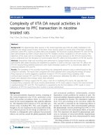
Phosphatidylinositol 3'-kinase (PI3K) has been demon-
strated to be critical in mediating several aspects of
PDGF actions in various cells [23,58-62]. To explore the
potential role of PI3K signaling in the signaling processes
involved in TPTC-induced disruption of GJIC in liver
cells, we measured GJIC in rat liver cells with and without
pre-treatment with the Pl3K inhibitor LY294002 (100
μmol/L) before exposure to TPTC (1.5 ppm) for 30 min.
As shown in Fig. 4, pre-incubation of rat liver cells with
LY294002 (100 μmol/L) for 30 min almost stopped com-
pletely the inhibition of GJIC caused by TPTC, although
the inhibitor itself did not exert much influence on GJIC,
as compared with the control. Similar result was also
found in the group exposed to TPTC and PD98059 as
compared with that exposed to TPTC alone (Fig. 3).
Thus, we conclude that TPTC blocked GJIC through
MAPK and PI3K pathways.
To study the involvement of protein kinase C (PKC) in
the inhibition of GJIC by TPTC, an inhibitor of PKC,
GF109203X (bisindolylmaleimide 1) was utilized to block
the activity of the enzyme before exposure to TPTC-
GF109203X inhibits the isozymes of PKC α, β
I
, β
II
, γ, δ,
and ε [63,64]. The cells were pre-exposed to the PKC
inhibitor (10 μM) for 30 min prior to co-exposure to
TPTC (1.5 ppm) and incubated further for 30 min. The
diffusion length of GJIC did not obviously decrease when
only GF109203X was added. On the other hand, cells
were treated with 10 μM GF109203X for 30 min, followed
by addition of TPTC. The diffusion length of GJIC
decreased obviously following the addition of TPTC or
TPTC with GF109203X (Fig. 5)., No change was observed
in the inhibition of GJIC by TPTC alone. Thus, the inhi-
bition of GJIC by TPTC was not mediated by PKC.
Neither GF109203X, LY294002 nor PD98059 alone at
the indicated concentration had any notable effects on
GJIC in these cells.
Effects of TPTC on connexin 43 protein level and
phosphorylation
One possible mechanism involved in the inhibition of
GJIC is abnormal phosphorylation of connexins [65-67].
WB-F433 cells express Cx43 predominantly as gap junc-
tion protein [68]. Western blot analysis was performed to
detect the state of Cx43 phosphorylation in WB-F344
cells after treatment with TPTC. In untreated cells, three
Figure 1 Cytotoxicity of TPTC in WB-F344 liver cell. (A) Cell viability of WB-F344 liver cells after exposure to TPTC of different concentrations for 30
min and 60 min. LC50 of TPTC in WB-F344 liver cells amounted to 5 ± 0.9 ppm, n = 5. (B) Colony-forming efficiency of WB-F344 cells treated with dif-
ferent concentrations of TPTC. When the concentration of TPTC was 12 ppb, the proliferation of WB-F344 liver cells was significantly inhibited. Data
indicate survival as a percentage relative to untreated cells. All values are represented as means ± S.D. of five independent experiments. Statistical
significance was determined using ANOVA (*p < 0.05).
Lee et al. Journal of Occupational Medicine and Toxicology 2010, 5:17
/>Page 5 of 10
isoforms of Cx43, which correspond to different phos-
phorylated forms of Cx43, are detectable as P0 (unphos-
phorylated form), P1 and P2 (phosphorylated forms),
respectively [69]. After 15-min and 30-min exposure to
TPTC, the P0 band disappeared, and a shift to bands of
higher molecular weight occurred (P1) (Fig. 6).
Effects of TPTC on connexin 43 in immunofluorescence
staining
The expression of Cx43 in WB-F344 cell under stained
with fluorescein isothiocyanate (FITC) and DAPI after
30-min exposure with1.5 ppm TPTC compared to the
control group (A) with 1.5% DMSO was showed (Fig. 7).
The fluorescent intensity did decrease in group (B) after
exposure with TPTC.
Discussion
Carcinogenesis is a multistep process, including "initia-
tion," "promotion," and "metastasis" ("progression") [70].
Potter suggested that the initiation process prevents
genetically altered stem cells from terminally differentiat-
ing [71], and, at the same time, GJIC restricts the growth
of these cells. However, when exposed to tumor promot-
ers, which inhibit GJIC, these transformed cells prolifer-
ate [37]. The results of this study indicate that the TPTC
inhibits GJIC in WB-F344 rat liver epithelial cells in a
concentration- and time-dependent manner. In the pres-
ent study, we demonstrate for the first time that exposure
TPTC results in downregulation of Cx43 expression in
liver cell cultures. Moreover, we show that TPTC modu-
lates Cx expression predominantly through activation of
MAPK and PI3K signaling pathways. Several in vivo and
in vitro studies have revealed potential effects of organo-
tins in broad spectrum including immunosuppressive,
neurotoxic, endocrinopathic, reproductive, teratogenic,
developmental, and possibly carcinogenic activity
[3,13,72-75]. Alterations in the phosphorylation status of
Figure 2 Inhibition of GJIC by TPTC using the modified scrape-loading/dye transfer method with the Lucifer yellow fluorescent dye. (A)
Dose-dependent inhibition of GJIC after 30-min TPTC exposure. (B) Time-dependent inhibition of GJIC exposed to TPTC. Cells were treated with 1.5
ppm TPTC. The results are represented as means ± S.D. of at least three independent experiments. Statistical significance was determined using ANO-
VA (*p < 0.05).
control A1 0.5 ppm A2 1.0 ppm A3 1.5 ppm A4 2.0 ppm
control B1 15 min B2 30 min B3 45 min B4 60 min
Lee et al. Journal of Occupational Medicine and Toxicology 2010, 5:17
/>Page 6 of 10
connexins are a consequence of the activities of the pro-
tein kinase and/or protein phosphatases. GJIC recovered
when pre-treated with PD 98059 (ERK inhibitor), and
LY294002 (PI3-kinase inhibitor), but did not recover
when GF109203X (Protein Kinase C inhibitor) was
added. The reactions of fluorescence of Cx43 in WB-
F344 cells after treatment with TPTC did decrease and
the phosphorylation of Cx43 was found in Western Blot
analysis. Some studies also showed that TPTC could
inhibit the phosphorylation and ATP formation in chlo-
roplasts and embryos of marine invertebrate [9,76].
The inhibition of GJIC by TPTC was independent of
PKC activity but clearly dependent upon the activation of
both MAPK and PI3-kinase pathways. The loss of GJIC
Figure 3 Effect of PD98059 (MEK 1 inhibitor) on TPTC-induced
disruption of GJIC in WB-F344 cells (mean values ± S.D.). The con-
trol group comprises negative controls. The PD98059 group comprises
cells treated with 50 μM PD98059 for 30 min. The TPTC group compris-
es cells treated with 1.5 ppm TPTC for 30 min. The PD98059 + TPTC
group comprises cells pre-treated with 50 μM PD98059 for 30 min and
then exposed to 1.5 ppm TPTC for 30 min. Asterisks indicate statistically
significant difference. (
#
p < 0.05 compared with the control group, *p
< 0.05 compared with the group exposed to PD98059 alone, and
&
p <
0.05 compared with the group exposed to TPTC alone.)
PD 98059
PD 98059+ TPTC
Figure 4 Effect of LY294002 (PI3K inhibitor) on TPTC-induced dis-
ruption of GJIC in WB-F344 cells (mean values ± S.D.). The control
group comprises negative controls. The LY294002 group comprises
cells treated with.100 μM LY294002 for 30 min. The TPTC group com-
prises cells treated with 1.5 ppm TPTC for 30 min. The LY294002 + TPTC
group comprises cells pre-treated with 100 μM LY294002 and exposed
to 1.5 ppm TPTC for 30 min. Asterisks indicate statistically significant
difference. (# p < 0.05 compared with the control group, * p < 0.05
compared with the group exposed to LY294002 alone, and p < 0.05
compared with the group exposed to TPTC alone.)
Figure 5 Effect of GF109302X (PKC inhibitor) on TPTC-induced
disruption of GJIC in WB-F344 cells (mean values ± S.D.). The con-
trol group comprises negative controls. The GF109302X group com-
prises cells treated with 10 μM GF109203X for 30 min. The TPTC group
comprises cells treated with 1.5 ppm TPTC for 30 min. The GF109302X
+ TPTC group comprises cells pre-treated with 10 μM GF109203X for
30 min and then exposed to 1.5 ppm TPTC for 30 min. Asterisks indi-
cate statistically significant difference. (# p < 0.05 compared with the
control group, *p < 0.05 compared with the group exposed to
GF109302X alone.)
Figure 6 Western blot analysis of Cx43, α-tublin, and E-cadherin
protein expression alterations in TPTC treated WB F344 liver cells.
A: T, TPTC 1.5 ppm (15-min exposure); D, negative control (DMSO). B: T,
TPTC 1.5 ppm (30-min exposure); D, negative control (DMSO). P0 grad-
ually decreased and density of P1 increased. The band of P0 totally dis-
appeared after exposure to 1.5 ppm TPTC for 30 min. No change of α-
tublin, and E-cadherin protein were found. MW: Connexin 43 was 43
kD, α-Tublin was 55 kD, and E-Cadherin was 120 kD.
A B
Exposure time 15 min 30 min
Control T D T D
Cx43
P2
P1
P0
α-tublin
E-cadherin
Lee et al. Journal of Occupational Medicine and Toxicology 2010, 5:17
/>Page 7 of 10
was also described in cancer cells [77,78]. Alteration in
expression of connexins may be involved in the expres-
sion of neoplastic phenotype [79] and changes in the
phosphorylation pattern of connexins are also associated
with GJIC inhibition by other tumor-promoting agents
and oncogenes [80-82].
Hence, there is no evidence of a causal cross-talk
between the two modulatory pathways, MAPK and PI3K.
However, both PD58059 and LY294002 abolished com-
pletely the effect of TPTC downregulation of Cx43, impli-
cating both MAPK and PI3K signaling cascades in a
common mechanism of Cx regulation. It is possible that
MAPK and PI3K act through a common downstream
pathway, such as GSK-3 activation [83-86], to control
endothelial cellular function through Cxs.
In conclusion, the present study shows that TPTC
inhibits GJIC in WB-F344 rat liver epithelial cells by
altering the Cx43 protein expression through the MAPK
Figure 7 The expression of Cx43 in WB-F344 cell under stained with FITC and DAPI. A. expression of Cx43 in WB-F344 cell with 1.5% DMSO; B:
expression of Cx43 in WB-F344 with 1.5 ppm TPTC after 30-min exposure. The fluorescent intensity did decrease in FITC stain after treatment with 1.5
ppm of TPTC for 30 min.
APhase
BPhase
A FITC
A FITC&DAPI
B FITC
B FITC&DAPI
Lee et al. Journal of Occupational Medicine and Toxicology 2010, 5:17
/>Page 8 of 10
and PI3-kinase pathways. However, to prove the carcino-
genicity of TPTC still needs further study. This prelimi-
nary study could provide the possible mechanism for
further evaluation of toxicity of TPTC.
Competing interests
The authors declare that they have no competing interests.
Authors' contributions
CHL participated in the study design, interpretation of results, analysis, and
manuscript writing. IHC participated in the study design and analysis. CRL par-
ticipated in the statistical analysis and manuscript writing. CHC participated in
the study design and coordination. MCT participated in the study design and
coordination. JLT carried out the immunoassays, the study design, analysis and
manuscript writing. HFL participated in the study design, interpretation of
results and manuscript preparation. All authors read and approved the final
manuscript.
Acknowledgements
This study was supported by a grant (NSC-93-2113-M-037-018) from the
National Science Council, Taiwan.
Author Details
1
Department of Emergency Medicine, National Cheng Kung University
Hospital, Tainan, Taiwan,
2
Graduate Institute of Occupational Safety and Health,
College of Health Science, Kaohsiung Medical University, Kaohsiung 80708,
Taiwan,
3
Department of Ophthalmology, Chang Gung Memorial Hospital,
Kaohsiung, Taiwan and
4
Chang Gung University, College of Medicine,
Kaohsiung, Taiwan
References
1. Ueno S, Susa N, Furukawa Y, Komatsu Y, Koyama S, Suzuki T: Butyltin and
phenyltin compounds in some marine fishery products on the
Japanese market. Archives of Environmental Health 1999, 54:20.
2. Duncan J: The toxicology of molluscicides. The organotins.
Pharmacology & Therapeutics 1980, 10:407-429.
3. Kimbrough RG: Toxicity and health effects of selected organotin
compounds: A review. Environmental Health Perspectives 1976, 14:51-56.
4. Golub M, Doherty J: Triphenyltin as a potential human endocrine
disruptor. Journal Of Toxicology And Environmental Health Part B, Critical
Reviews 2004, 7:281-295.
5. Santos MM, Reis-Henriques Armanda M, Natividade Vieira M, Sole M:
Triphenyltin and tributyltin, single and in combination, promote
imposex in the gastropod Bolinus brandaris. Ecotoxicology and
Environmental Safety 2006, 64:155-162.
6. Horiguchi T, Shiraishi H, Shimizu M, Morita M: Effects of triphenyltin
chloride and five other organotin compounds on the development of
imposex in the rock shell, Thais clavigera. Environmental Pollution 1997,
95:85-91.
7. Shim WJ, Kahng SH, Hong SH, Kim NS, Kim SK, Shim JH: Imposex in the
rock shell, Thais clavigera, as evidence of organotin contamination in
the marine environment of Korea. Marine Environmental Research 2000,
49:435-451.
8. Boyer IJ: Toxicity of dibutyltin, tributyltin and other organotin
compounds to humans and to experimental animals. Toxicology 1989,
55:253-298.
9. Cima F, Ballarin L, Bressa G, Martinucci G, Burighel P: Toxicity of Organotin
Compounds on Embryos of a Marine Invertebrate (Styela
plicata;Tunicata). Ecotoxicology and Environmental Safety 1996,
35:174-182.
10. Ohhira S, Enomoto M, Matsui H: In vitro metabolism of tributyltin and
triphenyltin by human cytochrome P-450 isoforms. Toxicology 2006,
228:171-177.
11. Sarpa M, De-Carvalho RR, Delgado IF, Paumgartten FJR: Development
toxicity of triphenyltin hydroxide in mice. Regulatory Toxicology and
Pharmacology 2007, 49:43-52.
12. Chicano JJ, Ortiz A, Teruel JA, Aranda FJ: Organotin compounds alter the
physical organization of phosphatidylcholine membranes. Biochimica
et Biophysica Acta (BBA) - Biomembranes 2001, 1510:330-341.
13. Snoeij NJ, Van Iersel AAJ, Penninks AH, Seinen W: Triorganotin-induced
cytotoxicity to rat thymus, bone marrow and red blood cells as
determined by several in vitro assays. Toxicology 1986, 39:71-83.
14. Snoeij NJ, Penninks AH, Seinen W: Biological activity of organotin
compounds An overview. Environmental Research 1987, 44:335-353.
15. Whalen MM, Hariharan S, Loganathan BG: Phenyltin Inhibition of the
Cytotoxic Function of Human Natural Killer Cells. Environmental
Research 2000, 84:162-169.
16. Whalen MM, Wilson S, Gleghorn C, Loganathan BG: Brief exposure to
triphenyltin produces irreversible inhibition of the cytotoxic function
of human natural killer cells. Environmental Research 2003, 92:213-220.
17. Chipman JK, Mally A, Edwards GO: Disruption of gap junctions in toxicity
and carcinogenicity. Toxicological Sciences: An Official Journal Of The
Society Of Toxicology 2003, 71:146-153.
18. Kumar NM, Gilula NB: The Gap Junction Communication Channel. Cell
1996, 84:381-388.
19. Willecke K, Eiberger J, Degen J, Eckardt D, Romualdi A, Goldenagel M,
Deutsch U, Suhl G: Structural and functional diversity of connexin
genes in the mouse and human genome. Biological Chemistry 2002,
383:725-737.
20. Trosko JE, Ruch RJ: Cell-cell communication in carcinogenesis. Frontiers
In Bioscience: A Journal And Virtual Library 1998, 3:d208-236.
21. Zahler S, Hoffmann A, Gloe T, Pohl U: Gap-junctional coupling between
neutrophils and endothelial cells: a novel modulator of
transendothelial migration. Journal Of Leukocyte Biology 2003,
73:118-126.
22. Loewenstein WR: Junctional intercellular communication and the
control of growth. Biochimica Et Biophysica Acta 1979, 560:1-65.
23. Huang R, Liu YG, Lin Y, Fan Y, Boynton A, Yang D, Huang RP: Enhanced
apoptosis under low serum conditions in human glioblastoma cells by
connexin 43 (Có43). Molecular Carcinogenesis 2001, 32:128-138.
24. Yamasaki H, Naus CCG: Role of connexin genes in growth control.
Commentary 1996, 17:1199-1213.
25. Krutovskikh V, Yamasaki H: The role of gap junctional intercellular
communication (GJIC) disorders in experimental and human
carcinogenesis. Histology And Histopathology 1997, 12:761-768.
26. Trosko JE, Chang CC, Upham B, Wilson M: Epigenetic toxicology as
toxicant-induced changes in intracellular signalling leading to altered
gap junctional intercellular communication. Toxicology Letters 1998,
102-103:71-78.
27. King TJ, Fukushima LH, Hieber AD, Shimabukuro KA, Sakr WA, Bertram JS:
Reduced levels of connexin43 in cervical dysplasia: inducible
expression in a cervical carcinoma cell line decreases neoplastic
potential with implications for tumor progression. Carcinogenesis 2000,
21:1097-1109.
28. Pelin K, Hirvonen A, Linnainmaa K: Expression of cell adhesion molecules
and connexins in gap junctional intercellular communication deficient
human mesothelioma tumour cell lines and communication
competent primary mesothelial cells. Carcinogenesis 1994,
15:2673-2675.
29. Lee SW, Tomasetto C, Paul D, Keyomarsi K, Sager R: Transcriptional
downregulation of gap-junction proteins blocks junctional
communication in human mammary tumor cell lines. The Journal of
Cell Biology 1992, 118:1213-1221.
30. Garber SA, Fernstrom MJ, Stoner GD, Ruch RJ: Altered gap junctional
intercellular communication in neoplastic rat esophageal epithelial
cells. Carcinogenesis 1997, 18:1149-1153.
31. Tsai H, Werber J, Davia MO, Edelman M, Tanaka KE, Melman A, Christ GJ,
Geliebter J: Reduced connexin 43 expression in high grade, human
prostatic adenocarcinoma cells. Biochemical And Biophysical Research
Communications 1996, 227:64-69.
32. Huang RP, Hossain MZ, Sehgal A, Boynton AL: Reduced connexin43
expression in high-grade human brain glioma cells. Journal Of Surgical
Oncology 1999, 70:21-24.
33. Schlemmer SR, Kaufman DG: Endometrial stromal cells regulate gap-
junction function in normal human endometrial epithelial cells but not
in endometrial carcinoma cells. Molecular Carcinogenesis 2000, 28:70-75.
Received: 8 March 2010 Accepted: 30 June 2010
Published: 30 June 2010
This article is available from: 2010 Lee et al; licensee BioMed Central Ltd. This is an Open Access article distributed under the terms of the Creative Commons Attribution License ( ), which permits unrestricted use, distribution, and reproduction in any medium, provided the original work is properly cited.Journal of Occupational Medicine and Toxicology 2010, 5:17
Lee et al. Journal of Occupational Medicine and Toxicology 2010, 5:17
/>Page 9 of 10
34. Umhauer S, Ruch RJ, Fanning J: Gap junctional intercellular
communication and connexin 43 expression in ovarian carcinoma.
American Journal Of Obstetrics And Gynecology 2000, 182:999-1000.
35. Murray SA, Davis K, Fishman LM, Bornstein SR: Alpha1 connexin 43 gap
junctions are decreased in human adrenocortical tumors. The Journal
Of Clinical Endocrinology And Metabolism 2000, 85:890-895.
36. Trosko JE, Upham BL: The emperor wears no clothes in the field of
carcinogen risk assessment: ignored concepts in cancer risk
assessment. Mutagenesis 2005, 20:81-92.
37. Yotti LP, Chang CC, Trosko JE: Elimination of metabolic cooperation in
Chinese hamster cells by a tumor promoter. Science (New York, NY)
1979, 206:1089-1091.
38. Conklin CMJ, Bechberger JF, MacFabe D, Guthrie N, Kurowska EM, Naus
CC: Genistein and quercetin increase connexin43 and suppress growth
of breast cancer cells. Carcinogenesis 2007, 28:93-100.
39. Nojima H, Ohba Y, Kita Y: Oleamide derivatives are prototypical anti-
metastasis drugs that act by inhibiting Connexin 26. Current Drug
Safety 2007, 2:204-211.
40. Sai K, Kanno J, Hasegawa R, Trosko JE, Inoue T: Prevention of the down-
regulation of gap junctional intercellular communication by green tea
in the liver of mice fed pentachlorophenol. Carcinogenesis 2000,
21:1671-1676.
41. Sun H, Liu G-t: Chemopreventive effect of dimethyl dicarboxylate
biphenyl on malignant transformation of WB-F344 rat liver epithelial
cells. Acta Pharmacologica Sinica 2005, 26:1339-1344.
42. Trosko JE, Chang CC: Mechanism of up-regulated gap junctional
intercellular communication during chemoprevention and
chemotherapy of cancer. Mutation Research 2001, 480-481:219-229.
43. Tsao M-S, Smith JD, Nelson KG, Grisham JW: A diploid epithelial cell line
from normal adult rat liver with phenotypic properties of 'oval' cells.
Experimental Cell Research 1984, 154:38-52.
44. Mosmann T: Rapid colorimetric assay for cellular growth and survival:
Application to proliferation and cytotoxicity assays. Journal of
Immunological Methods 1983, 65:55-63.
45. Shackelford RE, Innes CL, Sieber SO, Heinloth AN, Leadon SA, Paules RS:
The Ataxia telangiectasia Gene Product Is Required for Oxidative
Stress-induced G1 and G2 Checkpoint Function in Human Fibroblasts.
Journal of Biological Chemistry 2001, 276:21951-21959.
46. El-Fouly MH, Trosko JE, Chang C-C: Scrape-loading and dye transfer: A
rapid and simple technique to study gap junctional intercellular
communication. Experimental Cell Research 1987, 168:422-430.
47. Coons A, Kaplan M: Localization of antigen in tissue cells. II.
Improvements in a method for the detection of antigen by means of
fluorescent antibody. J Exptl Med 1950, 91:1-13.
48. Wang B-a, Li M, Mu Y-m, Lu Z-h, Li J-y: Effects of tributyltin chloride (TBT)
and triphenyltin chloride (TPT) on rat testicular Leydig cells. Zhonghua
Nan Ke Xue 2006, 12:516-519.
49. Ren P, Mehta PP, Ruch RJ: Inhibition of gap junctional intercellular
communication by tumor promoters in connexin43 and connexin32-
expressing liver cells: cell specificity and role of protein kinase C.
Carcinogenesis 1998, 19:169-175.
50. Leithe E, Rivedal E: Epidermal growth factor regulates ubiquitination,
internalization and proteasome-dependent degradation of
connexin43. Journal Of Cell Science 2004, 117:1211-1220.
51. Sirnes S, Leithe E, Rivedal E: The detergent resistance of Connexin43 is
lost upon TPA or EGF treatment and is an early step in gap junction
endocytosis. Biochemical And Biophysical Research Communications 2008,
373:597-601.
52. Vikhamar G, Rivedal E, Mollerup S, Sanner T: Role of Cx43
phosphorylation and MAP kinase activation in EGF induced
enhancement of cell communication in human kidney epithelial cells.
Cell Adhesion And Communication 1998, 5:451-460.
53. Ruch RJ, Trosko JE: Gap-junction communication in chemical
carcinogenesis. Drug Metabolism Reviews 2001, 33:117-124.
54. Ruch Randall J, Trosko James E, Madhukar BV: Inhibition of Connexin43
Gap Junctional Intercellular Communication by TPA Requires ERK
Activation. Journal of Cellular Biochemistry 2001, 83:163-169.
55. Dudley DT, Pang L, Decker SJ, Bridges AJ, Saltiel AR: A Synthetic Inhibitor
of the Mitogen-Activated Protein Kinase Cascade. Volume 92.
Proceedings of the National Academy Sciences of the United States of
America; 1995:7686-7689.
56. Yang S-R, Cho S-D, Ahn N-S, Jung J-W, Park J-S, Jo E-H, Hwang J-W, Jung J-
Y, Kim T-Y, Yoon B-S, et al.: Role of gap junctional intercellular
communication (GJIC) through p38 and ERK1/2 pathway in the
differentiation of rat neuronal stem cells. The Journal Of Veterinary
Medical Science/The Japanese Society Of Veterinary Science 2005,
67:291-294.
57. Hakulinen P, Rintala E, Maki-Paakkanen J, Komulainen H: Altered
expression of connexin43 in the inhibition of gap junctional
intercellular communication by chlorohydroxyfuranones in WB-F344
rat liver epithelial cells. Toxicology and Applied Pharmacology 2006,
212:146-155.
58. Yao J, Morioka T, Oite T: PDGF regulates gap junction communication
and connexin43 phosphorylation by PI 3-kinase in mesangial cells.
Kidney International 2000, 57:1915-1926.
59. Zhang F, Cheng J, Lam G, Jin DK, Vincent L, Hackett NR, Wang S, Young
LM, Hempstead B, Crystal RG, Rafii S: Adenovirus vector E4 gene
regulates connexin 40 and 43 expression in endothelial cells via PKA
and PI3K signal pathways. Circulation Research 2005, 96:950-957.
60. Zhao Y, Rivieccio MA, Lutz S, Scemes E, Brosnan CF: The TLR3 ligand
polyI: C downregulates connexin 43 expression and function in
astrocytes by a mechanism involving the NF-kappaB and PI3 kinase
pathways. Glia 2006, 54:775-785.
61. Barac YD, Zeevi-Levin N, Yaniv G, Reiter I, Milman F, Shilkrut M, Coleman R,
Abassi Z, Binah O: The 1,4,5-inositol trisphosphate pathway is a key
component in Fas-mediated hypertrophy in neonatal rat ventricular
myocytes. Cardiovascular Research 2005, 68:75-86.
62. Gao Q, Katakowski M, Chen X, Li Y, Chopp M: Human marrow stromal
cells enhance connexin43 gap junction intercellular communication in
cultured astrocytes. Cell Transplantation 2005, 14:109-117.
63. Gekeler V, Boer R, Uberall F, Ise W, Schubert C, Utz I, Hofmann J, Sanders
KH, Schachtele C, Klemm K, Grunicke H: Effects of the selective
bisindolylmaleimide protein kinase C inhibitor GF 109203X on P-
glycoprotein-mediated multidrug resistance. British Journal Of Cancer
1996, 74:897-905.
64. Gekeler V, Boer R, Ise W, Sanders KH, Schachtele C, Beck J: The specific
bisindolylmaleimide PKC-inhibitor GF 109203X efficiently modulates
MRP-associated multiple drug resistance. Biochemical And Biophysical
Research Communications 1995, 206:119-126.
65. Solan JL, Lampe PD: Connexin phosphorylation as a regulatory event
linked to gap junction channel assembly. Biochimica et Biophysica Acta
(BBA) - Biomembranes 2005, 1711:154-163.
66. Moreno AP, Lau AF: Gap junction channel gating modulated through
protein phosphorylation. Progress in Biophysics and Molecular Biology
2007, 94:107-119.
67. Lampe PD, Lau AF: The effects of connexin phosphorylation on gap
junctional communication. The International Journal of Biochemistry &
Cell Biology 2004, 36:1171-1186.
68. Ruch RJ, Bonney WJ, Sigler K, Guan X, Matesic D, Schafer LD, Dupont E,
Trosko JE: Loss of gap junctions from DDT-treated rat liver epithelial
cells. Carcinogenesis 1994, 15:301-306.
69. Matesic DF, Rupp HL, Bonney WJ, Ruch RJ, Trosko JE: Changes in gap-
junction permeability, phosphorylation, and number mediated by
phorbol ester and non-phorbol-ester tumor promoters in rat liver
epithelial cells. Molecular Carcinogenesis 1994, 10:226-236.
70. Trosko JE, Ruch RJ: Cell-cell communication in carcinogenesis. Front
Biosci 1998, 3:d208-236.
71. Potter VR: A new protocol and its rationale for the study of initiation
and promotion of carcinogenesis in rat liver. Carcinogenesis 1981,
2:1375-1379.
72. Matsui H, Wada O, Ushijima Y, Akuzawa T: Triphenyltin chloride inhibits
superoxide production by human neutrophils stimulated with a
surface active agent. FEBS Letters 1983, 164:251-254.
73. McCollister DD, Schober AE: Assessing toxicological properties of
organotin Compounds. Environmental Quality And Safety 1975, 4:80-95.
74. Snoeij NJ, van Iersel AAJ, Penninks AH, Seinen W: Toxicity of triorganotin
compounds: Comparative in vivo studies with a series of trialkyltin
compounds and triphenyltin chloride in male rats. Toxicology and
Applied Pharmacology 1985, 81:274-286.
75. Antizar-Ladislao B: Environmental levels, toxicity and human exposure
to tributyltin (TBT)-contaminated marine environment. A review.
Environment International 2008, 34:292-308.
Lee et al. Journal of Occupational Medicine and Toxicology 2010, 5:17
/>Page 10 of 10
76. Gould JM: Inhibition by triphenyltin chloride of a tightly-bound
membrane component involved in photophosphorylation. European
Journal Of Biochemistry/FEBS 1976, 62:567-575.
77. Loewenstein WR, Kanno Y: Intercellular Communication and the Control
of Tissue Growth: Lack of Communication between Cancer Cells.
Nature 1966, 209:1248-1249.
78. Vine AL, Bertram JS: Cancer chemoprevention by connexins. Cancer and
Metastasis Reviews 2002, 21:199-216.
79. Udaka N, Miyagi Y, Ito T: Connexin expression in mouse lung tumor.
Cancer Letters 2007, 246:224-229.
80. Kamibayashi Y, Oyamada Y, Mori M, Oyamada M: Aberrant expression of
gap junction proteins (connexins) is associated with tumor
progression during multistage mouse skin carcinogenesis in vivo.
Carcinogenesis 1995, 16:1287-1297.
81. Asamoto M, Takahashi S, Imaida K, Shirai T, Fukushima S: Increased gap
junctional intercellular communication capacity and connexin 43 and
26 expression in rat bladder carcinogenesis. Carcinogenesis
15:2163-2166.
82. Oyamada Y, Oyamada M, Fusco A, Yamasaki H: Aberrant expression,
function and localization of connexins in human esophageal
carcinoma cell lines with different degrees of tumorigenicity. Journal
of Cancer Research and Clinical Oncology 1995, 120:445-453.
83. Hideshima T, Nakamura N, Chauhan D, Anderson KC: Biologic sequelae
of interleukin-6 induced PI3-K/Akt signaling in multiple myeloma.
Oncogene 2001, 20:5991-6000.
84. Hoarau C, Martin L, Faugaret D, Baron C, Dauba A, Aubert-Jacquin C,
Velge-Roussel F, Lebranchu Y: Supernatant from bifidobacterium
differentially modulates transduction signaling pathways for biological
functions of human dendritic cells. Plos ONE 2008, 3:e2753-e2753.
85. Risbud MV, Fertala J, Vresilovic EJ, Albert TJ, Shapiro IM: Nucleus pulposus
cells upregulate PI3K/Akt and MEK/ERK signaling pathways under
hypoxic conditions and resist apoptosis induced by serum withdrawal.
Spine 2005, 30:882-889.
86. Yu C, Rahmani M, Dai Y, Conrad D, Krystal G, Dent P, Grant S: The lethal
effects of pharmacological cyclin-dependent kinase inhibitors in
human leukemia cells proceed through a phosphatidylinositol 3-
kinase/Akt-dependent process. Cancer Research 2003, 63:1822-1833.
doi: 10.1186/1745-6673-5-17
Cite this article as: Lee et al., Inhibition of gap junctional Intercellular com-
munication in WB-F344 rat liver epithelial cells by triphenyltin chloride
through MAPK and PI3-kinase pathways Journal of Occupational Medicine and
Toxicology 2010, 5:17