báo cáo hóa học:" Influence of horse stable environment on human airways" doc
Bạn đang xem bản rút gọn của tài liệu. Xem và tải ngay bản đầy đủ của tài liệu tại đây (378.55 KB, 7 trang )
BioMed Central
Page 1 of 7
(page number not for citation purposes)
Journal of Occupational Medicine
and Toxicology
Open Access
Research
Influence of horse stable environment on human airways
Lena Elfman*
1
, Miia Riihimäki
2
, John Pringle
2
and Robert Wålinder
1
Address:
1
Department of Medical Sciences, Occupational and Environmental Medicine, Uppsala University Hospital, SE-751 85 Uppsala, Sweden
and
2
Department of Clinical Sciences, Section of Large Animal Surgery and Medicine, Equine Internal Medicine, Swedish University of Agricultural
Sciences, SE-750 07 Uppsala, Sweden
Email: Lena Elfman* - ; Miia Riihimäki - ; John Pringle - ;
Robert Wålinder -
* Corresponding author
Abstract
Background: Many people spend considerable amount of time each day in equine stable
environments either as employees in the care and training of horses or in leisure activity. However,
there are few studies available on how the stable environment affects human airways. This study
examined in one horse stable qualitative differences in indoor air during winter and late summer
conditions and assessed whether air quality was associated with clinically detectable respiratory
signs or alterations to selected biomarkers of inflammation and lung function in stable personnel.
Methods: The horse stable environment and stable-workers (n = 13) in one stable were
investigated three times; first in the winter, second in the interjacent late summer and the third
time in the following winter stabling period. The stable measurements included levels of ammonia,
hydrogen sulphide, total and respirable dust, airborne horse allergen, microorganisms, endotoxin
and glucan. The stable-workers completed a questionnaire on respiratory symptoms, underwent
nasal lavage with subsequent analysis of inflammation markers, and performed repeated
measurements of pulmonary function.
Results: Measurements in the horse stable showed low organic dust levels and high horse allergen
levels. Increased viable level of fungi in the air indicated a growing source in the stable. Air particle
load as well as 1,3-β-glucan was higher at the two winter time-points, whereas endotoxin levels
were higher at the summer time-point. Two stable-workers showed signs of bronchial obstruction
with increased PEF-variability, increased inflammation biomarkers relating to reported allergy, cold
or smoking and reported partly work-related symptoms. Furthermore, two other stable-workers
reported work-related airway symptoms, of which one had doctor's diagnosed asthma which was
well treated.
Conclusion: Biomarkers involved in the development of airway diseases have been studied in
relation to environmental exposure levels in equine stables. Respirable dust and 1,3-β-glucan levels
were increased at winter stabling conditions. Some employees (3/13) had signs of bronchial
obstruction, which may be aggravated by working in the stable environment. This study contributes
to the identification of suitable biomarkers to monitor the indoor horse stable environment and
the personnel. An improved management of the stable climate will be beneficial for the health of
both stable workers and horses.
Published: 25 May 2009
Journal of Occupational Medicine and Toxicology 2009, 4:10 doi:10.1186/1745-6673-4-10
Received: 19 December 2008
Accepted: 25 May 2009
This article is available from: />© 2009 Elfman et al; licensee BioMed Central Ltd.
This is an Open Access article distributed under the terms of the Creative Commons Attribution License ( />),
which permits unrestricted use, distribution, and reproduction in any medium, provided the original work is properly cited.
Journal of Occupational Medicine and Toxicology 2009, 4:10 />Page 2 of 7
(page number not for citation purposes)
Background
About 5.5% of the population in Sweden ride at a riding
school or have their own horse. Consequently, many peo-
ple spend considerable amount of time each day in
equine stable environments either as employees in the
care and training of horses or in leisure activity. In
humans it is well known that exposure to organic dust,
microorganisms and endotoxins from different farm ani-
mal stabling systems can cause pulmonary disease. How-
ever, there are few studies available on how indoor horse
stable environment affects human airways [1-4]. Studies
in conventional stables have shown that recommended
levels of endotoxin, which may cause inflammation in
human airways, are often exceeded [3]. Moreover, in tem-
perate climates such as Sweden, stables are often closed in
winter times, why natural ventilation is poor, but greatly
improved during summer stabling. This can markedly
affect indoor air quality. However, to the best of our
knowledge there are no controlled human studies on air-
way symptoms and signs in stable personnel in relation to
horse stable air quality.
The aim of this study was to examine qualitative differ-
ences in indoor horse stable air at winter and late summer
conditions and assess whether air quality or season was
associated with clinically detectable respiratory signs or
alterations to selected biomarkers of inflammation and
lung function in stable personnel.
Methods
Study design
Indoor horse stable environment and personnel (13 indi-
viduals) were investigated twice during the stabling
period (Feb 2004, March 2005) and once after summer
(Sept 2004). The human study was approved by the ethics
committee at Uppsala University, Uppsala, Sweden (Ups
03-649), and the personnel gave verbal consent to partic-
ipate in the study.
Hygienic measurements in stable environment
The stable was 12 × 30 m, housed a total of 18 horses and
was without supplemental heating or mechanical ventila-
tion. It was constructed of a wooden frame with concrete
floor with insulated metal roof and wooden outer walls
with an inside lining of Plyfa™ (16 mm) board up to 2 m
height. The stalls were divided by wood plank walls with
upper steel bars, and with sliding doors of the same con-
struction. There were two entrances to the stable, one at
each end, with doors that were generally kept open during
cleaning and training sessions. The horses were bedded on
straw, fed three times a day with haylage and pelleted fod-
der and had their stalls cleaned daily each morning when
horses were outdoors.
Environmental measurements were collected during the
morning routines in the stable such as cleaning stalls,
feeding, cleaning and training horses. The sampling
points were both in the stalls during cleaning and in the
corridor. The indoor stable environment was assessed for
total and respirable dust, microorganisms, ammonia,
hydrogen sulphide, endotoxin, 1,3-β-glucan, horse aller-
gen, temperature and relative humidity. Sampling was
performed on three occasions at approximately 6-month
intervals (February 04, September 04, March 05). All sam-
plings were conducted over 4–7 hours, beginning at 07:00
while normal activities took place in the stable.
With the exception of respirable dust, all air sampling was
performed with stationary pumps (SKC Inc., Eighty Four,
PA, USA) placed approximately 1–1.5 m above ground
level at three points in the stable corridor, one at each exit
and one in the middle. Pumps were placed just outside a
stall with the filter unit attached to the steel bars. Total
and respirable dust in air was collected in a cassette with a
25 mm (pore size 0.8 μm) membrane filter. In the case of
respirable dust a metal cyclone (SKC Inc.; USA) was added
before the filter cassette and the equipment was attached
to the personnel's clothing in the breathing zone. Sam-
pling was done with a flow of 2 L/min for 4–7 hours. All
airborne dust samples were analysed by a gravimetric
method, and the organic proportion calculated after com-
bustion of the filter and weighing of the remaining inor-
ganic material (Occupational and Environmental
Medicine Laboratory, Orebro University Hospital, Swe-
den). The detection limit was 0.1 mg/sample and results
are expressed as mg/m
3
.
Samples for airborne microorganisms, CAMNEA method
[5], endotoxin and 1,3-β-glucan were collected with a sta-
tionary pump connected to a cassette with a 25 mm sterile
nucleopore filter (pore size 0.4 μm, 2.0 L/min, 4 hours).
Surface sampling (90 × 65 mm, 0.006 m
2
) was performed
with a Scotch-brite
R
on the outer wall in three stalls at
about 1.5 m above ground. The Scotch-brite has been
investigated for purity and was shown not to affect micro-
organisms collected on this material. Furthermore, the
Scotch-brite method has been compared with a tape lift-
method [6] for surface sampling and was shown to give
similar results (Wessén B, unpublished results). Sample
extraction was performed by scraping a specified area with
a dry sterile cloth (provided by Scotch Ltd) and eluted
with sterile particle-free Tween (0.05%). The total concen-
tration of airborne and surface moulds and bacteria were
analysed with a method based on acridine orange staining
and epifluorescence microscopy performed by Pegasus
Lab (Eurofins Environment Sweden AB, Sweden). Species
of viable moulds and bacteria were determined by incuba-
tion on two different media. The incubation time was 7
days on both media and all microorganisms, except for
Streptomyces sp. where the incubation time was 21 days.
The detection limit for viable organisms was 30 colony
forming units (cfu) per m
3
of air.
Journal of Occupational Medicine and Toxicology 2009, 4:10 />Page 3 of 7
(page number not for citation purposes)
For analysis of endotoxin and 1,3-β-glucan the filters were
extracted with pyrogen-free water. Samples from March
2005 were however not available for analysis. Endotoxin
was determined by the Department of Environmental
Medicine, University of Gothenburg (Feb 2004) and
Department of Infection Control, Uppsala University
Hospital (Sep 2004) using the kinetic turbidimetric
method with the Limulus test (Cape Cod Inc., MA, USA
and Endosafe, Charles River Endosafe, Charleston, USA,
respectively). The results are expressed as ng/m
3
and the
detection limit was 0.147 ng/m
3
. The amount of 1,3-β-
glucan was determined by the department of Environ-
mental Medicine using the Limulus test with glucan-spe-
cific lysate (Cape Cod Inc., MA, USA) in the chromogenic,
kinetic version. The results are expressed as ng/m
3
and the
detection limit was 0.1 ng/m
3
.
Airborne allergen particles were collected with an IOM-
sampler (SKC Inc., USA) with Fluoropore membrane fil-
ters (pore size 1.0 μm, Type FA, Millipore AB, Sweden).
The IOM-sampler was attached to a pump operating with
an airflow of 2.0 L/min and sampling was performed over
4–7 hours/day. Air samples were extracted with phos-
phate buffered saline containing 0.05% Tween 20 (PBS-T)
and 1% BSA (bovine serum albumin, Sigma, USA) at 4 to
8°C over-night under continuous rotation. The extracts
were centrifuged at 7000 × g for 10 min and supernatants
stored at -20°C until analysed.
Horse allergen levels were determined using a two-site
sandwich ELISA (monoclonal antibodies from Mabtech
AB, Stockholm, Sweden) [7,8] and expressed as Units/m
3
air, where 1 Unit is equal to 1 ng protein of a horsehair
and dander extract used as a standard (Allergon, Valinge,
Sweden). The detection limit for the assay was 2 U/ml,
which was the equivalent of 2 U/m
3
. The respiratory
health and fluctuation of markers of inflammation of the
stabled horses were also examined at each of the sampling
occasions and these results have been reported elsewhere
[9].
Personnel
The stable personnel (6 M, 7 F) completed a questionnaire
regarding respiratory related history and illness. Lavage of
the nasal mucosa was performed with a plastic syringe
attached to a nose olive with 0.9% sterile saline solution
which was introduced into the nasal cavity [10]. Each nos-
tril was rinsed with 5 ml of saline solution that was
retained in place for 30–60 seconds. The recovered fluid
was pooled, held on ice and within 360 minutes centri-
fuged at 800 × g for 5 min. The resulting supernatant was
then centrifuged at 1400 × g for 5 minutes and frozen at -
20°C until analysis. The following inflammation markers
were analysed; eosinophil cationic protein (ECP) (a
marker of eosinophil activity) [11], myeloperoxidase
(MPO) of the neutrophils in the mucosa [12], lysozyme (a
marker of neutrophil activity and secretion from parasym-
pathetically innervated mucosal glands) [13], and albu-
min (a marker of capillary leakage of plasma proteins)
[11]. Each person filled in a symptom diary and moni-
tored lung function with a Piko-1 electronic device
(Medeca Pharma, Uppsala, Sweden) for peak expiratory
flow record (PEF) and forced expiratory volume in 1 sec-
ond (FEV1), which was performed 1–3 times a day for 1–
4 weeks [14]. Due to compliance problems the number of
repeated lung function tests varied greatly between indi-
viduals. Two of the personnel did not participate in the
lung function test (no. 10 and 13). Since there was a high
turnover of personnel only individual 2 participated at all
three occasions.
Statistics
Concentrations of environmental parameters are gener-
ally presented as median values and, where appropriate,
as mean +/- standard deviation (SD). Lung function val-
ues among the personnel are given as mean +/- SD and
inflammation markers as median levels and inter-quartile
(IQ) range.
Results
Hygienic measurements in the horse stable environment
At the two winter sampling time-points the outdoor
morning temperature was approximately -5°C, and the
indoor stable temperature was only slightly higher, 3 to
6°C. When sampling in late summer the outdoor and
indoor temperature was the same, namely 15.0°C. The
relative humidity was similar in outdoor and indoor
measurements (43–71%).
At the two winter samplings, the temperature was too low
to correctly detect levels of both ammonia and hydrogen
sulphide in air samples (limit > 10°C). At Sep 2004 sam-
pling, the hydrogen sulphide was below the detection
limit whereas ammonia (20–27 ppm) was slightly over
the occupational limit for humans [15] (25 ppm) and
hygienic limits for horses (10 ppm) in stables [16]. At the
Feb 2004 sampling, levels of endotoxin and 1,3-β-glucan
were transiently higher in the morning when stable doors
were completely closed (31 ng/m
3
and 362 ng/m
3
, respec-
tively). These levels rapidly dropped to 5 ng/m
3
(median
value, range: 2–7 ng/m
3
) and 85 ng/m
3
(median value,
range: 24–121 ng/m
3
), respectively, once doors were
mostly open. In Sep 2004 sampling endotoxin was higher
at 15 ng/m
3
(range: 9–16 ng/m
3
) while 1,3-β-glucan was
lower at 21 ng/m
3
(median value, range 19–27 ng/m
3
).
The median value of horse allergen was 18 300 U/m
3
(range: 16 800 – 89 700 U/m
3
) in Feb 2004 and 12 700
U/m
3
(range: 10 100 – 13 600 U/m
3
) in Sep 2004. Due to
laboratory problems the determinations of horse allergen,
endotoxin and 1,3-β-glucan from March 2005 could not
be included. Total and respirable dust in the stable air
(Figure 1a) were generally low and consistently below
Journal of Occupational Medicine and Toxicology 2009, 4:10 />Page 4 of 7
(page number not for citation purposes)
upper acceptable limits [15], being only slightly higher
during winter sampling with organic dust levels constitut-
ing 60–70% of the total airborne dust concentration (data
not shown).
The levels of airborne bacteria were slightly increased in
Feb and Sep 04, but normal in March 05, while levels of
fungi were slightly increased (compared to reference envi-
ronments without microbial damage, Pegasus Lab., Euro-
fins, Uppsala, Sweden) at all three sampling times (Figure
1b). Alternaria and Cladosporium were the most abundant
fungal species. The number of colony forming units was
higher in the two winter measurements, indicating a
growing source. Samples from the indoor walls showed
slightly increased levels (compared with unaffected build-
ing material, Pegasus Lab) of bacteria in Feb 04, but they
were normal in Sep 04 and March 05. Levels of fungi on
the inner wall surfaces were slightly increased at all three
sampling times (Figure 1c), with Streptomyces being the
most abundant bacterial species. However, visual inspec-
tion of the horse stable did not indicate any moisture
damage of the building.
Personnel
Demographic factors and mean pulmonary function data
are presented in Additional file 1. Increased PEF-variabil-
ity (CV > 20%) was registered among 2 out of 13 subjects,
of which individual number 8 performed 18 PEF-meas-
urements during 14 days and number 11 performed 15
PEF-measurements during 6 days [see Additional file 2].
Levels of ECP in nasal lavage were increased in 3 subjects
(no. 2, 8 and 11) [see Additional file 3], of which number
8 and 11 were the same subjects who showed increased
PEF-variability, indicating bronchial obstruction and
allergic inflammation equivalent to allergic asthma. These
two individuals also reported partly work-related airway
symptoms [see Additional file 2]. Additionally, MPO lev-
els in nasal lavage fluid were increased in many measure-
ments (9/13 subjects) indicating enhanced activity of
neutrophil granulocytes in the airways of these subjects.
When compared to indices published for white collar
workers [17], concentration of lysozyme in nasal lavage
was also raised in 9 out of 13 subjects, indicating
enhanced mucosal secretion. Albumin was raised in only
two individuals, number 2 (smoker) at all three times and
number 11 (reported untreated bronchial asthma) in Sep
04 and March 05. Subject number 9 has a doctor's diag-
nosed asthma, which is well treated and therefore does
not show any increased PEF-variability and ECP levels
were below detection limit. However, she reported work-
related airway symptoms.
Discussion
Not surprisingly, air particle load was higher during sta-
bling period than after pasture, probably because of less
natural ventilation during winter times. These findings
were in a similar range to earlier studies [3,18,19]. How-
ever, in other stable environment studies there were differ-
ences in fodder, bedding material and management
routines, which makes comparison difficult since there
are so many contributing factors. In Sep 2004, the equiv-
alent figure for airborne total dust was 0.88 mg/m
3
and
respirable dust 0.27 mg/m
3
. Remarkably, this latter figure
is very close to the value of 0.25 mg/m
3
reported in stables
without either horses or fodder and bedding and with all
outside doors kept open continuously [20] and thus can
suggest only a small impact of presence of bedding and
feeds in our stable on the overall respirable dust levels.
The organic dust level approximated 70% (range 0.4–0.8
mg/m
3
) of the total dust level (data not shown), which is
well under the upper occupational limit for humans (5
mg/m
3
) [15] and hygienic limits for horse stables (10 mg/
m
3
) [16].
Bacterial and fungal spores are the main constituents of
respirable dust in stables, being released from fodder and
bedding material as well as possibly growing on inner
walls due to dampness from horses, especially when
washing horses after training. Normal to increased levels
of microorganisms were found in both air- and surface
samples. Generally, levels were higher in Feb and March
than in Sep, especially the colony forming units indicating
growth of microorganisms. The most common bacterial
species in both air- and surface samples (Figure 1b and 1c)
was Streptomyces spp, which when present on building
material releases geosmine and is recognised by an
"earthy smell". Air samples with the presence of Streptomy-
ces spp have been correlated with respiratory problems in
people [21]. The most common fungi found in this stable
were Cladosporium, Alternaria sp and Aspergillus fumigatus.
Cladosporium is very common on damp wood material
and can produce large quantities of spores that are
released to the air, which can lead to allergic reactions to
the fungi among sensitized subjects. Alternaria sp com-
monly grow on organic matter such as hay and fodder and
can produce mycotoxins. Alternaria is recognized as one of
the most important sources for human allergy to mould.
Dust from other animal confined buildings such as poul-
try houses have been shown to contain several inflamma-
tory agents such as bacterial endotoxin and 1,3-β-glucan
[22]. Endotoxin levels in that study ranged between 10–
1003 ng/m
3
with a mean of 410 ng/m
3
and 1,3-β-glucan
levels ranged between 0.01–70 ng/m
3
with a mean of 20
ng/m
3
. At present there are no official guidelines regard-
ing threshold values for endotoxin and 1,3-β-glucan.
Based on review of available epidemiological and experi-
mental data it has been suggested that endotoxin levels
below10 ng/m
3
induce no airway inflammation, whereas
200 ng/m
3
can induce toxic pneumonitis [23]. Based on
Journal of Occupational Medicine and Toxicology 2009, 4:10 />Page 5 of 7
(page number not for citation purposes)
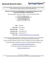
a) levels of total and respirable dust, b) levels of total microorganisms in air samples, c) levels of total microorganisms in surface samples, at the three sampling time-points; Feb 2004, Sep 2004 and March 2005Figure 1
a) levels of total and respirable dust, b) levels of total microorganisms in air samples, c) levels of total microor-
ganisms in surface samples, at the three sampling time-points; Feb 2004, Sep 2004 and March 2005.
0,00
0,20
0,40
0,60
0,80
1,00
1,20
1,40
Feb 2004 Sep 2004 Mar 2005
mg/m3
total
repirable
0,0
1,0
2,0
3,0
4,0
5,0
6,0
Feb 2004 Sep 2004 March 2005
x106/m3
Bacteria
Fungi
0,0
2,0
4,0
6,0
8,0
10,0
12,0
Feb 2004 Sep 2004 March 2005
x104/cm2
Bacteria
Fungi
Journal of Occupational Medicine and Toxicology 2009, 4:10 />Page 6 of 7
(page number not for citation purposes)
our findings even summer stable air can be slightly above
the lower level of 10 ng/m
3
, but the influence on markers
of airway inflammation at this level are unknown. For 1,3-
β-glucan, exposure levels of 20 ng/m
3
has been associated
with lymphocytosis [24], indicating a systemic inflamma-
tory response to this inhaled substance. Our results
showed that both summer and winter stable air clearly
exceeded this level, particularly during winter conditions.
The effect on airway health in people thus deserves closer
evaluation in relation to 1,3-β-glucan, as horses sampled
at the same occasions from this stable had significant up-
regulation of gene expression of inflammatory cytokines
during winter stabling [9].
Objective signs of asthma were found in two out of thir-
teen subjects measured by increased PEF-variability,
which is used clinically to monitor variable bronchial
obstruction [25,26]. One subject had asthma, but was
well controlled by medication, and thus showed no PEF-
variability. Additionally, ECP, as a biomarker of allergic
activity among asthmatic patients, was elevated in three of
the subjects. Only two workers reported work-related air-
way symptoms and two reported partly work-related
symptoms, which could be due to lack of self-awareness l.
There was also a large turnover among the employees
making comparisons over time difficult in the present
study. However, pulmonary function data and measure-
ments of inflammatory markers are consistent and con-
clusions in comparison with clinical data from previous
studies can be made. Compared with white-collar workers
the stable personnel in the present study also had
increased levels of lysozyme in the nasal mucosa [17].
Since lysozyme is a marker of nasal mucosal secretion this
could be a reaction to the relatively high levels of airborne
dust in the stables. Additionally, levels of MPO as a
marker of neutrophil activity were found to be increased
in the nasal mucosa, which could be due to a high expo-
sure to bacteria in straw bedding and horse dung. Albu-
min was increased in only two personnel indicating that
there was no general environmental effect on nasal
mucosal plasma protein leakage among the personnel.
Horse dander is considered a 'strong' human allergen and
since horses are fairly common in Sweden (300 000) and
comparable countries it may be a key trigger of hidden
asthma among stable workers. Our results are similar to
those obtained in the cross-sectional study performed
among grooms in a Hippodrome of Istanbul [4]. Thus,
further investigations, including objective physiologic air-
way measurements among stable workers appear to be
motivated.
Conclusion
We have studied biomarkers involved in the development
of airway diseases among both horses and humans in rela-
tion to environmental exposure levels in equine stables.
Respirable dust and 1,3-β-glucan were increased during
winter stabling conditions, with the latter well above lev-
els reported to induce systemic white blood cell reactions
in people. The compliance of the personnel in completing
diaries was too low to detect work-related effects over
time. However, some employees (3/13) showed signs of
bronchial obstruction, which may be worsened by work-
ing in the stable environment. This study describes altera-
tions in stable air quality, and relates them to findings of
suitable biomarkers to monitor the indoor stable environ-
ment and the personnel. An improved management of the
stable climate will be beneficial for the health of both sta-
ble workers and horses.
Competing interests
The authors declare that they have no competing interests.
Authors' contributions
All authors participated in the design of the study, read
and approved the final manuscript. LE coordinated the
project, performed all the environmental sampling, ana-
lysed some of the samples and evaluated the data and
drafted the manuscript. RW performed the sampling of
the personnel, evaluated the data and drafted part of the
manuscript.
Additional material
Acknowledgements
We thank C-H Bergström and his personnel at Lunda Gård, Uppsala, Swe-
den, for making this study possible. The monoclonal antibodies to horse
Additional file 1
Demographic description and clinical characteristics of stable work-
ers. The data provided describes the age, height, smoking status and lung
function of the stable workers
Click here for file
[ />6673-4-10-S1.doc]
Additional file 2
Results from lung function (PEF) and reported symptoms in stable
workers. The data provided represents mean, SD and V-coefficient of
PEF-values at two time-points as well as reported symptoms.
Click here for file
[ />6673-4-10-S2.doc]
Additional file 3
Nasal lavage markers of inflammation in stable workers. The data pro-
vided are levels of inflammation markers ECP, MPO, Lysozyme and Albu-
min in nasal lavage.
Click here for file
[ />6673-4-10-S3.doc]
Publish with Bio Med Central and every
scientist can read your work free of charge
"BioMed Central will be the most significant development for
disseminating the results of biomedical research in our lifetime."
Sir Paul Nurse, Cancer Research UK
Your research papers will be:
available free of charge to the entire biomedical community
peer reviewed and published immediately upon acceptance
cited in PubMed and archived on PubMed Central
yours — you keep the copyright
Submit your manuscript here:
/>BioMedcentral
Journal of Occupational Medicine and Toxicology 2009, 4:10 />Page 7 of 7
(page number not for citation purposes)
were a generous gift from Mabtech AB (Stockholm, Sweden). Funding for
the project was from the Uppsala City Council.
References
1. Kristiansen JD, Lahoz AX: Riding-school lung? Allergic alveolitis
in an 11-year-old girl. Acta Paediatr Scand 1991, 80:386-388.
2. Mackiewicz B, Prazmo Z, Milanowski J, Dutkiewicz J, Fafrowicz B:
[Exposure to organic dust and microorganisms as a factor
affecting respiratory function of workers of purebred horse
farms]. Pneumonol Alergol Pol 1996, 64(Suppl 1):19-24.
3. McGorum BC, Ellison J, Cullen RT: Total and respirable airborne
dust endotoxin concentrations in three equine management
systems. Equine Vet J 1998, 30:430-434.
4. Tutluoglu B, Atis S, Anakkaya AN, Altug E, Tosun GA, Yaman M: Sen-
sitization to horse hair, symptoms and lung function in
grooms. Clin Exp Allergy 2002, 32:1170-1173.
5. Palmgren U, Strom G, Blomquist G, Malmberg P: Collection of air-
borne micro-organisms on Nuclepore filters, estimation and
analysis – CAMNEA method. J Appl Bacteriol 1986, 61:401-406.
6. Mattsson J: Tape lift versus contact agar plates – evaluation of
analysis methods. Indoor Air: Proceedings of 10th Int Conference on
Indoor Air Quality and Climate 2005:2683-2685.
7. Emenius G, Larsson PH, Wickman M, Harfast B: Dispersion of
horse allergen in the ambient air, detected with sandwich
ELISA. Allergy 2001, 56:771-774.
8. Kim JL, Elfman L, Mi Y, Johansson M, Smedje G, Norback D: Current
asthma and respiratory symptoms among pupils in relation
to dietary factors and allergens in the school environment.
Indoor Air 2005, 15:170-182.
9. Riihimäki M, Raine A, Elfman L, Pringle J: Markers of respiratory
inflammation in horses in relation to seasonal changes in air
quality in a conventional racing stable. Can J Vet Res 2008,
72:432-439.
10. Walinder R, Norback D, Wieslander G, Smedje G, Erwall C, Venge P:
Nasal patency and biomarkers in nasal lavage – the signifi-
cance of air exchange rate and type of ventilation in schools.
Int Arch Occup Environ Health 1998, 71:479-486.
11. Venge P, Hakansson L, Peterson CG: Eosinophil activation in
allergic disease.
Int Arch Allergy Appl Immunol 1987, 82:333-337.
12. Venge P: Soluble markers of allergic inflammation. Allergy
1994, 49:1-8.
13. Raphael GD, Jeney EV, Baraniuk JN, Kim I, Meredith SD, Kaliner MA:
Pathophysiology of rhinitis. Lactoferrin and lysozyme in
nasal secretions. J Clin Invest 1989, 84:1528-1535.
14. Anees W, Gannon PF, Huggins V, Pantin CF, Burge PS: Effect of
peak expiratory flow data quantity on diagnostic sensitivity
and specificity in occupational asthma. Eur Respir J 2004,
23:730-734.
15. The Swedish Work Environment Authority, AFS 2005:17.
Swedish Board of Agriculture; 2003:12.
16. Regulations on farm animal housing DFS 2004:17. Volume 1.
Swedish Animal Welfare Agency; 2004:5. vol. 100. pp. 5
17. Walinder R, Wieslander G, Norback D, Erwall C, Venge P: Influence
of personal factors on nasal patency and lavage biomarkers
in white-collar workers. Rhinology 2000, 38:130-135.
18. Woods PS, Robinson NE, Swanson MC, Reed CE, Broadstone RV,
Derksen FJ: Airborne dust and aeroallergen concentration in
a horse stable under two different management systems.
Equine Vet J 1993, 25:208-213.
19. Rosenthal FS, Gruntman A, Couetil LL: A comparison of total,
respirable, and real-time airborne particulate sampling in
horse barns. J Occup Environ Hyg 2006, 3:599-605.
20. Crichlow EC, Yoshida K, Wallace K: Dust levels in a riding stable.
Equine Vet J 1980, 12:185-188.
21. Bornehag CG, Blomquist G, Gyntelberg F, Jarvholm B, Malmberg P,
Nordvall L, Nielsen A, Pershagen G, Sundell J: Dampness in build-
ings and health. Nordic interdisciplinary review of the scien-
tific evidence on associations between exposure to
"dampness" in buildings and health effects (NORDDAMP).
Indoor Air 2001, 11:72-86.
22. Rylander R, Carvalheiro MF: Airways inflammation among
workers in poultry houses. Int Arch Occup Environ Health 2006,
79:487-490.
23. Rylander R: Evaluation of the risks of endotoxin exposures. Int
J Occup Env Med 1997, 3:32-36.
24. Thorn J, Beijer L, Rylander R: Airways inflammation and glucan
exposure among household waste collectors. Am J Ind Med
1998, 33:463-470.
25. Quanjer PH, Lebowitz MD, Gregg I, Miller MR, Pedersen OF: Peak
expiratory flow: conclusions and recommendations of a
Working Party of the European Respiratory Society. Eur
Respir J Suppl 1997, 24:2S-8S.
26. Global strategy for asthma management and prevention. In
Global Initiative For Asthma (GINA) NHLBI/WHO Workshop Report;
2006.