báo cáo hóa học:" Ergonomic assessment of the posture of surgeons performing endoscopic transurethral resections in urology" docx
Bạn đang xem bản rút gọn của tài liệu. Xem và tải ngay bản đầy đủ của tài liệu tại đây (1.42 MB, 11 trang )
BioMed Central
Page 1 of 11
(page number not for citation purposes)
Journal of Occupational Medicine
and Toxicology
Open Access
Research
Ergonomic assessment of the posture of surgeons performing
endoscopic transurethral resections in urology
Alwin Luttmann*
†1
, Matthias Jäger
†1
and Jürgen Sökeland
1,2
Address:
1
IfADo - Leibniz Research Centre for Working Environment and Human Factors, Ardeystraße 67, 44139 Dortmund, Germany and
2
Urologische Klinik, Städtische Kliniken Dortmund (formerly), Münsterstraße 240, 44145 Dortmund, Germany
Email: Alwin Luttmann* - ; Matthias Jäger - ; Jürgen Sökeland -
* Corresponding author †Equal contributors
Abstract
Background: During transurethral endoscopic prostate and bladder operations the influence of
an ergonomic redesign of the arrangement of the operation equipment - including the introduction
of a video-assisted resection method ('monitor endoscopy') instead of directly viewing onto the
operation area via the endoscope ('direct endoscopy') - was studied with respect to the postures
of the surgeons.
Methods: Postures were analysed on the basis of video recordings of the surgeons performed in
the operation theatre during live operations and subsequent visual posture estimation executed by
an observer. In particular, head, trunk and arm positions were assigned to posture categories
according to a newly developed posture classification schema. 10 urological operations with direct
endoscopy and 9 with monitor endoscopy were included.
Results: Application of direct endoscopy coincides with distinct lateral and sagittal trunk and head
inclinations, trunk torsion and strong forearm and upper arm elevations of the surgeons whereas
operations with monitor endoscopy were performed with an almost upright head and trunk and
hanging arms. The disadvantageous postures observed during direct endoscopy are mainly caused
by the necessity to hold the endoscope continuously in close contact with the eye.
Conclusion: From an ergonomic point of view, application of the video-assisted resection method
should be preferred in transurethral endoscopic operations in order to prevent awkward postures
of the surgeons and to limit muscular strain and fatigue. Furthermore, the application of the
monitor method enables the use of a chair equipped with back support and armrests and benefits
the reduction of postural stress.
Background
Historical development of endoscopic operation methods
in urology
The application of endoscopic operation methods has a
long tradition especially in urology. As early as 1879 opti-
cal endoscopy began when the urologist Maximilian Nitze
demonstrated the first rod-shaped cystoscope equipped
with an optical lens system and an electrical light source
(for historical review of cystoscopy see [1,2]). In the fol-
lowing decades the instruments were improved by intro-
Published: 19 October 2009
Journal of Occupational Medicine and Toxicology 2009, 4:26 doi:10.1186/1745-6673-4-26
Received: 25 June 2009
Accepted: 19 October 2009
This article is available from: />© 2009 Luttmann et al; licensee BioMed Central Ltd.
This is an Open Access article distributed under the terms of the Creative Commons Attribution License ( />),
which permits unrestricted use, distribution, and reproduction in any medium, provided the original work is properly cited.
Journal of Occupational Medicine and Toxicology 2009, 4:26 />Page 2 of 11
(page number not for citation purposes)
ducing light transmitting glass fibres for the illumination
and so-called 'air-lens or rod-lens systems' for the visual
inspection of the operation area.
Since the first application of such a cystoscope in the last
decades of the 19
th
century until the eighties of the 20
th
century, i.e. for about 100 years, only "direct endoscopy"
was applied in urology. Using this method, one of the sur-
geon's eyes looks through the optical lens system directly
into the body and the eye is permanently in contact with
the ocular of the endoscope. In consequence, the head of
the surgeon has to follow all movements of the instru-
ment. From an ergonomic point of view, direct endoscopy
has to be assessed as disadvantageous, since awkward pos-
tures are inevitable, which are at least partially caused by
the construction of the instruments.
For transurethral applications in urology 'resectoscopes'
are used consisting of the endoscope and a wire loop
which is mounted together with the endoscope in the
same shaft. For the dissection of tissue from the bladder or
prostate the wire loop is - under control of a foot-switch -
charged with a high-frequency current and moved
through the tissue. The resectoscope is held with one hand
and the other is used to manipulate the wire loop. The
application of this method results in a 'close coupling'
between the head, the hands, and the endoscope. Since in
direct endoscope the eye has to be permanently in contact
with the instrument, the surgeon has to steeply incline the
upper body for long periods of the operation, in particular
during the resection of tissue from the ventral part of the
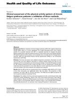
prostate or bladder. A typical posture of the surgeon with
inclined head and trunk while the eye is in close contact
with the resectoscope is shown in the left-hand photo of
figure 1. The performance of transurethral resections
requires high motoric dexterity on the part of the sur-
geons, since even in disadvantageous postures of the
upper body, fine movements of the hands and fingers
have to be executed.
Modern endoscopic instrumentation using video technique
With respect to postural load of the surgeons, improve-
ment is attained, if instead of the direct-view endoscopy
the "monitor endoscopy" is applied. In this method the
visual inspection of the operation area is performed via a
video camera mounted on the top of the endoscope and a
monitor. Such video devices are available since the begin-
ning of the eighties of the last century and meanwhile
Photos of the operating theatre before and after redesignFigure 1
Photos of the operating theatre before and after redesign. Photos illustrating the arrangement of the operating devices
and typical postures of the surgeon before (left) and after (right) ergonomic redesign of the workplace including the introduc-
tion of a video system for the control of the operation area.
Journal of Occupational Medicine and Toxicology 2009, 4:26 />Page 3 of 11
(page number not for citation purposes)
monitor-displayed endoscopy can be assessed to be the
golden standard for transurethral resections [3]. The ergo-
nomic advantage of this method is that the complication
of the close coupling between the eye and the endoscope
is solved and the surgeon's trunk and head can remain in
an almost upright posture during the operations (see
photo in the right-hand part of figure 1).
In the first years after their introduction, the video systems
were used only hesitantly in urology [3]. The delayed
application of the monitor method may at that time be
caused by an insufficient ergonomic arrangement of the
technical devices. In particular, the monitor was often
placed on a movable trolley at the side of the operating
table or was hanging on the ceiling outside the central vis-
ual field of the surgeon. So the visual inspection of the
monitor requires disadvantageous postures with long-
term twisting of the neck and trunk and the arrangement
results in deficiencies in the perception-action compati-
bility [4].
Ergonomic redesign of the urological operation theatre
The study on hand was performed before and after a com-
prehensive redesign of the operation theatre (see right-
hand part of figure 1) and was aimed to evaluate the rear-
rangement with respect to the surgeons' postures. Rede-
sign was performed for several reasons: One reason was to
support the routine application of the video system and to
reduce the load on the musculoskeletal system. A fast and
easy control of all visual readouts including the video
monitor should be possible without turning or twisting
the trunk and head. Furthermore, a flexible arrangement
of the tools and devices needed for transurethral opera-
tion was aspired. The placement of the equipment should
allow to have free access to the patient when performing
various operations. At least, the arrangement including
the operating chair should be adaptable to the individual
anthropometry of the surgeon and the patient. The
demands were fulfilled by applying a horizontal bar fixed
to the ceiling of the operation theatre; all devices includ-
ing the monitor were mounted on racks hanging on the
bar. The racks can slide horizontally on the bar and can be
arranged according to the actual requirements of a special
operation. The monitor position can also be changed in
the horizontal as well as in the vertical direction. In the
vertical direction, positioning between the hand and eye
level is strived for, according to the anthropometry of the
surgeon. Nevertheless positioning above eye level is often
inevitable, since a certain minimum distance of the mon-
itor above the patient's chest has to be maintained. Hori-
zontal fixation of the monitor in the mid-sagittal plane of
the surgeon allows the control of the operation area with-
out turning or twisting the head or trunk and benefits the
hand-eye coordination [5]. A special chair equipped with
adjustable back- and armrests was applied in order to
reduce postural stress further (for details, see [6]).
Posture recording
Even if the advantage of the monitor method seems to be
obvious with respect to ergonomic work design, a quanti-
tative analysis of the postures which surgeons adopted
during the performance of transurethral resections is
missing. Therefore, the aim of this study was to determine
the postures of urologists during the execution of
transurethral operations and, in particular, to compare the
postures during operations with direct and monitor
endoscopy.
For the recording of posture, sophisticated methods were
developed, consisting e.g. of goniometers and inclinome-
ters to measure the inclination and flexion of body seg-
ments (e.g. [7]) or optoelectronic devices using reflective
markers or light-emitting diodes to track relevant body
locations in combination with a set of video- or infrared
cameras. Such methods need high effort in applying the
posture-recording technique and can hardly be adapted to
the specific demands of an application in the operation
theatre during surgical treatments (e.g. to ensure sterility
and to avoid any restrictions of surgical actions). For such
reasons, continuous recording of posture data during real
surgical work was executed rarely. However, the possibil-
ity of such measurements was demonstrated in a pilot
study with optoelectronic posture measurements during
laparoscopic cholecystectomy under live conditions [8].
Similar measurements using optoelectronic devices were
performed during operation-simulation experiments [9].
In another approach, posture was determined for defined
sections of live operations on the basis of sequences of
photos [10] or characteristic long-lasting static postures
were selected using video recordings and assessed using a
computerised man model [11]. The method used in the
study on hand represents an extended version of a posture
estimation procedure applied by Berguer et al. [9]. It is
based on video recordings of the surgeons taped during
live surgical work in an operating room and a visual off-
line estimation of the surgeons' postures performed by an
observer. This posture-estimation method was favoured
in spite of its limited accuracy, since the surgeons activity
was not influenced by the measuring technique and the
procedure was easy to apply under live conditions in the
operating theatre.
Posture documentations were used to evaluate the pos-
tural stress of the surgeons during live surgical work in
urology before and after redesign of the operation theatre
in particular regarding the introduction of modern endo-
scopic instrumentation. It is concluded, that the use of a
video system results in a clear reduction of awkward posi-
Journal of Occupational Medicine and Toxicology 2009, 4:26 />Page 4 of 11
(page number not for citation purposes)
tions of the upper body segments and is therefore recom-
mended for transurethral endoscopic operations.
Methods
Study protocol and subjects
The study was performed before and after an ergonomic
redesign of the arrangement of the operational equip-
ment. Before redesign direct endoscopy was applied only.
More than one year after redesign, investigations regard-
ing monitor endocopy were executed, when the surgeons
were getting well accustomed to the new arrangement and
almost exclusively used the video system.
In total, 5 surgeons (male; age between 34 and 61 years,
mean 45 years; body height between 172 and 190 cm,
mean 182 cm) participated in the study. Analyses were
carried out for 10 operations with direct endoscopy (7
treatments of prostate adenoma; 3 resections of bladder
tumours) and for 9 operations with monitor endoscopy
(5 treatments of prostate adenoma; 4 resections of blad-
der tumours). The analysed operation periods lasted
between 18.1 and 79.4 min (mean 41.1 min) for direct
endoscopy and between 12.8 and 75.6 min (mean 37.8
min) for monitor endoscopy.
Posture recording and categorisation
Video recordings of the surgeons' postures were per-
formed in the operation theatre continuously throughout
the operations. A video camera was mounted in a height
of about 2 1/2 meters on a tripod standing at the right-
hand side behind the surgeon. The camera looked from
above on the surgeon and was focused on the upper part
of the body and the right arm-hand region. The view on
the left arm and hand was often restricted. The analysis of
the video recordings was executed subsequently in the
laboratory by visual inspection of the videos and applying
an encoding system for the description of the consecutive
postures the surgeon has adopted in the course of the
operation. Whenever the surgeon has changed his posi-
tion, the analysing person had to estimate the current
position of the surgeon and to create a numerical code. In
this code, the angular positions of various body segments
were described using a specific classification procedure.
This classification system is based on a method which was
originally developed for the posture evaluation of manual
handling tasks [12]; it was modified and adapted to the
actual problem. An overview of the classification system is
provided in table S1, additional file 1. It is based on a "16-
digit posture code" used to describe the posture of the
trunk (5 digits), head (4 digits) and the right shoulder and
arm (6 digits). Additionally the posture of the lower body
(1 digit) and the line of vision (1 digit) were documented.
The task of the analysing person was to assign the actual
posture of the body segments mentioned in table S1,
additional file 1 into given categories. With regard to sag-
ittal trunk inclination (digit 1) the actual position of the
trunk is classified in steps of 20° between 60° forward
and 20° backward, i.e. the categories '>60°', '40° to 60°',
'20° to 40°', '0° to 20°', 'around 0°', '-20° to 0°' and '<-
20°' are provided. For the category 'around 0°' a span
between about 5° forward and backward is assumed.
Accordingly, 7 expressions were used to quantify the sag-
ittal trunk inclination (see column 5 in table S1, addi-
tional file 1). Similarly, the trunk posture was categorised
in 20°-steps with respect to the lateral inclination and tor-
sion. (For clarification, see also the body-contour draw-
ings in figure 2 illustrating some posture categorisations
exemplarily.) Adoption of a hollow back and the use of
the back support of the chair ('trunk support') were cate-
gorised using two expressions ('yes' or 'no'). The postures
of the head were codified analogously using the digits 6 to
9; for the sagittal and lateral head inclination also 20°-
steps were chosen with the exception of the backward
inclination where categories of '0° to -10°' and '< -10°'
are provided. The positions of the right shoulder and arm
were classified using the digits 10 to 15. The posture of the
lower body was described in digit 16 with only 2 expres-
sions ('sitting' or 'standing'), and digit 17 was applied to
discriminate between different lines of vision and was
used in operations with monitor endoscopy, only. The
application of the encoding system to the 19 operations
considered in this study reveals a mean number of
encoded segment positions of about 7,500 per operation
ranging between 2,500 and 14,700.
The posture categorisation is based on the subjective esti-
mation of an analysing person and not on measurements
using technical sensors. For this reason, a restrained appli-
cation of the method is indispensable and a critical
appraisal is provided in the discussion section. In the con-
ception of the study, some measures are undertaken in
order to limit the risk of errors; e.g. only one person has
performed the posture encoding for all operations in
order to prevent differences in the posture categorisation
caused by different interpretations of various analysing
persons. Furthermore, the analysing person was inten-
sively trained before executing the posture encoding and
the reproducibility of her posture rating was checked.
Results
Example for the time courses of upper-body segments'
positions
The encoding procedure results in a series of consecutive
code numbers representing the time course of the posture
of relevant body segments. In figure 2 examples of the
resulting time sequences are shown graphically for the
head, trunk and upper arm postures during the first 30
minutes of two operations, one with direct endoscopy
(left-hand diagrams) and another with monitor endos-
copy (right-hand diagrams). Additionally, a graphical
Journal of Occupational Medicine and Toxicology 2009, 4:26 />Page 5 of 11
(page number not for citation purposes)
illustration of the posture categorisation is shown at the
left-hand side of the figure using body-contour drawings.
In the upper trace the time courses of the head inclination
in the sagittal plane are presented. Postures were assigned
to only three categories with angles between -10° and
20°. The category '0° to 20°', indicating a slight forward
inclination, was preferable quoted in case of direct endos-
copy, whereas for monitor endoscopy backward inclina-
tions between -10° and 0° were most frequently
mentioned. In the two middle traces in figure 2 the time
courses of the sagittal and lateral trunk inclinations are
indicated. For direct endoscopy, the highest number of
entries was found for sagittal as well as lateral inclinations
in the class of '0° to 20°'. For monitor endoscopy, the
class 'around 0°' was quoted most frequently for both
directions. In the lowest trace of figure 2 the time course
for the right-upper-arm elevation is presented. For both
operation techniques high fluctuations of the elevation
angle can be seen with a larger variation range for direct
endoscopy than for monitor endoscopy.
Summarising results regarding head, trunk and arm
positions
The findings demonstrated exemplarily in figure 2 for sin-
gle surgical treatments were summarised for all analysed
operations. Results are presented in figures 3, 4 and 5 in
the form of histograms for the fractions of time for the var-
ious categories of the head and trunk inclinations as well
as of the elevations of the right upper arm and forearm.
The fractions of time were determined for each operation
as the total time spent in the specific posture categories
and expressed as the percentage of the duration of the
respective operation. The fractions of time were averaged
over all operations and are presented in the figures 3, 4
and 5 together with the respective standard deviations and
the result of the statistical comparison between both oper-
ation methods (t-test).
Time course of posture indicatorsFigure 2
Time course of posture indicators. Examples of the time courses of various posture indicators for the first 30 min of an
operation performed with direct endoscopy (left) and monitor endoscopy (right). The used posture categorisation is illustrated
in the body-contour drawings.
20° to 40°
0° to 20°
around 0°
-20° to 0°
-40° to -20°
20° to 40°
0° to 20°
-20° to 0°
around 0°
> 20°
around 0°
0° to 20°
-10° to 0°
< -10°
> 20°
90°
60°
20°
0°
0°
20°
-10°
0°
20°
40°
-20°
-40°
monitor endoscopydirect endoscopy
sagittal trunk inclination
lateral trunk inclination
sagittal head inclination
0 102030
time in min
right-upper-arm elevation
0102030
60° to 90°
20° to 60°
0° to 20°
> 90°
around 0°
0°
-20°
20°
40°
60°
< -20°
Journal of Occupational Medicine and Toxicology 2009, 4:26 />Page 6 of 11
(page number not for citation purposes)
In figure 3 the histograms of the percentages of time spent
in postures with head inclinations are shown for both, the
sagittal (upper diagram) and lateral direction (lower dia-
gram). For direct endoscopy, the maximum in the fre-
quency distribution exists in the sagittal plane for forward
inclinations of 0° to 20° and, for monitor endoscopy, in
backward direction between -10° and 0°. For both opera-
tion techniques distinct skew frequency distributions were
found; this implies that during direct endoscopy forward
inclinations of between 0° and 20° occurred most often
but were observed rarely above 20°, whereas during mon-
itor endoscopy the highest frequency was found for back-
ward inclinations between -10° and 0° and inclinations
below -10° were avoided. For lateral head inclinations
(lower diagram) the maximum in the frequency distribu-
tion is located in the class 'around 0' for both operation
techniques. The distributions differ, however, in so far, as
the distribution is almost symmetrical for monitor endos-
copy, whereas for direct endoscopy inclinations were
found more often in rightward than in leftward direction.
Histograms for the sagittal and lateral trunk inclinations
are presented in figure 4. For the sagittal plane (upper dia-
gram), differences regarding the location of the maximum
Frequency distributions of head inclinationFigure 3
Frequency distributions of head inclination. Compari-
son of histograms of the fractions of time with head inclina-
tion in the sagittal (above) or lateral plane (below) for
operations with direct and monitor endoscopy - mean and
standard deviation (direct endoscopy: n = 10, monitor
endoscopy: n = 9), * = significantly different (p < 0.05). Speci-
fications of the head inclination angles are provided in the
insets.
sagittal head inclination
<-10° around 0° >20°
0°
-10°
20°
percentage of time
direct endoscopy monitor endoscopy
-10°to 0° 0°to 20°
backward forward
0
10
20
30
40
50
60
70
*
*
lateral head inclination
<-20°
leftward
-20° to 0° around 0° 0° to 20° >20°
rightward
direct endoscopy monitor endoscopy
0°
20°-20°
rightleft
percentage of time
*
*
*
0
10
20
30
40
50
60
70
Frequency distributions of trunk inclinationFigure 4
Frequency distributions of trunk inclination. Compari-
son of histograms of the fractions of time with trunk inclina-
tion in the sagittal (above) or lateral plane (below) for
operations with direct and monitor endoscopy - mean and
standard deviation (direct endoscopy: n = 10, monitor
endoscopy: n = 9), * = significantly different (p < 0.05). Speci-
fications of the trunk inclination angles are provided in the
insets.
sagittal trunk inclination
<-20° -20°to 0° around 0° 0°to 20° 20°to 40° 40°to 60° >60°
60°
40°
20°
0°
-20°
percentage of time
direct endoscopy monitor endoscopy
backward forward
0
20
40
60
80
**
*
lateral trunk inclination
<- 40° -40°to -20° -20°to 0° around 0° 0°to 20° 20°to 40° > 40°
percentage of time
0°
20°
40°
-20°
-40°
direct endoscopy monitor endoscopy
leftward rightward
right
left
0
20
40
60
80
*
*
Journal of Occupational Medicine and Toxicology 2009, 4:26 />Page 7 of 11
(page number not for citation purposes)
were found: During direct endoscopy most situations
coincide with slight forward inclinations in the category
'0° to 20°' and for monitor endoscopy upright positions
with an inclination angle 'around 0°' were preferably cho-
sen. In the lateral plane (lower diagram), the distribution
is almost symmetrical for monitor endoscopy whereas for
direct endoscopy the persons adopted postures with right-
ward inclinations for a higher percentage of time. Besides
the sagittal and lateral trunk inclination its torsion was
evaluated (not shown in the figure). The percentage of
time with trunk torsion amounted to about 10% for direct
endoscopy and was clearly reduced to about 2% when the
monitor method was applied.
In figure 5 the findings regarding the right-arm positions
are summarised. Differences between the operation tech-
niques were found in particular for the right-upper-arm
elevation (upper diagram) where distinct skew frequency
distributions were observed with a maximum in the class
'60° to 90°' for direct endoscopy and of '0° to 20°' for
monitor applications. Also the percentage of time with
right-forearm elevations of more than 20° is clearly
increased during direct endoscopy (lower diagram). Com-
parison of the postures for the different operation meth-
ods included the observation of elbow flexions (not
shown in the diagram). In particular for extreme flexions
of more than 90°, a reduction from about 50% to about
20% was found when the monitor method was applied
instead of direct endoscopy.
As a part of the redesign of the equipment, a newly devel-
oped operating chair equipped with a back support and
armrests was introduced. It was applied in the video-
assisted operations, only. Evaluation of the video record-
ings reveals, that the back support was intensively used
during monitor endoscopy for about 34% of the opera-
tion time whereas the right-hand armrest was used for
only 3% of the time. The newly developed chair was not
applied during the operations with direct endoscopy,
since the back support and the armrests may hamper the
free movement of the trunk and arms which is necessary
to enable the permanent contact between endoscope, eye
and hands.
In the figures 3, 4 and 5 the fractions of the operation
time, i.e. the sum of all time periods with defined postures
in relation to the total operation time, are provided as an
indicator of the postural stress of the surgeons. Besides
these fractions of time found by cumulating the duration
of all time periods with specific postures during an opera-
tion, the mean length of the time periods the surgeons
remained uninterruptedly in the various posture catego-
ries were determined for each operation. The findings
were averaged over all operations with direct and monitor
endoscopy, respectively. Results are presented in table S2,
additional file 2, for the various head and trunk inclina-
tions in the sagittal and lateral directions and for the ele-
vations of the right upper arm and forearm. Additionally,
the ranges for the means found for the single operations
are presented in brackets. Comparison of the mean dura-
tion of the time periods in the various postures table S2,
additional file 2, with the corresponding frequency distri-
butions of the cumulated periods (figures 3, 4 and 5)
yields a clear similarity between both measures of pos-
tural stress; in particular, the maximum values in the dura-
Frequency distributions of right-arm elevationFigure 5
Frequency distributions of right-arm elevation. Com-
parison of histograms of the fractions of time with elevation
of the right upper arm (above) or the right forearm (below)
for operations with direct endoscopy and monitor endos-
copy - mean and standard deviation (direct endoscopy: n =
10, monitor endoscopy: n = 9), * = significantly different (p <
0.05). Specifications of the arm elevation angles are provided
in the insets.
right-upper-arm elevation
around 0° 0° to 20° 20° to 60° 60° to 90° > 90°
60°
90°
20°
0°
direct endoscopy monitor endoscopy
percentage of time
upward
*
*
*
0
10
20
30
40
50
60
direct endoscopy monitor endoscopy
percentage of time
right-forearm elevation
<-20° -20° to 0° around 0° 0° to 20° > 20°
20°
0°
-20°
downward upward
*
0
10
20
30
40
50
60
70
Journal of Occupational Medicine and Toxicology 2009, 4:26 />Page 8 of 11
(page number not for citation purposes)
tions and the time fractions are to be found for the same
postures.
Discussion
Criticism of the method
The posture description in this study is based on an easy-
to-apply method of video recording and a subjective pos-
ture rating based on visual inspections of the screen
images. The use of such a seemingly simple method needs
critical assessment. The advantage of this procedure is
based on the low technical demands and the fact that the
work of the surgeons is not constrained in any way. A pos-
sible disadvantage originates from the limited accuracy of
the posture description which depends, on the one hand,
on the ability of the observer to distinguish between dif-
ferent postures and, on the other hand, on the step-width
of the angles used in the posture categorisation procedure.
When defining the classification steps a compromise has
to be found between these two factors. A relatively high
step-width of 20° was chosen in order to enable visual
inspection. Such a difference of 20° between segment
positions can clearly be differentiated, even if spatial
movements of the body segments have to be analysed and
only a two-dimensional reproduction on a screen is used.
With respect to the precision of the rating it has to be con-
sidered that the evaluation is not focussed on the absolute
inclination angles of the body segments but on the dis-
crimination between the postures in the two workings sit-
uations with direct and monitor endoscopy. Therefore,
the requirements regarding the resolution of the inclina-
tion angles and the need of correct angle values are lim-
ited and a step-width of 20° was assessed as being
sufficient for the classification.
In order to benefit the reproducibility of the categorisa-
tion and to prevent inter-individual differences in the pos-
ture estimation the encoding procedure was executed by
the same person for all operations. This person was inten-
sively trained by multiple executions of analyses of the
same video recordings and checking the reproducibility of
the ratings of identical video sequences. The differences
between the categorisation of identical segment positions
amounted - if at all - to one classification step, only. Inso-
far experiences from previous application of a visual
video-based posture rating [12] are confirmed. The rela-
tively high reproducibility of such a subjective posture rat-
ing is to a high degree based on the off-line encoding of
the video recordings which allows multiple inspections of
the same activity section and a stepwise rating of the pos-
ture of the various body segments.
Postural stress for different operation techniques
The differences in the postures found for the two opera-
tion methods can be explained mainly by the different
handling of the resectoscope in direct and monitor endos-
copy and the necessity for the eye to remain continuously
in contact with the ocular of the instrument when apply-
ing direct endoscopy.
Distinct forward and rightward inclinations of the trunk
and head as well as twisting of the trunk were found in
direct endoscopy, since in this operation technique a close
coupling of instrument, hands and eye is permanently
needed and the trunk and head have to track the endo-
scope movements during the total operation time. There-
fore, awkward postures cannot be avoided, particularly if
tissue has to be removed at the ventral part of the prostate
or bladder. During monitor endoscopy, a clear backward
inclination of the head was observed for considerable per-
centages of time, since the monitor was positioned above
eye level for a part of the surgeons. Positioning at a lower
level was, however, possible only to a limited extent, since
the bottom of the monitor has to stay away from the tho-
rax of the patient in a certain minimum distance. In the
sagittal plane, the trunk and head remained mostly in an
upright position when a monitor system was used and
sideward inclinations - frequently found during direct
endoscopy - were avoided. The angles of upper-arm and
forearm elevations were remarkably higher during direct
endoscopy than during monitor use. In direct endoscopy,
permanent arm elevation is required to hold the endo-
scope for prolonged periods in time in close contact with
the eye, whereas the use of a video system allows to work
with almost hanging upper arms.
In the figures 3, 4 and 5 the results of the statistical com-
parison of the fractions of time spent in the various pos-
ture categories during direct and monitor endoscopy are
indicated. For all body segments and most of the relevant
posture categories a significant reduction of the fraction of
time spent in awkward postures was found for operations
with monitor endoscopy. Thus, the ergonomic improve-
ment of the redesign of the workplace including the intro-
duction of the monitor endoscopy is substantiated.
Some of the standard deviations shown in the figures 3, 4
and 5 are relatively high and were matter of further analy-
ses, in particular, regarding the influence of the body
height of the surgeons. For this purpose the percentage of
time in the various posture categories were determined
separately for the surgeons with a body height below and
above 180 cm (2 and 3 persons, respectively). Significant
difference was found for the percentage of time with back-
ward inclinations of the head during monitor endoscopy
amounting to more than 60% for the smaller persons in
comparison to about 20% for the taller ones. Further sig-
nificant differences were found for the trunk and upper
arm postures during direct endoscopy: The necessity to
hold a permanent contact of the endoscope with the eye
and to grip the instrument with both hands (see the exam-
ple shown in the left-hand photo of figure 1) results for
taller persons in a lateral trunk inclination in the category
Journal of Occupational Medicine and Toxicology 2009, 4:26 />Page 9 of 11
(page number not for citation purposes)
'0° to 20°' for about 25% of the operation time, in com-
parison to less than 10% for the smaller persons. Right-
upper-arm elevation is enhanced for the smaller persons;
the percentage of time with upper-arm elevation above
60° amounts to more than 60% for the smaller persons in
comparison to about 30% for the taller ones. Both, the
long-term sideward inclination of the trunk and the eleva-
tion of the arm are significantly reduced when applying
the monitor technique.
Conclusion of the posture data reveals that the head,
trunk and arm positions are more disadvantageous during
direct endoscopy than during monitor endoscopy. These
findings match results from previous electromyographical
studies in the operation theatre on shoulder and back
muscles of surgeons (right and left m. trapezius, right m.
deltoideus, left m. erector spinae) during the performance
of urological operations [6,13]. Comparison of the myoe-
lectrical activities for the different operation techniques
demonstrates a significant decrease in particular for the
shoulder muscles, when monitor endoscopy is applied
instead of direct endoscopy. In the electromyographical
studies also the occurrence of muscular fatigue was stud-
ied [14]. In case of direct endoscopy for about 80% of the
operations muscular fatigue was verified, whereas for the
application of monitor endoscopy the percentage of oper-
ations with fatigue was reduced to about 42% [15].
The performance of the fine-motoric work of transurethral
tissue resection requires a stable positioning of the resec-
toscope. Steady holding of the instrument by means of the
hands is only possible, if the upper body including the
trunk, shoulders and arms represents a firm mechanical
basis for the manipulations with the wire loop. The stabi-
lisation of the positions of the upper body elements has to
be performed by the muscles spanning the inter-segmen-
tal joints. Fixation of the links between adjacent segments
can effectively be executed by co-contractions of the
respective flexor and extensor muscles. Correspondingly,
continuous muscular activation at a high level has to be
expected. This assumption was confirmed in the afore-
mentioned electromyographical studies [13] indicating
very high activation levels for the trapezius and deltoideus
muscles of up to about 60% of the maximum voluntary
activation during direct endoscopy. In case of direct
endoscopy, activation is enhanced, since the upper body
has to be fixed in constrained postures with increased lat-
eral and sagittal inclinations and long-term arm eleva-
tions up to shoulder height with the hands located in the
height of the eyes, whereas during monitor applications
the endoscope is held with almost hanging upper arms
and the activation level of the trapezius and deltoideus is
significantly lower [6]. Muscular strain was found to
depend on the anthropometry of the surgeons: For sur-
geons with a body height above 180 cm higher muscular
activities were observed in the shoulder region than for
smaller persons [6]. The more disadvantageous trunk
positions of the taller persons mentioned before and, in
particular, the increased time fraction with lateral trunk
inclinations are assumed as a possible reasen, even if the
height of the operation table and the seat of the operating
chair were commonly adjusted according to the individ-
ual anthropometry of the surgeon.
Reduction of postural stress by ergonomic work design
With respect to the reduction of postural stress, a chair
with back support was proven to be a successful tool. Back
support was used for a considerable time of more than
one third of the operation time and the reduction in the
myoelectrical activity of the erector spinae found in the
aforementioned studies may result at least partly from the
use of the back support.
For the armrests, it is difficult to make a quantitative
assessment regarding their benefit. Visual observation of
the surgeons' postures gives the impression that the sur-
geons used the left-hand armrest very often. However, the
time of use could not be quantified exactly in the off-line
analysis of this study, since the left-hand side of the body
was insufficiently displayed in the video recordings and
the posture evaluation of this study was therefore con-
fined to the right-hand body segments. The number of 3%
of the operation time mentioned before for the use of the
right-hand armrest is the only quantitative item deter-
mined in this regard. It is, however, not very meaningful
without further interpretations, since the right hand is
used to resect the tissue by means of the wire loop inserted
in the resectoscope. During the execution of such fine
hand and finger movements, the corresponding arm
should not be posed on the armrest and, consequently,
the right-hand armrest could be used for small periods of
time, only. Even if the time for the use of the left-hand
armrest was not determined quantitatively, its advantage
seems to be obvious, since the left hand is used to stabilise
the position of the resectoscope and the fixation of the
instrument is effectively supported when the left elbow is
placed on the armrest. The aforementioned reduction in
the myoelectrical activity of the left trapezius after the
redesign of the workplace including the use of an opera-
tion chair with armrests may also confirm this statement.
For the posture analyses performed in this study it seems
to be important to consider the sensorimotor lateral pref-
erence of the subjects, i.e. of the eye, hand and foot. In
particular the lateralisation of the eye in combination
with the handedness may influence the postures of the
upper body adopted during the operations [16]. It has to
be assumed that, in particular during direct endoscopy,
the head posture is affected by the eye preference, since
the dominant eye is used to perform such monocular
Journal of Occupational Medicine and Toxicology 2009, 4:26 />Page 10 of 11
(page number not for citation purposes)
tasks. Also the lateralisation of the hand may interact with
the trunk and shoulder posture since the resectoscope is
normally held by the non-dominant hand whereas the
dominant hand is used to manipulate the wire loop and
to perform the fine-motoric work of tissue resection [17].
With respect to the sensorimotor lateral preference the
studied group was consistent: All surgeons used the left
hand to hold the instrument, the right hand to manipu-
late the wire loop and the right eye to contact the aperture
of the endoscope when applying direct endoscopy. There-
fore a possible interference of the results with different lat-
eralisation of the subjects can be excluded in this study. In
general, however, a relationship between the individual
lateralisation status for eye, hand and foot and the pre-
ferred endoscopic operation technique (direct vs. monitor
endoscopy) seems to be relevant according to a nation-
wide survey on about 1350 urologists [16].
Conclusion
From the ergonomic point of view, endoscopic transure-
thral prostate and bladder operations should preferably
be performed using a video-assisted operation method
(monitor endoscopy) instead of directly viewing at the
operation area via the endoscope (direct endoscopy) for
several reasons:
- During direct endoscopy, the percentage of the operation
time and the duration of the periods with disadvanta-
geous postures of the upper body are increased in compar-
ison to monitor endoscopy. In particular, distinct lateral
and sagittal inclinations of the trunk and head as well as
torsion of the trunk are to be found. Furthermore, the
upper arm and forearm are held in clearly elevated posi-
tions for longer periods and larger portions of time.
- In direct endoscopy, long-term trunk and head inclina-
tion as well as arm elevation are inevitable, since perma-
nent contact between the eye and the endoscope is needed
and, in consequence, the head and trunk have to track the
endoscope movements during the total operation time.
- In monitor endocopy, the close coupling between the
endoscope, the eye and the hands is solved and the appli-
cation of the video system allows working in a more
advantageous posture with hanging arms and an upright
trunk and head.
- In direct endoscopy, muscular activity in the shoulder
and back regions is enhanced and clear signs of muscular
fatigue were established for the trapezius muscle in a
former study. A considerable reduction of the myoelectri-
cal activity was observed during monitor endoscopy. Fur-
thermore, the percentage of operations with fatigue of the
trapezius muscle was clearly reduced in comparison to the
application of the direct-endoscopy methodology.
- The use of the monitor method allows the application of
an operating chair equipped with back support and arm-
rests and - in consequence - a reduction of postural stress,
whereas in direct endoscopy the free movement of the
trunk and arm is required and any trunk or back support
is hindering.
Competing interests
The authors declare that they have no competing interests.
Authors' contributions
AL and MJ conceived and designed the study. JS suggested
the study and provided the clinical expertise. AL prepared
the manuscript. All authors have read and approved the
manuscript.
Additional material
Acknowledgements
Thanks to Ute von Hoerner for her collaboration regarding the posture
encoding.
References
1. Wolf R: 100 Jahre Cystoskop. Knittlingen, Wolf 1979.
2. Sökeland J, Schulze H: Entwicklung der Prostataforschung. In
Benigne Prostatahyperplasie Edited by: Sökeland J. Stuttart, New York,
Thieme; 1995:1.1-1.40.
3. Faul P: Video TUR: Raising the gold standard. Eur Urol 1993,
24:256-261.
4. Ehrenstein WH, Arnold-Schulz-Gahmen BE: Perception-action
compatibility and eye-dominance in using visually-displayed
information. In Advances in multimedia and simulation; Human-
machine-interface implications Edited by: Holzhausen KP. Bochum, Ver-
lag der FH Bochum; 1997:478-486.
5. Matern U, Faist M, Kehl K, Giebmeyer C, Buess G: Monitor posi-
tion in laparoscopic surgery. Surg Endosc 2005, 19:436-440.
6. Luttmann A, Sökeland J, Laurig W: Muscular strain and fatigue
among urologists during transurethral resections using
direct and monitor endoscopy. Eur Urol 1998, 34:6-14.
7. Ellegast RP, Kupfer J: Portable posture and motion measuring
system for use in ergonomic field analysis. Ergonomic software
tools in product and workplace design. Stuttgart, Ergon 2000:47-54.
Additional file 1
Table S1 - Encoding system for body segments' positions. System for the
categorisation of body segments' positions including the range of definition
for the various movements and the used step-width and numbers of cate-
gories.
Click here for file
[ />6673-4-26-S1.PDF]
Additional file 2
Table S2 - Duration of body segments' positions. Duration of the time
periods the surgeons remained uninterruptedly in various posture catego-
ries of the head, trunk and right arm; average and range (in brackets) of
the mean durations for the single operations (direct endoscopy: n = 10,
monitor endoscopy: n = 9).
Click here for file
[ />6673-4-26-S2.PDF]
Publish with Bio Med Central and every
scientist can read your work free of charge
"BioMed Central will be the most significant development for
disseminating the results of biomedical research in our lifetime."
Sir Paul Nurse, Cancer Research UK
Your research papers will be:
available free of charge to the entire biomedical community
peer reviewed and published immediately upon acceptance
cited in PubMed and archived on PubMed Central
yours — you keep the copyright
Submit your manuscript here:
/>BioMedcentral
Journal of Occupational Medicine and Toxicology 2009, 4:26 />Page 11 of 11
(page number not for citation purposes)
8. Person JG, Hodgson AJ, Nagy AG: Automated high-frequency
posture sampling for ergonomic assessment of laparoscopic
surgery. Surg Endosc 2001, 15:997-1003.
9. Berguer R, Rab GT, Abu-Ghaida H, Alarcon A, Chung J: A compar-
ison of surgeons' posture during laparoscopic and open sur-
gical procedures. Surg Endosc 1997, 11:139-142.
10. van Det MJ, Meijerink WJHJ, Hoff C, van Veelen MA, Pierie JPEN:
Ergonomic assessment of neck posture in the minimally
invasive surgery suite during laparoscopic cholecystectomy.
Surg Endosc 2008, 22:2421-2427.
11. Vereczkei A, Feussner H, Negele T, Fritzsche F, Seitz T, Bubb H, Hor-
váth ÖP: Ergonomic assessment of the static stress con-
fronted by surgeons during laparoscopic cholecystectomy.
Surg Endosc 2004, 18:1118-1122.
12. Jäger M, Jordan C, Luttmann A, Laurig W, DOLLY Group: Evalua-
tion and assessment of lumbar load during total shifts for
occupational manual materials handling jobs within the
Dortmund Lumbar Load Study - DOLLY. Int J Ind Ergonomics
2000, 25:553-571.
13. Luttmann A, Sökeland J, Laurig W: Electromyographical study on
surgeons in urology, Part I: Influence of the operating tech-
nique on muscular strain. Ergonomics 1996, 39:285-297.
14. Luttmann A, Jäger M, Sökeland J, Laurig W: Electromyographical
study on surgeons in urology, Part II: Determination of mus-
cular fatigue. Ergonomics 1996, 39:298-313.
15. Luttmann A, Sökeland J, Jäger M, Laurig W: Arbeitsgestaltung im
urologischen Operationssaal. In 75 Jahre multidisziplinäre Arbeits-
medizin in Berlin: Arbeitsphysiologie im Wandel Edited by: Caffier G,
Kössler F, Ullsperger P. Bremerhaven, Wirtschaftsverlag NW;
2002:120-138.
16. Siefer A, Ehrenstein WH, Arnold-Schulz-Gahmen BE, Sökeland J,
Luttmann A: Populationsstatistik und Assoziationsanalyse sen-
sumotorischer Seitenbevorzugung und deren Relevanz für
verschiedene berufliche Tätigkeitsfelder. Zbl Arbeitsmed 2003,
53:346-353.
17. Arnold-Schulz-Gahmen BE, Luttmann A, Sökeland J, Ehrenstein WH:
Endoskopische Sichtsysteme und visumotorische Seitenbev-
orzugung. Visualisierung in der Simulationstechnik. Aachen, Shaker
2001:27-35.