báo cáo hóa học:" Clinical report of cervical arthroplasty in management of spondylotic myelopathy in Chinese" pot
Bạn đang xem bản rút gọn của tài liệu. Xem và tải ngay bản đầy đủ của tài liệu tại đây (393.6 KB, 7 trang )
BioMed Central
Page 1 of 7
(page number not for citation purposes)
Journal of Orthopaedic Surgery and
Research
Open Access
Research article
Clinical report of cervical arthroplasty in management of
spondylotic myelopathy in Chinese
Yan Wang*, Xuesong Zhang, Songhua Xiao, Ning Lu, Zheng Wang and
Mi Zhou
Address: Department of Orthopaedic Surgery, the PLA General Hospital, Beijing, China
Email: Yan Wang* - ; Xuesong Zhang - ; Songhua Xiao - ;
Ning Lu - ; Zheng Wang - ; Mi Zhou -
* Corresponding author
Abstract
Objectives: To investigate clinical effects and manual operational point of Bryan cervical disc
prosthesis in Chinese, to observe the stability and range of movement (ROM) post-operatively.
Methods and materials: From 2003,12 to 2005,12, Bryan disc prosthesis replacement applied in
83 cases (102 levels) of cervical spondylotic myelopathy (CSM) after anterior decompression in our
hospital. Clinical (JOA grade and Odom's scale) and radiological (X-ray of flexion, extension; left
and right bending position) follow-up was performed. Systemic radiographic study about stability
and ROM of replaced level post operationally were measured. CT or MRI scans were applied in all
cases to evaluate the signs of the prosthesis deflexion and hetero-ossification in the replaced levels.
Results: At least 12 months follow-up were done in 65/83 of these paients. All of 83 patients were
improved according to Odsm's scale. JOA score increased from average 8.7 to 15.5. There was no
prosthesis subsidence. Replaced segment achieved stability and restored partial of normal ROM
4.73°(3.7°–5.9°) early postoperation and 8.12°(5.8°–13.6°) more than 12 months postoperation in
flex and extension position. No obvious loss of lordosis was found. CT or MRI follow-up shows
position deflexion of the prosthesis metal endplates (<1.5 mm) in 14/77 levels and (1.5~3 mm) in
4/77. heter-ossification was found in the replaced levels only in 2 cases.
Conclusion: Byran cervical disc prosthesis restored motion to the level of the intact segment in
flexion-extension and lateral bending in post-operative images. At the same time, it can achieve
good anterior decompression treatment effect and immediate stability in replaced 1 or 2 levels, and
which is a new choice for the treatment of CSM.
Background
Anterior cervical decompression and fusion have tradi-
tionally been considered one of the most successful of cer-
vical spine surgeries; however, the problems associated
with cervical fusion have been illuminated recently [1,2].
A reduction in range of motion and increasing stress at
adjacent levels are commonly accepted as pitfalls of
fusion surgery. Motion in the cervical spine occurs at both
the interbody. Fusion techniques typically aim to stop
motion at either one or both of these regions. Loss of
motion either anteriorly or posteriorly can lead to subse-
quent ankylosis at the other motion center [3].
Published: 04 November 2006
Journal of Orthopaedic Surgery and Research 2006, 1:13 doi:10.1186/1749-799X-1-13
Received: 17 August 2006
Accepted: 04 November 2006
This article is available from: />© 2006 Wang et al; licensee BioMed Central Ltd.
This is an Open Access article distributed under the terms of the Creative Commons Attribution License ( />),
which permits unrestricted use, distribution, and reproduction in any medium, provided the original work is properly cited.
Journal of Orthopaedic Surgery and Research 2006, 1:13 />Page 2 of 7
(page number not for citation purposes)
Cervical disc replacement became favorable in recent
years. Several studies on the effectiveness of Bryan cervical
disc prosthesis in treating radiculopathy, spondylotic
myelopathy have been conducted in Europe and Aus-
tralia. But to date, no study on the clinical result of Bryan
cervical disc prosthesis in China has been reported. No
systematic radiographic study among Chinese about the
post operation result on range of motion (ROM) of
replaced level or stability of the prosthesis has been
reported either. In order to find out the clinical result of
CSM of which among Chinese, a prospective but non-ran-
dom clinical and radiographic study was conducted in our
hospital (one of the largest orthopaedic department in
China). The main purpose of this study is to find out the
treatment result and key points in the operation proce-
dure of Bryan cervical disc prosthesis among Chinese. The
post-operation stability and range of motion were also
measured.
Methods
Patient sample
83 patients (102 levels) with CSM were selected for this
study. Detailed neurological examination and JOA (Japa-
nese Orthopaedic Association Scale; JOA) grading were
conducted to ensure that all the patients are qualified for
anterior cervical decompression in the space level. MRI
scan indicates that all patients have spinal cord compres-
sion with 1 or 2 segments. No instability or hypermobility
was identified by flexion and extension cervical x-rays.
The 83 patients were not randomly selected, but as a con-
tinuous group. Before the operation, cervical arthroplasty
had been explained to all patients, and were chosen by
those patients as treatment procedure.
Study conduction
Between December, 2003 and December, 2005, a group
of 4 surgeons performed the surgery for 83 patients (102
levels) with CSM. Before each operation, detailed clinical
examination and JOA grading were conducted. All of the
83 patients were qualified for the cervical arthroplasty
standard presented by Doctor Bryan and Sekhon.
1,3,6,12,24 months follow-up were done in all patients.
Among the 83 patients, 65 cases finished at least 12
months follow-up (12 to 28 months). Systematic radio-
graphic study about the stability and ROM of replaced
level was performed. Clinical (JOA grade and Odom's
scale) and radiological (X-ray of flexion, extension; left
and right bending position) were conducted. Computer-
Tomographie (CT) or Magnetic Resonance Imaging (MRI)
scan were applied to find out the right place of prosthesis
and hetero-ossification in the replaced levels (Figures 1, 2,
3, 4).
Surgical techniques
The patients were placed supine on a fluoroscopic imag-
ing table with their arms at their side. Shoulders taped to
allow for fluoroscopic imaging, which were obtained in
anterior-posterior and lateral planes to determine the
level of diseased disc. and the shoulders were pulled cau-
dally by a heavy bandage (2 kg), when necessary, to secure
a clear lateral visualization of the lower cervical spine. A
standard right-sided approach was undertaken to access
the anterior cervical spine. A set of proprietary surgical
retractor system, Medtronic Sofamor Danek, Memphis
TN, was used to assist in exposure. After initial discectomy
to achieve wide exposure, we used a gravitational referenc-
ing system to establish a virtual axis in the intervertebral
disc space to help us position a milling fixture. This mill-
ing fixture controls the location and movement of the
powered cutting instruments for preparing the vertebral
endplates that will be replaced by the prosthesis. To our
experience, if we keep the angle of tunnel instruments 2~3
degrees more than the measured angle, both the up and
lower milled endplates will be exactly parallel to the ver-
tebrae space. This also makes the geometry of the shell's
convex outer surface match the milled endplates better,
and consequently, the prosthesis is captured inside a ridge
of bone. This tight fit provides immediate stability.
Post-operation patient care
All patients were required to wear hard collar for 1 week
(wound healing period), and then freed from assistant
support. Clinical and radiological follow-ups were carried
out in 1,3,6,12,24 months postoperationly.
Results
Among these 83 patients, 43 were males, 40 were females.
The average age is 48.9 years old (range: 34~58 Yrs). No
motor neuron disease was found to any of the patients in
this group. The cervical disc prosthesis pattern number is
confirmed by CT-scans. (Please see Table 1 for details).
On average, each operation lasted 128 min (range 90–185
min). Neither neurological nor vascular complication was
identified during or after the surgeries. According to the
Odsm's scale, all of 65 patients (77 levels) followed up at
least 12 months improvement (47/65 excellent, 18/65
good). Average JOA score of the 65 patients increased
from 8.7 to 15.5 at last follow up. No prosthesis subsid-
ence or excursion was identified. Replaced segment
achieved stability and partially restored normal ROM
(4.73° (3.7°–5.9°) in 3 months postoperationly and
8.12° (5.8°–13.6°) more than 12 months postopera-
tionly in flexion and extension position, and 3.42° (2.3°–
4.4°), 3.16° (2.5°–4.1°) in 3 months postoperationly
and 5.08° (3.8°–6.25°), 4.87° (3.2°–7.1°) more than 12
months postoperationly in left and right bending posi-
tion). No obvious loss of lordosis was found. Result from
Journal of Orthopaedic Surgery and Research 2006, 1:13 />Page 3 of 7
(page number not for citation purposes)
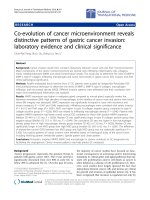
53 y, female, CSMFigure 1
53 y, female, CSM. Preoperative anteroposterior and lateral x-rays show straight alinement and degeneration. Lateral flexion/
extension x-rays show the range of motion before operation. And sagittal MRI T2 show that extensive disc degeneration in
cervical disc. Hight loss of disc space and spinal cord compression can be found in C5/6.
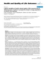
24 months postoperative anteroposterior (A) and lateral (B) x-rays after disc replaced at C5/6, Lateral flexion/extension x-rays(C, D) show restored ROM, and no prosthesis excusion or subsidenceFigure 2
24 months postoperative anteroposterior (A) and lateral (B) x-rays after disc replaced at C5/6, Lateral flexion/extension x-
rays(C, D) show restored ROM, and no prosthesis excusion or subsidence.
Journal of Orthopaedic Surgery and Research 2006, 1:13 />Page 4 of 7
(page number not for citation purposes)
CT or MRI follow-up shows position deflexion of the
prosthesis metal endplates (<1.5 mm) in 14/77 levels and
(1.5~3 mm) in 4/77. Heter-ossification was found in the
replaced levels only in 2 cases.
Complication
Complication following insertion may be divided into
those specific to the prosthesis and those associated with
anterior cervical discectomy. No cerebrospinal fluid leak
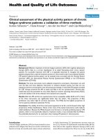
34 y, male, Preoperative anteroposterior and lateral x-rays show loss of the lordosisFigure 3
34 y, male, Preoperative anteroposterior and lateral x-rays show loss of the lordosis. And sagittal MRI T2 show that spinal cord
compression secondary to disc herniation in C4/5, C5/6. Disc degeneration exists in C6/7, but no anterior compression in
coronal plane.
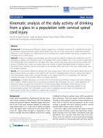
24 months postoperative anteroposterior and lateral x-rays after disc replaced at C4/5,5/6, Lateral flexion/extension x-rays show restored normal ROMFigure 4
24 months postoperative anteroposterior and lateral x-rays after disc replaced at C4/5,5/6, Lateral flexion/extension x-rays
show restored normal ROM. CT scan show no prosthesis excusion or heteostification.
Journal of Orthopaedic Surgery and Research 2006, 1:13 />Page 5 of 7
(page number not for citation purposes)
or wound hematomas in this group, but 1 case of esopha-
geal injury occurred, though 1 week of nasogastric feeding
tube and regional drainage, recoverd successfully. No no
end plate of Bryan cervical disc migration, 1 case of failure
to maintain motion because of spontaneous fusion in
replaced segment. No cases of subsidence of device fail-
ure. 2 case of worsened cervical regional kyphosis in 9
cases of with preoperative focal kyphosis at operated seg-
ments.
Discussion
Anterior single-level discectomy and/or intervertebral dis-
cdecompression and fusion is a common treatment for
symptomatic intervertebral cervical spondylosis with
myeloradiculopathy., in combination with/without inter-
nal fixation is still the most widely accepted treatment for
CSM today. This method has significantly improved the
treatment result in comparison with the traditional
method of posterior decompression. Although it
improves pathologic recovery of spinal cord, but fusion
leads to cervical ROM loss and increases load to adjacent
levels, which maybe will accelerate degeneration of adja-
cent segments. Hilibrand [4] et al reported that 2.9%
patients need additional surgical treatment each year due
to complications with adjacent segments after anterior
interbody fusion with cervical intervention. Due to the
high complication rate and adverse effect on adjacent seg-
ments by cervical interbody fusion, a new technique that
keeps advantages of anterior decompression while main-
taining the impaired motion level for cervical spondylotic
disease is required. Cervical disc prosthesis offers a new
method for CSM treatment, especially for patients of sin-
gle level [5]. Such adjacent segment degeneration may be
preventable if spinal motion can be maintained by a func-
tional disc prosthesis.
Disc replacement attempts started in the 1950s. In year
1966, Fernstorm [6] tried a method by placing a stainless
steel ball into lumber and cervical disc center. Although
his attempt did no harm to the patients, there was either
no positive treatment effect or the treatment result only
lasted for a short period of time. In 1990s, the invention
of cervical disc prosthesis makes disc replacement a viable
treatment method for CSM disease. Different types of
prosthesis, such as Prestige, Bryan, and ProDisc-C were
introduced. It is reported that approximately 6,000
patients have been treated with the Bryan disc in Europe
and North America until 2005. In 2002, Goffin [7] et al
reported for the first time that Bryan Disc (Spinal Dynam-
ics Corp.) was successfully used for treatment of cervical
radiculopathy, and achieved 85–90% satisfactory rate in
12 months follow-up. The next year, Sekhon [8] reported
that Bryan Disc is used for treating 7 patients with CSM
(5/7 single level and 2/7 adjacent two levels). According
to Odom's criteria, the result from follow-up between 1
and 17 months after operation shows 5/7 excellent and 2/
7 good. All the replaced levels restored motion, but loss of
lordosis took place in 4/7. Bryan [9] reported that a total
of 97 prosthesis were implanted in patients with single
level degenerative disc disease (DDD). Among the 97
patients, 49 were given 12 months follow-up and the
result is excellent: 70%, good: 4%, fair: 13%, poor: 13%
according to Odom's criteria. 10 patients had finished 24
months follow-up, and similar result was achieved.
Gwynedd [10] et al further studied biomechanical profile
of the cervical spine following 20 cases of cervical arthro-
plasty. They found that the overall cervical motion (C2–
C7) was moderately but significantly increased during late
follow-up. Sagittal rotation, anterior and posterior disc
height, translation, and center of motion coordinates did
not change significantly following surgery.
The Bryan Cervical Disc prosthesis consists of a low-fri-
tion poluurethane nucleus surrounded by a polyurethane
sheath and siuated between two titanium alloy shells. The
biarticulating metal-on-polymer disc possesses elasticity
and little compressibility and allows for unconstrained
motion and translation through normal ROM. The pros-
thesis is axially symmetric, allowing for similar ROM in
sagital plane motion and in lateral bending. Biomechani-
cal studies suggest a mobile center of rotation, allowing
the device to accommodate a range of preoperative center-
of-rotation values without subjecting the facets and liga-
ments to abnormal stresses [11]. A cohort of 20 patients
using quantitative motion analysis software to analyze
intervertebral motion, reaching the conclusion that sagit-
tal ROM, centers of rotation, horizontal translation, and
disc height were not significantly altered following disc
replacemt compared with the preoperative state [12]. So
the right place of the prosthesis is important, because that
abnormal shifting of the center of rotation following spi-
nal arthroplasty has been implicated in recent reports of
facet pain associated with prosthesis positioning[13]. A
functional disc prosthesis may be able to restore the func-
tional spine unit and prevent subsequent adjacent seg-
ment degeneration.
Table 1: All of 65 patients (77 levels) were adopted the cervical
disc replacement, the static of cervical disc prosthesis in the
replaced levels.
level Patient
C3/4 7
C4/5 19
C5/6 30
C6/7 8
C3/4, C4/5 3
C4/5, C5/6 9
C3/4, C5/6 5
C4/5, C6/7 2
Journal of Orthopaedic Surgery and Research 2006, 1:13 />Page 6 of 7
(page number not for citation purposes)
As described above, cervical arthroplasty has been an
alternative to cervical anterior discectomy with fusion. At
present, the short-term results are promising [14]. But to
date, no study on the clinical result of Bryan cervical disc
prosthesis among chinese patients has been reported, and
no systemic radiographic study about immediate stability
and short-term ROM after the operation has been
reported in China either. Does the prosthesis's design (the
same hight of 8.5 mm of each size prosthesis) matches
anatomic character of Chinese, especially in whose cervi-
cal vertebra is small? In 2 cases whose vertebra was small,
interspace is also narrow, when inserting the 8.5 mm
space enlarger, the lower vertebra leaked, and alignment
of posterior corner of vertebra which was in a straight line
just adjusted before operation, changed to angled, so the
milled track was not as the designed one, Which caused
adverse open of prosthesis space. In this group of 83 cases
(102 levels), one independent radiologist assessed all
radiographs. No subsidence and migration was observed
from X-ray photography and no obvious kyphosis was
found to any of the patients either. On the contrary, 2
patients, who had cervical kyphosis misalignment before
the operation, gained normal cervical lodorsis after disc
replacement. In order to be comparable to patient selec-
tion in Europe or North America [15,16], only CSM
patients with single or adjacent or jump 2 levels were cho-
sen this group. The prosthesis restores height of degener-
ated disc space and relieves the spasm of cervical muscle.
Post-operation Cobb angles for flexion/extension and lat-
eral bending of the function segment unit (FSU) at the
implant level indicates improved ROM. At last follow up
of those finished at least 12 months follow up, all of 65/
83 cases achieved flexion/extension ROM above 8.12
degrees in replaced site, and left/right lateral bending
ROM ranged between 5.08 and 4.87 degrees. According to
Odom's criteria, the result of those finished at least 12
months follow up after operation of the 65 patients
achieved 47/65 excellent and 18/65 good. To our experi-
ence, if we increase 2–3 degrees comparing to the meas-
ured angle, the position of the prosthesis will be parallel
to introvertebra space maybe due to the more curved
lower endplate of cervical vertebra among patients in
most Chinese. This finding requires additional anatomic
study on cervical vertebra in Chinese to confirm. Result
from CT or MRI shows that 14/77 levels with deflexion of
prosthesis less than 1.5 mm and 4/77 level with deflexion
of prosthesis between 1.5 mm and 3 mm, nonsteonotid
drug were not taken as routine postoperatively in this
group. Ossification in the replaced levels was identified
just in 2 cases. Rotation of cervical alignment in the oper-
ation bed during operation is the potential reason for
excursion of prosthesis.
Choosing appropriate prosthesis size and insertion posi-
tion is critical to the success of the operation. Details in
these procedures will not only affect instant stability of
prosthesis but also alter the axis of rotation and load shar-
ing [17-19]. Surgeon need to make adjustments according
to variations in anatomic structure and degenerative con-
ditions in different patients. A potential contraindication
of the cervical arthroplasty with metal endplates is oste-
oporosis which can potentially increase the risk of end-
plate subsidence into the vertebral bodies [20]. The long-
term impact of bone loss with aging due to subsidence of
metal endplates requires additional studies. A forward
long-term study with multi-center, randomly selected
patients among patients in Asia is to be carried out in our
hospital.
Conclusion
CT and MRI images more than 24 months post operation
indicate Byran cervical disc prosthesis partly restored
motion of the impaired segments in flexion-extension
and lateral bending while maintaining the advantage of
anterior decompression. Selecting appropriate prosthesis
size and insertion position is critical to treatment result. In
summary, cervical arthroplaty of Bryan disc can achieve
good anterior decompression treatment effect and stabil-
ity in replaced 1 or 2 levels, which makes it a new choice
for the treatment of CSM in Chinese.
References
1. Frank MP, Steven RG: Cervical Disc Replacement. Spine 2005,
30(17s):s27-s33.
2. Pickett GE, Sekhon LH, Sears WR, Duggal N: Complications with
cervical arthroplasty. J Neurosurg Spine 2006, 4(2):98-105.
3. Lali HS, Sekhon : Reversal of Anterior Cervical Fusion with a
Cervical Arthroplasty Prosthesis. J Spinal Disord Tech 2005,
18(Suppl 1):s125-128.
4. Hilibrand AS, Carlson GD, Palumbo MA, Jones PK, Bohlman HH:
Radiculopathy and myelopathy at segments adjacent to the
site of a previous anterior cervical arthrodesis. J Bone Joint Surg
(Am) 1999, 81:519-528.
5. Mayer HM, Wiechert K, Korge A, Qose I: Minimally invasive total
disc replacement: Surgical technique and preliminary clini-
cal results. Eur Spine J 2002, 11(suppl 2):124-130.
6. Fernstrom U: Arthroplasty with intercorporal endoprosthesis
in herniated disc and in painful disc. Acta Chir Scand 1966,
357(suppl):154-159.
7. Goffin : Proceedings of the Annual Meeting of the Spinal Arthroplasty Soci-
ety, May 2002, Montpellier, France .
8. Sekhon LH: Cervical arthroplasty in the management of
spondylotic myelopathy. J Spinal Disord Tech 2003,
16(4):307-313.
9. Bryan VE: Cervical motion segment replacement. Eur Spine J
2002, 11(Suppl 2):S92-97.
10. Azmi H, Schlenk RP: Surgery for postarthrodesis adjacent-cer-
vical segment degeneration. Neurosurg Focus 2003, 15:1-16.
11. Wigfield C, Gill S, Nelson R, Langdon I, Metcalf N, Pobertson J: Influ-
ence of an artificial cervical joint compared with fusion on
adjacent-level motion in the treatment of degenerative cer-
vical disc disease. J Neurosurg 2002:17-21.
12. Cunningham BW, Gordon JD, Dmitriev AE, Hu N, Mcafee PC: Bio-
mechanical evaluation of total disc replacement arthro-
plasty: an in vitro human cadaveric model. Spine
2003,
28:S110-7.
13. Gwynedd EP, Jeffrey PR, Neil D: Kinematic: Analysis of the Cer-
vical Spine Following Implantation of an Artificial Cervical
Disc. Spine 2005, 30(17):1949-1954.
14. John BP, Vincent CT: Treatment of the Painful Motion Seg-
ment: Cervical Arthroplasty. Spine 2005, 30(16s):s23-s32.
Publish with BioMed Central and every
scientist can read your work free of charge
"BioMed Central will be the most significant development for
disseminating the results of biomedical research in our lifetime."
Sir Paul Nurse, Cancer Research UK
Your research papers will be:
available free of charge to the entire biomedical community
peer reviewed and published immediately upon acceptance
cited in PubMed and archived on PubMed Central
yours — you keep the copyright
Submit your manuscript here:
/>BioMedcentral
Journal of Orthopaedic Surgery and Research 2006, 1:13 />Page 7 of 7
(page number not for citation purposes)
15. Wigfield CC, Gill SS, Nelson RJ, Metcalf NH, Roberson JT: The new
Frenchay artificial cervical joint: results from a two-year
pilot study. Spine 2002, 27(22):2446-2452.
16. Cummings BH, Robertson JT, Gill SS: Surgical experience with an
implanted artificial cervical joint. J Neurosurg 1998, 88:943-948.
17. Wiffield CC, Skrzypiec D, Jackowski A, Adams MA: Internal stress
distribution in cervical intervertebral discs: the influence of
an artificial cervical joint and simulated anterior interbody
fusion. J Spinal Disord Tech 2003, 16(5):441-449.
18. DiAngelo DJ, Roberston JT, Metcalf NH, McVay BJ, Davis RC: Bio-
mechanical testing of an artificial cervical joint and an ante-
rior cervical plate. J Spinal Disord Tech 2003, 16(4):314-323.
19. Pointillart V: Cervical disc prosthesis in humans: First failures.
Spine 2001, 26:E90-92.
20. Scott DB, Richard AB, John GH, Hanley EN Jr, Zigler JE: Disc
Replacements: This Time Will We Really Cure Low-Back
and Neck Pain? J Bone Joint Surg (Am) 2004, 86:411-423.