báo cáo hóa học:" Microdecompression for lumbar synovial cysts: an independent assessment of long term outcomes" pdf
Bạn đang xem bản rút gọn của tài liệu. Xem và tải ngay bản đầy đủ của tài liệu tại đây (300.69 KB, 5 trang )
BioMed Central
Page 1 of 5
(page number not for citation purposes)
Journal of Orthopaedic Surgery and
Research
Open Access
Research article
Microdecompression for lumbar synovial cysts: an independent
assessment of long term outcomes
Bradley K Weiner*
1
, Joel Torretti
2
and Michael Stauff
3
Address:
1
Division of Spinal Surgery The Methodist Hospital 6550 Fannin, Suite 2500 Houston, Texas 77030, USA,
2
Department of Orthopaedics
Dartmouth Hitchcock Medical Center Hanover, New Hampshire, USA and
3
Penn State College of Medicine Hershey, Pennsylvania, USA
Email: Bradley K Weiner* - ; Joel Torretti - ; Michael Stauff -
* Corresponding author
Abstract
Background: Outcomes of surgical intervention for lumbar synovial cysts have been evaluated in
the short and intermediate term. Concerns regarding cyst recurrence, the development of late
instability at the involved level, and instability/stenosis at adjacent levels (when concomitant) fusion
is performed suggest that long term follow-up is needed. This study aims to fill that void.
Methods: Forty-six patients operated by a single surgeon not involved in the study were followed
up long term at an average of 9.7 years (range 5 to 22 years) post-operatively. All patients
underwent decompression (+/- concomitant arthrodesis in the presence of associated
degenerative spondylolisthesis) using the operative microscope for magnification/illumination.
Outcomes were assessed using a customized questionnaire evaluating: relief of pain/claudicant
symptoms, numbness/parasthesias, and weakness; as well as late onset low back pain, new radicular
symptoms, need for additional surgery, and patient satisfaction. Outcomes in patients with or
without fusion were compared as well.
Results: 87% of patients noted resolution of their pre-operative pain, numbness, and weakness.
28% of patients developed late onset low back pain. 17% developed late onset radicular symptoms
in a new nerve root distribution. 15% required subsequent additional surgery. 89% of patients were
satisfied with the surgical outcome. No differences were found for any outcome measure between
patients undergoing concomitant fusion and those undergoing decompression alone using the two-
sample t-test.
Conclusion: This study provides outcome data at an average of nearly ten years post-operative.
This information should allow surgeons to provide realistic expectations for their patients
regarding outcomes and should enhance the informed consent and surgical decision-making
process.
Background
Although originally recognized in peripheral joints by
Baker in 1877[1,2], synovial cysts of the lumbar facet
joints were not described until 1950 in the German litera-
ture [3,4] and were first well-delineated in English by Kao
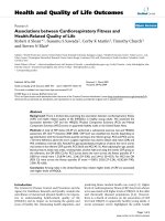
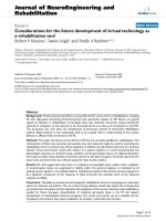
in the late 1960's/early 1970's [5,6]. Since then, CT and
MRI scanning (Figure 1) have afforded highly sensitive
and specific diagnosis of the cysts and the oft-associated
Published: 3 April 2007
Journal of Orthopaedic Surgery and Research 2007, 2:5 doi:10.1186/1749-799X-2-5
Received: 10 October 2006
Accepted: 3 April 2007
This article is available from: />© 2007 Weiner et al; licensee BioMed Central Ltd.
This is an Open Access article distributed under the terms of the Creative Commons Attribution License ( />),
which permits unrestricted use, distribution, and reproduction in any medium, provided the original work is properly cited.
Journal of Orthopaedic Surgery and Research 2007, 2:5 />Page 2 of 5
(page number not for citation purposes)
compression of neurological structures. Such compres-
sion can result in radiculopathy, neurogenic claudication,
and, rarely, cauda equina syndrome [7-11].
While multiple non-operative therapies have been imple-
mented [12-16], few have demonstrated significant or
lasting efficacy when used to treat patients with moderate
or severe symptoms such as intractable pain or neurolog-
ical deficit[12,14,16,17]. Accordingly, surgical interven-
tion is commonly performed on patients in this group
and several studies have demonstrated reasonable out-
comes at short to intermediate term follow-up
[7,11,15,18-23]. The longest follow-up published prior to
the current study has been forty months and concerns
about recurrence of the cysts, instability at the involved
level (when isolated decompression is undertaken), or
instability at adjacent levels (when concomitant fusion is
performed) suggest that a longer-term look is needed to
better understand the implications of our interventions.
The purpose of this study was to independently evaluate
the long-term clinical outcomes in patients who under-
went microdecompression with or without concomitant
arthrodesis for symptomatic lumbar synovial cysts unre-
sponsive to non-operative measures. The average follow-
up of 9.7 years (range five to 22 years) represents the long-
est follow-up to date; the minimum follow-up for inclu-
sion of five years being greater than the previously
reported maximum follow-up of 3.25 years.
Methods
Surgical Technique
Patients were placed under general endotracheal anesthe-
sia and placed in a kneeling position on a standard frame.
The involved level(s) was marked preoperatively using c-
arm imaging. A midline skin incision was made and the
dorsolumbar fascia incised just lateral to the midline ipsi-
lateral to the synovial cyst. Unilateral laminae were
exposed using the Cobb elevator to the mid-portion of the
facet joint. An intraoperative radiograph was used to con-
firm the level. A laminotomy on the undersurface of the
cephalad lamina was undertaken to mirror the cephalad
extent of the cyst as determined by pre-operative MRI. A
similar caudal laminotomy was performed, again to mir-
ror the extent of the cyst. Ligmantum flavum was then
excised and the subarticular and foraminal zones decom-
pressed via complete excision of soft-tissue/bony stenos-
ing lesions to include extirpation of the synovial cyst. If
the cyst was adherent to the dura (a common finding), it
was carefully teased free so that no cyst pseudocapsule
remained. The facet joint was opened and residual syno-
vial tissue excised. If the patient had neurogenic claudica-
tion, a contralateral microdecompression as previously
described[24] was undertaken. If the patient had an asso-
ciated degenerative spondylolisthesis, bilateral uninstru-
mented intertransverse fusion as well as facet joint fusion
was undertaken as previously described[25]. Magnifica-
tion/illumination was provided by the operative micro-
scope in all cases. The wound was irrigated, hemostasis
obtained, and closure carried out in standard fashion.
Patients (Table 1)
Forty-six patients operated between 1984 and 2001 were
available for follow-up. All surgeries were performed by a
single surgeon who was not involved in the study. Age at
surgery ranged from 25 to 96 years with a mean of 73
years. Twenty-nine were females and seventeen males.
Twenty-eight cysts were at the L4-L5 level, eight at L5-S1,
six at L3-L4, and one each at L1-L2 and L2-L3. Clinical
syndromes included unilateral monoradiculopathy in
eighteen patients and neurogenic claudication in twenty-
eight. Radiographically, twenty-three had an associated
degenerative spondylolisthesis and underwent concomi-
tant arthrodesis. This was the only indication for fusion in
the study population. This follow-up study was approved
by the institutional review board and oral consent was
obtained from all participants.
Data
Clinical outcomes and patient satisfaction were assessed
by two independent spine surgeons using the question-
naire in Table 2. All forty-six patients responded.
A typical case of synovial cyst at L5-S1Figure 1
A typical case of synovial cyst at L5-S1.
Journal of Orthopaedic Surgery and Research 2007, 2:5 />Page 3 of 5
(page number not for citation purposes)
Results (Table 3)
Follow-up averaged 9.7 years with a range of five to 22
years.
Same-Site Pain/Similar Symptoms
Forty of the forty-six patients (88%) reported relief of their
preoperative pain/symptoms. Six (12%) had persisting
complaints ranked in severity at an average of 5.5 on the
visual analog scale (VAS: range 2–10) versus an average of
9 on VAS preoperatively. All patients had pain or claudi-
cation preoperatively.
Same-site Numbness
Twenty-three patients (50%) reported preoperative
numbness. Of these, at follow-up, twenty (87%) reported
complete or near-complete resolution, two (9%) were the
same, and one (4%) was worse.
Same-Site Weakness
Nineteen (41%) had complained of weakness prior to sur-
gery. Of these, at follow-up, sixteen (84%) reported com-
plete resolution, two (11%) reported no significant
change in strength, and one (5%) was worse than preop-
eratively.
New Back Pain
After initially doing well, thirteen patients (28%) reported
the eventual development of new back pain ranked on
average at 7.5 on the VAS.
New Leg Pain
Eight patients (17%) reported the eventual onset of new
radicular leg pain (different root involved) with a mean
VAS severity of 7.4.
Additional Surgery
Seven patients (15%) reported the need for additional
lumbar spine surgery. Three patients who had not under-
gone fusion at the initial surgery required eventual revi-
sion decompression and fusion to include the operated
levels due to instablility. Four patients who had under-
gone concomitant arthrodesis at the primary surgery due
to presence of a degenerative spondylolisthesis eventually
developed juxtafusional stenosis/instability requiring sec-
ondary decompression and fusion at involved adjacent
levels.
Patient Satisfaction
Forty-one patients (89%) reported overall satisfaction
with the outcome of their initial procedure and would rec-
ommend it to a friend with the same problem.
Table 2: Questionnaire
1. Do you have numbness or tingling in your leg(s) similar to what you had before surgery? (Better, Same, Worse)
2. Do you have weakness in your leg(s) similar to what you had before surgery? (Better, Same, Worse).
3. Do you still have pain/symptoms in the same site that made you have surgery in the first place? (Rated on Visual Analog Scale)
4. Have you developed back pain over the years that is new/different than before surgery? (Rated on Visual Analog Scale)
5. Have you developed leg pain over the years that is new/different than before your surgery? (Rated on Visual Analog Scale)
6. Have you had additional surgery on your back? (Type of surgery, Reason for surgery)
7. Are you happy with the results of the surgery and would you recommend it to a friend with the same problem?
Positive responses on questionnaire were then followed up for specific details via telephone interview.
Table 1: Patient Characteristics
Number 46
Age in years 73 (range 25–96)
Sex 29 Females 17 Males
Anatomic Level of Cyst L4-L5 28
L5-S1 8
L3-L4 6
L2-L3 1
L1-L2 1
Clinical Syndromes Neurogenic Claudication 28
Monoradiculopathy 18
Associated Degenerative Spondylolisthesis 23
Journal of Orthopaedic Surgery and Research 2007, 2:5 />Page 4 of 5
(page number not for citation purposes)
Did Presence of a Spondylolisthesis/Need for Fusion Alter
Outcomes?
There were no statistically significant differences for any of
the outcome measures above between patients presenting
without a degenerative spondylolisthesis (decompression
alone) and those presenting with one (decompression
with concomitant fusion) using the two-sample t-test.
Discussion and Conclusion
This study demonstrates that at an average of nearly ten
years following decompressive surgery for symptomatic
lumbar synovial cysts (with associated fusion if a degener-
ative spondylolisthesis is present), patients can anticipate:
(a) about an 85% likelihood that their preoperative pain/
claudication, numbness, and weakness will be resolved,
(b) about a 25% likelihood of developing later onset back
pain, (c) about 15% likelihood of developing later onset
radicular symptoms in a new nerve root distribution, (d)
that they have a 15% likelihood of needing additional
lumbar surgery, and that (e) about nine out of ten patients
are happy with the results of surgery.
These findings are generally commensurate with those of
other studies having evaluated outcomes at much shorter
intervals. Howington[7] achieved 88% good/excellent
results at 40 months and Lyons[15] found similar results
but with much shorter term follow-up. Khan[23] reported
about 80% success rates at 26 months. At the extremes are
Sandhu[20], Metellus[19], Pirotte[22], and Trummer[18]
who reported between 95% and 100% success rates; and
Epstein[21] who reported 60% good/excellent results at
24 months. The former studies likely representing snap-
shots commonly encountered in short-term retrospective
studies, the latter probably representing cases associated
with the need for more extensive laminectomies given
that 16 of 66 patients in this study went onto develop sig-
nificant and progressive instability at the operated level.
The value of the current study, given that its follow-up is
dramatically longer than any previously published, is that
it demonstrates, generally, that the beneficial effects of
surgical intervention seen at shorter and intermediate
time frames appear to persist, however some patients will
develop late-onset low back pain, radicular pain, and may
need additional surgery long term. This additional infor-
mation should allow surgeons to provide realistic expec-
tations for their patients regarding outcomes and should
enhance the informed consent and surgical decision-mak-
ing process.
The potential weaknesses of long term studies such as this
are two-fold. First, over a period of ten to twenty years
patients may go on to further degeneration or develop
new medical comorbidities such that their overall health
status (SF-36) or disease specific status (ODI) may actu-
ally appear worse than their pre-operative status – despite
the fact that their specific reasons for surgery (e.g.; severe
L5 root pain) may well have been relieved by the interven-
tion. Long term studies are one of the rare cases where very
specific outcome measures are indicated to ferret out this
information, hence the choice of custom questionnaire
used. Second, over that ten to twenty year period of time,
the standards of care and the evidence base may have
changed such that the information provided by the study
is no longer relevant. This is a common problem in the
total hip/knee replacement literature where long term
outcomes are provided for prostheses no longer manufac-
tured and surgical approaches no longer used. Just as the
first potential weakness was avoided by intention, this
second potential weakness was avoided by good fortune.
Twenty-two years down the road, the standard of care,
commensurate with the current evidence base, remains
decompression of involved neurological tissue by com-
plete excision of the cyst (including residual synovial tis-
sue to avoid recurrence), excision of ligamentum flavum
and other soft tissue and bony compressive pathology,
and concomitant arthodesis in the presence of a degener-
ative spondylolisthesis.
References
1. Baker WM: Formation of synovial cysts in connection with
joints. St Bartholomews Hosp Rep 1885, 21:177-190.
2. Baker WM: On the formation of synovial cysts in the leg in
connection with disease of the knee joint. 1877. Clin Orthop
1994, 299:2-10.
3. Vosschulte K, Borger G: Anatomische and funktinonelle unter-
suchugen iiber ben bandsheibenprolaps. Langenbecks Arch Klin
Chir 1950, 265:329-355.
4. Schollner D: Ganglion on a vertebral joint. Z Orthop Ihre Gren-
zgeb 1967, 102:619-620.
Table 3: Summary of Results
Complaint % of patients having; % of patients symptom-free
Same Site Pain/Symptoms 12% averaging 5.5 on VAS; 88% resolved
Same Site Numbness/Tingling 9% same, 4% worse; 87% resolved
Same Site Weakness 11% same, 5% worse; 84% resolved
New Back Pain 28% averaging 7.5 on VAS; 72% pain free
New Radiculopathy 17% averaging 7.4 on VAS; 83% pain free
Additional Surgery 15%
Publish with BioMed Central and every
scientist can read your work free of charge
"BioMed Central will be the most significant development for
disseminating the results of biomedical research in our lifetime."
Sir Paul Nurse, Cancer Research UK
Your research papers will be:
available free of charge to the entire biomedical community
peer reviewed and published immediately upon acceptance
cited in PubMed and archived on PubMed Central
yours — you keep the copyright
Submit your manuscript here:
/>BioMedcentral
Journal of Orthopaedic Surgery and Research 2007, 2:5 />Page 5 of 5
(page number not for citation purposes)
5. Kao CC, Uilein A, Bickel WH, Turner JH: Lumbar intraspinal
extradural ganglion cyst. J neurosurg 1968, 29:168-172.
6. Kao CC, Winkler SS, Turner JH: Synovial cyst of the spinal facet.
J Neurosurg 1974, 41:372-376.
7. Howington JU, Connolly ES, Voorhies RM: Intraspinal synovial
cysts. J neurosurg 1999, 91:193-199.
8. Hsu KY: Lumbar intraspinal synovial and ganglion cysts. Spine
1995, 20:80-89.
9. Yarde WL, Arnold PM, Kepes JJ, Wilkinson SP, Batnitski S: Synovial
cysts of the lumbar spine. Surg Neurol 1995, 43:459-464.
10. Baum JA, Hanley EN Jr: Intraspinal synovial cyst simulating spi-
nal stenosis. Spine 1986, 11:487-489.
11. Kurz LT, Garfin SR, Ungar AS, Thorne RP, Rothman RH: Intraspinal
synovial cyst causing sciatica. J Bone Joint Surg 1985,
67A:865-871.
12. Abrahams JJ, Wood GW, Eames FA: CT-guided needle aspiration
biopsy of an intraspinal synovial cyst. Am J Neuroradiol 1988,
9:398-400.
13. Lemish W, Apsimon T, Chakera T: Lumbar intraspinal synovial
cysts. Spine 1989, 14:1378-1383.
14. Parlier-Cuau C, Wybier M, Nizard R, Champsaur P, Laredo JD:
Symptomatic lumbar synovial facet cysts. Radiology 1999,
210:509-513.
15. Lyons MK, Atkinson JL, Wharen RE, Dean HD, Zimmerman RS: Sur-
gical evaluation and management of lumbar synovial cysts. J
neurosurg 2000, 93:53-57.
16. Bjorkgren AG, Kurz LT, Resnick D, Sartorius DJ, Garfin SR: Symp-
tomatic intraspinal synovial cysts: percutaneous injection.
Am J Roent 1987, 149:105-107.
17. Shah RV, Lutz GE: Lumbar intraspinal synovial cysts:conserva-
tive management and review of the world's literature.
Spine
J 2003, 3:479-488.
18. Trummer M, Laschka F, Tillich M, Homann CN, Unger F: Diagnosis
and surgical management of intraspinal synovial cysts. J Neu-
rol Neurosurg Psych 2001, 70:74-77.
19. Metellus P, Fuentes S, Adetchessi T, Levrier O, Flores-Parra I, Taliano
D: Retrospective study of 77 patients harbouring lumbar syn-
ovial cysts. Acta Neurochir (Wien) 2006, 148:4754.
20. Sandhu FA, Santiago P, Fessler RG, Palmer S: Minimally invasive
surgical treatment of lumbar synovial cysts. Neurosurg 2003,
54:107-112.
21. Epstein NE: Lumbar laminectomy for resection of lumbar syn-
ovial cysts: an outcome study. Spine 2004, 29:1049-1056.
22. Pirotte B, Gabrovsky N, Massager N, Levivier M, David P: Synovial
cysts of the lumbar spine. J Neurosurg 2003, 99S:14-19.
23. Khan AM, Synnot K, Cammissa FP, Girardi FP: Lumbar synovial
cysts of the spine: and evaluation of surgical outcome. J Sp
Disord Tech 2005, 18:127-131.
24. Weiner BK, Walker M, Brower RS, McCulloch JA: Microdecom-
pression for lumbar spinal canal stenosis. Spine 1999,
24:2268-2272.
25. McCulloch JA: Microdecompression and uninstrumented sin-
gle-level fusion for spinal canal stenosis and degenerative
spondylolisthesis. Spine 1998, 23:2243-2252.