báo cáo hóa học:" Initial intramuscular perfusion pressure predicts early skeletal muscle function following isolated tibial fractures" pptx
Bạn đang xem bản rút gọn của tài liệu. Xem và tải ngay bản đầy đủ của tài liệu tại đây (244.99 KB, 8 trang )
BioMed Central
Page 1 of 8
(page number not for citation purposes)
Journal of Orthopaedic Surgery and
Research
Open Access
Research article
Initial intramuscular perfusion pressure predicts early skeletal
muscle function following isolated tibial fractures
Michael Müller*
†
, Aleaxander C Disch
†
, Nicole Zabel, Norbert P Haas and
Klaus D Schaser
Address: Charité – University Medicine Berlin, Center of Musculoskeletal Surgery, Berlin, Germany
Email: Michael Müller* - ; Aleaxander C Disch - ; Nicole Zabel - ;
Norbert P Haas - ; Klaus D Schaser -
* Corresponding author †Equal contributors
Abstract
Background: The severity of associated soft tissue trauma in complex injuries of the extremities
guides fracture treatment and decisively determines patient's prognosis. Trauma-induced
microvascular dysfunction and increased tissue pressure is known to trigger secondary soft tissue
damage and seems to adversely affect skeletal muscle function.
Methods: 20 patients with isolated tibial fractures were included. Blood pressure and
compartment pressure (anterior and deep posterior compartment) were measured continuously
up to 24 hours. Corresponding perfusion pressure was calculated. After 4 and 12 weeks isokinetic
muscle peak torque and mean power of the ankle joint in dorsal and plantar flexion were measured
using a Biodex dynamometer.
Results: A significant inverse correlation between the anterior perfusion pressure at 24 hours and
deficit in dorsiflexion at 4 weeks was found for both, the peak torque (R = -0.83; p < 0.01) and the
mean power (R = -0.84; p < 0.01). The posterior perfusion pressure at 24 h and the plantar flexion
after 4 weeks in both, peak torque (R = -0.73, p =< 0.05) and mean power (R = -0.7, p =< 0.05)
displayed a significant correlation.
Conclusion: The functional relationship between the decrease in intramuscular perfusion
pressures and muscle performance in the early rehabilitation period indicate a causative and
prognostic role of early posttraumatic microcirculatory derangements and skeletal muscle function.
Therapeutic concepts aimed at effective muscle recovery, early rehabilitation, and decreased
secondary tissue damage, should consider the maintenance of an adequate intramuscular perfusion
pressure.
Introduction
The severity of soft tissue trauma and the degree of sec-
ondary tissue damage, has a fundamental impact on the
mid- and longterm prognosis of complex injuries to the
extremities [1-3]. The extent of soft tissue injury is a result
of both the direct tissue destruction by the trauma and the
closely associated microvascular dysfunction and inflam-
matory response, as a secondary consequence to the ini-
tial trauma [4,5]. Derangements in capillary and nutritive
perfusion, along with endothelial dysfunction, aggravates
Published: 17 April 2008
Journal of Orthopaedic Surgery and Research 2008, 3:14 doi:10.1186/1749-799X-3-14
Received: 20 July 2007
Accepted: 17 April 2008
This article is available from: />© 2008 Müller et al; licensee BioMed Central Ltd.
This is an Open Access article distributed under the terms of the Creative Commons Attribution License ( />),
which permits unrestricted use, distribution, and reproduction in any medium, provided the original work is properly cited.
Journal of Orthopaedic Surgery and Research 2008, 3:14 />Page 2 of 8
(page number not for citation purposes)
tissue oedema and intramuscular compartment pressures
[6,7]. In turn, an increased compartment pressure beyond
a critical threshold (acute compartment syndrome) dete-
riorates the nutritive perfusion by external capillary com-
pression and restricts oxygen delivery. This causes
tremendous pain and finally converges into a fatal vicious
circle, of ischemia, inflammation and irreversible damage
to vital neuromuscular structures [6,8,9]. Based on these
underlying pathomechanisms, the established treatment
for acute compartment syndrome includes an emergency
fasciotomy, allowing the intramuscular pressure to
decline. Therefore, in normotonic individuals, compart-
ment pressure monitoring is recommended in order to
anticipate the transition from impending, to the manifes-
tation of compartment syndrome [8,10-12].
Among other factors, complete restitution of skeletal mus-
cle contraction force, and the restoration of intramuscular
energy resources are major determinants for the outcome.
These influence the return of muscle function and deter-
mine the speed and success of rehabilitation. In particular,
the direct impact of secondary fracture-associated soft tis-
sue damage on long-term isokinetic skeletal muscle per-
formance is only partly understood. Therefore, this study
was aimed to quantitatively analyze the effect of soft tissue
injury after isolated tibial fracture on the skeletal muscular
outcome. This was assessed by measuring of the intramus-
cular perfusion pressure and the post-traumatic isokinetic
muscle performance and recovery.
Methods
Study population and inclusion criteria
Between June 2004 and May 2005, 20 patients with iso-
lated unilateral, solitary closed and open fractures of the
tibia diaphysis, were prospectively studied (8 female, 12
males). The average age was 42 years (range: 25 to 65). All
the in- and exclusion criteria were preset in a prospective
study design prior to enrolment of patients. Due to the
temporal profile of posttraumatic increase in tissue pres-
sure, patients were only included if surgical treatment
(closed or open reduction with internal or external fixa-
tion), started within the first 24 hours after trauma. Previ-
ous studies have demonstrated that the temporal profile
of increase in intramuscular pressure in response to soft
tissue trauma and/or fracture peaks within the first 24–48
hours [13,14]. In order to include maximum increase in
intramuscular pressure and to correlate these changes to
later muscle function, patients with trauma more than 24
hours ago, i.e. who possibly have already passed the max-
imum peak pressure, were excluded and not studied for
tissue pressure monitoring. Before surgery, a time expo-
sure was necessary in order to obtain both, a focused his-
tory from the patient, and to perform an appropriate
examination to exclude additional injuries. Also, for the
premedication procedure, in order to obtain written
informed consent, and to organise surgical-capacity, addi-
tional time was necessary.
Patients with closed Tscherne G3- and open Gustilo typ
IIIB/C soft tissue damage, i.e. with impending/manifest
compartment syndrome or traumatic ischemia were not
entered into the study, as the often subsequently per-
formed emergency fasciotomy and compartment decom-
pression does not allow a valid intramuscular pressure
measurement. Patients with an age of less than 18 years,
or patients with multiple life-threatening injuries (poly-
trauma), or traumatic brain injury (no written consent
available), additional fractures of the ipsi- and/or contral-
ateral extremity, or patients who developed manifest com-
partment syndrome requiring fasciotomy within the first
24 hours, were also excluded. Due to the increased risk of
progressive hematoma and bleeding by percutaneous
insertion of the microsensor probe, patients with blood
coagulation disorders and/or anticoagulative medication
were not enrolled into the study. Exclusion and inclusion
criteria are summarized in Table 1.
The criteria to plan surgical treatment followed the guide-
lines of the AO foundation [15]. Decision was made on
the basis of clinical representation and the x-ray pictures.
An informed written consent was obtained prior to partic-
ipation in this study.
Fracture classification
Fractures were classified according to the AO classification
of long bones [15]. Soft tissue trauma was quantified by
Table 1: Peselected exclusion and inclusion criteria.
Exclusion criteria Inclusion criteria
Age < 18 years Written informed consent
Tscherne G3 or Gustilo Typ IIIB/C injuries Tscherne G0/G1/G2 or Gustilo I°/II°/IIIa° injuries
Multiple life-threatening injuries (polytrauma) Age > 18 years
Traumatic brain injury No previous inury of the fracture site
Additional fractures of the ipsi- and/or contralateral extremity Mono- injury
Manifested compartment syndrome Surgical treatment within the first 24 h
Blood coagulation disorders
Anticoagulative medication
Journal of Orthopaedic Surgery and Research 2008, 3:14 />Page 3 of 8
(page number not for citation purposes)
the Gustilo classification for open, and the Tscherne clas-
sification for closed fractures [16,17]. Patients with closed
tibial fractures, with a Tscherne grade of C0, C1 and C2,
and patients with a Gustilo grade of I to IIIA, were all
included. Both the classification, and the treatment proce-
dures of all patients, was evaluated by the senior author,
who was blinded to the results of pressure measurement.
All patients received standardized postoperative care, i.e.
NSAR-medication, cryotherapy and immobilization for
the first 24 hours.
Pressure parameters
Intramuscular compartment pressure (IMP) recordings
were assessed prior to surgery, directly postoperatively, 2,
4, 6, 8, 10, 12, 16, and 24 hours after surgery in the ante-
rior (IMP
ant
) and deep posterior compartment (IMP
post
).
Therefore, a CODMAN
®
microsensor (0.7 mm outside
diameter, Johnson & Johnson Professional, Inc., Rayn-
ham, MA, USA) was used, placed at the level of the frac-
ture line.
Systolic, diastolic and mean arterial blood pressures
(MAP) were monitored over 24 hours after trauma. The
intramuscular perfusion pressure (PP) was calculated
from the difference of mean arterial blood pressures, and
the compartment pressure (PP
ant/post
= MAP - IMP
ant/post
).
(As the blood pressure may change in response to local or
multiple trauma, continuous monitoring of perfusion
pressure, i.e., the difference between the mean arterial and
venous pressure at the end of the capillary, has been
proven to be a more valid adjunct in decision making for
an early decompression [8,18].)
Clinical appearance and blood parameters
Throughout the entire study period and postoperative
course, clinical signs of a compartment syndrome were
monitored continuously. The diagnosis of acute compart-
ment syndrome of the thigh, was based on the diagnostic
criteria previously described for acute compartment syn-
drome [19,20]. Diagnostic symptoms included thigh pain
out of proportion to the injury, massive swelling and
induration of the involved compartment, an increased
thigh circumference, local pain that was aggravated by
passive muscular stretch, weakness of the involved thigh
muscles, or sensory or motor deficits in the anatomic dis-
tribution of the nerves contained in the involved compart-
ment.
Serum levels of creatine kinase (CK), myoglobin, C-reac-
tive protein (CRP), white blood cell count (WBC), hae-
moglobin (Hb) and haematocrit (Hct), were determined
pre-operatively, one and four days after surgery.
Muscular function
Muscle function was assessed using a Biodex dynamome-
ter (Biodex Medical Systems Inc, New York, USA). Isoki-
netic peak torque and the mean power (considered as the
endurance parameter) of the ankle joint in dorsiflexion
and plantar flexion, were determined after 4 and after 12
weeks following injury. Peak torque was measured by five
repetitions at a slow speed, (60°/s) while the mean power
was assessed using 10 repetitions at an increased speed
(120°/s). These tests were performed for both the unin-
jured, and the injured limb. Determined functional
parameters for the uninjured limb were considered to be
the patients individual muscle strength. Muscle function
of the injured limb was expressed as a percentage of the
uninjured one. All kinematical tests, were carried out by a
research physiotherapist, who was blinded to the underly-
ing compartment and perfusion pressure values
Statistical analysis
The Kruskal-Wallis, Wilcoxon rank-sum, and Spearman's
rank correlation coefficient, were used for statistical anal-
ysis. A significance was specified for a p value lower than
0.05 for all statistical test methods.
Results
Patient characteristics and distribution to the fracture
classification
The patient characteristics and results of the AO fracture
classification are shown in table 2. According to the
underlying type and meta-/diaphyseal localization of the
fracture, (based on the guidelines of the AO foundation),
fourteen patients were treated with an intramedullary
interlocking nail (Expert Tibial Nail, ETN, Synthes, Ober-
dorf, Switzerland). One was treated with an external fixa-
tor, and in five patients, ostheosynthesis was performed
using percutaneously inserted angular stable plates, (LISS
or Locking Compression Plates, LCP, Synthes, Oberdorf,
Switzerland).
Clinical appearance and blood parameters
None of the 20 investigated patients developed a clinically
manifest compartment syndrome during the study
period. Neither clinical suspicion, nor relevant persistent
elevations of compartment pressures exceeding generally
accepted limits [21], were found.
A positive correlation was shown, between the increase of
serum levels of creatine kinase, and the perfusion pressure
in the posterior compartment, 24 hours postoperatively
(R = 0.61; p = 0.08). When studied, no further significant
correlations were found, between perfusion pressure val-
ues, and serum levels of evaluated blood parameters.
Journal of Orthopaedic Surgery and Research 2008, 3:14 />Page 4 of 8
(page number not for citation purposes)
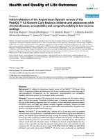
Intramuscular pressure parameters
Figure 1 shows, the mean course of the intramuscular
compartment pressure, and the muscular perfusion pres-
sure in the anterior and deep posterior compartment,
within the first 24 hours.
The IMP
ant
was significantly increased (P < 0.05) when
compared to the IMP
post
, while the corresponding per-
fusion pressure was decreased (P < 0.05) (i.e. the per-
fusion pressure in the anterior compartment was
significant decreased compared to perfusion pressure in
the posterior compartment). In 6 patients, compartment
pressures were temporary elevated over 40 mmHg, with
an anterior pressure maximum of 63 mmHg after 2 hrs in
one patient, which was measured in an anterior compart-
ment. During the first 24 hours, all 20 Patients showed
perfusion pressures higher than 40 mmHg.
Muscular function
Mean deficit (%) in dynamometric Biodex measurements
for peak torque, and mean power in dorsiflexion and
plantar flexion after 4 and 12 weeks, respectively, are
given in table 3.
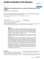
A significant correlation between the anterior perfusion
pressure (PP
ant
), 24 hours postoperatively, and the dorsi-
flexion after four weeks was shown for both the peak
torque (R = -0.83; p < 0.05) and the mean power
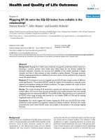
(R = -0.86; p < 0.05) (Figure 2). A reduction of PP
post
after
24 hours, was also significantly correlated, to a uniformly
decreased peak torque and mean power (R
peak
= -0.73;
R
mean
= -0.696; p < 0.05) in plantar flexion after four
weeks (Figure 3).
12 weeks following surgery no significant correlation was
evident between perfusion pressure values and dorsi- or
plantar flexion. The results are summarized in table 4 and
5.
Discussion
In this present study, we were able to demonstrate a signif-
icant functional relationship between the trauma-induced
reduction of perfusion pressure after 24 hours, in the ante-
Course of compartment pressure (IMP) and perfusion pres-sure (PP) in the anterior- (ant) and deep posterior (post) compartment within the first 24 hoursFigure 1
Course of compartment pressure (IMP) and per-
fusion pressure (PP) in the anterior- (ant) and deep
posterior (post) compartment within the first 24
hours.
Table 2: Demographic Characteristics and Injury Patterns
Patient Age, sex Side Aetiology AO Classification Tscherne classification
(for closed fractures)
Gustilo classification
(for open fractures)
Treatment method
1 47, m Left Traffic accident 42-B2 G1 nailing
2 25, f Right Traffic accident 42-C2 II° nailing
3 35, m Right Traffic accident 43-A2 G2 ORIF
4 26, f Right Traffic accident 42-B2 G1 nailing
5 37, f Left Sport accident 42-C1 I° nailing
6 48, f Right Traffic accident 42-B2 I° nailing
7 54, f Left Fall 42-B3 II° nailing
8 45, m Left Traffic accident 43-C1 II° ORIF
9 63, m Left Fall 42-B2 G2 nailing
10 39, m Right Industrial accident 43-A1 G2 ORIF
11 42, m Right Traffic accident 42-B2 G2 nailing
12 28, m Left Sport accident 41-C1 I° ORIF
13 40, m Right Traffic accident 42-C2 II° nailing
14 23, m Left Sport accident 42-B2 I° nailing
15 21, m Left Traffic accident 42-B2 G2 nailing
16 60, m Right Fall 43-C3 II° ORIF
17 54, f Left Traffic accident 42-B3 II° nailing
18 65, f Right Traffic accident 42-C2 IIIA° external Fixateur
19 42, f Right Traffic accident 42-B1 G1 nailing
20 37, m Right Traffic accident 42-A2 G1 nailing
Journal of Orthopaedic Surgery and Research 2008, 3:14 />Page 5 of 8
(page number not for citation purposes)
rior and posterior tibial compartment, and the skeletal
muscle function in the early rehabilitation phase, i.e. 4
weeks postoperatively. The decrease in perfusion pressure
after 24 hours, which was associated with a deficit in dor-
siflexion and plantar flexion of the ankle joint after 4
weeks, indicates a causal-prognostic role of early microcir-
culatory deteriorations for a manifestation/development
of skeletal muscle dysfunction, after four weeks post
trauma.
Previous experimental and clinical studies have shown
that tissue damage in response to soft tissue injury with
endothelial dysfunction, edema, local inflammation and
intramuscular pressure increase requires some time to
develop [22]. Consequently, preceding studies of our
group and others have shown that tissue pressure follow-
ing trauma shows maximum peaks not before 24 hours
after trauma [14,22]. Apart from these experimental rea-
sons, we have also correlated the measured time points
before 24 hrs. However, significant correlations were not
found before 24 hours after surgery. This indicates that
pressure increases at 24 hrs are most relevant and of prog-
nostic importance for resultant muscle performance and
muscle restoration 4 weeks after surgery. According to the
limitation of the study period to 24 hrs, further conclu-
sions about functional relationships between tissue pres-
sure and muscle function could not be drawn.
In vivo analysis of microcirculation following soft-tissue
injury demonstrated a interrelation between the severity
of soft-tissue trauma and nutritive capillary derangements
in skeletal muscle [14]. Progressive tissue damage, follow-
ing severe soft-tissue injury, was shown to be a result of
delayed and prolonged microvascular perfusion failure.
These results imply that post-traumatic muscle dysfunc-
tion may in fact be caused by the direct trauma, although
the extent of impairment seems mainly influenced by the
degree of posttraumatic perfusion disturbance. Crisco et
al. have investigated biomechanical, physiological and
histological alterations in a gastrocnemius muscle contu-
Regression analysis of perfusion pressure on the muscle deficit after 24 hours, in dorsiflexion at 4 weeks after traumaFigure 2
Regression analysis of perfusion pressure on the muscle deficit after 24 hours, in dorsiflexion at 4 weeks after
trauma. (a) for the peak torque (R = -0.83; p < 0.05) and (b) for the mean power (R = -0.86; p < 0.05). Muscle deficit is given
as a percentage of the non injured side, e.g. 80 percent means a 20 percent deficit.
Table 3: Dynamometric Biodex Measurements
Peak torque Mean power
Dorsiflexion Plantar flexion Dorsiflexion Plantar flexion
Mean deficit (%) 4 weeks 50.1 ± 13.1
a
61.3 ± 19.4 44.9 ± 17.7 55.3 ± 23.7
Mean deficit (%) 12 weeks 38.6 ± 20.1 45.7 ± 13 37.4 ± 16.3 27.9 ± 39.4
(Mean deficit (%) (to the uninjured side) in dynamometric Biodex measurements, for peak torque and mean power in dorsiflexion and plantar
flexion after 4 weeks and 12 weeks.
a
standard deviation)
Journal of Orthopaedic Surgery and Research 2008, 3:14 />Page 6 of 8
(page number not for citation purposes)
sion injury model, of male Wistar rats [23]. They also
demonstrated a significant deficit in contractile function,
in relation to the extent of contusion injury.
In addition, supporting the notion that the extent of mus-
cle trauma is a limitating co-factor to posttraumatic mus-
cle performance, Shaw and co-workers showed a
significant relationship between the severity of tibial frac-
tures, and the resulting rehabilitation time in football
players [24]. It could also be observed, that fracture mor-
phology, the presence of an open wound and the Tscherne
grade of closed fractures correlated with regained muscle
power [25]. Also, in addition to the severity of the initial
injury, the patient's age seems to be one of the main fac-
tors influencing muscle recovery following diaphyseal
tibia fractures [25,26]. The fact that in our study, no corre-
lation between muscle recovery and age was found may be
due both to the small variation in age of the included
patients, with the oldest patient being 65 years, and the
comparably small number of included patients.
Similar to our findings, Gaston et al. could show that
muscle function of the ankle and subtalar joints, recover
quickly from an initially low level [25]. They have further
found, that the differences in muscle power caused by age,
muscle damage, and the type of fracture, became more
obvious not before 15 to 20 weeks. The fact that our study
period was limited to 12 weeks, may explain why we did
not detect differences, in the outcome which depended on
age, or the type of fracture.
Our findings suggest that, the initial posttraumatic
changes in microcirculation within the first 24 hours have
a prognostic and predictive importance for muscle recov-
ery at 4 weeks after surgery. Early muscle recovery is in
turn, an absolute prerequisite for rapid mobilization, and
accelerated rehabilitation. In this context, effective treat-
ment strategies after lower leg injuries have to ensure the
restitution of nutritive perfusion, and the maintenance of
sufficient perfusion pressure, in order to prevent subse-
quently impaired muscle performance and delayed reha-
Table 5: Biodex measurements (Plantarflexion) after 4 and 12
weeks versus perfusion pressure in the posterior compartment
at 24 hours
4 Weeks 12 Weeks
Peak torque Mean power Peak torque Mean power
R = -0.73 R = -0.696 R = -0.28 R = -0.39
p < 0.001 p < 0.001 p = 0.293 p = 0.121
(Biodex measurements (Plantarflexion) after 4 and 12 weeks versus
perfusion pressure in the posterior compartment at 24 hours.
Statistical significance is given by p-values less than 0.05)
Regression analysis of perfusion pressure on the muscle deficit after 24 hours, in plantar flexion at 4 weeks after traumaFigure 3
Regression analysis of perfusion pressure on the muscle deficit after 24 hours, in plantar flexion at 4 weeks
after trauma. (a) for the peak torque and (b) for the mean power. (Rpeak = -0.73; Rmean = -0.696; p < 0.05) Muscle deficit
is given as a percentage of the non injured side, e.g. 80 percent means a 20 percent deficit.
Table 4: Biodex measurements (Dorsiflexion) after 4 and 12
weeks versus perfusion pressure in the anterior compartment at
24 hours
4 Weeks 12 Weeks
Peak torque Mean power Peak torque Mean power
R = -0.83 R = -0.86 R = -0.39 R = -0.48
p < 0.001 p < 0.001 p = 0.119 p = 0.07
(Biodex measurements (Dorsiflexion) after 4 and 12 weeks versus
perfusion pressure in the anterior compartment at 24 hours.
Statistical significance is given by p-values less than 0.05)
Journal of Orthopaedic Surgery and Research 2008, 3:14 />Page 7 of 8
(page number not for citation purposes)
bilitation. The short immobilization period for the first
couple of days is beneficial in providing a sufficient
phagocytosis of necrotized tissue and granulation tissue
formation. However, for regeneration of myofibers and
capillary ingrowth, a specifically early mobilization proce-
dure was shown to be essential [5,23,27,28]. Early, post-
operative mobilization was introduced in 1954 [29].
Apart from these positive mobilization-associated effects
of the regeneration of skeletal muscle morphology, bio-
mechanical in vitro investigations, also demonstrated a
faster return of muscle strength to the level of the unin-
jured contralateral muscle, following an active early mobi-
lization [27].
Our results confirm that perfusion pressure (calculated
from the difference of the mean arterial pressure and the
compartment pressure) correlates significantly with the
post traumatic muscle performance while absolute intrac-
ompartimental pressures alone did not. Perfusion pres-
sure is, by taking into account the arterial blood pressure,
i.e. the macrohemodynamic situation, a more valid
parameter to reflect posttraumatic muscle tissue damage.
As a result, an increased compartment pressure in combi-
nation with an adequate blood pressure appears to not be
unavoidably related with a greater extent of muscle cell
damage, risk of compartment syndrome, or an impaired
post traumatic muscle performance. In our study, 6
patients had a temporary compartment pressure higher
than 40 mmHg. In all of these patients, a sufficient per-
fusion pressure was calculated and existed. The later per-
formed Biodex measurements in these patients
corresponded to the perfusion pressure, while a relation-
ship to compartment pressures was not shown. Despite an
elevation in the compartment pressure, the evaluated
peak torque and mean power results were in the range of
the other patients. This notion is confirmed by an evalua-
tion of skeletal muscle metabolism with nuclear magnetic
resonance spectroscopy [30]. The authors demonstrated,
that metabolic derangements mainly depend on the dif-
ference between MAP and compartment pressure, rather
than on absolute compartment pressure [30]. It was
shown, that a perfusion pressure of less than 40 mm Hg
in bluntly traumatized muscle, was associated with tissue
acidosis and ischemia. Again, investigating the relation-
ship between compartment and perfusion pressure, Hart-
sock et al. demonstrated, that capillary perfusion in
skeletal muscle is equally and profoundly impaired, either
at a PP of 25.5 ± 14.3 mm Hg or a compartment pressure
exceeding 60 mmHg [31]. In addition, Whitesides et al.
were the first to recommend that differential perfusion
pressure, as opposed to absolute intramuscular pressures,
were of high importance [32]. This underlines the essen-
tial significance of local and distal tissue perfusion.
In a recent study, White et al. demonstrated, that a
decrease of perfusion pressure to a lower limit of 30 mm
Hg, and an elevated intramuscular pressure to an upper
level of 70 mm Hg, is tolerated without significant adverse
consequences [33]. Obviously, a parallel/simultaneous
elevation of both the diastolic blood and the intramuscu-
lar perfusion pressure, maintains an adequate capillary
perfusion. Thus enables the tissue to tolerate elevated
compartment pressures. Consideration should be given to
polytraumatized patients, where possibly prolonged peri-
ods of insufficient circulation coupled with a depressed
blood pressure and an inadequate oxygenation, may lead
to a shift of the critical threshold of tissue tolerance, into
decreased compartment pressures. However, the combi-
nation of clinical awareness, and the continuous differen-
tial perfusion pressure monitoring, as based on our
experience and that of other authors [34,35], is a much
more effective, specific and reliable method in detecting a
subsequent compartment syndrome, as opposed to just
measuring absolute intracompartimental pressure values.
Furthermore, the measurement of intramuscular pressure
alone, as a criterion for fasciotomy, has a lower specificity,
and was shown to result in an unnecessarily high fasciot-
omy rate and an increased rate of associated short- and
long term complications [36].
Conclusion
We were able to show a significant correlation between
the perfusion pressure after 24 hours and the functional
outcome in muscle performance after 4 weeks. There was
no correlation between muscle function and the intrac-
ompartimental pressure itself. Alterations in muscle per-
fusion, caused by primary and secondary soft-tissue
damage were responsible for the substantial muscle dys-
function seen for up to 4 weeks following trauma. Obvi-
ously, monitoring perfusion pressure is far more superior
and sensitive, in the assessment of post-traumatic effects
on muscle performance and recovery. Therefore, effective
treatment strategies must be made to ensure the restitu-
tion of nutritive perfusion and sufficient perfusion pres-
sure. This is in order to prevent future deficits in muscle
performance, and a delayed rehabilitation.
References
1. Claudle RJ, Stern PJ: Severe open fractures of the tibia. J Bone
Joint Surg [Am] 1987, 69:801-808.
2. Edwards CC, Simmons SC, Browner BD, MC W: Severe open tib-
ial fractures. Clinical orthopaedics and related research 1988 ,
230:98-115.
3. Gustilo RB, Mendoza RM, Williams DN: Problems in the manage-
ment of type III (severe) open fractures: a new classification
of type III open fractures. The Journal of trauma 1984, 24:742-746.
4. Levin LS, Condit DP: Combined injuries—soft tissue manage-
ment. Clin Orthop Relat Res 1996, 327:172-181.
5. Jarvinen TA, Jarvinen TL, Kaariainen M, Kalimo H, Jarvinen M: Muscle
injuries: biology and treatment. The American journal of sports
medicine 2005, 33(5):745-764.
6. Harris K, Walker PM, Mickle DA, Harding R, Gatley R, Wilson GJ,
Kuzon B, McKee N, AD. R: Metabolic response of skeletal mus-
cle to ischemia. Am J Physiol 1986, 250(2 Pt 2):H213-H220
Publish with BioMed Central and every
scientist can read your work free of charge
"BioMed Central will be the most significant development for
disseminating the results of biomedical research in our lifetime."
Sir Paul Nurse, Cancer Research UK
Your research papers will be:
available free of charge to the entire biomedical community
peer reviewed and published immediately upon acceptance
cited in PubMed and archived on PubMed Central
yours — you keep the copyright
Submit your manuscript here:
/>BioMedcentral
Journal of Orthopaedic Surgery and Research 2008, 3:14 />Page 8 of 8
(page number not for citation purposes)
7. Hill AG, Hill GL: Metabolic response to severe injury. Br J Surg
1998, 85:884-890.
8. McQueen MM, Christie J, Court-Brown CM: Acute compartment
syndrome in tibial diaphyseal fractures. J Bone Joint Surg Br
1996, 78(1):95-98.
9. Zhang L, Bail H, Mittlmeier T, Haas NP, Schaser KD: Immediate
microcirculatory derangements in skeletal muscle and peri-
osteum following closed tibial fracture. J Trauma 2003,
54(5):979-985.
10. Schwartz JT Jr., Brumback RJ, Lakatos R, Poka A, Bathon GH, Burgess
AR: Acute compartment syndrome of the thigh. A spectrum
of injury. J Bone Joint Surg Am 1989, 71(3):392-400.
11. Tischenko GJ, Goodman SB: Compartment syndrome after
intramedullary nailing of the tibia. J Bone Joint Surg Am 1990,
72(1):41-44.
12. Mars M, Hadley GP: Raised intracompartmental pressure and
compartment syndromes. Injury 1998, 29(6):403-411.
13. Rorabeck CH: The treatment of compartment syndromes of
the leg. J Bone Joint Surg Br 1984, 66(1):93-97.
14. Schaser KD, Vollmar B, Menger MD, Schewior L, Kroppenstedt SN,
Raschke M, Lubbe AS, Haas NP, Mittlmeier T: In vivo analysis of
microcirculation following closed soft-tissue injury. J Orthop
Res 1999, 17(5):678-685.
15. Rüedi TP, Buckley RE, Moran CG: AO Principles of Fracture
Management. Edited by: Publishing AO. Thieme; 2007:1102.
16. Tscherne H, Oestern HJ: A new classification of soft-tissue dam-
age in open and closed fractures (author's transl). Unfall-
heilkunde 1982, 85(3):111-115.
17. Gustilo RB: Current concepts in the management of open
fractures. Instructional course lectures 1987, 36:359-366.
18. Blick SS, Brumback RJ, Poka A, Burgess AR, Ebraheim NA: Compart-
ment syndrome in open tibial fractures. J Bone Joint Surg Am
1986, 68(9):
1348-1353.
19. Matsen FA 3rd, Rorabeck CH: Compartment syndromes. Instruc-
tional course lectures 1989, 38:463-472.
20. Hargens AR, Mubarak SJ: Current concepts in the pathophysiol-
ogy, evaluation, and diagnosis of compartment syndrome.
Hand clinics 1998, 14(3):371-383.
21. Heppenstall RB, Scott R, Sapega A, Park YS, Chance B: A compara-
tive study of the tolerance of skeletal muscle to ischemia.
Tourniquet application compared with acute compartment
syndrome. J Bone Joint Surg Am 1986, 68(6):820-828.
22. Seekamp A, Van Griensven M, Blankenburg H, Regel G: Intramus-
cular partial oxygen tension monitoring in compartment
syndrome an experimental study. Eur J Emerg Med 1997,
4(4):185-192.
23. Crisco JJ, Jokl P, Heinen GT, Connell MD, Panjabi MM: A muscle
contusion injury model. Biomechanics, physiology, and his-
tology. The American journal of sports medicine 1994, 22(5):702-710.
24. Shaw AD, Gustilo T, Court-Brown CM: Epidemiology and out-
come of tibial diaphyseal fractures in footballers. Injury 1997,
28(5-6):365-367.
25. Gaston P, Will E, McQueen MM, Elton RA, Court-Brown CM: Anal-
ysis of muscle function in the lower limb after fracture of the
diaphysis of the tibia in adults. J Bone Joint Surg Br 2000,
82(3):326-331.
26. Giannoudis PV, Nicolopoulos C, Dinopoulos H, Ng A, Adedapo S,
Kind P: The impact of lower leg compartment syndrome on
health related quality of life. Injury 2002, 33(2):117-121.
27. Jarvinen MJ, Lehto MU: The effects of early mobilisation and
immobilisation on the healing process following muscle inju-
ries. Sports Med 1993, 15(2):78-89.
28. Jarvinen M, Aho AJ, Lehto M, Toivonen H: Age dependent repair
of muscle rupture. A histological and microangiographical
study in rats. Acta orthopaedica Scandinavica 1983, 54(1):64-74.
29. Woodard C: What is active treatment? In Sports Medicine Edited
by: Woodard C. London, England: Max Parrish & Co ; 1954:1-14.
30. Heppenstall RB, Sapega AA, Scott R, Shenton D, Park YS, Maris J,
Chance B: The compartment syndrome. An experimental
and clinical study of muscular energy metabolism using
phosphorus nuclear magnetic resonance spectroscopy. Clin
Orthop Relat Res 1988:138-155.
31. Hartsock LA, O'Farrell D, Seaber AV, Urbaniak JR: Effect of
increased compartment pressure on the microcirculation of
skeletal muscle. Microsurgery 1998, 18(2):67-71.
32. Whitesides TE, Haney TC, Morimoto K, Harada H: Tissue pressure
measurements as a determinant for the need of fasciotomy.
Clinical orthopaedics and related research 1975:43-51.
33. White TO, Howell GE, Will EM, Court-Brown CM, McQueen MM:
Elevated intramuscular compartment pressures do not
influence outcome after tibial fracture. The Journal of trauma
2003, 55(6):1133-1138.
34. Janzing HM, Broos PL: Routine monitoring of compartment
pressure in patients with tibial fractures: Beware of over-
treatment! Injury 2001, 32(5):415-421.
35. McQueen MM, Court-Brown CM: Compartment monitoring in
tibial fractures. The pressure threshold for decompression. J
Bone Joint Surg Br 1996, 78(1):99-104.
36. Ovre S, Hvaal K, Holm I, Stromsoe K, Nordsletten L, Skjeldal S:
Compartment pressure in nailed tibial fractures. A thresh-
old of 30 mmHg for decompression gives 29% fasciotomies.
Arch Orthop Trauma Surg 1998, 118(1-2):29-31.