Báo cáo hóa học: " Overexpression of microRNA-206 in the skeletal muscle from myotonic dystrophy type 1 patients" docx
Bạn đang xem bản rút gọn của tài liệu. Xem và tải ngay bản đầy đủ của tài liệu tại đây (1.15 MB, 9 trang )
Gambardella et al. Journal of Translational Medicine 2010, 8:48
/>Open Access
RESEARCH
BioMed Central
© 2010 Gambardella et al; licensee BioMed Central Ltd. This is an Open Access article distributed under the terms of the Creative Com-
mons Attribution License ( which permits unrestricted use, distribution, and reproduc-
tion in any medium, provided the original work is properly cited.
Research
Overexpression of microRNA-206 in the skeletal
muscle from myotonic dystrophy type 1 patients
Stefano Gambardella*
1,2
, Fabrizio Rinaldi
1
, Saverio M Lepore
3
, Antonella Viola
1
, Emanuele Loro
4
, Corrado Angelini
4
,
Lodovica Vergani
4
, Giuseppe Novelli
1,5,2
and Annalisa Botta
1
Abstract
Background: MicroRNAs are highly conserved, noncoding RNAs involved in post-transcriptional gene silencing. They
have been shown to participate in a wide range of biological processes, including myogenesis and muscle
regeneration. The goal of this study is to test the hypothesis that myo-miRs (myo = muscle + miR = miRNA) expression
is altered in muscle from patients affected by myotonic dystrophy type 1 (DM1), the most frequently inherited
neuromuscular disease in adults. In order to gain better insights about the role of miRNAs in the DM1 pathogenesis, we
have also analyzed the muscular expression of miR-103 and miR-107, which have been identified in silico as attractive
candidates for binding to the DMPK mRNA.
Methods: To this aim, we have profiled the expression of miR-133 (miR-133a, miR-133b), miR-1, miR-181 (miR-181a,
miR-181b, miR-181c) and miR-206, that are specifically induced during myogenesis in cardiac and skeletal muscle
tissues. miR-103 and miR-107, highly expressed in brain, heart and muscle have also been included in this study. QRT-
PCR experiments have been performed on RNA from vastus lateralis biopsies of DM1 patients (n = 7) and control
subjects (n = 4). Results of miRNAs expression have been confirmed by Northern blot, whereas in situ hybridization
technique have been performed to localize misexpressed miRNAs on muscle sections from DM1 and control
individuals.
Results: Only miR-206 showed an over-expression in 5 of 7 DM1 patients (threshold = 2, fold change between 1.20 and
13.22, average = 5.37) compared to the control group. This result has been further confirmed by Northern blot analysis
(3.37-fold overexpression, R
2
= 0.89). In situ hybridization localized miR-206 to nuclear site both in normal and DM1
tissues. Cellular distribution in DM1 tissues includes also the nuclear regions of centralized nuclei, with a strong signal
corresponding to nuclear clumps.
Conclusions: This work provides, for the first time, evidences about miRNAs misexpression in DM1 muscle tissues,
adding a new element in the pathogenesis of this complex genetic disease.
Background
Myotonic dystrophy type 1 (DM1; MIM #160900), the
most frequent autosomal dominant myopathy in adults,
is associated with an expansion of (CTG)n repetitions in
the 3'UTR of the DMPK gene (DMPK; MIM#605377), on
chromosome 19q13.3 [1-3]. Common clinical findings
are myotonia, muscle wasting and weakness. Additional
features of the disease typically include heart conduction
defects, cataracts, hypogonadism, and cognitive impair-
ment [4].
The expanded DMPK mRNA play a trans-dominant
effect on RNA metabolism through its binding to the
Muscleblind-like 1 (MBNL1) splicing regulator, leading to
abnormal alternative splicing for a set of genes mainly
expressed in skeletal muscle and heart [5,6]. Several
expression studies have also been applied to further
understand the pathological mechanism occurring in
DM1 muscle and they support the idea that the toxic
effect of CUG
exp
RNA may occur also at the level of tran-
scription [7,8]. Less is known about the expression of
microRNA genes and DM1. MicroRNAs (miRNAs) are a
class of naturally occurring small noncoding RNAs that
control gene expression by targeting mRNAs for transla-
* Correspondence:
1
Biopathology Department, Tor Vergata University, Rome, Italy
Full list of author information is available at the end of the article
Gambardella et al. Journal of Translational Medicine 2010, 8:48
/>Page 2 of 9
tional repression or cleavage [9]. Primary miRNA tran-
scripts are cleaved into 70- to 80-nucleotide precursor
miRNAs (pre-miRNAs) hairpins by RNase III Drosha in
the cell nucleus and transported to the cytoplasm, where
pre-miRNAs are processed by RNA Dicer into 19- to 25-
nucleotide miRNA duplexes. One strand of each duplex is
degraded, and the other strands become mature miRNA,
which recognize sites in the 3'-UTR of the target mRNAs
and cause translational repression or mRNA cleavage.
miRNAs are a new player among gene regulation mecha-
nisms, and their functions have not been fully explored
but are known to include the regulation of cellular differ-
entiation, proliferation, and apoptosis [10]. They have
been shown to participate in a wide range of biological
processes, including myogenesis and muscle regenera-
tion. The three muscle-specific miRNAs, miR-1, miR-
133, and miR-206 have been shown to play important
roles in the regulation of muscle development [11].
miR-1 and miR-133 are expressed in cardiac and skele-
tal muscle and are transcriptionally regulated by the myo-
genic differentiation factors and serum response factor
(SRF). The myogenic transcription factors myogenin and
myogenic differentiation 1 (MyoD) bind to regions
upstream of the miR-1 and miR-133 stem loop, providing
a molecular explanation for their observed induction dur-
ing myogenesis [12-14]. Moreover, miR-1 promotes dif-
ferentiation of cardiac and skeletal progenitors and their
exit from the cell cycle in mammals [15], while miR-133
inhibits their differentiation and maintains them in a pro-
liferative state. miR-206 is expressed only in skeletal mus-
cles, and promotes muscle differentiation if induced by
MyoD and myogenin during myogenesis [16,17]. These
muscle-specific miRNAs seem to participate in muscle
diseases, including cardiac hypertrophy, heart failure,
cardiac arrhythmias, congenital heart disease, and mus-
cular dystrophy [18-22]. Other miRNAs, not specifically
expressed in muscle, have been proposed to be involved
in DM1 pathogenesis. A computational analysis on the
repression effects of CTG-repeat binding miRNAs,
revealed that miR-103 and 107 are attractive candidates
for binding to DMPK transcript in a length-dependent
manner [23]. In this model, mir-107 and mir-103 which
contain CAG repeats in their seed regions, preferentially
bind to the mutated DMPK mRNA. This could have a
miRNA-leaching effect on the amount of unbound
miRNA which is reduced and could no longer repress
other target genes. miRNAs involvement could therefore
have significant consequences on the expression of pro-
teins important in DM1 disease pathogenesis and pro-
gression.
The main goal of this study is to test the hypothesis that
myo-miRs expression is altered in muscle biopsies from
DM1 patients with comparable expansion size. In order
to gain better insights about the role of miRNAs in DM1,
we have also analyzed the muscular expression of the
miR-103 and miR-107 CTG-repeat binding miRNAs.
A combination of Northern blot and QRT-PCR experi-
ments have been utilized to quantify the expression levels
of miRNAs, while in situ hybridization performed on
muscle sections revealed the intracellular localization of
misexpressed miRNAs. This is the first report investigat-
ing the potential involvement of miRNAs in the patho-
genesis of DM1 and shows a significant overexpression of
miRNA-206, whose functional significance remains to be
elucidated.
Methods
Patient recruitment
Seven unrelated DM1-patients, aged 30-50 years, were
diagnosed at the Department of Neurology, University of
Padua, Padua, Italy. The diagnosis of DM1 was based on
clinical, electromyographic (high frequency repetitive
discharges), ophthalmologic and cardiac investigations.
After written informed consent, DM1 muscle samples
were obtained by diagnostic needle biopsies from vastus
lateralis. Control samples (vastus lateralis) were obtained
from 4 subjects deemed free of neuromuscular disorders,
aged 35 and 42 years. All muscle biopsies were frozen in
liquid nitrogen immediately after surgery, and stored at -
80°C until used. Histological analysis of DM1 biopsies
showed the typical pathology of the disease, including
atrophic fibres with increased fibre size variation and
marked proliferation of centrally located nuclei. Hema-
toxylin-eosin and Ghomory thricome stains showed
absence of inflammatory aspects in all the DM1 samples
analyzed. The main pathohistological features of each
DM1 specimens used in this study are reported in Table
1. (CTG) repeat expansion sizes were determined in mus-
cle tissues and resulted to be included into the E2 class.
The study was approved by the local ethical committee of
Tor Vergata University and all the procedures have been
performed in compliance with the Helsinki Declaration.
Quantitative reverse transcription-PCR of miRNAs and
mRNAs
Total RNA was extracted from 500 mg of frozen vastus
lateralis tissue using TRIZOL reagent (Life Technologies,
Inc.) following the manufacturer's instructions. cDNA
was reverse transcribed from 3 μg of total RNA samples
using specific miRNA primers and reagents from the
TaqMan MicroRNA Reverse Transcription kit and Assays
(Applied Biosystems). The resulting cDNA was amplified
by PCR using TaqMan MicroRNA Assay primers with the
Taq Man Universal PCR Master Mix (code 4324018) and
analyzed with a 7500 ABI PRISM Sequence Detector Sys-
tem according to the manufacturer's instructions
(Applied Biosystems). We analyzed the expression of the
following miRNAs: hsa- mir-1 (Assay n. 4373161), hsa-
Gambardella et al. Journal of Translational Medicine 2010, 8:48
/>Page 3 of 9
mir-206 (Assay n. 4373092), hsa- mir-181a (Assay n.
4373117), hsa- mir-181b (Assay n. 4373116), hsa- mir-
181c (Assay n. 4373115), hsa- mir-133a (Assay n.
4373142), hsa- mir-133b (Assay n. 4373172), hsa- mir-103
(Assay n. 4373158), hsa- mir-107 (Assay n. 4373154). 7
DM1 patients and 4 control subjects have been included
in this study. Values of DM1 patients were compared to
the medium value of control subjects analyzed separately.
The relative levels of miRNA expression were calculated
and normalized using the 2
-ΔΔCt
method relative to HSA-
let-7a miRNA (Assay n. 4373169). All TaqMan-PCRs
were performed in triplicates. Both let-7a and U6-sn-
RNA were considered initially as possible control miR-
NAs for normalization of samples. Let-7a miRNA is fre-
quently used as internal control because of its stable
expression across human tissues and cell lines [24,25],
even though some studies report its misregulation espe-
cially in cancer conditions (lung cancer, chronic lympho-
cytic leukemia, breast cancer, prostate cancer,
hepatocellular carcinoma) not related with muscular dis-
orders [26-28]. QRT-PCR analysis (data not shown)
showed a similar expression level of U6 and let-7a miR-
NAs in all the samples included in the study. Finally, let-
7a has been chosen as control miRNA because its Ct (Ct
= cycle threshold, defined as the number of cycles
required for the fluorescent signal to cross the threshold)
value is more comparable to the Ct values of myo-miRs
considered in this study. For quantification of Utrophin
transcritpt, 2 μg of total RNA was reverse transcribed
using high capacity cDNA reverse transcription kit
(Applied Biosystem). The resulting cDNA was amplified
using the ABI Prism 7000 Real-Time Sequence Detection
System quantification employing the Syber-Green assay.
Primers sequence for Utrophin was taken from Arning et
al. [29] (Forward 5' aaggacctggtcaacgttcca 3', Reverse 5'
acccgtgtcatagacattgagca 3'). The Beta-Actin mRNA level
was used as control for normalization of samples (For-
ward 5#8242; gacaggatgcagaaggagattact 3', Reverse
5#8242; tgatccacatctgctggaaggt 3').
Nothern Blot analysis
Given the limited amount of RNA available, total RNAs
from DM1 muscle biopsies were pooled into two groups:
DMa (DM1-1, DM1-2, DM1-3, DM1-4) and DMb (DM1-
5, DM1-6, DM1-7). RNAs from 4 healthy subjects were
pooled and used as control. RNA was separated on dena-
turating polyacrylamide gels in TBE buffer, transferred to
a nylon membrane (Hybond-N+, GE Biosciences) with
Trans-blot SD Semi-dry Transfer Cell (Bio-Rad) and fixed
in the membrane by UV crosslinking, with 1200 μJ.
Hybridization probes were prepared with 20 μM oligonu-
clotides, whose sequences were complementary to inves-
tigated miRNAs. Probes were labeled with [32P] γ-ATP
(5000 ci/mmol; 10 mCi/ml, from Hartmann Analytic
GmBH, Germany) using polynucleotide kinase (New
England Biolabs). The labeled probes were purified with
Sephadex G25 spin columns (GE Biosciences). After add-
ing the probe, hybridization was carried out overnight at
42°C. After hybridization, membranes were washed with
SSPE 6×. Dried membranes were exposed to Phosphoim-
aging plates (Kodak), which were read out in a Storm
scanner (Amersham- GE Biosciences). For Northern blot
analysis miRNA U6 were used as control for normaliza-
tion of samples. U6, widely used in Northern Blot analy-
sis, has been chosen as control miRNA because its band
intensity is more comparable to the those of myo-miRs
considered in this study. Densitometry of autoradiograms
was performed using OptiQuant image analysis software
(Packard). A linear regression has been applied in order
to correlate expression values obtained with QRT-PCR
and Northern Blot analysis.
Western blotting
Muscle 20 μm sections were collected from frozen bioptic
samples, lysed in Laemmli buffer and run in a 4-12%
Table 1: Pathohistological features of each DM1 specimens
Patients Sex Muscle Lysosomal activity* Muscle pathohistological aspects
DM1-1 M VL xxxx atrophy
DM1-2 M VL xxx atrophy
DM1-3 M VL xx mild atrophy
DM1-4 M VL xxxx atrophy
DM1-5 M VL x few atrophic fibers
DM1-6 F VL xxxx severe atrophy
DM1-7 M VL absent few atrophic fibers
Are shown the main pathohistological features of each DM1 specimens used in this study (sex, muscle, lysosomal activity and muscle
pathohistological aspects). Muscle biopsies from VL of 1 female and 3 males have been used as control samples. (VL = Vastus Lateralis; M =
Male; F = Female; * = Phosphatase activity present in atrophic fiber where autophagy is active.)
Gambardella et al. Journal of Translational Medicine 2010, 8:48
/>Page 4 of 9
T30C4 SDS-PAGE. Proteins were then blotted into nitro-
cellulose membrane, probed with specific Utrophin
(Novacastra, NCL-DRP2) and α-Tubulin (Santa Cruz, B7
sc-5286) antibodies. After incubation with secondary
HRP-conjugated antibodies, recognized bands were visu-
alized by chemiluminescence (GE HealthCare). Inte-
grated optical density of each band was calculated with
commercial software and normalized compared to Tubu-
lin amounts.
In situ hybridisation
In situ hybridization was performed on transversal sec-
tions of vastus lateralis muscles from DM1 patients show-
ing a significant up-regulation of miR-206 and from two
control subjects included in this study. A locked nucleic
acid (LNA) detection probe for miR-206 (Exiqon Cod.
EX100008999901), a LNA U6 positive control probe
(Exiqon Cod. EX9900201) and a LNA negative control
probe with a scramble sequence (Exiqon Cod.
EX9900401) have been used in this analysis. All probes
were labeled with digoxigenin (DIG) (Roche). Cryosec-
tion prepared from quadriceps vastus lateralis of human
biopsies in normal and DM1 patients fixed with 4% PFA,
were treated with proteinase K, re-fixed with PFA and
then acetylated with acetylation buffer (0.1 M trietha-
nolamine pH 8.0).
After washing with PBS and pre-hybridization, slides
were incubated with DIG-labeled LNA miR-206 probe,
DIG-labeled LNA U6 probe (positive contol) and a DIG-
labeled LNA scrambled sequence (negative control) at
49°C overnight. Washes were done at 49°C in 5× SSC,
50% formamide, 2× SSC and at room temperature in 0.2×
SSC and then PBS 1×/0.1% Tween-20, then slides were
incubated with blocking solution (PBS 1×/0.5% BSA/1 -
5% inactivated FCS), followed incubation with FITC-cou-
pled anti-digoxygenin antibody (Roche) at 4°C overnight.
After washes with PBS 1×/0.1% Tween 20, slides were
rinsed with DAPI, mounted and analyzed by fluorescent
microscopy Olympus BX51 at 40× magnification.
Results
miR-206 expression is increased in DM1 muscle
In this work we have profiled the expression of miR-133
(miR-133a, miR-133b), miR-1, miR-181 (miR-181a, miR-
181b, miR-181c) and miR-206, specifically induced dur-
ing myogenesis, in muscle biopsies from 7 DM1 patients,
compared with 4 control subjects. In order to gain better
insights about the role of miRNAs in the DM1 pathogen-
esis, we have also analyzed the muscular expression of
miR-103 and miR-107, which have been identified in sil-
ico as attractive candidates for binding to the DMPK tar-
get mRNA.
We first calculated the relative amount of miRNAs
expression using the HSA-let-7a miRNA for normaliza-
tion of samples in three independent QRT-PCR reac-
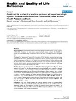
tions. As shown in Figure 1, QRT-PCR experiments
showed no differences in the expression of miR-1, miR-
133, miR-181, miR-103 and miR-107 between DM1 and
control muscles. In striking contrast, miR-206 expression
was increased in 5 of 7 DM1 patients (threshold = 2, fold
change between 1.20 and 13.22, average = 5.37) compared
to median value of controls group set as 1 (Fold change of
Ctr-2, Ctr-3 and Ctr-4 normalized with Ctr-1 are 1.3, 0.98
and 1,15 respectively). To validate the over-expression of
miR-206 in DM1 muscles, we performed Northern blot
analysis using pooled DM1 and control samples and U6
as control sn-RNA. We decided to pool samples because
the quantity of RNA derived from patients' biopsies was
not enough to analyze each sample separately. Figure 2a
shows Northern blot results of the four myo-miRs con-
sidered (miR 181, miR 1, miR 206 and miR 133) com-
pared to U6-snRNA in DM1 and controls muscle
samples. Densitometry analysis of autoradiograms (Fig-
ure 2b) further confirmed the results obtained through
QRT-PCR. miR-206 was over-expressed in both DMa and
DMb pool (DMa = 3,36 +/- 0.11, DMb = 3,39 +/- 0.10).
Linear regression demonstrates a statistically significant
positive correlation between QRT-PCR and Nothern blot
analyses results (R
2
DMa = 0.98; R
2
DMb = 0.82).
mRNA and protein level of Utrophin are not decreased in
DM1 muscle lysates
A predicted target gene of miR-206 is the Utrophin gene
(Utrn)
. Rosenberg et al.
[16] confirmed this prediction with multiple lines of evi-
dence indicating that miR-206 acts at post-trascriptional
level in repressing Utophin expression. We therefore per-
formed Western blot analysis to test the expression levels
of the Utrn protein in DM1 patients vs. controls. Figure
3a shows a Western blot image for the quantification of
Utrophin and Tubulin (used as housekeeping protein) in
Figure 1 QRT-PCR quantification of myo-miRs and miR-133 and
miR-107 in biopsies from vastus lateralis of 7 DM1 patients com-
pared with 4 controls. Fold change values of miR-206: DM1-1 = 3,73;
DM1-2 = 7,02; DM1-3 = 7,77; DM1-4 = 2,70; DM1-5 = 1,20; DM1-6 =
13,22; DM1-7 = 1,95
Gambardella et al. Journal of Translational Medicine 2010, 8:48
/>Page 5 of 9
5 DM1 patients and 4 controls. We included in this
experiment only DM1 patients showing a significant
over-expression of miR-206 (DM1-1 fold change 3,73,
DM1-2 fold change 7,02, DM1-3 fold change 7,77, DM1-4
fold change 2,70, DM1-6 fold change 13,22). After densi-
tometric analysis of each band, we found a high variabil-
ity in the Utrophin level both in controls and DM1
patients (Figure 3a). Utrophin/Tubulin ratios range from
0,31 and 1,66 (medium value = 0,80) in DM1 muscles and
from 0,15 to 0,85 (medium value = 0,46) in control sam-
ples, with no significant differences between the two
groups (Figure 3b). A linear regression analysis compar-
ing Utrophin quantification and miR-206 expression did
not show any correlation (R
2
= 0.132, not shown) indicat-
ing that, in our DM1 samples, the levels of Utrophin are
not under the direct control of miR-206 expression.
Although miRNAs are believed to regulate their targets
primarily through translational inhibition, there is
increasing evidence that miRNAs can also influence the
abundance of target mRNAs [30]. On this basis, we have
also studied the abundance of Utrophin mRNA in our
muscle specimens. QRT-PCR experiments, using Syber-
Green assay and the Beta-Actin mRNA as control for nor-
malization of samples, showed no significant differences
between DM1 and controls groups (Fold change DM1-1
= 1,27, DM1-2 = -1,82, DM1-3 = 1,77, DM1-4 = 2,1,
DM1-5 = 1,06, DM1-6 = -1,92, DM1-7 = 1,29). Again, lin-
ear regression analysis comparing Utrophin mRNA quan-
tification and miR-206 expression did not show any
correlation (R
2
= 0.138).
miRNA-206 localizes to centralized nuclei and nuclear
clumps in DM1 muscle sections
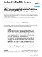
To detect the intracellular localization of miR-206, we
performed in situ hybridization using locked nucleic acid
(LNA) probes on cryostat vastus lateralis muscle sections
from controls and DM1 patients. Figure 4 shows the
hybridization pattern of miR-206 in transversal muscle
sections from a DM1 (Figure 4b) and a control (Figure 4a)
subject using a DIG-labeled LNA probe detected with a
FITC coupled anti-digoxygenin antibody. miR-206 local-
izes most exclusively to the nuclear region both in normal
and DM1 tissues. However, in DM1 muscles a strong sig-
nal was detected also in correspondence to centralized
nuclei and nuclear clumps (Figure 4b, see red arrow),
which are pathological hallmarks of dystrophic muscles.
We also investigated expression of miR-206 in cytoplasm
both in normal and DM1 tissue, but no signals were visi-
ble, indicating a nuclear specific function of miR-206 in
the muscle tissue. As controls of hybridization, the mus-
cle sections were hybridized with the LNA U6 positive
control probe (Figure 4c), which recognize a small and
stable ribonucleoprotein in all human cells. The specific-
ity of hybridization was assessed using an LNA probe
with a scrambled sequence not present in the human
genome (Figure 4d).
Discussion
miR-206 is a member of the muscle-specific miR-1 family,
that consists of six members clustered into three bicis-
tronic pairs arising from an initial local gene duplication
Figure 2 Northern blot analysis of myo-miRs expression. DM1 pa-
tients were pooled into two groups: DMa (DM1-1, DM1-2, DM1-3,
DM1-4) and DMb (DM1-5, DM1-6, DM1-7), 4 healthly subjects were
pooled as well. The U6-snRNA was used as control for normalization of
samples. Figure 2a: Northern Blot results of the 3 pooled samples (Ctr,
DMa and DMb) for the 4 miRNA analyzed (miR 181, miR 1, miR 206 and
miR 133) compared to U6-snRNA. Figure 2b: Densitometry of autora-
diograms performed using OptiQuant image analysis software (Pack-
ard) showing miR/U6 ratios.
Figure 3 Western blot analysis showing the Utrophin and α-Tu-
bulin protein expression levels in 5 DM1 patients and 4 controls.
Utophin/Tubulin ratios in the analyzed samples are: DM1-1 = 0,63,
DM1-2 = 0,74, DM1-3 = 0,31, DM1-4 = 1,66, DM1-5 = 0,66, CTR 1 = 0,15,
CTR 2 = 0,85, CTR 3 = 0,50, CTR 4 = 0,34. Figure 3a: Western blot of Utro-
phin and α-Tubulin protein expression in the 5 DM1 patients showing
the miR-206 upregulation and in 4 controls. 50 μg of sample was load-
ed on each lane. Figure 3b: Densitometric analysis of Western blot au-
toradiograms performed using OptiQuant image analysis software
(Packard) showing Utophin/Tubulin ratios.
Gambardella et al. Journal of Translational Medicine 2010, 8:48
/>Page 6 of 9
which produced the original paralogous gene cluster
(miR-1 and miR-133). Then two "non-local" genomic
duplications resulted in the new clusters located on dif-
ferent chromosomes [31]. It is the unique myomiR exclu-
sively expressed in skeletal muscle [32-36] and has been
rarely detectable in the heart [37-41]. The skeletal mus-
cle-specific expression of miR-206 was first clearly dem-
onstrated by microarray analysis and later confirmed by
Northern blot [16].
Additional muscle-enriched miRNAs have also been
identified and shown to be involved in cardiogenesis,
myogenic, differentiation and growth [18]. Several studies
were performed to analyze the expression of miRNAs, in
general, and myo-miRs, specifically, in muscolar dystro-
phies. Eisenberg et al. [22] performed a microarray analy-
sis on 10 muscular disorders in humans, not including
DM1. They identified 185 miRNAs with differential
expression, but myomiRs were not included in this list.
Microarray analyses of muscle from the dystrophin-defi-
cient (mdx) mouse, an animal model of Duchenne mus-
cular dystrophy (DMD), suggest that changes in miRNAs
expression may contribute to the pathophysiology of
muscular dystrophy [42-45]. Therefore, McCarthy et al.
[46] analyzed the expression of the muscle-enriched miR-
NAs in the mdx diaphragm, the most severely affected
muscle in the dystrophin-deficient mouse. They observed
an increase in miR-206 expression in this muscle, associ-
ated with a similar increase in Myod1 expression. These
results suggested that miR-206 expression contributes to
the chronic pathology observed in the mdx diaphragm by
repressing expression of genes that otherwise would
serve a compensatory function, limiting the severity of
the disease, as in the hindlimb musculature [46].
Figure 4 In situ hybridisation showing miR-206 localization in transversal section of vastus lateralis muscle from one DM1 patient and one
control subject. 4a: Tissue distribution of miR-206 in an healthy subject. miR-206 was expressed mostly in nuclear regions. 4b: Tissue distribuition of
miR-206 in a DM1 patient. The miR-206 strongest signal corresponds to nuclear clumps (red arrow). Expression of miR-206 was also observed in nu-
clear regions of centralized nuclei. 4c: hybridization of U6-siRNA LNA used as positive control. 4d: LNA probe with a scrambled sequence, which is not
present in the human genome, has been used to test the specificity of the probes. Green signal corresponds to lipofuscin-derived autofluorescence
of the muscle tissue and does not localize with the nuclei.
Gambardella et al. Journal of Translational Medicine 2010, 8:48
/>Page 7 of 9
The main goal of this paper was to investigate the
pathophysiological roles of muscle-specific miRNAs in
DM1, the most frequent autosomal dominant myopathy
in adults. We therefore profiled the expression of miR-
133, miR-1, miR-181 and miR-206, in 7 vastus lateralis
biopsies from DM1 patients compared with 4 control
subjects. We have also included in our study the muscular
expression of the miR-103 and miR-107 CTG-repeat
binding miRNAs which are highly expressed in brain,
heart and muscle [23]. These two miRNAs contain CAG
repeats in their seed regions and have been identified,
through computational analysis, as potential repressor
factors of the wild type and mutant DMPK transcripts.
The binding of miR-103 and miR-107 to the 3'UTR of the
DMPK expanded mRNAs could therefore affects the stoi-
chiometry of free to bound CTG-repeat binding miR-
NAs, or otherwise disrupt the CTG-repeat binding
miRNA function in DM1 muscle tissues.
After a combination of QRT-PCR and Northern blot
experiments, only miR-206 was found to be over-
expresssed in 5 of 7 DM1 patients compared with the
controls group. Interestingly, samples DM1-5 and DM1-
7, which did not show upregulation of miR-206, demon-
strated lower phosphatase activity and milder atrophy
compared to the other DM1 specimens. The misregula-
tion of miR-206 in DM1 is consistent with what observed
by McCarthy et al. [46] in the affected diaphragm of mdx
mouse. Since the vastus lateralis from DM1 patients
exhibits all the pathological hallmarks of a dystrophic tis-
sue, miR-206 may contribute to the chronic course of
both muscular dystrophies. Several computational and
functional studies identified the putative targets of miR-
206. Rosenberg et al. [17] have predicted its targets based
on sequence match, and indicated the p180 subunit of
DNA polymerase α and three other genes as direct tar-
gets. Down-regulation of the polymerase inhibits DNA
synthesis, an important component of the differentiation
program, connecting miR-206 function to the cell quies-
cence in the differentiation process. Moreover they
showed that miR-206 was capable of post-transcription-
ally repressing Utrophin expression. They concluded that
these data could be used to develop specific therapies
aimed at increasing or maintaining Utrn expression in
Duchenne muscular dystrophy.
To determine whether miR-206 might function in a
similar fashion under dystrophic conditions, John J.
McCarthy et al. measured Utrophin protein levels in mdx
diaphragm [46]. In this study the Utrophin transcript
level has also been evaluated, since there is increasing
evidence that miRNAs can also accelerate target mRNA
degradation [30] with the consequent decreasing of target
mRNA abundance. Results indicate that Utrophin is post-
transcriptionally regulated in the mdx diaphragm, but are
not consistent with regulation by miR-206 as Utrophin
protein increased, not decreased as would be expected if
regulated by miR-206. Similarly, we tested the protein
and mRNA levels of Utrophin in muscle biopsies from
DM1 patients showing a miR-206 over-expression. West-
ern blot and QRT-PCR analyses did not demonstrate sig-
nificant differences between DM1 and controls groups.
Our observation further support the idea that the Utro-
phin gene is not target of miR-206 in vivo in our DM1
muscle samples.
Hypothetical mRNA targets of miR-206 can also be
derived, trough computational analysis, from microarray
studies of mRNA differentially expressed in DM1 tissues.
Osborne at al. [8] performed a global mRNA profiling in
transgenic mice that express CUGexp RNA to identify
DM1-affected genes and study mechanisms for dysregu-
lation. 175 transcripts were dysregulated in this mice
models, comprising 110 transcripts that were upregu-
lated and 65 that were downregulated. In-silico analysis
through Targetscan
indicate
that five of the downregulated transcripts are potential
target of miR-206: RETSAT (all trans retinol 13,14
reductase), GNPNAT1 (glucosamine-phosphate N-
acetyltransferase 1), LAPTM4B (lysosomal-associated
protein transmembrane 4B), IGFBP5 (insulin-like growth
factor binding protein 5) and VASP (vasodilator-stimu-
lated phosphoprotein) mRNAs. It is therefore possible
that the effect of miR-206 upregulation found in our DM1
sample could influence the expression of additional genes
not reported so far in literature.
Even if data about the target of miRNAs are increasing,
less is know about the distribution and localization of
miRNAs in the cells. Politz et al. described the intracellu-
lar localization of miR-206 in single cultured myogenic
cells using in situ hybridization followed by high-resolu-
tion imaging microscopy. They found that miR-206 is not
only distributed throughout the cytoplasm as expected
but also is concentrated in the nucleolus [47].
To detect and localize miR-206 in our DM1 and control
muscle biopsies, we exploited the higher specificity and
hybridization efficiency of locked nucleic acid (LNA)
probes. These LNA-modified molecules exhibit unprece-
dented thermal stability when hybridized with their RNA
target molecules. The analysis of miRNAs accumulation
in frozen tissue sections using (DIG)-labeled LNA probes
resulted in the generation of comprehensive miRNA
expression atlases that have proven highly useful for
functional studies of individual miRNA [48].
We therefore utilized the same technology to deter-
mine the tissue localization of miR-206 in transversal sec-
tion of vastus lateralis from DM1 and control subjects.
Interestingly, we found that miR-206 is prevalently
expressed in the nuclear regions, with a tissue distribu-
tion in DM1 muscles characterized by a strong signal cor-
responding also to the nuclear clumps and centralized
Gambardella et al. Journal of Translational Medicine 2010, 8:48
/>Page 8 of 9
nuclei. The localization of miR-206 in DM1 atrophic
fibers may indicate a possible involvement of miR-206 in
the process of atrophy which already involves the activa-
tion of the MyomiRs network in the regulation of slow
myosin expression [46].
Also if deeper studies need to be performed in order to
improve our knowledge on miR-206 involvement in
DM1, it is possible to speculate that miR-206 could con-
tribute to the chronic course of the pathology and need to
be considered for future molecular therapies.
Abbreviations
QRT-PCR: Quantitative Real Time-Polymerase Chain Reaction; DM1: Myotonic
Dystrophy Type 1; UTR: Untranslated Region; DMPK: Dystrophia Myotonica Pro-
tein Kinase; MBNL1: Muscleblind-like 1; EXP: Expansion; SRF: Serum response
factor; UTRN: Utrophin
Competing interests
The authors declare that they have no competing interests.
Authors' contributions
SG: conceived the study design, handled biological samples, performed qrt-
PCR, analysis and drafted the manuscript, FR participated in the design of the
study and performed Northern Blot analysis, SML performed in-situ hybridisa-
tion, AV participated in the design of the study and collected the clinical data
of patients, EL performed western blot analysis, CA and LV performed clinical
analysis and sample collection, GN and AB coordinated the study and partici-
pated in manuscript writing and editing. All authors read and approved the
final manuscript.
Acknowledgements
Study supported by Telethon grant #GPP07250 and AFM grant #13360. Muscle
samples were provided by Telethon Biobank N° GTB07001.
Author Details
1
Biopathology Department, Tor Vergata University, Rome, Italy,
2
Fondazione
Livio Patrizi, Rome, Italy,
3
Pharmacobiological Science Department, "Magna
Grecia" University, Catanzaro, Italy,
4
Neurosciences Department, University of
Padua, Padua, Italy and
5
Fatebenefratelli Hospital, Villa S. Pietro,. Rome, Italy
References
1. Brook JD, McCurrach ME, Harley HG, Buckler AJ, Church D, Aburatani H,
Hunter K, Stanton VP, Thirion JP, Hudson T: Molecular basis of myotonic
dystrophy: expansion of a trinucleotide (CTG) repeat at the 3' end of a
transcript encoding a protein kinase family member. Cell 1992,
68:799-808.
2. Mahadevan M, Tsilfidis C, Sabourin L, Shutler G, Amemiya C, Jansen G:
Myotonic dystrophy mutation: an unstable CTG repeat in the 3'
untranslated region of the gene. Science 1992, 255:1253-5.
3. Fu YH, Pizzuti A, Fenwick RG Jr, King J, Rajnarayan S, Dunne PW, Dubel J,
Nasser GA, Ashizawa T, de Jong P: An unstable triplet repeat in a gene
related to myotonic muscular dystrophy. Science 1992, 255:1256-8.
4. Amack JD, Mahadevan MS: Myogenic defects in myotonic dystrophy.
Dev Biol 2004, 15:294-301.
5. Lin X, Miller JW, Mankodi A, Kanadia RN, Yuan Y, Moxley RT, Swanson MS,
Thornton CA: Failure of MBNL1-dependent post-natal splicing
transitions in myotonic dystrophy. Hum Mol Genet 2006,
15(13):2087-97.
6. Kanadia RN, Johnstone KA, Mankodi A, Lungu C, Thornton CA, Esson D,
Timmers AM, Hauswirth WW, Swanson MS: A muscleblind knockout
model for myotonic dystrophy. Science 2003, 302(5652):1978-80.
7. Botta A, Vallo L, Rinaldi F, Bonifazi E, Amati F, Biancolella M, Gambardella S,
Mancinelli E, Angelini C, Meola G, Novelli G: Gene expression analysis in
myotonic dystrophy: indications for a common molecular pathogenic
pathway in DM1 and DM2. Gene Expr 2007, 13(6):339-51.
8. Osborne RJ, Lin X, Welle S, Sobczak K, O'Rourke JR, Swanson MS, Thornton
CA: Transcriptional and post-transcriptional impact of toxic RNA in
myotonic dystrophy. Hum Mol Genet 2009, 18(8):1471-81.
9. Chen K, Rajewsky N: The evolution of gene regulation by transcription
factors and microRNAs. Nat Rev Genet 2007, 8:93-103.
10. Kondo N, Toyama T, Sugiura H, Fujii Y, Yamashita H: miR-206 Expression is
down-regulated in estrogen receptor alpha-positive human breast
cancer. Cancer Res 2008, 68(13):5004-8.
11. Davin Townley-Tilsona WH, Callis TE, Wang D: MicroRNAs 1, 133, and 206:
Critical factors of skeletal and cardiac muscle development, function,
and disease. The International Journal of Biochemistry & Cell Biology 2009
in press.
12. Rao PK, Kumar RM, Farkhondeh M, Baskerville S, Lodish HF: Myogenic
factors that regulate expression of muscle-specific microRNAs. Proc
Natl Acad Sci USA 2006, 103:8721-8726.
13. Chen JF, Mandel EM, Thomson JM, Wu Q, Callis TE, Hammond SM, Conlon
FL, Wang DZ: The role of microRNA-1 and microRNA-133 in skeletal
muscle proliferation and differentiation. Nat Genet 2006, 38:228-233.
14. Zhao Y, Samal E, Srivastava D: Serum response factor regulates a
muscle-specific microRNA that targets Hand2 during cardiogenesis.
Nature 2005, 436:214-220.
15. Kwon C, Han Z, Olson EN, Srivastava D: MicroRNA1 influences cardiac
differentiation in Drosophila and regulates Notch signaling. Proc Natl
Acad Sci USA 2005, 102:18986-18991.
16. Kim HK, Lee YS, Sivaprasad U, Malhotra A, Dutta A: Muscle-specific
microRNA miR-206 promotes muscle differentiation. J Cell Biol 2006,
174:677-687.
17. Rosenberg MI, Georges SA, Asawachaicharn A, Analau E, Tapscott SJ:
MyoD inhibits Fstl1 and Utrn expression by inducing transcription of
miR-206. J Cell Biol 2006, 175:77-85.
18. Carè A, Catalucci D, Felicetti F, Bonci D, Addario A, Gallo P, Bang ML,
Segnalini P, Gu Y, Dalton ND, Elia L, Latronico MV, Høydal M, Autore C,
Russo MA, Dorn GW, Ellingsen O, Ruiz-Lozano P, Peterson KL, Croce CM,
Peschle C, Condorelli G: MicroRNA-133 controls cardiac hypertrophy.
Nat Med 2007, 13:613-618.
19. van Rooij E, Sutherland LB, Qi X, Richardson JA, Hill J, Olson EN: Control of
stress-dependent cardiac growth and gene expression by a microRNA.
Science 2007, 316:575-579.
20. Yang B, Lin H, Xiao J, Lu Y, Luo X, Li B, Zhang Y, Xu C, Bai Y, Wang H, Chen
G, Wang Z: The muscle-specific microRNA miR-1 regulates cardiac
arrhythmogenic potential by targeting GJA1 and KCNJ2. Nat Med 2007,
13:486-491.
21. McCarthy JJ, Esser KA, Andrade FH: MicroRNA-206 is overexpressed in
the diaphragm but not the hindlimb muscle of mdx mouse. Am J
Physiol Cell Physiol 2007, 293:C451-457.
22. Eisenberg I, Eran A, Nishino I, Moggio M, Lamperti C, Amato AA, Lidov HG,
Kang PB, North KN, Mitrani-Rosenbaum S, Flanigan KM, Neely LA, Whitney
D, Beggs AH, Kohane IS, Kunkel LM: Distinctive patterns of microRNA
expression in primary muscular disorders. Proc Natl Acad Sci USA 2007,
104:17016-17021.
23. Hon LS, Zhang Z: The roles of binding site arrangement and
combinatorial targeting in microRNA repression of gene expression.
Genome Biol 2007, 8(8):R166.
24. D Jiang J, Lee EJ, Gusev Y, Schmittgen TD: Real-time expression profiling
of microRNA precursors in human cancer cell lines. Nucleic Acids Res
2005, 33:5394-5403.
25. Mattie MD, Benz CC, Bowers J, Sensinger K, Wong L, Scott GK, Fedele V,
Ginzinger D, Getts R, Haqq C: Optimized high-throughput microRNA
expression profiling provides novel biomarker assessment of clinical
prostate and breast cancer biopsies. Mol Cancer 2006, 5:24.
26. Volinia S, Calin GA, Liu CG, Ambs S, Cimmino A, Petrocca F, Visone R, Iorio
M, Roldo C, Ferracin M, Prueitt RL, Yanaihara N, Lanza G, Scarpa A,
Vecchione A, Negrini M, Harris CC, Croce CM: A microRNA expression
signature of human solid tumors defines cancer gene targets. Proc Natl
Acad Sci USA 2006, 103:2257-61.
27. Iorio MV, Ferracin M, Liu CG, Veronese A, Spizzo R, Sabbioni S, Magri E,
Pedriali M, Fabbri M, Campiglio M, Ménard S, Palazzo JP, Rosenberg A,
Musiani P, Volinia S, Nenci I, Calin GA, Querzoli P, Negrini M, Croce CM:
MicroRNA gene expression deregulation in human breast cancer.
Cancer Res 2005, 65:7065-70.
Received: 23 December 2009 Accepted: 20 May 2010
Published: 20 May 2010
This article is available from: 2010 Gambardella et al; licensee BioMed Central Ltd. This is an Open Access article distributed under the terms of the Creative Commons Attribution License ( ), which permits unrestricted use, distribution, and reproduction in any medium, provided the original work is properly cited.Journal of Translational Medicine 2010, 8:48
Gambardella et al. Journal of Translational Medicine 2010, 8:48
/>Page 9 of 9
28. Kutay H, Bai S, Datta J, Motiwala T, Pogribny I, Frankel W, Jacob ST, Ghoshal
K: Downregulation of miR-122 in the rodent and human hepatocellular
carcinomas. J Cell Biochem 2006, 15:671-8.
29. Arning L, Jagiello P, Schara U, Vorgerd M, Dahmen N, Gencikova A, Mortier
W, Epplen JT, Gencik M: Transcriptional profiles from patients with
dystrophinopathies and limb girdle muscular dystrophies as
determined by qRT-PCR. Journal of Neurology 2003, 251:72-78.
30. Jackson RJ, Standart N: How do microRNAs regulate gene expression?
Sci STKE 2007:re1.
31. Hertel J, Lindemeyer M, Missal K, Fried C, Tanzer A, Flamm C, Hofacker IL,
Stadler PF: The expansion of the metazoan microRNA repertoire. BMC
Genomics 2006, 7:25.
32. Lee RC, Ambros V: An extensive class of small RNAs in Caenorhabditis
elegans. Science 2001, 294:862-864.
33. Lagos-Quintana M, Rauhut R, Yalcin A, Meyer J, Lendeckel W, Tuschl T:
Identification of tissue-specific microRNAs from mouse. Curr Biol 2002,
12:735-739.
34. Babak T, Zhang W, Morris Q, Blencowe BJ, Hughes TR: Probing microRNAs
with microarrays: Tissue specificity and functional inference. RNA 2004,
10:1813-1819.
35. Thomson JM, Parker J, Perou CM, Hammond SM: A custom microarray
platform for analysis of microRNA gene expression. Nat Methods 2004,
1:47-53.
36. Wienholds E, Kloosterman WP, Miska E, Alvarez-Saavedra E, Berezikov E, de
Bruijn E, Horvitz HR, Kauppinen S, Plasterk RH: MicroRNA expression in
zebrafish embryonic development. Science 2005, 309:310-311.
37. Sempere LF, Freemantle S, Pitha-Rowe I, Moss E, Dmitrovsky E, Ambros V:
Expression profiling of mammalian microRNAs uncovers a subset of
brainexpressed microRNAs with possible roles in murine and human
neuronal differentiation. Genome Biol 2004, 5:R13.
38. Baskerville S, Bartel DP: Microarray profiling of microRNAs reveals
frequent coexpression with neighboring miRNAs and host genes. RNA
2005, 11:241-247.
39. Beuvink I, Kolb FA, Budach W, Garnier A, Lange J, Natt F, Dengler U, Hall J,
Filipowicz W, Weiler J: A novel microarray approach reveals new tissue-
specific signatures of known and predicted mammalian microRNAs.
Nucleic Acids Res 2007, 5(7):e52.
40. Shingara J, Keiger K, Shelton J, Laosinchai-Wolf W, Powers P, Conrad R,
Brown D, Labourier An E: optimized isolation and labeling platform for
accurate microRNA expression profiling. RNA 2005, 11:1461-1470.
41. Liang Y, Ridzon D, Wong L, Chen C: Characterization of microRNA
expression profiles in normal human tissues. BMC Genomics 2007,
8:166.
42. Haslett JN, Kunkel LM: Microarray analysis of normal and dystrophic
skeletal muscle. Int J Dev Neurosci 2002, 20:359-365.
43. Porter JD, Khanna S, Kaminski HJ, Rao JS, Merriam AP, Richmonds CR,
Leahy P, Li J, Guo W, Andrade FH: A chronic inflammatory response
dominates the skeletal muscle molecular signature in
dystrophindeficient mdx mice. Hum Mol Genet 2002, 11:263-272.
44. Porter JD, Merriam AP, Leahy P, Gong B, Feuerman J, Cheng G, Khanna S:
Temporal gene expression profiling of dystrophin-deficient (mdx)
mouse diaphragm identifies conserved and muscle group-specific
mechanisms in the pathogenesis of muscular dystrophy. Hum Mol
Genet 2004, 13:257-269.
45. Rouger K, Le Cunff M, Steenman M, Potier MC, Gibelin N, Dechesne CA,
Leger JJ: Global/temporal gene expression in diaphragm and hindlimb
muscles of dystrophin-deficient (mdx) mice. Am J Physiol Cell Physiol
2002, 283:C773-C784.
46. McCarthy JJ, Esser KA, Peterson CA, Dupont-Versteegden EE: Evidence of
MyomiR network regulation of beta-myosin heavy chain gene
expression during skeletal muscle atrophy. Physiol Genomics 2009,
39:219-26.
47. Politz JC, Zhang F, Pederson T: MicroRNA-206 colocalizes with
ribosome-rich regions in both the nucleolus and cytoplasm of rat
myogenic cells. Proc Natl Acad Sci USA 2006, 103(50):18957-62.
48. Naguibneva I, Ameyar-Zazoua M, Polesskaya A, Ait-Si-Ali S, Groisman R,
Souidi M, Cuvellier S, Harel-Bellan A: The microRNA miR-181 targets the
homeobox protein Hox-A11 during mammalian myoblast
differentiation. Nat Cell Biol 2006, 8(3):278-84.
doi: 10.1186/1479-5876-8-48
Cite this article as: Gambardella et al., Overexpression of microRNA-206 in
the skeletal muscle from myotonic dystrophy type 1 patients Journal of Trans-
lational Medicine 2010, 8:48