báo cáo hóa học:" Treatment of neuromuscular scoliosis with posterior-only pedicle screw fixation" pptx
Bạn đang xem bản rút gọn của tài liệu. Xem và tải ngay bản đầy đủ của tài liệu tại đây (2.41 MB, 8 trang )
BioMed Central
Page 1 of 8
(page number not for citation purposes)
Journal of Orthopaedic Surgery and
Research
Open Access
Research article
Treatment of neuromuscular scoliosis with posterior-only pedicle
screw fixation
Hitesh N Modi
†1
, Seung-Woo Suh*
†1
, Hae-Ryong Song
†2
,
Harry M Fernandez
†1
and Jae-Hyuk Yang
†1
Address:
1
Scoliosis Research Institute, Department of Orthopedics, Korea University Guro Hospital, Seoul, Korea and
2
Rare Disease Institute,
Department of Orthopedics, Korea University Guro Hospital, Seoul, Korea
Email: Hitesh N Modi - ; Seung-Woo Suh* - ; Hae-Ryong Song - ;
Harry M Fernandez - ; Jae-Hyuk Yang -
* Corresponding author †Equal contributors
Abstract
Background: To determine whether posterior-only approach using pedicle screws in
neuromuscular scoliosis population adequately addresses the correction of scoliosis and maintains
the correction over time.
Methods: Between 2003 and 2006, 26 consecutive patients (7 cerebral palsy, 10 Duchenne
muscular dystrophy, 5 spinal muscular atrophy and 4 others) with neuromuscular scoliosis
underwent posterior pedicle screw fixation for the deformity. Preoperative, immediate
postoperative and final follow-up Cobb's angle and pelvic obliquity were analyzed on radiographs.
The average age of the patients was 17.5 years (range, 8–44 years) and the average follow-up was
25 months (18–52 months).
Results: Average Cobb's angle was 78.53° before surgery, 30.70° after surgery (60.9% correction),
and 33.06° at final follow-up (57.9% correction) showing significant correction (p < 0.0001). There
were 9 patients with curves more than 90° showed an average pre-operative, post operative and
final follow up Cobb's angle 105.67°, 52.33° (50.47% correction) and 53.33° (49.53% correction)
respectively and 17 patients with curve less than 90° showed average per operative, post operative
and final follow up Cobb's angle 64.18, 19.24(70% correction) and 21.41(66.64 correction); which
suggests statistically no significant difference in both groups (p = 0.1284). 7 patients underwent
Posterior vertebral column resection due to the presence of a rigid curve. The average spinal-pelvic
obliquity was 16.27° before surgery, 8.96° after surgery, and 9.27° at final follow-up exhibited
significant correction (p < 0.0001). There was 1 poliomyelitis patient who had power grade 3 in
lower limbs pre-operatively, developed grade 2 power post-operatively and gradually improved to
the pre-operative stage. There was 1 case of deep wound infection and no case of pseud-arthrosis,
instrument failures or mortality.
Conclusion: Results indicate that in patients with neuromuscular scoliosis, acceptable amounts of
curve correction can be achieved and maintained with posterior-only pedicle screw
instrumentation without anterior release procedure.
Published: 10 June 2008
Journal of Orthopaedic Surgery and Research 2008, 3:23 doi:10.1186/1749-799X-3-23
Received: 6 November 2007
Accepted: 10 June 2008
This article is available from: />© 2008 Modi et al; licensee BioMed Central Ltd.
This is an Open Access article distributed under the terms of the Creative Commons Attribution License ( />),
which permits unrestricted use, distribution, and reproduction in any medium, provided the original work is properly cited.
Journal of Orthopaedic Surgery and Research 2008, 3:23 />Page 2 of 8
(page number not for citation purposes)
Background
The prevalence of severe spinal deformity in patients with
neuromuscular disorders is estimated between 50% and
80% [1-3]. The progression of untreated neuromuscular
spinal deformities can cause aggravation of pain [4-6],
decreased sitting balance [6-10], pressure sores, psycho-
logical problems (in patients without mental retarda-
tion), compromised pulmonary functions [11,12] and
increased mortality [13]. Surgical management has been a
reliable option for these patients since introduction of spi-
nal instrumentation by Harrington and subsequent
advances by others; most notably by Luque and Cotrel-
Dubousset [14,15]. The use of hooks in the thoracic spine
has been considered as a gold standard for the treatment
of neuromuscular scoliosis. There has been a movement
toward the use of thoracic pedicle screws in deformity sur-
gery, based on the reports regarding clinical advantages of
pedicle screw fixation in the lumbar spine in terms of
enhanced correction and stabilization, when compared
with a hook construct [5,16,17]. Uses of Luque rods or
unit rod instrumentation have their own disadvantages
such as loosening of wires, cutting out of wires, loss of fix-
ation and loss of correction over time and in addition,
delayed sitting and ambulation in post-operative phase.
Controversy persists, which patient requires anterior
release and/or fusion in combination with posterior
instrumentation and arthrodesis to improve curve correc-
tion.
Treatment of the neuromuscular scoliosis with posterior-
only pedicle screw instrumentation is a recent concept,
which obviates the need for additional anterior release,
and thus risk to life. Main purpose of this study was to
evaluate the outcome of single-stage posterior-only pedi-
cle screw instrumentation and fusion for the definitive
management of neuromuscular scoliosis. Specific empha-
sis was kept on correction of scoliosis and maintenance of
that correction over time.
Methods
Between 2003 and 2006, Twenty-six consecutive patients
(17 male and 9 female) with progressive neuromuscular
scoliosis underwent posterior only pedicle screw fixation
and fusion by a single spine surgeon. We did not perform
anterior surgery in any of these patients in the study.
Seven patients had CP (cerebral palsy), 10 had DMD
(Duchenne muscular dystrophy), 5 had SMA (Spinal
muscular atrophy) and four (2 post poliomyelitis residual
paralysis, 1 each multiple sclerosis and traumatic paraple-
gia) had other pathologies. Twelve, out of 26 patients, had
right side curve and 14 had left side curve. Fifteen, out of
the 26 patients, had apex at thoraco-lumbar junction.
Fourteen patients had single curves and 12 had double
curves. Pre-operatively, 18 patients were wheel chair
bound while eight were in ambulatory status (walking
independently or with the help of either crutches or
walker).
Pre-operative planning of each case was based on a total
of eight radiographs, i.e. antero-posterior and lateral radi-
ographs in the sitting and supine positions, lateral bend-
ing view and in maximal flexion and extension views.
Entire group underwent pulmonary function tests (PFT)
preoperatively. We retrospectively reviewed medical
records, operative records and sequential radiographs of
all patients. Radiographic measurements were recorded in
terms of number of curves, amount of preoperative flexi-
bility, preoperative, post-operative and final follow-up
Cobb's angle, pelvic obliquity, thoracic kyphosis and lum-
bar lordosis. Skeletal maturity was assessed by document-
ing the Risser's sign. Pelvic obliquity was measured as the
angle between the line joining two iliac crests and hori-
zontal line. Thoracic kyphosis was measured form upper
end-plate of T4 to lower end-plate of T12 and lumbar lor-
dosis was measured form upper end-plate of L1 to lower
end-plate of L5. The results of pulmonary function tests,
duration of anesthesia, operating time and amount of
blood loss were obtained from inpatient charts while
postoperative complications and improvement in sitting
balance and parents' or care takers' satisfaction were doc-
umented from follow up sheets.
Operative procedure
All patients were operated in prone position with poste-
rior-only approach. Spine was dissected, subperiosteally,
up to the tip of the transverse processes at all levels. Pedi-
cle screw inserted bilaterally with free hand technique at
all preoperatively decided levels. Bilateral facetectomies
was done at all levels,, including the apex, to facilitate the
maximum rotational correction. Contouring of the rods
were done manually with rod bender and mounted over
pedicle screws bilaterally, with concave side being the first
followed by the convex. Derotation maneuver was subse-
quently done, with or without in situ bending of rods,
simultaneously on both sides. Rods were fixed by tighten-
ing of the caps over screws and posterior fusion achieved,
after thorough decortications of posterior laminae, using
bone grafts mixed with cancellous allograft. Wound was
closed, over two-drainage tube, in layers. The entire
patients underwent for radiogram and CT scan postoper-
atively, which were stored in our computerized PACS sys-
tem with preoperative data. Posterior vertebral column
resection (PVCR) was performed in those patients who
had a stiff or rigid spine with a large curve (more than
90°). Pelvic fixation was done using poly-axial ilio-lum-
bar connectors developed by the authors in patients who
had severe pelvic obliquity or contractures in lower
extremities.
Journal of Orthopaedic Surgery and Research 2008, 3:23 />Page 3 of 8
(page number not for citation purposes)
We have statistically analyzed the results of pre operative
and postoperative corrections in Cobb's angle and pelvic
obliquity using paired t- test. In addition, we have divided
the study group in two categories depending upon the
severity of curve: group 1 (curve < 90°) and group 2 (curve
> 90°). The correction rate of Cobb's angle and pelvic
obliquity between group1 and group 2 were compared
using unpaired t-test. P value less than 0.05 was consid-
ered the significant for all the tests.
Results
The average age at the time of operation was 17.5 years
(range 8 – 44 years) and the average follow-up was 25
months (range, 18 – 52 months) (table 1). The average
percentage of pre operative flexibility was 41% (11%–
74%). Average preoperative, postoperative and final fol-
low-up Cobb's angle, pelvic obliquity, thoracic kyphosis
and lumbar lordosis are shown in table 2. The average cor-
rection rate in Cobb's angle was 60.9% post operatively
and 57.9% at final follow up. Similarly, the average cor-
rection rate in pelvic obliquity was 44.92% and 43.02%
postoperatively and at final follow-up respectively. There
has been statistically significant correction achieved, in
Cobb's angle (p < 0.0001, paired t-test) and pelvic obliq-
uity (p < 0.0001, paired t-test), post operatively which are
maintained at final follow-up. The difference in average
preoperative, postoperative and final follow-up thoracic
kyphosis did not reveal any significant difference (p =
0.74, ANOVA) while the difference in preoperative, post-
operative and final follow-up lumbar lordosis showed sig-
nificant improvement (p = 0.001, ANOVA).
There were 17 patients with curve less than 90° (group 1)
(figure 1) and 9 with curve more than 90° (Group 2) (fig-
ure 2) with average pre operative Cobb's angle of 64.18°
and 105.67° respectively. Average preoperative pelvic
obliquity was 14.94° for group 1 and 18.78° for group 2.
The average post operative correction in Cobb's angle was
70.02% and 50.31% for group I and group II respectively;
which did not show significant difference (p = 0.1284) in
correction between both groups. Similarly, the post oper-
ative correction in pelvic obliquity did not reveal any sig-
nificant difference (p = 0.3239) in correction between
both groups. Seven patients underwent PVCR from group
1. Their average pre-operative Cobb angle was 106.71°,
with 24% flexibility, had an average post-operative
Cobb's angle 55.86° showing 47.65% correction, which is
similar to other patients from group 1.
Table 1: Patients' demographics. (Age, diagnosis and level of apex)
No Age (years) Diagnosis Apex O peration F-U (months) Fixation Level
1 25 CP L1 PVCR 25 T5-Pelvis
2 17 CP L2 C & F 25 T2-Pelvis
318 CPL4C & F 21 T3-L5
416 CPL1C & F 22 T4-L4
514 CPT9C & F 22 T2-L4
616 CPT8C & F 25 T3-L2
7 20 CP T10 PVCR 23 T4-L4
812 DM DL2C & F 52 T3-L5
9 11 DM D T12 C & F 27 T2-L5
10 8 DM D L1 C & F 24 T3-L4
11 14 DM D L2 C & F 24 T4-Pelvis
12 17 DM D L1 PVCR 26 T3-Pelvis
13 13 DM D L1 C & F 26 pPelvis
14 15 DM D L1 C & F 21 T4-L5
15 10 DM D L1 C & F 21 T2-L5
16 17 DM D T12 C & F 18 T3-L5
17 14 DM D T12 C & F 19 T2-Pelvis
18 9 SM A L1 C & F 33 T3-Pelvis
19 13 SM A L1 PVCR 31 T3-Pelvis
20 21 SM A L2 C & F 27 T3-Pelvis
21 10 SM A L3 C & F 27 T2-Pelvis
22 29 SM A T12 PVCR 23 T4-L5
23 34 PO LIO T7 PVCR 22 T3-L4
24 44 PO LIO L1 PVCR 20 T6-L5
25 13 M S T8 C & F 25 T3-L5
26 26 PARA T12 C & F 21 T5-L5
Abbreviations: F-U: follow-up, CP: cerebral palsy, DMD: Duchenne muscular dystrophy, SMA: spinal muscular atrophy, MS: multiple sclerosis,
PARA: post traumatic paraplegia, PVCR: posterior vertebral column resection, C & F: correction and fusion.
Journal of Orthopaedic Surgery and Research 2008, 3:23 />Page 4 of 8
(page number not for citation purposes)
The average number of levels fused was 15. Out of the 26
patients, 23 underwent pulmonary function tests and
three patients, all of them being cerebral palsy patients,
were non co-operative. Average FVC was 51% (range,
22%–138%), average FEV was 53.5% (range, 15%–
147%) and average PEFR was 70.3% (range, 21%–148%).
The FVC, FEV1 and PEFR of patients with less than 90°
curves were 58%, 63% and 65 % and more than 90° were
38%, 39% and 52% respectively (table 4). Ten patients (4
DMD, 2 CP, 2 SMA, 1 Poliomyelitis, 1 paraplegia) had
Table 2: Average preoperative, postoperative and final follow-up values for Cobb's angle, pelvic obliquity, thoracic kyphosis and
lumbar lordosis.
Cobb's angle Pelvic obliquity thoracic kyphosis lumbar lordosis
No. pre-op post-op final pre-op post-op Final pre-op post-op Final pre-op post-op Final
1 108 76 80 37 23 24 3 16 15 -90 22 20
255 19 2010 3 5 20 16 14-21 38 44
369101126 68392733172926
4 52 23 25 7 2 2 33 22 23 40 36 37
595313010108413633625038
6 48 14 36 4 1 2 26 10 10 39 32 37
7 94 60 62 6 6 5 30 23 27 34 27 28
865 25245 2 2 1 1720-4 1723
968 36 355 0 3 21 23 22-38 24 24
10 51 15 16 15 7 9 44 35 33 34 35 36
11 70 21 23 33 12 13 2 18 14 -7 28 24
12 91 45 46 29 12 2 -13 4 3 -79 -15 -15
13 48 2 4 4 1 2 8 25 21 -16 24 23
14 88 32 33 33 20 20 5 3 5 -71 2 5
15 51 7 8 11 4 6 48 38 40 22 38 35
16109 495132 32308 23223 1718
17 82 29 28 17 14 15 20 10 12 -40 26 27
1859 5 615 8 1013 17166 1920
19101 262525 141625 2425-50 2223
20 63 33 33 9 3 5 122 65 63 67 45 44
21 84 16 18 7 0 2 7 15 14 -7 28 30
22123 505540 323031 252638 3134
23 123 70 68 7 9 7 26 32 33 47 35 39
24 107 64 62 4 2 2 65 29 30 27 37 40
2555 4 66 2 234 22236 1517
26 83 36 38 26 8 11 20 16 15 30 25 26
Note: minus mark in lumbar lordosis indicates lumbar kyphosis and minus mark in thoracic kyphosis indicates thoracic lordosis.
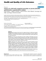
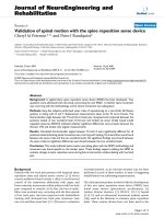
14 years male with CPFigure 1
14 years male with CP. shows a) preoperative AP and lateral radiogram; b) postoperative AP and lateral radiogram and c)
final follow-up AP and lateral radiogram of spine in a fourteen years boy with cerebral palsy. (Patient 5)
Journal of Orthopaedic Surgery and Research 2008, 3:23 />Page 5 of 8
(page number not for citation purposes)
FVC less than 35% (22%–33%) and six of them needed
post-op ventilation. However, we did not perform pulmo-
nary function tests postoperatively.
Abbreviations: WC: wheel-chair bound, WWS: walking
without support, Crutch: walking with crutch, walker:
walking with walker, PFT: pulmonary function test, PEF:
peak expiratory flow, FVC: forced vital capacity, FEV1:
forced expiratory volume during first second.
The average operating time was 6 hours 45 minutes (4
hours 30 minutes–10 hours). The average duration of
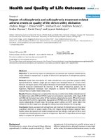
13 years girl with SMAFigure 2
13 years girl with SMA. shows a) preoperative AP and lateral radiogram; b) postoperative AP and lateral radiogram and c)
final follow-up AP and lateral radiogram of spine in a thirteen years girl with spinal muscular atrophy. (Patient 19)
Table 3: Values for duration of anesthesia, duration for operation, post operative ICU stay, ventilator support, hospital stay and
documentation of infection.
No Diagnosis Anaes time
(Hour:Min)
Op time
(Hour:Min)
EBL (mililiters) ICU (time) Ventilator
(time)
Infection Hospital Stay
(Days)
1 CP 11:10 10:00 4550 1 Day NO NO 32
2 CP 07:30 06:10 1250 NO NO NO 24
3 CP 06:50 04:30 1400 NO NO NO 30
4 CP 07:40 06:10 2100 NO NO NO 14
5 CP 08:00 06:40 1750 NO NO NO 26
6 CP 08:00 07:00 1000 NO NO NO 10
7 CP 09:00 07:20 5000 3 Days 36 Hours NO 42
8 DMD 08:10 06:30 2550 NO NO NO 24
9 DMD 07:45 05:30 1950 2 Days NO NO 15
10 DMD 06:50 04:50 1500 NO NO NO 10
11 DMD 07:00 06:00 2100 NO NO NO 17
12 DMD 11:35 09:00 4850 3 Days 25 Hours NO 19
13 DMD 08:40 07:30 2300 5 Hours NO NO 22
14 DMD 07:10 05:45 2750 6 Hours NO YES 7
15 DMD 06:40 04:50 1750 5.5 Days NO NO 18
16 DMD 08:20 06:40 2350 NO NO NO 18
17 DMD 08:25 06:10 2300 NO NO NO 17
18 SMA 05:50 04:50 1350 3 Hours NO NO 31
19 SMA 11:00 08:30 9000 NO NO NO 36
20 SMA 10:15 08:15 3750 NO NO NO 49
21 SMA 09:40 08:10 2600 NO NO NO 19
22 SMA 08:15 06:30 3150 1 Day 26 Hours NO 23
23 POLIO 10:50 08:50 3100 14 Days NO NO 18
24 POLIO 10:10 08:15 2100 1 Day 8 Days NO 38
25 MS 06:50 05:50 2100 2 Days NO NO 15
26 PARA 07:00 06:00 3500 10 Days 24 hours YES 60
Abbreviations: Anaes time: anaesthesia time, Op time: operation time, EBL: estimated blood loss, ICU: intensive care unit.
Journal of Orthopaedic Surgery and Research 2008, 3:23 />Page 6 of 8
(page number not for citation purposes)
anesthesia was 8 hours 24 min (5 hours 50 minutes–11
hours and 30 minutes). There was not much difference in
operating time between the group I (6 hours 7 minutes)
and group II (7 hrs 58 min) (table 3). The average intra-
operative blood loss was 2773 mililiters (1000–9000
mililiters). The average blood loss for patients who under-
went PVCR was 4535 mililiters, whereas it was 2123
mililiters for those who did not undergo PVCR. A total of
13 patients needed postoperative ICU care for an average
of 28.3 hours, except one poliomyelitis patient who was
in ICU for 2 weeks secondary to DIC and another patient
with traumatic paraplegia who had severe bleeding from
epidural vessels, was in ICU for 10 days. Six patients
required postoperative ventilation (all of them underwent
PVCR) for 24 to 36 hours except one poliomyelitis patient
who required ventilation for 8 days. The average duration
of hospitalization was 24.36 days.
Postoperatively all patients exhibited improvement in sit-
ting balance. Two patients who were wheelchair bound
preoperatively were able to walk with the help of walker
postoperatively and one wheel chair bound patient was
able to walk with the help of crutches (table 4). One SMA
patient who was able to walk with the help of walker pre-
operatively was able to walk with the help of crutches
postoperatively. None of the patient had deterioration in
sitting balance at final follow-up. Parents or care-takers of
all patients exhibited better personal and hygienic care
postoperatively.
Complications
Deep wound infection was seen in one patient with para-
plegia who had continuous bleeding from the operated
site for which exploration of the wound revealed bleeding
from an epidural vessel, which was cauterized. She devel-
oped wound dehiscence and deep infection and pus cul-
ture grew vancomycin resistant staphylococcus for which
she received teicoplanin and regular dressings. One
patient with poliomyelitis who had grade 3 power of the
lower limbs pre-operatively, developed grade 2 power
post-operatively but gradually improved to the pre-opera-
tive stage. He also had severe blood loss in the peri-oper-
ative period, went into DIC, and was in the ICU for 2
weeks. There was no mortality, pseudarthrosis or implant
failurein the study.
Discussion
Despite the magnitude of this surgery, successful outcome
of an operation for spinal deformity, secondary to neu-
romuscular disease, is considered beneficial by most
patients and/or their principal care providers
[7,15,18,19]. Aims of the surgery for neuromuscular scol-
iosis are safe correction of deformity, to stop curve pro-
gression, to maintain or recreate sitting balance and to
achieve a solid fusion of the balanced spine in the frontal
and sagittal planes [20,21]. The average age of the patients
in this study was 17.5 years. The increase in age results in
increasing stiffness and rigidity of the curve and difficulty
to achieve acceptable correction. The mean age in this
study was much higher than majority of studies, where
patients underwent earlier correction at around 12 years
of age or earlier [20,22,23]. However we achieved accept-
able correction in both Cobb's angle and pelvic obliquity
over al period of 25 months without significant loss of
postoperative correction.
We found that there were significant differences in PFT
values between curves greater than 90° and less than 90°.
Sussman noted an increased risk of postoperative pulmo-
nary problems when the FVC was less than 35% [13,24].
In our study, 10 patients had FVC less than 35% and 6 of
them needed postoperative ventilation, supporting Suss-
man's [25] findings. Postoperative PFT assessment is not
being done as a routine basis in our patients. Post-opera-
tive PFT values would indicate whether there was any
improvement of these parameters following surgery.
Therefore, we think, this is the limitation of present study.
Average blood loss for the patients who underwent PVCR
was 4535 milliliters, while it was only 2123 milliliters for
Table 4: Pre operative and postoperative ambulatory status with
preoperative pulmonary function tests.
Walking status pre-operative PFT
No Diagnosis pre-op post-op PEF FVC FEV1
1 CP WC Crutch FAIL FAIL FAIL
2 CP WWS WWS FAIL FAIL FAIL
3 CP WWS WWS 44 28 28
4 CP WWS WWS 121 97 95
5 CP WWS WWS 62 51 53
6 CP WWS WWS 66 46 Un Co-op
7 CP WWS WWS 41 24 25
8 DMD WC WC 46 33 29
9 DMD WC WC 63 71 74
10 DMD WC WC 32 14 15
11 DMD WC WC 61 49 53
12 DMD WC WC 42 31 32
13 DMD WC WC 73 60 61
14 DMD WC WC 73 69 72
15 DMD WC WC 148 138 147
16 DMD WC WC 43 38 41
17 DMD WC WC 43 23 21
18 SMA WWS WWS 80 91 82
19 SMA Walker Crutch 85 64 68
20 SMA WC WC 67 31 36
21 SMA WC WC 95 89 88
22 SMA WC WC 49 22 27
23 POLIO Crutch Crutch 51 33 31
24 POLIO WC Walker 70 65 63
25 MS WC WC 43 37 39
26 PARA WC Walker 37 24 28
Journal of Orthopaedic Surgery and Research 2008, 3:23 />Page 7 of 8
(page number not for citation purposes)
those who did not undergo PVCR. Other studies [21] with
comparable number of fusion levels had average blood
loss of 2.4 liters to 2.6 liters in normal fusions and 2.5 lit-
ers to 3.4 liters in patients who had associated PVCR. This
was the disadvantage of PVCR because it resulted in severe
blood loss, and in addition, all of them required postop-
erative ICU stay and six of the seven patients needed ven-
tilation. Kannan et al [26] comparing blood loss during
operation, found that the neuromuscular group had
greater blood loss than idiopathic scoliosis.
Average preoperative pelvic obliquity was 16° which
decreased to 9° after the surgery, and remained same even
at final follow-up. There is a growing controversy regard-
ing the distal extent of fusion in patients with neuromus-
cular scoliosis (figure 1). There was a general trend to
include the pelvis in all cases of neuromuscular scoliosis
to correct pelvic obliquity or to prevent its development
[20,26-30]. With all the problems described in the litera-
ture associated with pelvis fusion [9,31] few patients who
had pelvic obliquity greater than 15° and other patients
with pelvic obliquity less than 15° had severe lower
extremity contractures were chosen for pelvic fusion [32].
Recently, Tsirikos, et al [33,34] challenged the long-term
belief that fusion should be avoided in ambulatory
patients with CP. In our experience, we have noticed that
patients who have gross pelvic obliquity, do not exhibit
any problem with sitting balance without much progres-
sion over a short term. Therefore presently we do pelvic
fixation in patients with pelvic obliquity more than 15°
(figure 2) or with severe lower extremity contractures.
Westerlund et al [35] reported 66% correction in Cobb's
angle and 75% correction in pelvic obliquity in twenty-six
neuromuscular scoliosis with posterior-only unit rod
instrumentation. They did not perform any anterior pro-
cedure in their series and reported excellent results in
immature spine. Boachie-Adjei, et al [4] in their study
with 46 patients of neuromuscular scoliosis had an equal
number of patients in both the spastic and flaccid group
and concluded that corrections for scoliosis and pelvic
obliquity were similar in both the groups. In present
study, we have also found similar correction in both
Cobb's angle and pelvic obliquity without any anterior
procedure over a follow-up of 25 months. In addition,
none of the patient displayed deterioration in thoracic
kyphosis or lumbar lordosis at final follow-up and there-
fore we did not feel any need for anterior procedure for
the correction. Improvements in thoracic kyphosis and
lumbar lordosis resulted in to improved sitting balance.
Various authors [1,2,36] have used combined anterior
and posterior approaches to correct scoliosis, usually in
the presence of a very large or stiff curve. Their curves were
all less than 90° and they achieved correction rates from
41% to 71%, mostly by using Luque or Unit rod systems.
In the group with curve greater than 90° and the group
who underwent PVCR, we observed curve correction of
50.31% and 47.65% respectively. To achieve more correc-
tion, we prefer PVCR [21] at another extra level. The pedi-
cle screw system has an advantage of being a consolidated
fixation including all three columns [5,16,17]. This
greatly enhances the ability to simultaneously correct the
three dimensional nature of these complex spinal deform-
ities. Using the advantages of both pedicle screw and
PVCR, better correction can be expected. Various studies
have shown that posterior instrumentation with fusion
alone is sufficient to correct and to maintain even larger
and stiffer curves in neuromuscular scoliosis and it also
prevents crankshaft phenomenon in skeletally immature
patients [20,28,37,38]. Reports have also shown that the
addition of an anterior procedure, whether staged or
same-day, potentially contributes to the risk profile and
morbidity in these patients which further supports our cri-
teria of avoiding such measures unless clear benefit can be
supported. Our results support that neither anterior
release nor anterior arthrodesis is generally indicated to
obtain acceptable curve correction in even severe cases.
There are various reports suggesting use of hooks in tho-
racic level. The main purpose of using hook was to avoid
any neurological complications intraoperatively. How-
ever other reports [5,16,17] suggested that use of pedicle
screw provides stronger purchase and better rotational
correction in thoracic spine. Therefore we have used pedi-
cle screws all levels in the subjects and did not notice any
major neurological injury postoperatively. Our results are
also comparable with the hooks or any other implants.
In present study, all patients underwent for posterior-only
pedicle screw and fusion for neuromuscular scoliosis were
consecutive and not randomized which is, we think, the
limitation of study. If operations had been done in
selected patients, results would have been better than this.
However, majority of patients achieved acceptable correc-
tion in thoracic kyphosis and lumbar lordosis with
improvement in sitting balance showed the success of
treatment. In addition patients' parents or care takers have
also reported better nursing care after operation.
Conclusion
In conclusion, our series demonstrates the efficacy of pos-
terior-only spinal fusion using the pedicle screw fixation
for the definitive management of neuromuscular scolio-
sis. Curves greater than 90 degrees or rigid curves may
require additional PVCR at single level or if necessary, at
second level to achieve better outcome. Appropriate care
requires the availability of specialized personnel acting as
a multidisciplinary team. Our results demonstrate that
posterior-only pedicle screw fixation with PVCR if neces-
sary is effective in obtaining and maintaining alignment
Journal of Orthopaedic Surgery and Research 2008, 3:23 />Page 8 of 8
(page number not for citation purposes)
in the neuromuscular scoliosis population. This tech-
nique may avoid those risks incumbent with the addition
of an anterior approach.
Competing interests
The authors declare that they have no competing interests.
Authors' contributions
HNM has contributed in conception and design and
acquisition of data, analysis and interpretation of data,
drafting the manuscript and revising it critically, SWS has
contributed in conception and design of data, drafting the
manuscript and given the final approval of manuscript,
HRS has contributed in acquisition of data, revising the
manuscript critically and given the final approval, HMF
has contributed in drafting the manuscript and designing
of data and revising it critically and JHY has contributed
in acquisition of data and analysis and interpretation of
data.
All authors read and approved the final manuscript.
Acknowledgements
No acknowledgements
Each author certifies that he has no commercial associations (e.g. consul-
tancies, stock ownership, equity interests, patent/licensing arrangements,
etc) that might pose a conflict of interest in connection with the submitted
article.
References
1. Banta JV: Combined anterior and posterior fusion for spinal
deformity in Myelomeningocele. Spine 1990, 15:946-52.
2. Brown J, Zeller J, Swank S, et al.: Surgical and functional results
of spine fusion in spinal muscular atrophy. Spine 1988,
14:763-70.
3. Pehrsson K, Larsson S, Oden A, et al.: Long term follow-up of
patients with untreated scoliosis. A study of mortality,
causes of death, and symptoms. Spine 1992, 17:1091-6.
4. Bonnett C, Brown JC, Grow T: Thoracolumbar scoliosis in cer-
ebral palsy. J Bone Joint Surg [Am] 1976, 58:328-36.
5. Ferguson R, Allen B Jr: Considerations in the treatment of cer-
ebral palsy patients with spinal deformities. Orthop Clin North
Am 1988, 19(2):419-25.
6. Mubarak SJ, Morin WD, Leach J: Spinal fusion in Duchenne mus-
cular dystrophy:fixation and fusion to the sacropelvis? J Pedi-
atr Orthop 1993, 13:752-7.
7. Askin GN, Hallet R, Hare N, Webb JK: The outcome of scoliosis
surgery in the severely physically handicapped child. Spine
1997, 22:44-50.
8. Dewald RL, Faut MM: Anterior and posterior spinal fusion for
paralytic scoliosis. Spine 1979, 4:401-9.
9. Maloney W, Rinsky LA, Gamble JG: Simultaneous correction of
pelvic obliquity, Frontal plane and sagittal plane deformities
in neuromuscular scoliosis using a unit rod with segmental
sublaminar wires. J Pediatr Orthop 1990, 10:742-9.
10. O'Brien T, Akmakjian J, Ogin G, et al.: Comparison of one-staged
versus two-staged anterior/posterior spinal fusion of neu-
romuscular scoliosis. J Pediatr Orthop 1992, 12:610-5.
11. Bell D, Moseley C, Koreska J: Unit rod segmental spinal instru-
mentation in the management of patients with progressive
neuromuscular spinal deformity. Spine 1988, 14:1301-7.
12. Lonstein J: The Galveston technique using Luque or Cotrel-
Dubousset rods. Orthop Clin North Am 1994, 25:311-20.
13. Lonstein JE, Akbarnia BA: Operative treatment of spinal
deformities in patients with cerebral palsy or mental retar-
dation: an analysis of one hundred and seven cases. J Bone Joint
Surg [Am] 1983, 65:43-55.
14. Luque ER: Segmental Spinal Instrumentation for correction
of scoliosis. Clin Orthop Relat Res 1982:192-8.
15. Hamill CL, Lenke LG, Bridwell KH, et al.: The use of pedicle screw
fixation to improve correction in the lumbar spine of
patients with idiopathic scoliosis: is it warranted? Spine 1996,
21:1241-9.
16. Brown CA, Lenke LG, Bridwell KH, et al.: Complications of pedi-
atric thoracolumbar and lumbar pedicle screws. Spine 1998,
23:1566-71.
17. Granata C, Merlini L, Cervellati S, et al.: Long term results of spine
surgery in Duchenne muscular dystrophy. Neuromuscul Disord
1996, 6:61-8.
18. Cassidy C, Craig CL, Perry A, et al.: A reassessment of spinal sta-
bilization in severe cerebral palsy. J Pediatr Orthop 1994,
14:731-9.0.
19. Comstock CP, Leach J, Wenger DR: Scoliosis in total body
involvement cerebral palsy: analysis of surgical treatment
and patient and caregiver satisfaction. Spine 1998, 23:1412-25.
20. Broom MJ, Banta JV, Renshaw TS: Spinal fusion augmented by
Luque-rod segmental instrumentation for neuromuscular
scoliosis. J Bone Joint Surg [Am] 1989, 71:32-44.
21. Suk Se-Il, Kim JH, Kim WJ, et al.
: Posterior vertebral column
resection for severe spinal deformities. Spine 27(21):2374-82.
2002, Nov 1
22. Rang M: Cerebral palsy. In Lovell and Winter's Pediatric Orthopaedics
Edited by: Morrissey PT. Philadelphia: JB Lippincott; 1990:465-507.
23. Sussman M: Is fusion to the sacrum necessary in patients with
progressive flaccid neuromuscular disease? Orthop Trans 1987,
11:123-4.
24. Storer SK, Vitale MG, Hyman JE, et al.: Correction of adolescent
idiopathic scoliosis using thoracic pedicle screw fixation ver-
sus hook constructs. J Pediatr Orthop 2005, 25:415-9.
25. Sussman M: Advantage of early spinal stabilization and fusion
in patients with Duchenne muscular dystrophy. J Pediatr
Orthop 1984, 4:532-7.
26. Kannan S, Meert KL, Mooney JF, et al.: bleeding and coagulation
changes during spinal fusion surgery: a comparision of neu-
romuscular and idiopathic scoliosis patients. Pediatr Crit Care
Med 2002, 3(4):364-369.
27. Boachie-Adjei O, Lonstein JE, Winter RB, et al.: Management of
neuromuscular spinal deformities with Luque segmental
instrumentation. J Bone Joint Surg [Am] 1989, 71:548-62.
28. Dias R, Freeman M, Dabney K, et al.: Surgical correction of spinal
deformity using a unit rod in children with cerebral palsy. J
Pediatr Orthop 1996, 16:734-40.
29. Ferguson RL, Allen BL: Staged correction of neuromuscular
scoliosis. J Pediatr Orthop 1983, 3:555-62.
30. Larsson EL, Aaro S, Obery B: Activities and functional assess-
ment 1 year after spinal fusion for paralytic scoliosis. Eur Spine
J 1999, 8:100-9.
31. Marsh A, Edge G, Lehovsky J, et al.
: Spinal fusion in patients with
Duchenne's muscular dystrophy and a low forced vital capac-
ity. Eur Spine J 2003, 12:507-512.
32. Sussman M: Duchenne muscular dystrophy. J Am Acad Orthop
Surg 2002, 10:138-151.
33. Tsirikos AI, Chang WN, Shah SA, et al.: Preserving ambulatory
potential pediatric patients with cerebral palsy who undergo
spinal fusion using unit rod instrumentation. Spine 2003,
28:480-483.
34. Tsirikos AI, Chang WN, Dabney KW, et al.: Comparison of par-
ents' and caregivers' satisfaction after spinal fusion in chil-
dren with cerebral palsy. J Pediatr Orthop 2004, 24:54-58.
35. Westerlund IE, Gill SS, Jarosz TS, Abel MF, Blanco JS: Posterior-only
unit rod instrumentation and fusion for neuromuscular scol-
iosis. Spine 2001, 26(18):1984-89.
36. Sussman M: Posterior instrumentation and fusion of the tho-
racolumbar spine for treatment of neuromuscular scoliosis.
J Pediatr Orthop 1996, 16:304-13.
37. Burton DC, Asher MA, Lai SM: Scoliosis correction maintenance
in skeletally immature patients with idiopathic scoliosis: is
anterior fusion really necessary? Spine 2000, 25:61-8.
38. Renshaw T: Severe scoliosis in cerebral palsy: a comparison of
operative and nonoperative treatment. J Pediatr Orthop 1993,
13:412.