báo cáo hóa học:" Staged surgical treatment for severe and rigid scoliosis" docx
Bạn đang xem bản rút gọn của tài liệu. Xem và tải ngay bản đầy đủ của tài liệu tại đây (2.84 MB, 9 trang )
BioMed Central
Page 1 of 9
(page number not for citation purposes)
Journal of Orthopaedic Surgery and
Research
Open Access
Research article
Staged surgical treatment for severe and rigid scoliosis
Shi Yamin*, Li Li, Wei Xing, Gao Tianjun and Zhang Yupeng
Address: Department of Orthopedics, The 1st Affiliated Hospital to the General Hospital of PLA, Beijing, PR China
Email: Shi Yamin* - ; Li Li - ; Wei Xing - ; Gao Tianjun - ;
Zhang Yupeng -
* Corresponding author
Abstract
Background: A retrospective study of staged surgery for severe rigid scoliosis. The purpose of
this study was to evaluate the result of staged surgery in treatment of severe rigid scoliosis and to
discuss the indications.
Methods: From 1998 to 2006, 21 cases of severe rigid scoliosis with coronal Cobb angle more
than 80° were treated by staged surgeries including anterior release and halo-pelvic traction as first
stage surgery and posterior instrumentation and spinal fusion as second stage. Pedicle subtraction
osteotomy(PSO) was added in second stage according to spine rigidity. Among the 21 patients, 8
were male and 13 female with an average age of 15.3 years (rang from 4 to 23 years). The mean
pre-operative Cobb angle was 110.5° (80°-145°) with a mean spine flexibility of 13%. Radiological
parameters at different operative time points were analyzed (mean time of follow-up: 51 months).
Results: External appearance of all patients improved significantly. The average correction rate
was 65.2% (ranging from 39.8% to 79.5%) with mean correction loss of 2.23° at the end of follow-
up. No decompensation of trunk has been found. Mean distance between the midline of C7 and
midsacral line was 1.19 cm ± 0.51. Two patients had neurological complications: one patient had
motor deficit and recovered incompletely.
Conclusion: Staged operation and halo-pelvic traction offer a safe and effective way in treatment
of severe rigid scoliosis. Patients whose Cobb angle was more than 80° and the flexibility of the
spine was less than 20% should be treated in this way, and those whose flexibility of the spine was
less than 10% and the Cobb angle remained more than 70° after 1st stage anterior release and halo-
pelvic traction should undergo pedicle subtraction osteotomy (PSO) in the second surgery.
Background
Excellent outcomes of hemi vertebra excision, vertebral
body resection, and spinal osteotomy have been reported
for angular kyphosis or kyphoscoliosis. However, their
safety and effectiveness of these procedures have not been
estimated. It would be difficult to correct severe and rigid
spinal deformities satisfactorily by a single procedure in
consideration of the neurological safety. In consequence,
staged surgeries have been widely used in the treatment of
severe rigid scoliosis. Nevertheless, in few papers the
method of anterior releases followed by halo-pelvic trac-
tion has been mentioned.
There is a high risk in the surgical correction for severe
rigid scoliosis. A 5.3% incidence of permanent neurologi-
cal injury has been reported by Dutoit and the incidence
Published: 9 July 2008
Journal of Orthopaedic Surgery and Research 2008, 3:26 doi:10.1186/1749-799X-3-26
Received: 9 March 2007
Accepted: 9 July 2008
This article is available from: />© 2008 Yamin et al; licensee BioMed Central Ltd.
This is an Open Access article distributed under the terms of the Creative Commons Attribution License ( />),
which permits unrestricted use, distribution, and reproduction in any medium, provided the original work is properly cited.
Journal of Orthopaedic Surgery and Research 2008, 3:26 />Page 2 of 9
(page number not for citation purposes)
of transient neurological deficit was as high as 46% in
Luque's records. [1,2] Staged surgery has been used in the
treatment of severe rigid scoliosis to prevent neurological
compromise. The conventional staged surgery consists of
anterior release as first stage procedure and posterior spi-
nal fusion and instrumentation as second stage [3-6].
Nevertheless, in few papers the method of anterior
releases followed by halo-pelvic traction has been men-
tioned, and the indication of staged surgical methods is
discrepancy. With the development of the surgical and
anesthesia technology, combined anterior and posterior
procedure has been used in recent decade. However, its
advantage of reduced hospital stay and costs was not com-
parable to its higher complication rate. [7,8] This paper
evaluated the outcome of 21 cases with severe and rigid
scoliosis retrospectively treated with staged surgery and
the indication was discussed.
Materials and methods
From 1998 to 2006, 21 cases of severe rigid scoliosis were
treated with staged surgery. among the 21 patients, 8 were
male and 13 female with an average age of 15.3 years
(rang from 4 to 23 years). The scoliosis was classified as
congenital in 11 cases, idiopathic in 7 and neuroinomato-
sis in 2. The mean preoperative coronary Cobb angle was
110.5° (range from 80° to 145°), the mean Cobb angle
was 94.5° (70°-133°) on suspension view. Flexibility was
used to estimate the rigidity of the curve, it means (Preop-
erative Cobb's angle – Bending Cobb's angle)/Preopera-
tive Cobb angle × 100%. The curve was considered
stiffness when it was more than 30%. The mean flexibility
of the spine was 13% (range 1.5% to 27.3%)in this group.
2 cases of congenital scoliosis were confirmed diastemat-
omyelia in the spinal canal by CT and MRI. Mild or severe
limited dysfunctions of ventilation existed in all the cases.
All cases were grouped into two. 13 cases in group A were
performed with anterior release and halo-pelvic traction
in first stage; and then posterior spinal fusion and instru-
mentation in second stage. In group B, 9 cases were
treated with the same procedure as in group one in the
first stage; and then posterior spinal fusion and instru-
mentation plus wedge resection. The vertebral osteotomy
was done from T7 to L2. SEP and wake-up test were used
in all patients during operation.
Principal Surgical Techniques and Highlights
Anterior spine release and deformity correction with halo-pelvic
distraction apparatus
Apex and adjacent vertebra were exposed from convex
side through thoracic pathway, usually only 4 to 6 discec-
tomy could be performed because of the limitation of
exposure. Significant abnormal intervertebral mobility
should be confirmed. during operation.
4 pelvic screws were inserted at sites 2 to 2.5 cm posterior-
inferior to bilateral anterior-superior iliac spine and pos-
terior superior iliac spine respectively, which were linked
to pelvic ring and fixed. 4 cranial screws were inserted at
sites 1 cm superior-lateral to bilateral arcus superciliaris
and 2 cm superior to bilateral mamillary process respec-
tively.4 connector bars were linked between halo and pel-
vic ring.
Lengthening of Halo-pelvic distraction apparatus began 3
to 5 days after operation, with the extent of 2 times a day
and 3 to 5 mm each time. The indications of traction limit
include early appearance of clinical symptoms of cranial
nerves or spinal cord, muscular pain, gastrointestinal
symptoms which affected food intake even if the length-
ening was stopped, and severe pin tract infection caused
by the loose of screws.
Insertion of segmental pedicle screw system and correction of
deformity
During the second stage pedicle screws were inserted con-
tinuously or interruptedly in the concave side of the sta-
bility region [9], 2 screws should be inserted consecutively
at up end and low end and apex vertebra to decrease the
regional stress and strengthen correction force on the apex
vertebra. Fasten screws were tapped into the tugs of pedi-
cle screws after the pre-bending rod was put into tugs of
pedicle screws. Correction force to the spine was achieved
by rotating of the rod. The direction of rod rotation was
based on the types of scoliosis. The rod should be rotated
from the convex side to concave side in thoracic scoliosis
to transform the scoliosis in coronal plan to kyphosis in
sagital plan. For the lumbar scoliosis, the rod should be
rotated from concave side to convex side to transform the
scoliosis in coronal plan to lordosis in sagital plan. The
rod should be rotated in the same direction in thoraco-
lumbar double-curve scoliosis to achieve correction of
double-curve scoliosis deformity and restoration of sagital
curve of thoracic and lumbar spine.
Wedge-shape osteotomy of apex vertebra and deformity correction
Single vertebra or intervertebral disk space can be selected
as the center of wedge-shape osteotomy according to the
type of apex vertebra. To prevent possible neurological
symptoms caused by shrinkage of spinal cord, adjacent
half-laminectomy superior and inferior to the osteotomy
site should be performed for canal decompression.
Abnormities in spinal canal (bony crista or septation) or
spinal stenosis should be managed before osteotomy and
deformity correction. Exposure of osteotomy region
began from the convex side of apex vertebra, with the soft
tissue dissected sub-periosteally. The anterior-lateral side
of the apex vertebra or intervertebral disk space should be
exposed completely. According to the preoperative
Journal of Orthopaedic Surgery and Research 2008, 3:26 />Page 3 of 9
(page number not for citation purposes)
design, osteotomy of apex vertebra or adjacent vertebras
with apex center in disk space was performed.
The nerve root at the level of excision should be identified
and protected, particularly in the lumbar region, but some
severe cases in the thoracic region, the nerve root had to
be cut and left in place, this helps protect the dural tube
because the curette can be levered safely on this bone and
avoiding traction forces to the cord. And then, osteotomy
site was temporally fixed with rod to prevent abnormal
movement. The osteotomy of concavity side was per-
formed in the same way. Temporary rod in the convex side
was removed after the pre-bending rod was put in concave
side. The osteotomy space in the convex side was gradu-
ally closed by slowly rotating the rod in the concave side,
then pre-bending rod in the convex side was fixed, the
osteotomy space in the convex side might be closed by
proper compression the adjacent pedicle screws on the
basis of the size of the osteotomy space and the extent of
spinal cord shrinkage. Some epidural and bone bleeding
is to be expected and can be controlled with gel foam,
bone wax, and bipolar cautery.
Results
In a total of 21 patients, the average Cobb angle was 62°
(range 40° to 89°) after first-stage release and traction sur-
gical procedures, the average correction rate was
44.2%(range 23.9 to 63.9%). After second-stage correc-
tion with instrumentation, the average Cobb angle was
39.4° (range 22° to 73°). The average correction rate was
65.2% (range 39.8% to 79.5%).
Preoperative deformity degree and clinical effects were
investigated and analyzed with SPSS 11.0(SPSS, Inc., Chi-
cago, IL) (Tab 1): Because of heterogeneity of variance in
age of two groups, WilCoxon rank sum test was used and
demonstrated no significant difference in age (P > 0.05),
analysis of variance demonstrated no significant differ-
ence in preoperative Cobb angle (P > 0.05), curve correc-
tion rate after traction surgical procedure (P > 0.05), But
there was significant difference in spine flexibility (P <
0.05) between the 2 groups. (Table 1)
A total of 21 patients were followed up after operation.
The average follow up period was 51 months (range 5 to
81). At one year after surgery, 20 patients' showed a solid
segments fusion with no hardware failure. Average loss of
correction rate was 2.1% (range 1.3% to 6.1%). No
decompensation findings have been observed. Mean dis-
tance between the midline of C7 and midsacral line was
1.16 cm ± 0.54. One pedicle screw come out at 3 year after
surgery, the pseudoarthroses were resected in the revision
surgery. The rate of neurological complications was 9.5%
(2/21 patients); and these two patients all were subjects of
congenital scoliosis. One patient showed temporary para-
plegia at the level of the osteotomy site, but completely
recovered within 10 days after the additional decompres-
sion of vertebral canal and treated with hormone and
dehydration; the other one showed permanent neurolog-
ical deficit. At both lower extremities during the derota-
tion procedure. He recovered to III-IV muscle grade, but
there were not significant changes at 67 months after sur-
gery.
Radiographic assessment for sagittal balance was per-
formed by measuring thoracic kyphosis, lumbar lordosis,
distance between the vertical line on anterosuperior point
of T1 and that of S1, and sacral inclination. Clinical out-
comes were assessed by questionnaire measuring changes
in physical function, indoor activity, outdoor activity, psy-
chosocial activity, pain, and patient satisfaction with sur-
gery.
The mean trunk shift in global sagittal balance was 21 mm
before surgery, becoming 3 mm after surgery.
Final follow-up radiograph showed an increase in lumbar
lordosis from 20.1 degrees to 44.6 degrees (an increase of
24 degrees), whereas thoracic kyphosis remained stable
from 87 degrees to 54 degrees. Sagittal imbalance signifi-
Table 1: Evaluation of the outcome in staged surgical methods for severe rigid scoliosis
Group Cases Age Pre-OP
Cobb
Suspension
Cobb
Flexibility
▲
PO-Traction
Cobb
Traction
Rate
PO-OP
Cobb
Correction
rate(%)
Staged
Operation
12 4~23
(14.4)
80~145°
(112.5°)*
70~133°
(94.1°)
1.5~27.3%
(14.9%)
40~89°
(60.0°)
23.9~63.9%
(47.8%)
22~58°
(40.6°)
50.4~79.5%
(65.4%)
Staged+
Osteotomy
96~21
(16.8)
90~132°
(107.5°)
78~124°
(95.5°)
5.6~16.1%
(10.1%)
46~85°
(65.0°)
27.8~54.9%
(38.9%)
30~49°
(37.5°)
59.3~72.3
(65.0%)
Total 21 4~23
(15.3)
80~145°
(110.5°)
70~133°
(94.5°)
1.5~27.3%
(13.0%)
40~89°
(62.0°)
23.9~63.9%
(44.2%)
22~73°
(39.4°)
39.8~79.5%
(65.2%)
*Significant difference (p < 0.05) between two groups in the flexibility of the spine and no significant difference in age, Cobb angle and correction
rate.
Number in sign of aggregation is average value.
▲ Flexibility degree = (Pre-OP Cobb- Hang-up Cobb)/Pre-OP Cobb*100%
Journal of Orthopaedic Surgery and Research 2008, 3:26 />Page 4 of 9
(page number not for citation purposes)
cantly improved, whereas sacral inclination increased
from 8 degrees to 24 degrees. Satisfactory clinical outcome
was achieved; however, clinical improvements did not
correlate with changes in radiological measurements.
Discussion
The therapeutic efficacy of scoliosis is influenced by many
factors, such as the severity of deformity, spine flexibility,
patient's age, type of deformity, and combined other
deformities. Severe scoliosis is more difficult to treat than
usual ones. As the spine deformity is severe and stiff, and
the spinal cord has poor tolerance to the traction. It is dif-
ficult to complete the correction, and the probability of
nerve deficit increases. Moreover, because severe scoliosis
is usually combined with heart or lung disfuncitons, the
operation is of relatively high risk.
The scoliosis severity is the chief factor that may affect the
outcomes of deformity correction. Usually, if the coronal
Cobb's angle is more than 80° and the spine flexibility is
less than 20%, anterior loosen combined with halo-pelvic
traction should be accepted, then followed by posterior
correction in the second stage. (Fig 1, 2, 3, 4, 5, 6, 7, 8) For
the patients with neurological symptoms preoperatively,
halo-pelvic traction can also be used to prevent the neuro-
logical deficit from aggravating. As the spinal cord can
creep slowly, and the halo-pelvic traction can provide gen-
tle correction on spine, a good correction can be achieved.
Furthermore, even if neurological complication appears
during traction, the halo-pelvic device can be adjusted to
relieve it. Therefore, although the halo-pelvic device has
some disadvantages (e.g. hardware complicated and nurs-
ing problems), it is an alternative method to the preven-
tion of neurovascular complications in the treatment of
severe and rigid scoliosis without any major or permanent
neurological deficit.
For those cases with the spine flexibility less than 10%,
remained Cobb angle more than 60~70° after halo-pelvic
traction, nerve deficit reappear in the later stage of trac-
tion, and most severe congenital scoliosis up the adoles-
cent age, it is difficult to get a good correction only using
the posterior bar rotation in the second stage, so osteot-
omy should be used(Fig 9, 10, 11, 12, 13, 14, 15). Accord-
ing to this research, the spine flexibility of the first group
is obviously less than the second group; however, the cor-
M,12Y, neurofibromatosis scoliosis, double thoracic curveFigure 1
M,12Y, neurofibromatosis scoliosis, double thoracic curve.
Suspension view shows the flexibility of spineFigure 2
Suspension view shows the flexibility of spine.
Bending view shows the change of deformityFigure 3
Bending view shows the change of deformity.
Journal of Orthopaedic Surgery and Research 2008, 3:26 />Page 5 of 9
(page number not for citation purposes)
rection rate has no significant difference between the two
teams. It is proved that osteotomy is very effective for the
correction of the severe scoliosis.
Though osteotomy is useful in the treatment of scoliosis,
it can bring some complications, especially the nerve def-
icit. Bradford etc. had performed 24 cases of osteotomy,
and 3 of those had nerve deficit (12.5%) [10]. Among the
3 cases, muscle strength of ankle flexion weakened in 2
cases, and quadriceps femoris weakened in 1 case. Con-
sidering the possible rather too big local lumbar curve,
vertebral canal decompression was performed, and a good
recovery was achieved 6 months later. Berven etc. reported
a series of 13 cases undergoing osteotomy [11]. Leg palsy
happened in 4 cases (30.8%). These cases got complete
reablement half a year later. As to our research, of the 2
cases with leg sensory motor dysfunction, 1 case had
undergone osteotomy. The reason was probably that too
big range of osteotomy, the spinal cord shrinked after the
18 days after anterior release and halo-pelvic tractionFigure 4
18 days after anterior release and halo-pelvic traction. The
correction rate is 37%.
The correction rate is 51% after second operationFigure 5
The correction rate is 51% after second operation.
No correction loss at follow-up 6 months laterFigure 6
No correction loss at follow-up 6 months later.
M, 21Y, Idiopathic kyphoscoliosisFigure 9
M, 21Y, Idiopathic kyphoscoliosis.
Journal of Orthopaedic Surgery and Research 2008, 3:26 />Page 6 of 9
(page number not for citation purposes)
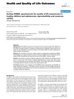
Body image a: pre-operation b: after anterior release and halo-pelvic tractionFigure 8
Body image a: pre-operation b: after anterior release and halo-pelvic traction. c: after second stage correction d: follow-up 6
months later.
Body image a: pre-operation b: after anterior release and halo-pelvic tractionFigure 7
Body image a: pre-operation b: after anterior release and halo-pelvic traction. c: after second stage correction d: follow-up 6
months later.
Journal of Orthopaedic Surgery and Research 2008, 3:26 />Page 7 of 9
(page number not for citation purposes)
gap closed, so the vertebral canal was relatively narrow.
For this case, SEP showed the latent period increased
(>30%), and the wave amplitude decreased (>50%) dur-
ing operation monitoring. The symptoms disappeared 1
week later after the enlarged decompression of vertebral
canal and other treatment postoperatively. All of the cases
in this research and other literatures had no nonreversible
nerve deficit due to osteotomy.
Current literatures say on the standard of care for severe
scoliosis that the treatment approach is different to the
subjects in this paper. Dr.Luhmann SJ, and Dr.Lenke LG
recently address that anterior and posterior spinal fusion
of large thoracic curves allows greater coronal correction
of thoracic curves between 70 degrees and 100 degrees,
when compared with PSF alone using thoracic hook con-
structs, but not with the use of thoracic pedicle screw con-
structs[12]. Scoliosis surgeons not using pedicle screw
constructs need to decide if the modest improvement in
The correction rates are 65.2% and 74.1% after second stage osteotomy and instrumentationFigure 13
The correction rates are 65.2% and 74.1% after second stage
osteotomy and instrumentation.
Bending view shows the change of deformityFigure 11
Bending view shows the change of deformity.
Suspension view shows the flexibility of spineFigure 10
Suspension view shows the flexibility of spine.
days after anterior release and halo-pelvic tractionFigure 12
days after anterior release and halo-pelvic traction. The cor-
rection rate is 35.6% and 50%.
Journal of Orthopaedic Surgery and Research 2008, 3:26 />Page 8 of 9
(page number not for citation purposes)
Body image a: pre-operation b: after anterior release and halo-pelvic tractionFigure 15
Body image a: pre-operation b: after anterior release and halo-pelvic traction. c: after second stage correction.
Body image a: pre-operation b: after anterior release and halo-pelvic tractionFigure 14
Body image a: pre-operation b: after anterior release and halo-pelvic traction. c: after second stage correction.
Publish with BioMed Central and every
scientist can read your work free of charge
"BioMed Central will be the most significant development for
disseminating the results of biomedical research in our lifetime."
Sir Paul Nurse, Cancer Research UK
Your research papers will be:
available free of charge to the entire biomedical community
peer reviewed and published immediately upon acceptance
cited in PubMed and archived on PubMed Central
yours — you keep the copyright
Submit your manuscript here:
/>BioMedcentral
Journal of Orthopaedic Surgery and Research 2008, 3:26 />Page 9 of 9
(page number not for citation purposes)
coronal correction with a combined approach justifies its
routine use in this patient population.
Dobbs MB and Lenke LG said in their patient population
with often restrictive preoperative pulmonary function
[13], a posterior-only approach with the use of an all-
pedicle screw construct has the advantage of providing the
same correction as an anterior/posterior spinal fusion,
without the need for entering the thorax and more nega-
tively impacting pulmonary function.
One of the main technical problems we encountered in
this mode of treatment is how to protect spinal cord dur-
ing pedical subtraction osteotomy. Procedures such as
osteotomy may be associated with a significant threat of
neurological complications. In my experiences, we must
stick to 3 key points (1) A temporary rod must be used
inserting to the convex side after osteotomy on this side to
prevent shear forces; (2) Do the additional decompres-
sion after derotation and closing of the osteotomy gap to
confirm there is no compression to the cord; (3) SEP or
MEP monitoring and wake-up test during and after the
derotation correction. Because experiences with this pro-
cedure are fairly recent, longer follow-up is required to
confirm whether this technique is reliable and efficacious.
Conclusion
As the spinal cord of the cases with severe rigid scoliosis
has poor tolerance to the traction, there is a high risk dur-
ing the correction, and the staged operation, especially the
Halo-pelvic distraction is an effective method to prevent
neurological complications. Usually, if the coronal
Cobb's is more than 80°, and the flexibility is less than
20%, anterior release with halo-pelvic traction should be
suggested, and followed by posterior correction with
instrumentations in the second stage. For the severe and
rigid cases with the flexibility less than 10%, and the mag-
nitude of curve more than 60~70° after halo-pelvic trac-
tion, the patients should undergo pedical subtraction
osteotomy(PSO) with instrumentations in the second sur-
gery.
Consent
Written informed consent was obtained from the patient
for publication of this case report and accompanying
images.
Authors' contributions
SY in charge of all the study and perform all operations,
LL perform all operations and complete the manuscript,
WX, GT, ZY complete data collection and radiograph
measurement.
Acknowledgements
The authors thank Professor Hou ShuXun, for his guidance, and Wang Hua-
Dong, Li QingMei, for their warmly assistances.
No funds were received in support of this work.
References
1. Luque ER: The correction of postural curves of Spine 1982, 7:270.
2. Dutoit M, Rigault P, Pouliquen JC, Padovani JP, Beneux J, Pasteyer J,
Merckx J, Guyonvarch G: Surgical treatment of scoliosis of 100
degrees and greater in children and adolescent (neurological
and myopathic scoliosis excluded). Apropos of a series of 66
cases. Rev Chir orthop Reparatrice Appar Mot 1985, 71:549-562.
3. Shi YM, Hou SX, Li L, Wang HD, Gao TJ, Wei X: Prevention and
management of the neurological complications during the
treatment of severe scoliosis. Zhonghua Wai Ke Za Zhi
45(8):517-9. 2007 Apr 15
4. Zhang YG, Wang Y, Zhang XS: [Clinical study on transpedicular
spinal osteotomy and vertebrectomy in 125 cases of severe
rigid spinal deformity]. Zhonghua Wai Ke Za Zhi 45(8):525-8.
2007 Apr 15
5. McMaster MJ, Singh H: The surgical management of congenital
kyphosis and kyphoscoliosis. Spine 26(19):2146-54. 2001 Oct 1
6. yong Qu, lihua Zhu, jinhua Liu: 90° yi shang ji zhu ce tu de shou
shu ce lue ji fang fa. Zhong Hua Wai Ke Za Zhi 2001, 39:102-10.
7. Tsirikos AI, Chang WN, Dabney KW, Miller F: Comparison of one-
stage versus tow-stage anteroposterior spinal fusion in pedi-
atric patients with cerebral palsy and neuromuscular scolio-
sis. Spine 2003, 28:1300-5.
8. Shen Jianxiong, Qiu Guixing, Wang Yipeng, Zhang Zhehai, Zhao Yu:
Comparison of 1-stage versus 2-stage anterior and posterior
spinal fusion for severe and rigid idiopathic scoliosis -a rand-
omized prospective study. Spine 2006, 31:2525-2528.
9. Suk SI, Lee CK, Kim WJ, Chung YJ, Park YB: Segmental pedicle
screw fixation in the treatment of thoracic idiopathic scolio-
sis. Spine 1995, 20:1399-1405.
10. Bradford DS, Tribus CB: Vertebral column resection for the
treatment of rigid coronal decompensation. Spine 1997,
22:1590-1599.
11. Berven SH, Deviren V, Smith JA, Emami A, Hu SS, Bradford DS: Man-
agement of fixed sagittal plane deformity: resules of the
transpedicular wedge resection osteotomy.
Spine 2001,
26:2036-2043.
12. Luhmann SJ, Lenke LG, Kim YJ, Bridwell KH, Schootman M: Tho-
racic adolescent idiopathic scoliosis curves between 70
degrees and 100 degrees: is anterior release necessary? Spine
30(18):2061-7. 2005 Sep 15
13. Dobbs MB, Lenke LG, Kim YJ, Luhmann SJ, Bridwell KH: Anterior/
posterior spinal instrumentation versus posterior instru-
mentation alone for the treatment of adolescent idiopathic
scoliotic curves more than 90 degrees. Spine 31(20):2386-91.
2006 Sep 15