báo cáo hóa học:" Post-traumatic flexion contractures of the elbow: Operative treatment via the limited lateral approach" doc
Bạn đang xem bản rút gọn của tài liệu. Xem và tải ngay bản đầy đủ của tài liệu tại đây (669.63 KB, 5 trang )
BioMed Central
Page 1 of 5
(page number not for citation purposes)
Journal of Orthopaedic Surgery and
Research
Open Access
Research article
Post-traumatic flexion contractures of the elbow: Operative
treatment via the limited lateral approach
Mark D Brinsden*, Andrew J Carr and Jonathan L Rees
Address: The Nuffield Department of Orthopaedic Surgery, University of Oxford, Oxford, UK
Email: Mark D Brinsden* - ; Andrew J Carr - ;
Jonathan L Rees -
* Corresponding author
Abstract
Varying surgical techniques, patient groups and results have been described regards the surgical
treatment of post traumatic flexion contracture of the elbow. We present our experience using
the limited lateral approach on patients with carefully defined contracture types.
Surgical release of post-traumatic flexion contracture of the elbow was performed in 23 patients
via a limited lateral approach. All patients had an established flexion contracture with significant
functional deficit. Contracture types were classified as either extrinsic if the contracture was not
associated with damage to the joint surface or as intrinsic if it was.
Overall, the mean pre-operative deformity was 55 degrees (95%CI 48 – 61) which was corrected
at the time of surgery to 17 degrees (95%CI 12 – 22). At short-term follow-up (7.5 months) the
mean residual deformity was 25 degrees (95%CI 19 – 30) and at medium-term follow-up (43
months) it was 32 degrees (95%CI 25 – 39). This deformity correction was significant (p < 0.01).
One patient suffered a post-operative complication with transient dysaesthesia in the distribution
of the ulnar nerve, which had resolved at six weeks. Sixteen patients had an extrinsic contracture
and seven an intrinsic. Although all patients were satisfied with the results of their surgery, patients
with an extrinsic contracture had significantly (p = 0.02) better results than those with an intrinsic
contracture. (28 degrees compared to 48 degrees at medium term follow up).
Surgical release of post-traumatic flexion contracture of the elbow via a limited lateral approach is
a safe technique, which reliably improves extension especially for extrinsic contractures. In this
series all
patients with an extrinsic contracture regained a functional range of movement and were
satisfied with their surgery.
Introduction
Elbow Stiffness with loss of function is a common disa-
bling problem that usually arises as a complication of
trauma [1-5], but may also occur following burns[6,7]. or
head injury [8,9] or in association with degenerative,
inflammatory or haemophiliac [10] arthropathy and con-
genital malformations [11]. The degree of stiffness is
related to the severity of the injury and the duration of
immobilisation at initial treatment [12,13]. Loss of elbow
extension commonly produces a significant functional
deficit [14]. Elbow contractures can be classified as extrin-
sic or intrinsic according to the underlying aetiology [15].
Published: 10 September 2008
Journal of Orthopaedic Surgery and Research 2008, 3:39 doi:10.1186/1749-799X-3-39
Received: 29 February 2008
Accepted: 10 September 2008
This article is available from: />© 2008 Brinsden et al; licensee BioMed Central Ltd.
This is an Open Access article distributed under the terms of the Creative Commons Attribution License ( />),
which permits unrestricted use, distribution, and reproduction in any medium, provided the original work is properly cited.
Journal of Orthopaedic Surgery and Research 2008, 3:39 />Page 2 of 5
(page number not for citation purposes)
Extrinsic contractures involve the peri-articular soft-tis-
sues with a normal or near normal articular surface.
Intrinsic factors include disruption of the normal articular
surface, osteophytes, intra-articular loose bodies and sec-
ondary osteoarthritis.
When non-operative treatments such as static or dynamic
splinting [16-22] fail then surgery is often considered.
Many surgical techniques have been described for estab-
lished contractures with significant functional impair-
ment. These include: manipulation-under-anaesthesia
[23]; arthroscopic release [24-26]; open capsulectomy via
anterior [27-31], posterior [13], medial [32,33], lateral
[30,34-37], or combined approaches [38].
We present our experience of the 'mini-open' lateral
approach to the elbow to correct an extension deficit in a
series of patients with an established post-traumatic flex-
ion contracture of both intrinsic and extrinsic types [35].
This approach facilitates access to the anterior capsule, the
lateral ligament complex and radio-capitellum joint. It is
also possible to access the posterior part of the elbow joint
and olecranon of required.
Methods
Between 1998 and 2004, 23 patients referred to our unit
were treated surgically for a post traumatic flexion con-
tracture of the elbow. The indication for surgery in all was
an established functionally significant extension deficit
that had failed non-operative treatment with at least 9
months having elapsed since injury. In each case the con-
tracture was classified as extrinsic or intrinsic after assess-
ment with clinical examination and plain radiographs
and the pre-operative flexion contracture recorded. All
patients consented to have their surgery under general
anaesthesia and regional block with a tourniquet. The lat-
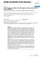
eral column approach was used with a small 8 cm (10 cm
if larger patient) incision centred over the lateral epi-
condyle (Figure 1). The same operative sequence was fol-
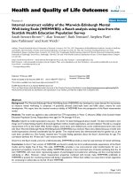
lowed for all patients. All patients had a section of anterior
capsule, extending across the entire anterior aspect of the
joint, excised under direct vision (Figure 2). If the radial
head was significantly damaged and determined at this
point to be a block to extension then it was excised. Next
if extension was still limited and the lateral collateral liga-
ment complex appeared tight it was z-lengthened rather
than sacrificed. Cases of intrinsic contracture also had any
intra-articular lesion addressed. Any implanted metal-
work that was easily accessible and may influence move-
ment or cause pain was also removed as were any
olecranonosteophytes identified on pre-operative imag-
ing. If ulnar nerve symptoms and signs were present then
an ulnar nerve release with subcutaneous transposition
was performed via a separate medial incision. No distract-
ing devices were used. The tourniquet was released, hae-
mostasis secured with electro-cautery and a drain placed
in the peri-articular soft-tissues. The residual "on-table"
passive deformity was assessed after wound closure and
before the application of dressings.
Post-operatively the limb was immobilised overnight in
maximum extension using a plaster slab. The drain was
removed and the cast replaced by a static, extension ther-
moplastic splint the next day. All patients were discharged
on the first post-operative day. No prophylaxis was given
to prevent heterotopic bone formation. The splint was
worn continuously for two weeks and then at night for six
A clinical photograph showing the anterior capsule of the elbow through the lateral approachFigure 1
A clinical photograph showing the anterior capsule of
the elbow through the lateral approach.
A clinical photograph showing the excised anterior capsuleFigure 2
A clinical photograph showing the excised anterior
capsule.
Journal of Orthopaedic Surgery and Research 2008, 3:39 />Page 3 of 5
(page number not for citation purposes)
weeks. Physiotherapy with active extension exercises com-
menced after two weeks in the presence of satisfactory
wound healing. Short-term results were assessed by clini-
cal review while medium-term follow-up was conducted
using a telephone questionnaire and patient based
deformity outlines as previously used by Morrey [39]. The
telephone questionnaire consisted of two questions; 'Are
you happy with the results of your surgery?' and 'In retro-
spect would you have the surgery again?'. These assess-
ment methods were used as most patients were tertiary
referrals to our unit, living many miles away and were
reluctant to return for a further appointment as they were
satisfied and doing well.
Results
In the study group there were 15 males and 8 females. The
median age was 35 yrs (range 16 – 52 yrs). The contracture
was post-traumatic in all cases (fracture with dislocation n
= 9; fracture n = 9; dislocation n = 3; and soft-tissue injury
n = 2). Sixteen patients had an extrinsic contracture and 7
patients had an intrinsic aetiology.
All patients underwent anterior capsulectomy and addi-
tional procedures included: Z-lengthening of lateral col-
lateral ligament n = 8; excision of radial head n = 3;
removal of metalwork n = 3; excision of olecranon osteo-
phyte n = 2; and ulna nerve transposition (via a separate
medial incision) n = 2. Patient demographics, operative
procedures and serial elbow deformities are listed in Table
1.
Short term follow-up was available at 7.5 months (95%CI
4 – 11) in all patients and medium term follow-up at 43
months (95%CI 30 – 56) in 20 patients (87%). Overall,
the mean pre-operative flexion deformity was 55 degrees
(95%CI 48 – 61). Surgery reduced the mean "on-table"
deformity to 17 degrees (95%CI 12 – 22). The short term
mean residual deformity was 25 degrees (95%CI 19 – 30)
and 32 degrees (95%CI 25 – 39) at medium term follow-
up. The improvement in the fixed-flexion deformity was
significant at both short-term and medium-term follow-
up (paired t-test – p < 0.01).
Sub group analysis of extrinsic and intrinsic groups
revealed:
Group One (extrinsic) patients had a mean pre-operative
flexion deformity of 53 degrees (95%CI 47 – 59); a mean
"on-table" correction to 13 degrees (95%CI 7 – 19); short
term deformity of 20 degrees (95%CI 16 – 25); and
medium term deformity of 28 degrees (95%CI 22 – 34).
Table 1: Demographics of patients who underwent surgical correction of post-traumatic flexion contracture of the elbow
Deformity (degrees)
Patient Age Diagnosis Classification Operation Pre-op Peri-op Short Term Medium Term
1 30 Soft Tissue Injury Extrinsic AC 40 0 20 35
2 44 Dislocation Extrinsic AC 40 0 10 10
3 16 Fracture/Dislocation Extrinsic AC 60 5 15 15
4 38 Fracture Extrinsic AC 65 30 30 30
5 29 Fracture/Dislocation Extrinsic AC, ZLCL 55 30 30 40
6 48 Dislocation Extrinsic AC, ZLCL 60 20 30 30
7 31 Fracture Extrinsic AC 40 5 10 20
8 49 Dislocation Extrinsic AC 70 0 30 30
9 29 Fracture Extrinsic AC, ZLCL 45 20 20 30
10 41 Fracture/Dislocation Extrinsic AC 60 15 20 30
11 35 Fracture/Dislocation Extrinsic AC 60 10 15 40
12 16 Fracture Extrinsic AC 50 20 20 30
13 26 Soft Tissue Injury Extrinsic AC, ZLCL 70 10 10 N/A
14 52 Fracture/Dislocation Extrinsic AC 40 10 20 20
15 40 Fracture/Dislocation Intrinsic AC, ZLCL, EOO 60 20 40 40
16 29 Fracture/Dislocation Extrinsic AC, ZLCL 50 30 30 45
17 18 Fracture Intrinsic AC, ERH 70 30 40 45
18 37 Fracture/Dislocation Intrinsic AC, ERH 20 0 0 N/A
19 41 Fracture/Dislocation Intrinsic AC, EOO 60 20 40 N/A
20 26 Fracture Extrinsic AC, ROM 50 30 30 10
21 50 Fracture Intrinsic AC, ZLCL, ROM, UNT 60 40 50 40
22 43 Fracture Intrinsic AC, ERH 50 30 30 45
23 32 Fracture Intrinsic AC, ZLCL, ROM, UNT 90 30 40 70
Key: AC = Anterior Capsulectomy; ZLCL = Z-lengthening Lateral Collateral Ligament; ERH = Excision of Radial Head; EOO = Excision of
OlecranonOsteophyte; ROM = Removal of Metalwork; UNT = Ulna Nerve Transposition.
Journal of Orthopaedic Surgery and Research 2008, 3:39 />Page 4 of 5
(page number not for citation purposes)
Group Two (intrinsic) patients had a mean pre-operative
flexion deformity of 57 degrees (95%CI 40 – 74); a mean
"on-table" correction to 25 degrees (95%CI 15 – 35);
short term deformity of 33 degrees (95%CI 21 – 46); and
medium term deformity of 48 degrees (95%CI 32 – 64).
The difference between the groups was significant at short
term (two sample independent t-test – p = 0.02) and
medium term (p = 0.05) follow-up.
All patients were satisfied with their surgery and would
undergo it again. No patients reported a loss or change in
their maximum flexion. One patient had a post-operative
complication with transient dysaesthesia in the distribu-
tion of the ulnar nerve that lasted for six weeks. There were
no cases of haematoma, infection or post-operative insta-
bility.
Discussion
Historically, open release was performed via extensive sur-
gical approaches such as the anterior approach that also
included a biceps tenotomy [28,31]. Urbaniak used the
anterior approach to perform a capsulectomy [40], but
this does not allow access to the posterior structures of the
elbow and is therefore not as useful. The medial approach
does permit access to the anterior and posterior parts of
the joint and exposes the ulnar nerve [32] but the radial
head and lateral ligament complex are beyond its reach.
Contracture release via the lateral approach exposes all the
relevant pathology [30] and in patients with an isolated
extension deficit can be performed through a "mini" lat-
eral incision [35].
Whatever the approach, the goal of surgical treatment is to
restore a functional range of movement. Morrey showed
that a flexion contracture of greater than 30° has a signif-
icant effect on elbow function [14] and Kraushaar pro-
posed that patients participating in gymnastics, racquet or
throwing sports were even less tolerant of an extension
deficit [41]. In our series, all but one of the patients had a
pre-operative flexion contracture greater than 30° and
complained of functional restriction with daily activities.
The patient with a deformity of 20° felt that her func-
tional requirements were such that this represented a sig-
nificant limitation.
We used deformity outlines for medium term follow up as
most patients were tertiary referrals to our unit, living
many miles away and were reluctant to return for a further
appointment to report a favourable outcome. Patients
were asked to get a family member draw around the
affected upper limb with the elbow in maximum exten-
sion and the forearm in neutral rotation, Morrey has suc-
cessfully reported on this previously [39].
While the ability of surgery to restore a functional range of
movement is documented in a number of studies results
have been variable. Morrey [36] and Wada [32] managed
to restore a functional arc in 50%, while Schindler [42]
only achieved this in 30% of cases. The patients in our
study did not have significant restriction of flexion and
were therefore only treated for lack of extension. In 18 of
the 23 cases (79%) the flexion contracture was corrected
to less than 30° providing a functional range. In the sub-
group of patients with extrinsic contracture all patients
had a correction to less than 30°.
There remains some controversy regarding the optimal
post-operative regimen following surgery. Continuous
passive motion (CPM) has been advocated as an adjunct
to surgery [27,30]. Morrey initially used a regimen of CPM
followed by dynamic splinting [15]. This programme
required a protracted in-patient stay and has been subse-
quently revised to three days of CPM as an in-patient fol-
lowed by dynamic splinting upon discharge [12]. Wada,
in a non-randomised trial, found no difference in the out-
come of patients receiving CPM after surgery [32], a find-
ing corroborated by Chantelot who reviewed the factors
influencing surgery for elbow contracture [43]. In our
series, the patients were splinted in maximum extension
at the end of surgery. A thermoplastic moulded splint was
custom-made and the patients were discharged on the first
post-operative day. The splint remained in place for two
weeks, after which they progressed to physiotherapy and
night splinting for six weeks. Despite having a compre-
hensive post-operative regimen in place, the final correc-
tion at last clinical review was, on average, 5–10° less than
that achieved at the time of surgery with further deteriora-
tion in the medium-term. Similar deterioration has been
observed in other series [43-45], and patients need to be
warned that final deformity correction is likely to fall
short of that achieved at the time of surgery and discharge.
Despite this all patients in our series were satisfied with
their outcome.
The ulnar nerve is at risk during retraction and with one
patient having a transientulnar nerve palsy, we recom-
mend careful positioning of retractors during this proce-
dure.
We agree with others that all pathology pertinent to this
type of flexion contracture can be addressed via the lim-
ited lateral approach. We also found that patients recov-
ered quickly with an attendant short in-patient stay (<24
hours). While careful consideration of the potential out-
come should be given when using this technique for
intrinsic contractures, our results show that for extrinsic
contractures with an extension deficit, the limited lateral
approach provides a safe reliable way of restoring a func-
tional range in a high percentage of patients.
Publish with BioMed Central and every
scientist can read your work free of charge
"BioMed Central will be the most significant development for
disseminating the results of biomedical research in our lifetime."
Sir Paul Nurse, Cancer Research UK
Your research papers will be:
available free of charge to the entire biomedical community
peer reviewed and published immediately upon acceptance
cited in PubMed and archived on PubMed Central
yours — you keep the copyright
Submit your manuscript here:
/>BioMedcentral
Journal of Orthopaedic Surgery and Research 2008, 3:39 />Page 5 of 5
(page number not for citation purposes)
Competing interests
The authors declare that they have no competing interests.
Authors' contributions
MDB collected data, analysed results and aided manu-
script writing. AJC collected data and aided manuscript
writing. JLR wrote the paper. All authors read and
approved the final manuscript.
References
1. Weiss AP, Sachar K: Soft tissue contractures about the elbow.
Hand Clin 1994, 10:439-51.
2. Josefsson PO, Johnell O, Gentz CF: Long-term sequelae of simple
dislocation of the elbow. J Bone Joint Surg Am 1984, 66:927-30.
3. Mehlhoff TL, Noble PC, Bennett JB, Tullos HS: Simple dislocation
of the elbow in the adult. Results after closed treatment. J
Bone Joint Surg Am 1988, 70:244-9.
4. Wheeler DK, Linscheid RL: Fracture-dislocations of the elbow.
Clin Orthop 1967, 50:95.
5. Tucker K: Some aspects of post-traumatic elbow stiffness.
Injury 1978, 9:216-20.
6. Hoffer MM, Brody G, Ferlic F: Excision of heterotopic ossifica-
tion about elbows in patients with thermal injury. J Trauma
1978, 18:667-70.
7. Seth MK, Khurana JK: Bony ankylosis of the elbow after burns.
J Bone Joint Surg Br 1985, 67:747-9.
8. Garland DE, O'Hollaren RM: Fractures and dislocations about
the elbow in the head-injured adult. Clin Orthop 1982:38-41.
9. Mendelson L, Grosswasser Z, Najenson T, Sandbank U, Solzi P: Peri-
articular new bone formation in patients suffering from
severe head injuries. Scand J Rehabil Med 1975, 7:141-5.
10. Dietrich SL: Rehabilitation and nonsurgical management of
musculoskeletal problems in the hemophilic patient. Ann N Y
Acad Sci 1975, 240:328-37.
11. Amadio PC, Dobyns JH: Congenital Abnormalities of the
Elbow. In The elbow and its disorders Edited by: Morrey BF. Philadel-
phia, Pennsylvania, W.B. Saunders Company; 2000.
12. Mansat P, Morrey BF, Hotchkiss RN: Extrinsic Contracture: "The
Column Procedure," Lateral and Medial Capsular Releases.
In The elbow and its disorders Edited by: Morrey BF. Philadelphia, Penn-
sylvania, W.B. Saunders Company; 2000.
13. King GJ, Faber KJ: Posttraumatic elbow stiffness. Orthop Clin
North Am 2000, 31:129-43.
14. Morrey BF, Askew LJ, Chao EY: A biomechanical study of normal
functional elbow motion. J Bone Joint Surg Am 1981, 63:872-7.
15. Morrey BF: Post-traumatic contracture of the elbow. Opera-
tive treatment, including distraction arthroplasty. J Bone Joint
Surg Am 1990, 72:601-18.
16. Morrey BF: The use of splints for the stiff elbows. Perspect
Orthop Surg 1990, 1:141-144.
17. Pittenger DE: Heterotopic ossification. Orthop Rev 1991, 20:33-9.
18. Bonutti PM, Windau JE, Ables BA, Miller BG: Static progressive
stretch to reestablish elbow range of motion. Clin Orthop
1994:128-34.
19. Green DP, McCoy H: Turnbuckle orthotic correction of elbow-
flexion contractures after acute injuries. J Bone Joint Surg Am
1979, 61:1092-5.
20. MacKay-Lyons M: Low-load, prolonged stretch in treatment of
elbow flexion contractures secondary to head trauma: a case
report. Phys Ther 1989, 69:292-6.
21. Hepburn GR, Crivelli KJ: Use of elbow dynasplint for reduction
of elbow flexion contractures: a case report. J Orthop Sports
Phys Ther 1984, 5:269-274.
22. Dickson RA: Reversed dynamic slings. A new concept in the
treatment of post-traumatic elbow flexion contractures.
Injury 1976, 8:35-8.
23. Duke JB, Tessler RH, Dell PC: Manipulation of the stiff elbow
with patient under anesthesia. J Hand Surg [Am] 1991, 16:19-24.
24. Timmerman LA, Andrews JR: Arthroscopic treatment of post-
traumatic elbow pain and stiffness. Am J Sports Med 1994,
22:230-5.
25. Jones GS, Savoie FH 3rd: Arthroscopic capsular release of flex-
ion contractures (arthrofibrosis) of the elbow. Arthroscopy
1993, 9:277-83.
26. Kim SJ, Kim HK, Lee JW:
Arthroscopy for limitation of motion
of the elbow. Arthroscopy 1995, 11:680-3.
27. Breen TF, Gelberman RH, Ackerman GN: Elbow flexion contrac-
tures: treatment by anterior release and continuous passive
motion. J Hand Surg [Br] 1988, 13:286-7.
28. Glynn JJ, Niebauer JJ: Flexion and Extension Contracture of the
Elbow. Clin Orthop 1976, 117:289-291.
29. Gates HS 3rd, Sullivan FL, Urbaniak JR: Anterior capsulotomy and
continuous passive motion in the treatment of post-trau-
matic flexion contracture of the elbow. A prospective study.
J Bone Joint Surg Am 1992, 74:1229-34.
30. Husband JB, Hastings H 2nd: The lateral approach for operative
release of post-traumatic contracture of the elbow. J Bone
Joint Surg Am 1990, 72:1353-8.
31. Wilson PD: Capsulectomy for Relief of Flexion Contractures
of the Elbow following Fracture. J Bone Joint Surg 1944, 26:71-86.
32. Wada T, Ishii S, Usui M, Miyano S: The medial approach for oper-
ative release of post-traumatic contracture of the elbow. J
Bone Joint Surg Br 2000, 82:68-73.
33. Willner P: Anterior Capsulectomy for Contractures of the
Elbow. J InternatColl Surg 1948, 11:359-361.
34. Weizenbluth M, Eichenblat M, Lipskeir E, Kessler I: Arthrolysis of
the elbow. 13 cases of posttraumatic stiffness. Acta Orthop
Scand 1989, 60:642-5.
35. Mansat P, Morrey BF: The column procedure: a limited lateral
approach for extrinsic contracture of the elbow. J Bone Joint
Surg Am 1998, 80:1603-15.
36. Morrey BF: Surgical treatment of extraarticular elbow con-
tracture. Clin Orthop 2000:57-64.
37. Cohen MS, Hastings H 2nd: Post-traumatic contracture of the
elbow. Operative release using a lateral collateral ligament
sparing approach. J Bone Joint Surg Br 1998, 80:805-12.
38. Itoh Y, Saegusa K, Ishiguro T, Horiuchi Y, Sasaki T, Uchinishi K:
Operation for the stiff elbow. Int Orthop 1989, 13:263-8.
39. Schneeberger AG, Adams R, Morrey BF: Semiconstrained total
elbow replacement for the treatment of post-traumatic
osteoarthrosis. J Bone Joint Surg Am 1997, 79:1211-22.
40. Urbaniak JR, Hansen PE, Beissinger SF, Aitken MS: Correction of
post-traumatic flexion contracture of the elbow by anterior
capsulotomy. J Bone Joint Surg Am 1985, 67:1160-4.
41. Kraushaar BS, Nirschl RP, Cox W: A modified lateral approach
for release of posttraumatic elbow flexion contracture. J
Shoulder Elbow Surg 1999, 8:476-80.
42. Schindler A, Yaffe B, Chetrit A, Modan M, Engel J: Factors influenc-
ing elbow arthrolysis. Ann Chir Main Memb Super 1991, 10:237-42.
43. Heirweg S, De Smet L: Operative treatment of elbow stiffness:
evaluation and outcome. Acta Orthop Belg 2003, 69:18-22.
44. Chantelot C, Fontaine C, Migaud H, Remy F, Chapnikoff D, Duquen-
noy A: [Retrospective study of 23 arthrolyses of the elbow for
post-traumatic stiffness: result predicting factors]. Rev Chir
Orthop ReparatriceAppar Mot 1999, 85:823-7.
45. Park MJ, Kim HG, Lee JY: Surgical treatment of post-traumatic
stiffness of the elbow. J Bone Joint Surg Br 2004, 86:1158-62.