báo cáo hóa học:" In vivo properties of the proangiogenic peptide QK" pdf
Bạn đang xem bản rút gọn của tài liệu. Xem và tải ngay bản đầy đủ của tài liệu tại đây (809.74 KB, 10 trang )
BioMed Central
Page 1 of 10
(page number not for citation purposes)
Journal of Translational Medicine
Open Access
Research
In vivo properties of the proangiogenic peptide QK
Gaetano Santulli
1,2
, Michele Ciccarelli
1
, Gianluigi Palumbo
1
,
Alfonso Campanile
1
, Gennaro Galasso
2
, Barbara Ziaco
3
,
Giovanna Giuseppina Altobelli
4
, Vincenzo Cimini
4
, Federico Piscione
2
,
Luca Domenico D'Andrea
5
, Carlo Pedone
3
, Bruno Trimarco
1
and
Guido Iaccarino*
1
Address:
1
Dipartimento di Medicina Clinica, Scienze Cardiovascolari ed Immunologiche, Cattedra di Medicina Interna, Università degli Studi
"Federico II" di Napoli, Italy,
2
Dipartimento di Medicina Clinica, Scienze Cardiovascolari ed Immunologiche, Cattedra di Cardiologia, Università
degli Studi "Federico II" di Napoli, Italy,
3
Dipartimento di Scienze Biologiche, Università degli Studi "Federico II" di Napoli, Italy,
4
Dipartimento
di Scienze Biomorfologiche e Funzionali, Università degli Studi "Federico II" di Napoli, Italy and
5
Istituto di Biostrutture e Bioimmagini, Consiglio
Nazionale delle Ricerche, Napoli, Italy
Email: Gaetano Santulli - ; Michele Ciccarelli - ;
Gianluigi Palumbo - ; Alfonso Campanile - ; Gennaro Galasso - ;
Barbara Ziaco - ; Giovanna Giuseppina Altobelli - ; Vincenzo Cimini - ;
Federico Piscione - ; Luca Domenico D'Andrea - ; Carlo Pedone - ;
Bruno Trimarco - ; Guido Iaccarino* -
* Corresponding author
Abstract
The main regulator of neovascularization is Vascular Endothelial Growth Factor (VEGF). We
recently demonstrated that QK, a de novo engineered VEGF mimicking peptide, shares in vitro the
same biological properties of VEGF, inducing capillary formation and organization. On these
grounds, the aim of this study is to evaluate in vivo the effects of this small peptide. Therefore, on
Wistar Kyoto rats, we evaluated vasomotor responses to VEGF and QK in common carotid rings.
Also, we assessed the effects of QK in three different models of angiogenesis: ischemic hindlimb,
wound healing and Matrigel plugs. QK and VEGF present similar endothelium-dependent
vasodilatation. Moreover, the ability of QK to induce neovascularization was confirmed us by digital
angiographies, dyed beads dilution and histological analysis in the ischemic hindlimb as well as by
histology in wounds and Matrigel plugs. Our findings show the proangiogenic properties of QK,
suggesting that also in vivo this peptide resembles the full VEGF protein. These data open to new
fields of investigation on the mechanisms of activation of VEGF receptors, offering clinical
implications for treatment of pathophysiological conditions such as chronic ischemia.
Introduction
Therapeutic vascular growth is a novel rising area for the
treatment of ischemic vascular diseases. Limited options
for treatment of chronic ischemic diseases, in particular in
patients with severe atherosclerosis, have induced to study
new therapeutic approaches based on the possibility to
increase the development of collateral circulation [1]. This
complex process involves both angiogenesis (creation of
Published: 8 June 2009
Journal of Translational Medicine 2009, 7:41 doi:10.1186/1479-5876-7-41
Received: 19 March 2009
Accepted: 8 June 2009
This article is available from: />© 2009 Santulli et al; licensee BioMed Central Ltd.
This is an Open Access article distributed under the terms of the Creative Commons Attribution License ( />),
which permits unrestricted use, distribution, and reproduction in any medium, provided the original work is properly cited.
Journal of Translational Medicine 2009, 7:41 />Page 2 of 10
(page number not for citation purposes)
new capillaries) and arteriogenesis (enlargement and
remodeling of pre-existing collaterals) [2]. In detail, the
term angiogenesis refers to the sprouting, enlargement, or
intussusceptions of new endothelialized channels and is
tightly associated to endothelial cells proliferation and
migration in response to angiogenic stimuli, in particular
hypoxia. Arteriogenesis is, instead, a result of growth and
positive remodeling of pre-existing vessels, forming larger
conduits and collateral bridges between arterial networks
via recruitment of smooth muscle cells. Unlike angiogen-
esis, this process is linked to shear stress and local activa-
tion of endothelium rather than hypoxia [3].
Nevertheless, these two mechanisms interplay during
conditions of chronic ischemia and can be modulated by
several growth factors, transcription factors and cytokines
[3,4].
In particular, the main regulator of neovascularization in
adult life is the system of vascular endothelial growth fac-
tor (VEGF), that is expressed as several spliced variants.
Among its several isoforms, VEGF
165
is the one that until
now has shown the ability to regulate mechanisms of neo-
vascularization both in vitro and in vivo. The two main
VEGF receptors are VEGFR-1 or fms-like tyrosine kinase 1
(Flt-1) and VEGFR-2 or fetal liver kinase 1 (Flk-1) also
known as kinase-insert domain-containing receptor
(KDR) [2].
In animal models of chronic ischemia, manoeuvres that
increase VEGF levels by intramuscular injection or vascu-
lar infusion of adenoviral vectors encoding for VEGF
[5,6], or indirectly, for example by physical training or β
2
adrenergic receptor overexpression in ischemic hindlimb
(HL), have shown to improve collateral flow [3,5-7]. In
spite of all, clinical trials using gene or protein therapy
with VEGF isoforms for treatment of myocardial or
peripheral ischemia have been somewhat disappointing
indicating the needs to develop new approaches in this
field [1,8].
We recently demonstrated that a de novo synthesized VEGF
mimetic, named QK, shares the same biological proper-
ties of VEGF and shows the ability to induce capillary for-
mation and organization in vitro [9], and showed to be
active in gastric ulcer healing in rodents when adminis-
tered either orally or systemically [10]. This mimetic is a
15 amino acid peptide which adopts a very stable helical
conformation in aqueous solution [11] that resembles the
17–25 α-helical region of VEGF
165
, and binds both
VEGFR-1 and 2.
The main purpose of this study is to evaluate in vivo the
effects of this de novo engineered VEGF mimicking peptide
on neovascularization, in normotensive Wistar Kyoto
(WKY) rats. Therefore, we first assessed the properties of
QK performing ex vivo experiments of vascular reactivity
in WKY common carotid rings [12], and then we evalu-
ated in vivo the role of this small peptide studying the ang-
iogenic models of ischemic HL, wound healing and
Matrigel plugs.
Methods
Peptides
The VEGF mimetic, referred to as QK, is a pentadecapep-
tide (KLTWQELYQLKYKGI) previously described [9]. We
also assessed the effects of a peptide without biological
activity and so used as control, VEGF
15
(KVKFMD-
VYQRSYCHP) [11], corresponding to the unmodified 14–
28 region of VEGF
165
, that remains unstructured and does
not bind to VEGFRs, indicating that the helical structure is
necessary for the biological activity. The N-terminus of
these peptides is capped with an acetyl group, while the C-
terminus ends in an amide group. Both peptides were syn-
thesized as previously described [9].
Animal studies
All animal procedures were performed on 12-week-old
(weight 280 ± 19 g) normotensive WKY male rats (Charles
River Laboratories, Milan, Italy; n = 66). The animals were
coded so that analysis was performed without any knowl-
edge of which treatment each animal had received. Rats
were cared for in accordance with the Guide for the Care
and Use of Laboratory Animals published by the National
Institutes of Health in the United States (NIH Publication
No. 85-23, revised 1996) and approved by the Ethics
Committee for the Use of Animals in Research of "Feder-
ico II" University.
Vascular Reactivity Determined on Common Carotid Rings
After isolation from WKY rats (n = 12), common carotids
were suspended in isolated tissue baths filled with 25 mL
Krebs-Henseleit solution (in mMol/L: NaCl 118.3, KCl
4.7, CaCl
2
2.5, MgSO
4
1.2, KH
2
PO
4
1.2, NaHCO
3
25, and
glucose 5.6) continuously bubbled with a mixture of 5%
CO
2
and 95% O
2
(pH 7.38 to 7.42) at 37°C as previously
described [13,14]. Endothelium-dependent vasorelaxa-
tion was assessed in vessels preconstricted with phenyle-
phrine (10
-6
Mol/L) in response to VEGF
15
, VEGF
165
, or
QK (10
-8
to 10
-6
Mol/L), prepared daily. The concentra-
tion is reported as the final molar concentration in the
organ bath. Endothelium-independent vasorelaxation
was tested after mechanical endothelium removal of the
endothelial layer.
Surgical Induction of Hindlimb Ischemia
Animals (n = 21) were anesthetized with tiletamine (50
mg/kg) and zolazepam (50 mg/kg); the right common
femoral artery was isolated [3,15] and permanently closed
with a non re-absorbable suture while the femoral vein
was clamped; through an incision on the artery made dis-
Journal of Translational Medicine 2009, 7:41 />Page 3 of 10
(page number not for citation purposes)
tal to the suture, with a plastic cannula connected to an
osmotic pump (Alzet 2002, Alza Corporation, Palo Alto,
California, USA) placed in peritoneum, we performed a
chronic (14 days) intrafemoral artery infusion (10
-7
Mol/
L) of VEGF
15
(n = 6), VEGF
165
(n = 7), or QK (n = 8).
Digital Angiographies and Collateral Blood Flow
Determination
Rats were anaesthetized as described above and the left
common carotid exposed as previously described [3]. A
flame stretched PE50 catheter was advanced into the
abdominal aorta right before the iliac bifurcation, under
fluoroscopic visualization (Advantix LCX, General
Electrics, Milwaukee, Wisconsin, USA). An electronic reg-
ulated injector (ACIST Medical Systems Eden Prairie, Min-
nesota, USA) was used to deliver with constant pressure
(900 psi) 0.2 ml of contrast medium (Iomeron 400,
Bracco Diagnostics, Milan, Italy). The cineframe number
for TIMI frame count (TFC) assessment was measured
with a digital frame counter on the suitable cine-viewer
monitor as previously described [15-17]. After angiogra-
phy, we injected into descending aorta 10
5
orange dyed
microbeads (15 μm diameter, Triton Technologies, San
Diego, California, USA) diluted in 1 ml NaCl 0.9% and
then animals were euthanized [16]. Tibialis anterior mus-
cles of ischemic HL were collected, fixed by immersion in
phosphate buffered saline (PBS, 0.01 M, pH 7.2–7.4)/for-
malin and then embedded in paraffin to be processed for
immunohistology. Gastrocnemious samples of the
ischemic and non-ischemic HL were collected and frozen
with liquid nitrogen and then were homogenized and
digested; the microspheres were collected and suspended
in N,N-dimethylthioformamide. The release of dye was
assessed by light absorption at 450 nm [7,16]. Data are
expressed as ischemic to non-ischemic muscle ratio.
Animal Wound Healing
The animals (n = 22) were anesthetized as above and the
dorsum was shaved by applying a depilatory creme (Veet,
Reckitt-Benckiser, Milano, Italy) and disinfected with pov-
idone iodine scrub. A 20 mm diameter open wound was
excised through the entire thickness of the skin, including
the panniculus carnosus layer [15]. Pluronic gel (30%) con-
taining (10
-6
M) VEGF
15
(n = 6), VEGF
165
(n = 8), or QK (n
= 8) was placed directly onto open wounds, then covered
with a sterile dressing. An operator blinded to the identity
of the sample measured wound areas every day, for 8 days.
Direct measurements of wound region were determined
by digital planimetry (pixel area), and subsequent analy-
sis was performed using a computer-assisted image ana-
lyzer (ImageJ software, version 1.41, National Institutes of
Health, Bethesda, MD, USA). Wound healing was quanti-
fied as a percentage of the original injury size.
Matrigel Plugs
Rats (n = 11), anesthetized as described above, were
injected subcutaneously midway on the right and left dor-
sal sides, using sterile conditions, with 0.8 ml of Matrigel
®
(BD Biosciences, Bedford, MA, USA), mixed with 16 U
heparin and either 10
-6
M VEGF
15
(n = 3), VEGF
165
(n = 4),
or QK (n = 4). After seven days, the animals were eutha-
nized and the implants were isolated along with adjacent
skin to be fixed in 10% neutral-buffered formalin solution
and then embedded in paraffin. All tissues were cut in 5
μm sections and slides were counterstained with a stand-
ard mixture of hematoxylin and eosin [4]. Quantitative
analysis was done by counting the total number of
endothelial cells, identified by lectin staining (see immu-
nohistology), in the Matrigel plug in each of 20 randomly
chosen cross-sections per each group, at ×40 magnifica-
tion, using digitized representative high resolution photo-
graphic images, with a dedicated software (Image Pro
Plus; Media Cybernetics, Bethesda, Maryland, USA).
Immunohistology
After re-hydration, sections were incubated with Griffonia
(Bandeiraea) simplicifolia I (GBS-I) biotinylated lectin
(Sigma, St. Louis, Missouri, USA) overnight (1:50). GBS-I
specific adhesion to capillary endothelium was revealed
by a secondary incubation for 1 hour at room temperature
with (1:400) horseradish peroxidase conjugated streptavi-
din (Dako, Glostrup, Denmark), which in presence of
hydrogen peroxide and diaminobenzidine gives a brown
reaction product. Five tissue sections of each animal from
each experimental group were examined. The number of
capillaries per 20 fields was measured on each section by
two independent operators, blind to treatment [3,15,16].
The differences between groups were evaluated by analy-
sis of variance (ANOVA).
Statistical Analysis
All data are presented as the mean value ± SEM. Statistical
differences were determined by one-way or two-way
ANOVA and Bonferroni post hoc testing was performed
where applicable. A p value less than 0.05 was considered
to be significant. All the statistical analysis and the evalu-
ation of data were performed using GraphPad Prism ver-
sion 5.01 (GraphPad Software, San Diego, California,
USA).
Results
Properties of QK were first assessed in ex vivo experiments
of vascular reactivity (Figure 1), and then in three different
in vivo regenerative models (Figures 2, 3 and 4), so to
show the ability of QK to induce neovascularization.
Vascular reactivity
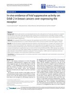
Vasomotor responses showed a similar relaxation induced
by 10
-6
M VEGF
165
and QK while, as expected, substan-
Journal of Translational Medicine 2009, 7:41 />Page 4 of 10
(page number not for citation purposes)
Effects of VEGF
15
, VEGF
165
and QK on the vasomotor responses of 12 common carotid arteries from normotensive rats (A)Figure 1
Effects of VEGF
15
, VEGF
165
and QK on the vasomotor responses of 12 common carotid arteries from normo-
tensive rats (A). Both VEGF
165
and QK induced a comparable vasorelaxation, while VEGF
15
, has no evident effect. After
removal of the endothelial layer there is no appreciable vasorelaxation (B). * = p < 0.05 vs VEGF
15
. Error bars show SEM.
Journal of Translational Medicine 2009, 7:41 />Page 5 of 10
(page number not for citation purposes)
tially no action was detected after VEGF
15
administration.
(Figure 1A). The endothelium was mechanically removed
from the aortic rings to assess endothelium-independent
vasomotor responses. Gentle endothelium denudation
prevented QK and VEGF
165
vasorelaxation, indicating that
these responses are endothelium dependent (Figure 1B).
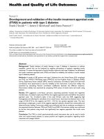
Ischemic hindlimb
Ischemic HL perfusion was assessed by TFC score of dig-
ital microangiographies. Both VEGF
165
and QK amelio-
rated the TFC score (VEGF
165
:17 ± 2; QK:16 ± 2)
compared to the scramble peptide-infused HL (VEGF
15
:38
± 3; p < 0.05, ANOVA) as depicted in Figure 2A.
Regional gastrocnemius blood flow was also measured by
dyed microspheres entrapment after intra-aortic infusion.
After muscle digestion, dye elution is properly related to
HL perfusion (ischemic/not-ischemic) [3]. Once again
(Figure 2B), VEGF
165
and QK treatment achieved a better
ischemic HL perfusion than VEGF
15
treatment
(VEGF
165
:0.92 ± 0.1; QK:0.95 ± 0.1; VEGF
15
:0.59 ± 0.2; p
< 0.05, ANOVA).
Capillary density was assessed on the tibialis anterior mus-
cle of the ischemic HL by means of lectin istochemistry.
VEGF
165
and QK increased capillaries to muscle fibers
ratio in comparison with VEGF
15
(VEGF
15
:0.5 ± 0.04;
VEGF
165
:0.7 ± 0.06; QK:0.72 ± 0.07; p < 0.05, ANOVA), as
shown in Figure 2C, D.
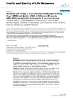
Wound healing
The examination of full-thickness wounds in the back
skin shows that both QK and VEGF
165
accelerate healing
In the model of ischemic hindlimb, VEGF
165
as well QK enhanced and ameliorated regenerative responses, as assessed by TIMI Frame Count (TFC, Panel A), dyed beads dilution from gastrocnemious muscles (B) and of histological analysis, with representa-tive images (C) of lectin GBS-I staining of capillaries in the tibialis anterior muscleFigure 2
In the model of ischemic hindlimb, VEGF
165
as well QK enhanced and ameliorated regenerative responses, as
assessed by TIMI Frame Count (TFC, Panel A), dyed beads dilution from gastrocnemious muscles (B) and of
histological analysis, with representative images (C) of lectin GBS-I staining of capillaries in the tibialis anterior
muscle. (Magnification ×40; bar = 10 μm) and the evaluation as number of capillaries per number of fibers (D) * = p < 0.05 vs
VEGF
15
. Error bars show SEM.
Journal of Translational Medicine 2009, 7:41 />Page 6 of 10
(page number not for citation purposes)
Diagram of the kinetics of wound closure (A)Figure 3
Diagram of the kinetics of wound closure (A). VEGF
165
and QK accelerate the closure of full thickness punch biopsy
wounds. Three to five rats were analyzed at each time point. Gross appearance after 5 days of the wound treated with VEGF
15
,
VEGF
165
, QK (10
-6
M); * = p < 0.05 vs VEGF
15
. Representative digital photographs (B) 5 days after wound. Error bars show
SEM.
Journal of Translational Medicine 2009, 7:41 />Page 7 of 10
(page number not for citation purposes)
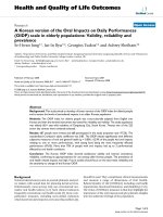
Representative images of Matrigel plugs subcutaneously injected at a magnification of ×60; bar = 40 μmFigure 4
Representative images of Matrigel plugs subcutaneously injected at a magnification of ×60; bar = 40 μm.
Endothelial cells are identified by lectin staining, that gives a brown reaction product. Different background is due to counter-
staining, performed with a standard mixture of hematoxylin and eosin, as described in Methods (A). Quantification of micro-
vessels infiltrating Matrigel plugs (B). * = p < 0.05 vs VEGF
15
. Error bars show SEM.
Journal of Translational Medicine 2009, 7:41 />Page 8 of 10
(page number not for citation purposes)
by enhancing angiogenesis in the granulation tissue (Fig-
ure 3).
Matrigel plugs
After injection, Matrigel containing the angiogenic stimuli
forms a plug into which blood vessels can migrate.
Matrigel pellets evidenced a significant greater peripheral
capillaries infiltration in VEGF
165
(86 ± 3.0) and QK (91 ±
4.5) treated rats than in VEGF
15
ones (26 ± 2.0; p < 0.05 vs
VEGF
165
and QK, ANOVA), as shown in Figure 4.
Discussion
In the present study, we examinated the in vivo effects of a
VEGF
165
mimetic, named QK, modeled on the region of
the VEGF protein responsible for binding to and activat-
ing the VEGFRs that are known to trigger angiogenesis. We
previously showed that QK can bind to the VEGFRs, initi-
ate VEGF-induced signaling cascades and stimulate angio-
genesis in vitro [9]. This is the first report to show that this
peptide is able to recapitulate the in vivo responses of
VEGF.
Angiogenesis is known to be a process of new blood vessel
formation from a pre-existing endothelial structure. It is
tuned by proangiogenic and antiangiogenic factors, and
the shift from this equilibrium may lead to pathological
angiogenesis [18,19]. Indeed, deregulation of angiogen-
esis is involved in several conditions including cancer,
ischemic, and inflammatory diseases (atherosclerosis,
rheumatoid arthritis, or age-related macular degenera-
tion). Therefore, the research for drugs able to regulate
angiogenesis constitutes a pivotal research field. In partic-
ular, occlusive vascular disease remains an important
cause for death and morbidity in industrialized society
[1,20], despite efforts to design new and efficient treat-
ment strategies [19,21].
Unfortunately, numerous reports indicate that in labora-
tory animals over-expression of VEGF may lead to meta-
bolic dysfunction, formation of leaky vessels and transient
edema [1,22]. Indeed, VEGF actions include the induction
of endothelial cells proliferation and migration; it is also
known as a vascular permeability factor, based on its abil-
ity to induce vascular leakage and vasodilatation in a dose
dependent fashion as a result of endothelial cell-derived
nitric oxide [12,23].
In humans, various clinical trials were designed to verify
new vessel growth by exogenous administration of proan-
giogenic factors in patients with refractory ischemic symp-
toms. Albeit initial small open-labeled trials yielded
promising results, subsequent larger double-blind rand-
omized placebo-controlled clinical trials have failed to
show much clinical benefit [19,24,25]. These largely dis-
appointing results may in part be explained by subopti-
mal delivery of genetic material to target cells or tissue.
Moreover, although adenoviral vectors provide high levels
of gene transfer and expression, there are well known
virus-related adverse effects, such as the induction of
immune and inflammatory response [6,21,26]. Recently,
several side effects have been reported for VEGF adminis-
tration in human subjects [1,8,25] such as increase in
atherosclerotic plaques, lymphatic edema or uncontrolled
neoangiogenesis leading to the development of function-
ally abnormal blood vessels, so to preclude its use in a
large share of ischemic population [21,27].
A hopeful alternative could be to use angiogenic stimula-
tors of smaller size, such as peptides, with a well-charac-
terized biologic mechanism of action. Indeed, recent
reports revealed a specific antagonistic relationship
between VEGF and other vascular growth factors, such as
the placental growth factor (PlGF), the basic fibroblast
growth factor (bFGF) and the platelet-derived growth fac-
tor (PDGF), with a dichotomous role for VEGF and VEG-
FRs [28-30]. So, the function of VEGF is far more intricate:
it can also negatively regulate angiogenesis and tumori-
genesis, by impeding the function of the PDGF receptor
on pericytes, leading to a loss of pericyte coverage of
blood vessels [31]. Moreover, several studies demon-
strated a more efficacious action obtained with a specific
stimulation of VEGFRs [32,33] if compared to VEGF over-
expression [22,34]. These findings suggest that the multi-
faceted array of the biological responses linked to VEGF
may be ascribable to its proneness to dimerize or interact
with other molecules [29]. Thus, because of lower molec-
ular and biological complexity, peptides that ensure only
the needed interaction with specific receptors could be
candidate lead compounds for a safer proangiogenic drug,
also to avoid adverse effects.
Perspectives
We show that the VEGF mimetic QK is able to increase
neoangiogenesis and collateral flow in WKY rats. Our
findings evidence the proangiogenic properties of this
small peptide, suggesting that also in vivo QK resembles
the full VEGF protein. Thus, a single peptide, that would
not be expected to dimerize, is still able to induce VEGF
specific angiogenic responses. Clearly, further studies are
needed to fully understand this mechanism, that appears
of intriguing interest. Anyway, these data open to new
fields of investigation on the mechanisms of activation of
VEGFRs, also to clarify complex angiogenesis pathways,
with strong clinical implications for treatment of patho-
physiological conditions such as chronic ischemia.
Competing interests
The authors declare that they have no competing interests.
Journal of Translational Medicine 2009, 7:41 />Page 9 of 10
(page number not for citation purposes)
Authors' contributions
GS, GI, MC, LDDA, CP and BT designed research, GS, MC,
GP, AC, GG, BZ, GGA, VC, and FP, carried out the experi-
ments; GS and GI performed the statistical analysis; GS,
GI and BT drafted the manuscript. All authors read and
approved the final manuscript.
References
1. Schaper W: Collateral circulation: past and present. Basic Res
Cardiol 2009, 104:5-21.
2. Testa U, Pannitteri G, Condorelli GL: Vascular endothelial
growth factors in cardiovascular medicine. J Cardiovasc Med
(Hagerstown) 2008, 9:1190-1221.
3. Iaccarino G, Ciccarelli M, Sorriento D, Galasso G, Campanile A, San-
tulli G, Cipolletta E, Cerullo V, Cimini V, Altobelli GG, Piscione F, Pri-
ante O, Pastore L, Chiariello M, Salvatore F, Koch WJ, Trimarco B:
Ischemic neoangiogenesis enhanced by beta2-adrenergic
receptor overexpression: a novel role for the endothelial
adrenergic system. Circ Res 2005, 97:1182-1189.
4. Li J, Post M, Volk R, Gao Y, Li M, Metais C, Sato K, Tsai J, Aird W,
Rosenberg RD, Hampton TG, Sellke F, Carmeliet P, Simons M: PR39,
a peptide regulator of angiogenesis. Nat Med 2000, 6:49-55.
5. Takeshita S, Zheng LP, Brogi E, Kearney M, Pu LQ, Bunting S, Ferrara
N, Symes JF, Isner JM: Therapeutic angiogenesis. A single
intraarterial bolus of vascular endothelial growth factor aug-
ments revascularization in a rabbit ischemic hind limb
model. J Clin Invest 1994, 93:662-670.
6. Vajanto I, Rissanen TT, Rutanen J, Hiltunen MO, Tuomisto TT, Arve
K, Narvanen O, Manninen H, Rasanen H, Hippelainen M, Alhava E,
Ylä-Herttuala S: Evaluation of angiogenesis and side effects in
ischemic rabbit hindlimbs after intramuscular injection of
adenoviral vectors encoding VEGF and LacZ. J Gene Med 2002,
4:371-380.
7. Leosco D, Rengo G, Iaccarino G, Filippelli A, Lymperopoulos A, Zin-
carelli C, Fortunato F, Golino L, Marchese M, Esposito G, Rapacciuolo
A, Rinaldi B, Ferrara N, Koch WJ, Rengo F: Exercise training and
beta-blocker treatment ameliorate age-dependent impair-
ment of beta-adrenergic receptor signaling and enhance car-
diac responsiveness to adrenergic stimulation. Am J Physiol
Heart Circ Physiol 2007, 293:H1596-1603.
8. Lei Y, Haider H, Shujia J, Sim ES: Therapeutic angiogenesis.
Devising new strategies based on past experiences. Basic Res
Cardiol 2004, 99:121-132.
9. D'Andrea LD, Iaccarino G, Fattorusso R, Sorriento D, Carannante C,
Capasso D, Trimarco B, Pedone C: Targeting angiogenesis:
structural characterization and biological properties of a de
novo engineered VEGF mimicking peptide. Proc Natl Acad Sci
USA 2005, 102:14215-14220.
10. Dudar GK, D'Andrea LD, Di Stasi R, Pedone C, Wallace JL: A vascu-
lar endothelial growth factor mimetic accelerates gastric
ulcer healing in an iNOS-dependent manner. Am J Physiol Gas-
trointest Liver Physiol 2008, 295:G374-381.
11. Diana D, Ziaco B, Colombo G, Scarabelli G, Romanelli A, Pedone C,
Fattorusso R, D'Andrea LD: Structural determinants of the unu-
sual helix stability of a de novo engineered vascular endothe-
lial growth factor (VEGF) mimicking peptide. Chemistry 2008,
14:4164-4166.
12. Fukumura D, Gohongi T, Kadambi A, Izumi Y, Ang J, Yun CO, Buerk
DG, Huang PL, Jain RK: Predominant role of endothelial nitric
oxide synthase in vascular endothelial growth factor-induced
angiogenesis and vascular permeability. Proc Natl Acad Sci USA
2001, 98:2604-2609.
13. Ciccarelli M, Cipolletta E, Santulli G, Campanile A, Pumiglia K, Cer-
vero P, Pastore L, Astone D, Trimarco B, Iaccarino G: Endothelial
beta2 adrenergic signaling to AKT: role of Gi and SRC. Cell
Signal 2007, 19:1949-1955.
14. Iaccarino G, Ciccarelli M, Sorriento D, Cipolletta E, Cerullo V, Iovino
GL, Paudice A, Elia A, Santulli G, Campanile A, Arcucci O, Pastore L,
Salvatore F, Condorelli G, Trimarco B: AKT participates in
endothelial dysfunction in hypertension. Circulation 2004,
109:2587-2593.
15. Sorriento D, Ciccarelli M, Santulli G, Campanile A, Altobelli GG,
Cimini V, Galasso G, Astone D, Piscione F, Pastore L, Trimarco B, Iac-
carino G: The G-protein-coupled receptor kinase 5 inhibits
NFkappaB transcriptional activity by inducing nuclear accu-
mulation of IkappaB alpha. Proc Natl Acad Sci USA 2008,
105:17818-17823.
16. Ciccarelli M, Santulli G, Campanile A, Galasso G, Cervero P, Altobelli
GG, Cimini V, Pastore L, Piscione F, Trimarco B, Iaccarino G:
Endothelial alpha1-adrenoceptors regulate neo-angiogen-
esis. Br J Pharmacol 2008, 153:936-946.
17. Galasso G, Schiekofer S, Sato K, Shibata R, Handy DE, Ouchi N,
Leopold JA, Loscalzo J, Walsh K: Impaired angiogenesis in glu-
tathione peroxidase-1-deficient mice is associated with
endothelial progenitor cell dysfunction. Circ Res 2006,
98:254-261.
18. Carmeliet P: VEGF gene therapy: stimulating angiogenesis or
angioma-genesis? Nat Med 2000, 6:1102-1103.
19. Khurana R, Simons M, Martin JF, Zachary IC: Role of angiogenesis
in cardiovascular disease: a critical appraisal. Circulation 2005,
112:1813-1824.
20. Sirico G, Brevetti G, Lanero S, Laurenzano E, Luciano R, Chiariello M:
Echolucent femoral plaques entail higher risk of echolucent
carotid plaques and a more severe inflammatory profile in
peripheral arterial disease. J Vasc Surg 2009, 49:346-351.
21. Epstein SE, Kornowski R, Fuchs S, Dvorak HF: Angiogenesis ther-
apy: amidst the hype, the neglected potential for serious side
effects. Circulation 2001, 104:115-119.
22. Karpanen T, Bry M, Ollila HM, Seppanen-Laakso T, Liimatta E, Lesk-
inen H, Kivela R, Helkamaa T, Merentie M, Jeltsch M, Paavonen K,
Andersson LC, Mervaala E, Hassinen IE, Ylä-Herttuala S, Oresic M,
Alitalo K: Overexpression of vascular endothelial growth fac-
tor-B in mouse heart alters cardiac lipid metabolism and
induces myocardial hypertrophy. Circ Res 2008, 103:1018-1026.
23. Gigante B, Morlino G, Gentile MT, Persico MG, De Falco S: Plgf-/-
eNos-/- mice show defective angiogenesis associated with
increased oxidative stress in response to tissue ischemia.
FASEB J 2006, 20:970-972.
24. Henry TD, Annex BH, McKendall GR, Azrin MA, Lopez JJ, Giordano
FJ, Shah PK, Willerson JT, Benza RL, Berman DS, Gibson CM, Baja-
monde A, Rundle AC, Fine J, McCluskey ER, VIVA Investigators: The
VIVA trial: Vascular endothelial growth factor in Ischemia
for Vascular Angiogenesis. Circulation 2003, 107:1359-1365.
25. Isner JM, Vale PR, Symes JF, Losordo DW: Assessment of risks
associated with cardiovascular gene therapy in human sub-
jects. Circ Res 2001, 89:389-400.
26. Brevetti LS, Sarkar R, Chang DS, Ma M, Paek R, Messina LM: Admin-
istration of adenoviral vectors induces gangrene in acutely
ischemic rat hindlimbs: role of capsid protein-induced
inflammation. J Vasc Surg 2001, 34:489-496.
27. Celletti FL, Waugh JM, Amabile PG, Brendolan A, Hilfiker PR, Dake
MD: Vascular endothelial growth factor enhances atheroscle-
rotic plaque progression. Nat Med 2001, 7:425-429.
28. Cao Y, Linden P, Shima D, Browne F, Folkman J: In vivo angiogenic
activity and hypoxia induction of heterodimers of placenta
growth factor/vascular endothelial growth factor. J Clin Invest
1996, 98:2507-2511.
29. Eriksson A, Cao R, Pawliuk R, Berg SM, Tsang M, Zhou D, Fleet C,
Tritsaris K, Dissing S, Leboulch P, Cao Y: Placenta growth factor-
1 antagonizes VEGF-induced angiogenesis and tumor
growth by the formation of functionally inactive PlGF-1/
VEGF heterodimers. Cancer Cell 2002, 1:99-108.
30. Greenberg JI, Shields DJ, Barillas SG, Acevedo LM, Murphy E, Huang
J, Scheppke L, Stockmann C, Johnson RS, Angle N, Cheresh DA: A
role for VEGF as a negative regulator of pericyte function
and vessel maturation. Nature 2008, 456:809-813.
31. Stockmann C, Doedens A, Weidemann A, Zhang N, Takeda N,
Greenberg JI, Cheresh DA, Johnson RS: Deletion of vascular
endothelial growth factor in myeloid cells accelerates tum-
origenesis. Nature 2008, 456:814-818.
32. Smadja DM, Bieche I, Helley D, Laurendeau I, Simonin G, Muller L,
Aiach M, Gaussem P: Increased VEGFR2 expression during
human late endothelial progenitor cells expansion enhances
in vitro angiogenesis with up-regulation of integrin alpha(6).
J Cell Mol Med 2007, 11:1149-1161.
33. Wang D, Donner DB, Warren RS: Homeostatic modulation of
cell surface KDR and Flt1 expression and expression of the
vascular endothelial cell growth factor (VEGF) receptor
mRNAs by VEGF. J Biol Chem 2000, 275:15905-15911.
Publish with Bio Med Central and every
scientist can read your work free of charge
"BioMed Central will be the most significant development for
disseminating the results of biomedical research in our lifetime."
Sir Paul Nurse, Cancer Research UK
Your research papers will be:
available free of charge to the entire biomedical community
peer reviewed and published immediately upon acceptance
cited in PubMed and archived on PubMed Central
yours — you keep the copyright
Submit your manuscript here:
/>BioMedcentral
Journal of Translational Medicine 2009, 7:41 />Page 10 of 10
(page number not for citation purposes)
34. Masaki I, Yonemitsu Y, Yamashita A, Sata S, Tanii M, Komori K, Nak-
agawa K, Hou X, Nagai Y, Hasegawa M, Sugimachi K, Sueishi K: Ang-
iogenic gene therapy for experimental critical limb ischemia:
acceleration of limb loss by overexpression of vascular
endothelial growth factor 165 but not of fibroblast growth
factor-2. Circ Res 2002, 90:966-973.