báo cáo hóa học:" Biocompatibility of Poly-ε-caprolactone-hydroxyapatite composite on mouse bone marrow-derived osteoblasts and endothelial cells" doc
Bạn đang xem bản rút gọn của tài liệu. Xem và tải ngay bản đầy đủ của tài liệu tại đây (1.74 MB, 9 trang )
BioMed Central
Page 1 of 9
(page number not for citation purposes)
Journal of Orthopaedic Surgery and
Research
Open Access
Research article
Biocompatibility of Poly-ε-caprolactone-hydroxyapatite composite
on mouse bone marrow-derived osteoblasts and endothelial cells
Haiying Yu
1,2
, Paul H Wooley
1,2
and Shang-You Yang*
1,2,3
Address:
1
Department of Biomedical Engineering, Wayne State University, Detroit, Michigan, USA,
2
Department of Orthopaedic Surgery, Wayne
State University, Detroit, Michigan, USA and
3
Orthopaedic Research Institute, Via Christi Health System, Department of Biological Sciences,
Wichita State University, 1845 Fairmount Street, Wichita, KS 67260, USA
Email: Haiying Yu - ; Paul H Wooley - ; Shang-You Yang* -
* Corresponding author
Abstract
Background: Tissue-engineered bone may be developed by seeding the cells capable of both
osteogenesis and vascularization on biocompatible composite scaffolds. The current study
investigated the performance of mice bone marrow-derived osteogenic cells and endothelial cells
as seeded on hydroxyapatite (HA) and poly-ε-caprolactone (PCL) composite scaffolds.
Methods: Mononuclear cells were induced to osteoblasts and endothelial cells respectively, which
were defined by the expression of osteocalcin, alkaline phosphatase (ALP), and deposits of calcium-
containing crystal for osteoblasts, or by the expression of vascular endothelial growth factor
receptor-2 (VEGFR-2) and von Willebrand factor (vWF), and the formation of a capillary network
in Matrigel™ for endothelial cells. Both types of cell were seeded respectively on PCL-HA scaffolds
at HA to PCL weight ratio of 1:1, 1:4, or 0:1 and were evaluated using scanning electron
microscopy, ALP activity (of osteoblasts) and nitric oxide production (of endothelial cells) plus the
assessment of cell viability.
Results: The results indicated that HA led to a positive stimulation of osteoblasts viability and ALP
activity, while HA showed less influence on endothelial cells viability. An elevated nitric oxide
production of endothelial cells was observed in HA-containing group.
Conclusion: Supplement of HA into PCL improved biocompatible for bone marrow-derived
osteoblasts and endothelial cells. The PCL-HA composite integrating with two types of cells may
provide a useful system for tissue-engineered bone grafts with vascularization.
Background
One approach to tissue engineering consists of seeding
appropriate cells on a biodegradable scaffold, stimulating
cell growth and differentiation in vitro, and then implant-
ing the engineered complex in vivo to achieve functional
tissue [1,2]. However seeding a single cell type into a bio-
material scaffold to replace an injured tissue that consists
of multiple cell types is usually inapplicable. An alterna-
tive strategy is the generation of a composite graft, which
contains not only the tissue specific cell types, but also
other supportive cells, such as endothelial cells (ECs) to
promote vascularization of the grafts.
ECs may be incorporated into bioengineered tissue[3,4]to
promote the tissue revascularization, and transportation
of oxygen and nutrients. Unfortunately, differentiated ECs
Published: 26 February 2009
Journal of Orthopaedic Surgery and Research 2009, 4:5 doi:10.1186/1749-799X-4-5
Received: 17 May 2008
Accepted: 26 February 2009
This article is available from: />© 2009 Yu et al; licensee BioMed Central Ltd.
This is an Open Access article distributed under the terms of the Creative Commons Attribution License ( />),
which permits unrestricted use, distribution, and reproduction in any medium, provided the original work is properly cited.
Journal of Orthopaedic Surgery and Research 2009, 4:5 />Page 2 of 9
(page number not for citation purposes)
isolated from most tissues including aortas, dermal capil-
laries and umbilical veins have inadequate proliferating
capacities and are less responsive to angiogenic survival
factors or anti-angiogenic signals [5,6]. In contrast, bone
marrow-derived endothelial progenitor cells (EPCs) pos-
sess high potential for neovascularization and reendothe-
lialization [7]. EPCs isolated from bone marrow or
peripheral blood have been observed to undergo more
than 1000 division cycles [8], indicating that even the
comparatively low numbers of adult EPCs may provide
sufficient seeding cells for tissue engineering applications.
Bone marrow stromal cells (MSCs) are multipotent stem
cells originating from the bone marrow stroma, and rep-
resent a particularly promising cell source for bone tissue
engineering. They can be easily harvested, expanded in
vitro and induced to differentiate to bone-forming cells
[9]. We have therefore selected MSCs as the source of oste-
ogenic precursors for tissue engineered bone in this
project. Polycaprolactone (PCL), an FDA-approved poly-
ester commonly as drug delivery devices used in clinical
practice, has been shown to be non-toxic to cells [10,11],
possessing many of the desirable properties such as
degradability and plasticity. Hydroxyapatite (HA) is the
inorganic part of the naturally occurring bone, and is
known to be both biocompatible and osteoconductive. It
suggests that the addition of HA to PCL will improve the
biocompatibility and osteoconductivity of the polymer
[12-14]. However, the precise dose-response relationship
of HA in PCL on viability and osteogenic functions of
bone marrow-derived osteoblasts remains to be eluci-
dated. Although ECs-initiating vascularization in the engi-
neered bone is critical [15], the survival and bioactivity of
EPCs-originated ECs in biomaterials of bone graft is fre-
quently neglected. Therefore, the objective of this study
was to evaluate the biocompatibility of the HA-PCL bio-
materials to both bone marrow-derived osteogenic and
endothelial cells.
Methods
Cell Culture and Induction
Bone marrow cells were obtained from male BALB/c mice
(6–8 weeks of age). Low-density bone marrow mononu-
clear cells (MNCs) were isolated by density centrifugation
over Histopaque
®
-1083 (Sigma-Aldrich, US). Cells were
then cultured in flask at 37°C and 5% CO
2
atmosphere
for differentiation of osteoblasts and endothelial cells,
respectively. For osteogenic cell induction, cells were cul-
tured in complete media [16,17] consisting of DMEM
supplemented with 10% fetal bovine serum (FBS) (Invit-
rogen, US), 10 mM β-glycerol phosphate (Sigma-Aldrich,
US), 10
-4
M L-ascorbic acid (Sigma-Aldrich, US), and 10
nM dexamethasone (Sigma-Aldrich, US), 2 mM
glutamine (Invitrogen, US), 100 U/ml penicillin (Invitro-
gen, US), 100 μg/ml streptomycin (Invitrogen, US). To
promote the endothelial phenotype of EPCs, the mono-
nuclear cells were plated onto flasks coated with fibronec-
tin (Sigma-Aldrich, US) and cultured in endothelial cell
basal medium-2 (Cambrex, US) supplemented with
EGM-2 MV SingleQuot
®
kit containing 5% FBS, human
epidermal growth factor (hEGF), human vascular
endothelial growth factor (VEGF), human insulin-like
growth factor-1 (IGF-1), hydrocortisone, penicillin (Invit-
rogen, US), and streptomycin (Invitrogen, US). After 4
days of culture, non-adherent cells were discarded by
washing with PBS. When 60% confluence was achieved,
cells were subcultured.
Cell Characterization
Immunocytofluorescence studies were performed to
detect the induced endothelial phenotypes. The induced
ECs were fixed in 4% paraformaldehyde, permeated with
0.01% Triton X-100 in PBS, and incubated in 1% block
serum for 1 h at 37°C. The cells were then incubated for 1
hour with monoclonal antibody against either mouse
VEGFR-2 or mouse vWF (Santa Cruz, US). Bound anti-
bodies were detected by incubation with fluorescein-5-
isothiocyanate (FITC)-conjugated (Jackson ImmunoRe-
search, US) (for VEGFR-2) or Alexa Fluor 488-conjugated
(Molecular Probes, US) (for vWF) secondary antibody.
The cells were examined in fluorescence microscope. 300
μl of Matrigel™ (BD Biosciences, US) mixed with 4 × 10
4
EPCs-derived ECs at 4°C was dispensed into a 24-well
plate and incubated at 37°C until solid. Photographs of
capillary-like formation were taken at 7 days of culture in
normal condition.
Similar fixation, permeabilization, and blocking proc-
esses were performed on bone marrow-derived osteob-
lasts, followed by the incubation with anti-osteocalcin
(Santa Cruz) for 1 hour, and visualization was achieved
using avidin-peroxidase complex (ABC kit, Santa Cruz
Biotechnology, US). Cells were counterstained with Gill's
hematoxylin solution. Calcium deposit produced by oste-
oblasts was demonstrated using von Kossa staining. After
fixation in 4% paraformaldehyde, the cells were incu-
bated with 1% silver nitrate solution (Sigma-Aldrich, US)
under ultraviolet light for 20 minutes, and unreacted sil-
ver was removed by 5% sodium thiosulfate (Sigma-
Aldrich, US). The alkaline phosphatase (ALP) activity of
osteoblasts was assayed using an ALP kit (Sigma-Aldrich,
US). The induced osteoblasts on slides were fixed in cit-
rate-acetone-formaldehyde solution at room temperature
for 1 minute. Following incubation in alkaline-dye mix-
ture for 15 minutes and rinsing in distilled water, the
slides were counterstained with hematoxylin solution.
Preparation of HA-PCL Scaffolds
PCL-HA scaffolds were prepared using a particulate leach-
ing technique as described previously [18]. The HA-PCL
composite at 2 different component ratios were prepared
Journal of Orthopaedic Surgery and Research 2009, 4:5 />Page 3 of 9
(page number not for citation purposes)
respectively, with HA (Sigma-Aldrich, US) to PCL
(Aldrich, US) at 1:1 (Group A) or 1:4 (Group B) wt/wt.
PCL scaffolds without HA were used as a control (Group
C). In each group, NaCl particles (particle size 212–355
μm) were used to generate a controlled level of porosity in
the matrix with weight ratio to PCL at 16:1 (Group A), 8:1
(Group B) and 4:1 (Group C). PCL (Mn 80000) was dis-
solved in tetrahydrofuran (Sigma-Aldrich, US) at 10% wt/
vol for 12 hours. HA powder (≤ 40 μm particle size) and
NaCl particles were mixed to homogeneity in the PCL
solution, which was sonicated for 60 seconds until vis-
cous slurry developed. Mixtures were poured into glass
dishes to a thickness of 4 mm, and dried at 37°C. After
evaporation of the solvent, 1.5 × 1.5 cm squares were cut
out and washed in excessive distilled water to leach out
the NaCl. All materials were then sterilized in 70% etha-
nol and dried before biological evaluation. Samples of the
PCL-HA scaffolds were gold sputter coated and their mor-
phology was observed using SEM (Hitachi S-2400, Japan)
at 15 kV. Energy Dispersive X-ray (EDX) analysis was also
conducted to confirm the existence of HA particles on the
composite scaffolds. The atomic percentages of calcium
and phosphorus were calculated.
Cells Culture on Scaffolds
Induced osteoblasts or endothelial cells in 50 μl suspen-
sions (3.5 × 10
6
cells/ml) were respectively loaded onto
each scaffold in 6-well plates. The scaffolds were left
undisturbed in a 37°C incubator for 3 hours to allow cells
to attach to the scaffold, after which the cells-materials
complex were kept in culture using the original osteogenic
or endothelial media. At day 7 the samples were harvested
for and biochemical evaluation.
For morphological examination, cells-materials complex
were fixed with 1.5% glutaraldehyde (Fisher Scientific,
US) for 30 min at 4°C. The samples were exposed to 2%
osmium tetroxide (Sigma-Aldrich, US) for 30 min. Fol-
lowing rinse in distilled water, they were dehydrated
through a graded series of ethanol (50, 70, 90, and 100%)
for 2–5 min. The dehydration was completed in hexame-
thyl disilazane (Fluka, Germany) for 10 minutes. After air-
drying and sputter coating with gold, the cells morphol-
ogy on the PCL-HA scaffolds was evaluated using SEM at
10 kV.
Assessment of Cell Viabilities and Functions on Scaffolds
For biochemistry assay, each type of cells was seeded on
30 scaffolds per group. Cell viability was evaluated by ana-
lyzing the mitochondrial activities of the cells. The Alamar
Blue assay (BioSource, US) was used to determine the
mitochondrial activity after 7 days of cell culture. The
cells-materials complexes were washed in Phosphate-
Buffered Saline (PBS) in 6-well plate. 3 ml of new condi-
tioned media supplemented with 200 μl of Alamar blue
was added to each well. Incubation was continued at
37°C, 5% CO
2
, for 3 hours. The culture medium was then
transferred to a 96-well plate and read on a spectrofluor-
ometer (excitation wavelength 530 nm, emission wave-
length 590 nm). The Alamar blue absorbance/mg of DNA
values was calculated for each sample.
Cell amount were determined by a fluorometric quantifi-
cation of DNA in the cells-materials complexes. After the
Alamar Blue assay, the cell-scaffolds were rinsed with PBS,
followed by 4 times of freezing (-80°C) and thawing
(37°C) cycles for 15 minutes each. The scaffolds were
then homogenized in 1.4 ml of cold 10 mM EDTA solu-
tion (Sigma-Aldrich, US). The pH of the samples was
adjusted to 7.0 by adding 1 M KH
2
PO
4
prior to the addi-
tion of 1.5 ml of the 200 ng/ml Hoechst 33258 fluores-
cent dye (Sigma-Aldrich, US). 100 μl of supernatant
sample were read with an excitation set at 350 nm and an
emission at 455 nm on a spectrofluorometer. The DNA
concentration in the samples was determined against a
DNA standard curve that was plotted according to a series
of 100 μl of calf thymus DNA (Sigma, US) in a range of
concentrations from 0.15265 to 20 μg/ml. The DNA val-
ues were used to normalize the cell viability and other cell
function parameters.
Production of ALP by osteogenic cells was measured using
a spectrophotometer. After the previous freeze-thaw cycle
and DNA assay, 50 μl of the sample was transferred to a
fresh 96-well plate and 50 μl of p-nitrophenyl phosphate
solution (Sigma, US) was added to each sample. Follow-
ing incubation for 5 min at 37°C, the production of p-
nitrophenol in the presence of ALP was measured by
monitoring light absorbance at 405 nm. The measure-
ment of the ALP assay was normalized against the amount
of total DNA in each sample.
The nitric oxide generated by endothelial cells on scaf-
folds was assessed using the Nitric Oxide Colorimetric
Assay kit (Calbiochem, Germany) in accordance with the
manufacturer's instructions. The presence of nitric oxide
in the culture media of endothelial cells-materials com-
plex was determined by detecting the colored product
spectrophotometrically. The absorbance was read at 540
nm. The nitric oxide concentrations for samples were cal-
culated according to the standard curve. Cellular nitric
oxide amount was normalized by total DNA each sample.
Statistical Analysis
All experiments were replicated three times to ensure the
reproducibility, and all data was presented as the mean ±
standard deviation. Single factor analysis of variance
(ANOVA) with a post hoc LSD from SPSS™ (Student Ver-
sion 10.0.5, Chicago, IL) was used to assess the statistical
significance among groups, which was defined as p < 0.05.
Journal of Orthopaedic Surgery and Research 2009, 4:5 />Page 4 of 9
(page number not for citation purposes)
Results
Cells Culture and Induction
Cell colonies were detected in the primary passage using
culture conditions with either osteogenic medium or
endothelial medium. These cells differentiated and prolif-
erated, and gradually exhibited homogeneous and spe-
cific cell morphologies. The differentiated cells displayed
osteoblasts-like spindle morphology in osteogenic
medium (Figure 1A), while endothelial cells presented
typical cobblestone morphology (Figure 1B). These cells
retained stable morphologies for more than 5 passages.
We choose the 2
nd
passage osteoblasts and endothelial
cells for subsequent cell characterization and the investi-
gation of biocompatibility.
Cell Characterization
The capacity of the induced osteoblasts to express osteo-
calcin was examined by immunocytochemistry (IHC)
(Figure 2A). The expression of osteocalcin was detected in
over 95% osteogenic-wise induced cells. These cells were
also identified by positive staining for ALP (Figure 2B),
which indicated that the induced cells possessed distin-
guishable osteoblastic phenotype. To demonstrate the
ability of cells to mineralize matrix, cells cultured on Petri
dishes were subjected to von Kossa stain to reveal calcium
deposition (Figure 2C) where the darkly stained mineral-
ized nodule were visualized by silver nitrate, indicating
normal osteoblasts function in conditioned culture. Dif-
ferentiation ability of ECs induced from bone marrow was
determined by the expression of endothelial markers,
VEGFR-2 and vWF, using immunocytofluorescence. The
95% endothelial-wise-induced cells expressed VEGFR-2
and vWF at 2
nd
passage of culture (Figure 3A, B), indicat-
ing the induced cells having normal endothelial pheno-
type. Matrigel™ culture was performed to monitor the
capability of capillary formation. Three-dimensional cap-
illary-like networks from EPCs-derived endothelial cells
were clearly established at one-week incubation (Figure
3C).
Cells Culture on Scaffolds
PCL scaffolds incorporated with or without HA were fab-
ricated with controlled porosity (70% ~ 75%) and pore
sizes. Interconnected pore morphologies were present in
all scaffolds, resulting in the high porosity of the scaffolds.
Different microtopographies of scaffolds were revealed by
scanning electron microscopy (SEM) (Figure 4). The
roughness of pore wall appeared dependent on the ratio
of HA to PCL. High HA concentration led to extensive pro-
trusions of HA particles and rough surfaces (Figure 4C),
while an almost smooth pore wall was achieved in the
PCL scaffold without HA incorporation (Figure 4A). EDX
analysis indicated atomic ratio of calcium to phosphorus
(Ca: P = 1.58) on both low HA ratio (HA: PCL = 1:4) and
high HA ratio (HA: PCL = 1:1) composite scaffolds, which
is comparable to a natural hydroxyapatite (Figure 4D).
The specific cell morphology of either osteoblasts or
endothelial cells displayed rarely difference response to
various groups of PCL-HA scaffold. The continuous cul-
ture of osteoblasts on HA-containing scaffolds for 7 days
revealed that cells retained their spindle morphology (Fig-
ure 5A) similar to the osteoblasts grown on tissue culture
flasks. Extracellular matrix (collagen-like fibers) was
clearly present at intercellular regions, where cellular pro-
jections were evident (Figure 5B). For the cultures of PCL-
HA scaffolds with bone marrow-derived endothelial cells,
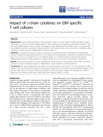
Morphology of osteoblasts and endothelial cellsFigure 1
Morphology of osteoblasts and endothelial cells. Dur-
ing the 2nd passage, the mice bone marrow-derived mono-
nuclear cells differentiated to osteoblasts exhibiting the
spindle morphology (Panel A), or differentiated to endothe-
lial cells presenting the typical cobblestone morphology
(Panel B). Magnification 200×.
Journal of Orthopaedic Surgery and Research 2009, 4:5 />Page 5 of 9
(page number not for citation purposes)
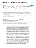
Characterizations of osteoblastsFigure 2
Characterizations of osteoblasts. Over 95% of induced
osteoblasts expressed osteocalcin visualized by immunocyto-
chemistry stains (Panel A, 200×). Alkaline phosphatase (ALP)
activity of osteoblasts was assayed using an ALP kit and visu-
alized as the pink color (Panel B, 200×). In addition, von
Kossa staining was performed to reveal ossification nodules
in the culture dishes of induced cells, as an indicator of oste-
oblasts function (Panel C, 200×).
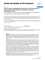
Characterization of endothelial cellsFigure 3
Characterization of endothelial cells. Panel (A) illus-
trated the VEGFR-2 expression on the induced ECs (200×).
Over 95% of cells lighted up in dark-field microscope for
cytoplasmic vWF following cultures in EC conditional
medium (Panel B, 200×). After one-week incubation in a
Matrigel™ basement membrane system, these cells prolifer-
ated and developed capillary-like 3-D structures (Panel C,
200×), suggesting functional endothelial phenotype.
Journal of Orthopaedic Surgery and Research 2009, 4:5 />Page 6 of 9
(page number not for citation purposes)
the cells completely covered the surface of the scaffolds at
7 days (Figure 5C). The spreading and paving endothelial
cells remained as typically cobblestone like shapes with
high cell-to-cell contact (Figure 5D). Cellular extensions
on the cells surface were also detected.
The biochemical tests would provide quantitive compari-
son on compatibility of biomaterials across various
groups. Comparisons were made among cell-scaffold
complexes that varied only in HA ratio to PCL. Addition
of HA resulted in a positive stimulation of osteoblasts via-
bility (Figure 6A). Compared to the HA-free PCL material,
osteoblasts viability was increased by 740% (p < 0.001) in
high HA ratio (HA: PCL = 1:1) group and by 570% (p <
0.001) in low HA ratio (HA: PCL = 1:4) group, revealed by
the Alamar Blue assay. Similarly, as a marker of osteoblast
differentiation, ALP activity was increased by 240% in the
high HA ratio group (p < 0.01) and by 150% in low HA
ratio group (p < 0.05), suggesting the promotion of oste-
oblasts function due to the osteoconductivity of HA (Fig-
ure 6B). There were no statistically significant difference
between the two HA-containing groups in terms of oste-
oblasts viabilities (p = 0.083) and ALP activities (p =
0.119). Although the addition of HA into PCL did not
show significant influence on endothelial cell viability
(Figure 6C), the HA involvement into PCL did lead to
36% (at HA: PCL = 1:1) (p < 0.05) and 80% (at HA: PCL
= 1:4) (p < 0.01) increase of nitric oxide production com-
pared to the HA-free scaffolds (Figure 6D). There was not
statistically significant difference on NO production
between low and high HA ratio groups (p = 0.292, Figure
6D).
Discussion
Bone tissues are essentially composite materials consist-
ing of various components tissues with different structural
arrangements and functions. Repairing bone defects using
tissue engineered grafts involves the interplay of several
variables, including scaffold materials with excellent bio-
compatibility, osteogenic cells capable of assembling the
complicated and functional bone tissue, and an appropri-
ate vascular bed to support the metabolic needs of new
bone tissue. We have been developing a composite scaf-
fold consisting of PCL and HA to enhance the biomechan-
ical properties as the potential bone graft substitute [19].
Furthermore, following introducing MSCs-derived oste-
oblast (responsible for osteogenesis) and EPCs-derived
endothelial cells (accountable for vascularization), we
attempted in the current study to evaluate the biological
impacts of various component ratios in PCL-HA scaffolds
on the two types of cells.
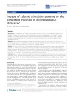
Microstructure of scaffolds exhibited by SEMFigure 4
Microstructure of scaffolds exhibited by SEM. The HA
were embedded in PCL, or exposed on the surface. Appar-
ently the roughness of pore-wall surfaces increased with
increasing the HA ratio. Panel (A) (800×) was an example of
HA-free PCL scaffolds. Panel (B) (1000×) showed composite
with low HA ratio, HA: PCL (w/w) = 1:4; while Panel (C)
(500×) revealed a sample with high HA concentration, HA:
PCL (w/w) = 1:1. The protruded components were con-
firmed as the HA by EDX (Panel D). The peaks of calcium
and phosphorus were prominent and quantified the atomic
ratio (Ca: P = 1.58).
Cells morphology on the composite scaffolds (HA: PCL = 1:1)Figure 5
Cells morphology on the composite scaffolds (HA:
PCL = 1:1). Panel (A) showed osteoblasts proliferating on
PCL-HA scaffolds (400×). Panel (B) was an enlargement of
Panel (A) showing the details of the attached osteoblasts
with meshwork of extracellular matrix (arrows), and cellular
projection (1500×). Panels (C) and (D) revealed the growth
of endothelial cells on a PCL-HA scaffold. The Panel (C) pic-
tured ECs proliferating and forming a cell sheet (600×), and
Panel (D) magnified attached ECs to detail their cobblestone
shape and cellular extensions (4000×).
Journal of Orthopaedic Surgery and Research 2009, 4:5 />Page 7 of 9
(page number not for citation purposes)
Large numbers of MSCs-originated osteoblasts and EPCs-
originated endothelial cells could be obtained in dish cul-
ture in vitro, making them possible to construct trans-
plantable tissues scaffolds. It has been shown that in vitro
stem/progenitor cells possess the capability of self-
renewal and differentiation into organ-specific cell types
[20]. Our results indicated that MSCs and EPCs could be
harvested from mice bone marrow and differentiate into
osteoblasts and endothelial cells respectively. Besides the
expression of specific molecular markers like osteocalcin,
VEGFR-2, and vWF, induced MSCs and EPCs functioned
normally, as indicated by both fine crystals of ossification
(comparable to natural bone mineral) and ALP activity in
osteoblasts, and capillary-like formation by endothelial
cells in Matrigel™. Due to the capacity of capillary forma-
tion, it was speculated that induced endothelial cells
might participate or mediate the neovascularization in tis-
sue engineered bone in vivo, which had been elucidated in
a study [15]. It demonstrated that these differentiated
bone marrow-derived endothelial cells and osteoblasts
had the potential to create a tissue-engineered bone graft
with microvascular network. Therefore, it is necessary to
evaluated biological effects PCL-HA scaffolds on these
cells seeded on it.
PCL and HA have been shown non-toxic and non-muta-
genic biomaterials [11,21-24]. In particular, the biocom-
patibility of HA due to its similarities to natural bone
mineral have led to HA widespread use in bone recon-
structive surgery. Degradation and metabolism of PCL
was completed in vivo, and ε-hydroxycaproic acid and
water were the only metabolites [22,25]. Our study indi-
cated that inclusion of HA into PCL significantly increased
the mitochondrial activity and expression of ALP by oste-
oblasts in a dose-dependent manner, which is in agree-
ment with previous studies [12,26]. The SEM images
demonstrated that bone marrow-derived osteoblasts were
spread on the scaffold surfaces with exposed HA particles
and synthesized extracellular matrix. Nitric oxide (NO) is
a biologically active molecule in the maintenance of vas-
cular homeostasis and predominately produced by
endothelial cells. We examined the levels of NO in cell
media and in cell-scaffold composite to confirm and
quantify the function of bone marrow-derived endothe-
lial cells. Our data showed that the addition of HA ele-
vated NO production in comparison with the HA-free
PCL scaffolds, suggesting that HA particles promoted
functions of the endothelial cells. Although Pezzatini [27]
reported excellent biocompatibility of HA nanocrystals
for endothelial cells, cell viability experiments of this
study resulted no difference among endothelial cell
groups. The different culture conditions and cell origins,
disparate HA dimensions, and biomaterials architectures
between our and Pezzatini's investigations may partially
explain the diverse outcomes.
In this study, adding of HA into PCL led to heterogeneous
surface properties. Variety of HA ratio to PCL generated
distinct exposure of HA particle and diverse topography,
which have been evidenced by SEM images. The exposed
HA particles were more predominated on the high HA
ratio scaffold than those on the lower ones, where the HA
free scaffold just exhibited smoother surface on the pore
walls. It appears that the rough surfaces due to HA embed-
ding provided more anchorage for cell process and
spreading, adhesion and orientation. Therefore, osteob-
lasts and endothelial cells were inclined to attach to the
rougher surface and appeared better viability and strong
cellular phenotypes, which supplement the literature
reports that cell number and attachment force were
increased on textured polymer substrates [26,28,29]. Fur-
thermore HA is a well known sorbent for molecules.
Involvement of HA particles on the PCL surface may
Effects of composite scaffolds on the cells viability and func-tionFigure 6
Effects of composite scaffolds on the cells viability
and function. The induced osteoblasts and endothelial cells
were separately cultured with the 3 groups of scaffold (30
scaffolds per group for each type of cell) for 7 days before
testing. Panels (A) and (C) summarized the Alamar Blue assay
for osteoblast and endothelial cell viabilities. Panel (B) plotted
alkaline phosphatase activity of osteoblasts on various scaf-
folds. Panel (D) showed the NO production of endothelial
cells on various scaffolds.
Journal of Orthopaedic Surgery and Research 2009, 4:5 />Page 8 of 9
(page number not for citation purposes)
change the surface charge, reduce the hydrophobicity of
PCL and promote the adsorption of proteins and other
molecules from surrounding environment [30,31]. In
hard tissues, proteins such as osteopontin, bone sialopro-
tein, and osteocalcin were able to recognize HA through
highly acidic domains, resulting in the attachment and
distribution of osteogenic cells on the surface of protein-
coated HA, and subsequently improving cell proliferation
and differentiation, and promoting new bone formation
[32-35]. Additionally the calcium ions released from the
dissolution of HA were able to neutralize PCL acidic prod-
ucts, so the adverse response due to the PCL degradation
could be overcame [14,36,37].
Conclusion
In conclusion, our data indicated that supplement of HA
into PCL provided a compatible environment for osteob-
lasts and endothelial cells to replicate and function. The
HA surface exposure accounted for the positive cellular
responses. Optimal component ratio in PCL-HA scaffold
could be selected in term of bioactivities of osteoblasts
and endothelial cells. These outcomes would contribute
to the construction of vascularized engineered bone in
vitro and implantation in vivo.
Competing interests
The authors declare that they have no competing interests.
Authors' contributions
HY, PHW and SYY contributed to the design of the study
and the writing of the manuscript. HY carried out the cell
culture, scaffold preparation, biochemistry assessment,
acquisition, and analysis of data. SYY participated in the
image and statistical analysis. All authors read and
approved the final manuscript.
References
1. Soker S, Machado M, Atala A: Systems for Therapeutic Angio-
genesis in Tissue Engineering. World J Urol 2000, 18:10-18.
2. Stahl A, Wu X, Wenger A, Klagsbrun M, Kurschat P: Endothelial
Progenitor Cell Sprouting in Spheroid Cultures Is Resistant
to Inhibition by Osteoblasts: A Model for Bone Replacement
Grafts. FEBS Lett 2005, 579:5338-5342.
3. Nör JE, Peters MC, Christensen JB, Sutorik MM, Linn S, Khan MK,
Addison CL, Mooney DJ, Polverini PJ: Engineering and Character-
ization of Functional Human Microvessels in Immunodefi-
cient Mice. Lab Invest 2001, 81:453-463.
4. Vailhé B, Vittet D, Feige J-J: in vitro Models of Vasculogenesis
And Angiogenesis. Lab Invest 2001, 81:439-452.
5. Lin Y, Weisdorf DJ, Solovey A, Hebbel RP: Origins of Circulating
Endothelial Cells and Endothelial Outgrowth from Blood. J
Clin Invest 2000, 105:71-77.
6. Brey EM, Uriel S, Greisler HP, Patrick CW, McIntire LV: Therapeu-
tic Neovascularization: Contributions from Bioengineering.
Tissue Eng 2005, 11:567-584.
7. Urbich C, Dimmeler S: Endothelial Progenitor Cells: Charac-
terization and Role in Vascular Biology. Circ Res 2004,
95:343-353.
8. Peichev M, Naiyer AJ, Pereira D, Zhu Z, Lane WJ, Williams M, Oz
MC, Hicklin DJ, Witte L, Moore MAS, Rafii S: Expression of
VEGFR-2 and AC133 by circulating human CD34
+
cells iden-
tifies a population of functional endothelial precursors. Blood
2000, 95:952-958.
9. Huang Y-C, Kaigler D, Rice KG, Krebsbach PH, Mooney DJ: Com-
bined Angiogenic and Osteogenic Factor Delivery Enhances
Bone Marrow Stromal Cell-Driven Bone Regeneration. J
Bone Miner Res 2005, 20:848-857.
10. Kweon HY, Yoo MK, Park IK, Kim TH, Lee HC, Lee H-S, Oh J-S,
Akaike T, Cho C-S: A novel degradable polycaprolactone net-
works for tissue engineering. Biomaterials 2003, 24:801-808.
11. Huatan H, Collett JH, Attwood D, Booth C: Preparation and char-
acterization of poly(ε-caprolactone) polymer blends for the
delivery of proteins. Biomaterials 1995, 16:1297-1303.
12. Rizzi SC, Heath DJ, Coombes AGA, Bock N, Textor M, Downes S:
Biodegradable polymer/hydroxyapatite composites: Surface
analysis and initial attachment of human osteoblasts. J Biomed
Mater Res 2001, 55:475-486.
13. Calandrelli L, Immirzi B, Malinconico M, Luessenheide S, Passaro I,
Pasquale RD, Oliva A: Natural and Synthetic Hydroxyapatite
Filled PCL: Mechanical Properties and Biocompatibility
Analysis. J Bioact Compat Polym 2004, 19:301-313.
14. Kim H-W, Knowles JC, Kim H-E: Effect of biphasic calcium phos-
phates on drug release and biological and mechanical prop-
erties of poly (ε-caprolactone) composite membranes. J
Biomed Mater Res 2004, 70A:467-479.
15. Yu H, Vord PJ Vande, Gong W, Wu B, Song Z, Matthew HW, Wooley
PH, Yang S-Y: Promotion of osteogenesis in tissue-engineered
bone by pre-seeding endothelial progenitor cells-derived
endothelial cells. J Orthop Res 2008, 26:1147-1152.
16. Ishaug SL, Crane GM, Miller MJ, Yasko A, Yaszemski MJ, Mikos AG:
Bone formation by three-dimensional stromal osteoblast
culture in biodegradable polymer scaffolds. J Biomed Mater Res
1997, 36:17-28.
17. Cheng SL, Yang JW, Rifas L, Zhang SF, Avioli LV: Differentiation of
human bone marrow osteogenic stromal cells in vitro: induc-
tion of the osteoblast phenotype by dexamethasone. Endo-
crinology 1994, 134:277-286.
18. Mikos AG, Sarakinos G, Vacanti JP, Langer RS, Cima LG: Biocom-
patible polymer membranes and methods of preparation of
three dimensional membrane structures. 1996.
19. Yu H, Matthew HW, Wooley PH, Yang S-Y: Effect of porosity and
pore size on microstructures and mechanical properties of
poly-ε-caprolactone-hydroxyapatite composites. J Biomed
Mater Res B Appl Biomater 2008.
20. Cancedda R, Dozin B, Giannoni P, Quarto R: Tissue Engineering
and Cell Therapy of Cartilage and Bone. Matrix Biol 2003,
22:81-91.
21. van Blitterswijk CA, Grote JJ, Kuypers W, Blok van Hoek CJG, Daems
WT: Bioreactions at the tissue/hydroxyapatite interface. Bio-
materials 1985, 6:243-251.
22. Pitt CG: Poly-ε-Caprolactone and Its Copolymers. In Biodegrad-
able Polymers as Drug Delivery Systems Edited by: Chasin M, Langer R.
New York: Maicel Dekker, Inc; 1990:71-120.
23. Jarcho M, Kay JF, Gumaer KI, Doremus RH, Drobeck HP: Tissue,
cellular and subcellular events at a bone-ceramic hydroxyla-
patite interface. J Bioeng 1997, 1(2):79-92.
24. Eggli PS, Muller W, Schenk RK: Porous hydroxyapatite and trical-
cium phosphate cylinders with two different pore size ranges
implanted in the cancellous bone of rabbits: A comparative
histomorphometric and histologic study of bony ingrowth
and implant substitution. Clin Orthop 1988, 232:127-138.
25. Griffith LG: Polymeric biomaterials. Acta Materialia 2000,
48:263-277.
26. Boyan BD, Hummert TW, Dean DD, Schwartz Z: Role of material
surfaces in regulating bone and cartilage cell response. Bio-
materials 1996, 17:137-146.
27. Pezzatini S, Solito R, Morbidelli L, Lamponi S, Boanini E, Bigi A, Ziche
M: The effect of hydroxyapatite nanocrystals on microvascu-
lar endothelial cell viability and functions. J Biomed Mater Res
(part A)
2006, 76:656-663.
28. Groessner-Schreiber B, Tuan RS: Enhanced extracellular matrix
production and mineralization by osteoblasts cultured on
titanium surfaces in vitro. J Cell Sci 1992, 101:209-217.
29. Bowers KT, Keller JC, Randolph BA, Wick DG, Michaels CM: Opti-
mization of surface micromorphology for enhanced osteob-
last responses in vitro. Int J Oral Maxillofac Implants 1992,
7:302-310.
Publish with Bio Med Central and every
scientist can read your work free of charge
"BioMed Central will be the most significant development for
disseminating the results of biomedical research in our lifetime."
Sir Paul Nurse, Cancer Research UK
Your research papers will be:
available free of charge to the entire biomedical community
peer reviewed and published immediately upon acceptance
cited in PubMed and archived on PubMed Central
yours — you keep the copyright
Submit your manuscript here:
/>BioMedcentral
Journal of Orthopaedic Surgery and Research 2009, 4:5 />Page 9 of 9
(page number not for citation purposes)
30. Furuzono T, Yasuda S, Kimura T, Kyotani S, Tanaka J, Kishida A:
Nano-scaled hydroxyapatite/polymer composite IV. Fabrica-
tion and cell adhesion properties of a three-dimensional scaf-
fold made of composite material with a silk fibroin substrate
to develop a percutaneous device. J Artif Organs 2004,
7:137-144.
31. Midy V, Hollande E, Rey C, Dard M, Plouët J: Adsorption of vascu-
lar endothelial growth factor to two different apatitic mate-
rials and its release. J Mater Sci Mater Med 2001, 12:293-298.
32. Stayton PS, Drobny GP, Shaw WJ, Long JR, Gilbert M: Molecular
Recognition at the Protein-Hydroxyapatite Interface. Crit Rev
Oral Biol Med 2003, 14:370-376.
33. Uemura T, Nemoto A, Liu Y-k, Kojima H, Dong J, Yabe T, Yoshikawa
T, Ohgushi H, Ushida T, Tateishi T: Osteopontin involvement in
bone remodeling and its effects on in vivo osteogenic poten-
tial of bone marrow-derived osteoblasts/porous hydroxyap-
atite construct. Mater Sci Eng C 2001, 17:33-36.
34. Sun J-S, Tsuang Y-H, Liao C-J, Liu H-C, Hang Y-S, Lin F-H: The
effects of calcium phosphate particles on the growth of oste-
oblasts. J Biomed Mater Res 1997, 37:324-334.
35. Ripamonti U: Soluble, insoluble and geometric signals sculpt
the architecture of mineralized tissues. J Cell Mol Med 2004,
8:169-180.
36. Schiller C, Epple M: Carbonated calcium phosphates are suita-
ble pH-stabilising fillers for biodegradable polyesters. Bioma-
terials 2003, 24:2037-2043.
37. Agrawal CM, Athanasiou KA: Technique to control pH in vicinity
of biodegrading PLA-PGA implants. Journal of Biomedical Mate-
rials Research 1997, 38:105-114.